
Integrative Biomedical Research

Integrative Biomedical Research (Journal of Angiotherapy) | Online ISSN  3068-6326
463
Citations
1.9m
Views
799
Articles
Your new experience awaits. Try the new design now and help us make it even better
Switch to the new experience
Figures and Tables
CASE STUDY   (Open Access)

Long-Term Clinical Course of Metastatic Colorectal Cancer Following Sequential Hepatic and Pulmonary Metastasectomy With Adjunctive Standardized Orthosiphon stamineus Extract (Nuvastatic™): A Case Report

Amin Malik Shah Abdul Majid 1,2*, Adam Shah Abdul Majid 2, Muhammad Asyraf Abduraman 2*

+ Author Affiliations

Integrative Biomedical Research 10 (2) 1-13 https://doi.org/10.25163/biomedical.10210912

Submitted: 05 August 2026 Revised: 22 September 2026  Published: 29 September 2026 


Abstract

Background: Selected patients with oligometastatic colorectal cancer (CRC) may achieve prolonged survival following complete resection of metastatic disease, although recurrence remains common. Nuvastatic™ is a standardized Orthosiphon stamineus botanical formulation under clinical development in oncology with preclinical anti-angiogenic and other biologically relevant activities. We describe the longitudinal course of metastatic CRC during conventional curative-intent management and adjunctive Nuvastatic™ use.Case presentation: A 47-year-old woman underwent high anterior resection for pT2N0 sigmoid adenocarcinoma in November 2020. By March–April 2021, carcinoembryonic antigen increased to 29 µg/L, and imaging revealed a large right-lobe hepatic metastasis with small pulmonary nodules. Nuvastatic™ was initiated at 4,000 mg/day approximately one month before right hepatectomy in May 2021. Following R0 hepatic resection, the patient completed six months of FOLFOX, using Nuvastatic™ between chemotherapy cycles. According to patient-reported treatment history, the dose was reduced to 2,000 mg/day approximately three months before enlargement of pre-existing pulmonary lesions was detected in March 2022, after which 4,000 mg/day was resumed. Pulmonary metastasectomy confirmed metastatic colorectal adenocarcinoma. A further 7-mm pulmonary metastasis was resected in August 2024. Genomic profiling demonstrated KRAS G12V, APC and TP53 alterations, microsatellite-stable disease, and low tumour mutational burden. CT imaging on 4 August 2026 showed no evidence of recurrence or new metastatic disease. The patient remained clinically well (ECOG 0) while continuing Nuvastatic™ 2,000 mg/day.Conclusion: This case describes prolonged survival and repeated periods of disease control during multimodal treatment and long-term adjunctive Nuvastatic™ exposure. Although causality cannot be established from a single case, the temporal relationship between dose reduction and pulmonary progression, together with the preclinical biological rationale for O. stamineus, warrants prospective investigation of Nuvastatic™ using pharmacokinetic, biomarker, and oncological endpoints.

Keywords: colorectal cancer; liver metastasis; pulmonary metastasis; KRAS G12V; metastasectomy; Orthosiphon stamineus; Nuvastatic™; CEA; botanical drug; case report

1. Introduction

Colorectal cancer (CRC) is a major cause of cancer morbidity and mortality worldwide, and the liver and lungs are among the most frequent sites of metastatic spread. In selected patients with technically resectable metastatic disease, multidisciplinary treatment incorporating systemic therapy and complete local treatment of metastatic deposits can achieve prolonged survival and, in a subset, long-term cure. Recurrence after colorectal liver or pulmonary metastasectomy remains common, however, reflecting occult micrometastatic disease and heterogeneous tumour biology. (Siegel et al., 2023; Morris et al., 2023)

RAS pathway alterations are important determinants of metastatic CRC biology and treatment selection. Activating KRAS mutations predict lack of benefit from anti-EGFR monoclonal antibodies and have been associated with less favourable outcomes following curative-intent metastasectomy in some series. Microsatellite-stable (MSS), low-tumour-mutational-burden disease also lacks the biomarker profile associated with the pronounced immune-checkpoint responsiveness seen in mismatch-repair-deficient/MSI-high CRC. (Morris et al., 2023; Roncato et al., 2024; Rhaiem et al., 2024)

Nuvastatic™ is an oral effervescent formulation containing 1,000 mg per sachet of a standardized extract of Orthosiphon stamineus Benth. (O. aristatus; Misai Kucing/Cat's Whiskers). Rosmarinic acid is used as a principal quantitative standardization marker, while the phytochemical profile also contains characterized polymethoxylated flavones including sinensetin, eupatorin and 3′-hydroxy-5,6,7,4′-tetramethoxyflavone (TMF). Preclinical work on standardized O. stamineus preparations and constituent phytochemicals has provided biological rationale for investigation of antioxidant, anti-inflammatory, antiproliferative and anti-angiogenic activities, including effects involving VEGF/VEGFR-2-related signalling. These mechanisms were not directly measured in the patient described here and should be regarded as mechanistic hypotheses rather than demonstrated explanations for the clinical outcome. (Hashim et al., 2016; Akowuah et al., 2004)

A multicentre randomized, double-blind, placebo-controlled Phase II study of Nuvastatic™ in 110 patients with stage II-IV solid tumours receiving chemotherapy reported improvement in cancer-related fatigue and quality-of-life measures, reduction in urinary F2-isoprostane and a favourable tolerability profile. The trial was registered as NCT04546607. These findings support clinical investigation of the standardized extract in supportive oncology but do not establish a direct antitumour effect. (Ng et al., 2024)

This report describes the long-term clinical course of a patient initially diagnosed with pathological Stage I sigmoid adenocarcinoma who subsequently displayed unexpectedly aggressive metastatic behaviour, with rapid hepatic metastatic recurrence and pulmonary dissemination despite an initially pT2N0 primary. Particular attention is given to the timing and dose of Nuvastatic™ exposure, the presence of pulmonary nodules before Nuvastatic™ initiation, the source-verified CEA trajectory, recurrent but anatomically limited pulmonary disease, the 2024 pulmonary recurrence, molecular tumour characteristics and the latest objective disease assessment in August 2026. The case is presented as a hypothesis-generating clinical observation in which a contribution from Nuvastatic™ is biologically plausible but cannot be quantified or separated from conventional treatment using a single-patient design.
2. Clinical Case and Outcomes

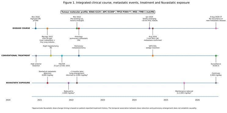
Clinical dates, imaging findings, pathological diagnoses, genomic findings, treatment records, and CEA measurements reported in this case were verified against the available medical records. Information regarding Nuvastatic™ dosing, timing, dose modifications, and perioperative interruptions was obtained from the patient’s reported treatment history unless otherwise specified. An overview of the full clinical course is shown in Figure 1.

2.1 Initial diagnosis and primary treatment

A 47-year-old woman with no significant documented past medical history presented in November 2020 with symptoms leading to colonoscopic evaluation. A sigmoid lesion was identified and biopsy demonstrated moderately differentiated adenocarcinoma. Her ECOG Performance Status was 0.

High anterior resection was performed in November 2020. Final pathology showed moderately differentiated adenocarcinoma invading the muscularis propria (pT2), with no metastatic involvement in 24 examined lymph nodes (pN0) and no confirmed distant metastatic disease at the time of surgery. The primary tumour was therefore classified as pathological Stage I (pT2N0M0). Adjuvant chemotherapy was not recommended and postoperative surveillance was initiated.

2.2 Detection of metastatic disease and initiation of Nuvastatic™

By March-April 2021, surveillance demonstrated a marked increase in serum CEA to 29.0 µg/L. Cross-sectional imaging identified a large right hepatic lesion. PET/CT on 16 April 2021 demonstrated a hypermetabolic right hepatic lobe metastasis (SUVmax 12.4) measuring 6.1 × 5.5 cm and involving segments 5, 6 and 8; other clinical imaging described the lesion as approximately 7.5 cm. Surgical pathology subsequently documented a substantially larger macroscopic metastatic deposit.

At the same time, small pulmonary nodules were already radiologically detectable. PET/CT described a 1-mm left-upper-lobe nodule and a new 3-mm nodule in the apical segment of the left lower lobe. These lesions were not distinctly FDG-avid and were considered likely to be below PET resolution. Early pulmonary metastatic involvement therefore could not be excluded, but was not histologically established at that stage.

Nuvastatic™ was initiated when the hepatic metastasis was detected, approximately one month before hepatic surgery. The patient took an effervescent formulation containing 1,000 mg standardized O. stamineus extract per sachet at a dose of one sachet four times daily (4,000 mg/day) (Table 1). Thus, Nuvastatic™ was introduced in the setting of established hepatic metastatic CRC and radiologically detectable but initially indeterminate pulmonary nodules. No other complementary medicines or supplements were taken regularly.

2.3 Hepatic metastasectomy and systemic chemotherapy

The patient underwent right hepatectomy with cholecystectomy on 10 May 2021. Histopathological examination confirmed metastatic moderately differentiated adenocarcinoma consistent with colorectal origin. Surgical margins were microscopically negative (R0 resection).

Following postoperative recovery, FOLFOX chemotherapy commenced on 21 June 2021 and was completed on 13 December 2021. Nuvastatic™ was used during the FOLFOX treatment period at a reported target dose of 4,000 mg/day; however, the patient reports timing intake between chemotherapy cycles rather than taking it continuously through each cycle. She also temporarily withheld Nuvastatic™ immediately before surgical procedures and resumed it after returning home. During chemotherapy, three pulmonary nodules reduced in size; the left-upper-lobe nodule was no longer visible and a tiny right-upper-lobe nodule remained stable. Oxaliplatin-associated peripheral neuropathy was documented.

2.4 First pulmonary metastatic progression and metastasectomy

After completion of FOLFOX, CT scan on 8 March 2022 demonstrated enlargement of two left-lower-lobe lesions: an apical lesion increased to approximately 11 mm and a basal inferior lesion to approximately 18 mm. Subsequent PET/CT scan showed improvement in previously questioned peritoneal changes, which were considered benign at multidisciplinary review.

Pulmonary metastasectomy was performed on 30 March 2022. Histopathological examination confirmed metastatic colorectal adenocarcinoma. The sequence of initial radiological detection, reduction during FOLFOX, subsequent enlargement and pathological confirmation support the interpretation that pulmonary metastatic dissemination had already occurred before Nuvastatic™ was initiated, although the earliest lesions were too small for definitive characterization.

Following resection of the pulmonary metastases, no further systemic anticancer therapy was administered. The patient entered structured surveillance while continuing Nuvastatic™.

2.5 Molecular characterization

Comprehensive genomic profiling of the hepatic metastasis using FoundationOne® CDx reported KRAS G12V, APC E1309* and TP53 P36fs*7 alterations. The tumour was microsatellite stable (MSS), tumour mutational burden was 1 mutation/Mb, NRAS was wild type and there was no reportable BRAF alteration. The KRAS alteration was clinically relevant because activating RAS mutations predict lack of benefit from EGFR-directed monoclonal antibodies such as cetuximab and panitumumab.

2.6 Surveillance and second pulmonary recurrence

Surveillance CT scan examinations on 27 February 2023

Table 1. Patient-Reported Nuvastatic™ Dose History

Treatment period

Nuvastatic™ regimen

Total daily extract

Initial metastatic disease phase

1,000 mg four times daily

4,000 mg/day

~3 months before March 2022 pulmonary enlargement*

Reduced to 1,000 mg twice daily

2,000 mg/day

Immediately after pulmonary progression identified*

Returned to 1,000 mg four times daily

4,000 mg/day

~1 year before manuscript preparation to present*

Reduced to 1,000 mg twice daily

2,000 mg/day

Table 2. Integrated Clinical Timeline

Date/period

Clinical event

Conventional management

Nuvastatic™ exposure

Nov 2020

Sigmoid adenocarcinoma; pT2N0, 0/24 nodes

High anterior resection; surveillance

Not started

Mar-Apr 2021

CEA 29 µg/L; large right hepatic metastasis; very small pulmonary nodules including new 3-mm LLL nodule

CT/MRI/PET-CT; MDT assessment

Started at metastatic detection, ~1 month before hepatectomy; 1,000 mg QID

10 May 2021

Histologically confirmed hepatic metastasis

Right hepatectomy + cholecystectomy; R0 resection

Target 4,000 mg/day; briefly withheld immediately before surgery and resumed after returning home*

21 Jun-13 Dec 2021

Pulmonary nodules reduced/stable during treatment

FOLFOX for ~6 months

Target 4,000 mg/day during FOLFOX period; patient reports use between chemotherapy cycles; patient-reported reduction to 2,000 mg/day approximately 3 months before March 2022 pulmonary enlargement

8 Mar 2022

Two LLL lesions enlarged to 11 mm and 18 mm

PET/CT; MDT reassessment

Pulmonary enlargement detected ~3 months after reduction to 2,000 mg/day; immediately returned to 4,000 mg/day*

30 Mar 2022

Pulmonary metastatic CRC confirmed

Pulmonary metastasectomy

Continued 4,000 mg/day*

2023-Jan 2024

Serial surveillance

CT: no evidence of relapse

Continued 4,000 mg/day*

25 Jun 2024

Slowly enlarging ~6-mm RUL nodule

Thoracic surgical referral

Continued 4,000 mg/day*

2 Aug 2024

7-mm RUL colorectal metastasis confirmed

VATS RUL wedge resection

Continued 4,000 mg/day*

7 Apr 2025

Post-metastasectomy surveillance

No new metastatic disease

Long-term 4,000 mg/day; later reduced to 2,000 mg/day approximately 1 year before manuscript preparation*

9 Feb 2026

Surveillance CT

Stable postsurgical appearances

2,000 mg/day maintenance*

4 Aug 2026

Latest CT

No evidence of recurrence or new metastatic disease

2,000 mg/day maintenance*

Aug 2026

Clinically well; ECOG 0

Ongoing surveillance

Continues 1,000 mg BID (2,000 mg/day)*

and 31 July 2023 showed no evidence of relapse, and CT scan on 8 January 2024 likewise showed no evidence of recurrent disease.

CT scan on 25 June 2024 demonstrated a slowly enlarging approximately 6-mm right-upper-lobe subpleural nodule adjacent to the oblique fissure. The patient was referred for thoracic surgical assessment. She underwent thoracoscopic (VATS) right-upper-lobe wedge resection on 2 August 2024. Histopathology confirmed a 7-mm colorectal pulmonary metastasis. At postoperative oncology review, it was noted that a nodule at this location had in fact been visible from the beginning of the disease course.

No additional adjuvant systemic chemotherapy was administered after the 2024 pulmonary metastasectomy; her oncology team noted that systemic chemotherapy had previously been delivered in 2021. Continued CT surveillance was planned.

2.7 Long-term Nuvastatic™ exposure

Nuvastatic™ exposure extended across the treatment and surveillance period, with brief patient-reported interruptions around surgical procedures and timing of intake between chemotherapy cycles. It was initiated at 1,000 mg four times daily (4,000 mg/day) when the hepatic metastasis was detected, approximately one month before hepatic surgery. The patient reports withholding it immediately before each operation and resuming it after returning home. According to the patient-reported treatment history, the dose was later reduced to 2,000 mg/day. Approximately three months after this reduction, surveillance imaging demonstrated interval enlargement of the pre-existing pulmonary lesions in March 2022. Immediately after pulmonary progression was identified, the patient increased the dose back to 4,000 mg/day and continued this higher dose during the subsequent treatment and surveillance period. Approximately one year before manuscript preparation, after a prolonged period of follow-up, the dose was again reduced to 2,000 mg/day. She continues 1,000 mg twice daily (2,000 mg/day) at present. These dose changes were not prospectively assigned or pharmacokinetically monitored and should therefore be interpreted as treatment-history observations rather than evidence of a dose-response relationship. The timing of these dose changes relative to clinical events is summarized in Table 2.

2.8 Source-verified CEA course

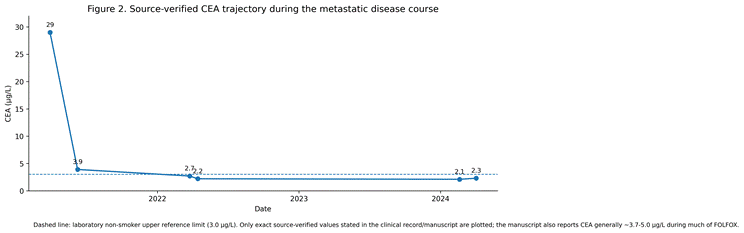
CEA was markedly elevated at the time hepatic metastatic disease was identified: 29.0 µg/L on 30 March 2021 (Figure 2). CEA was markedly elevated at the time hepatic metastatic disease was identified: 29.0 µg/L on 30 March 2021. It fell to 3.9 µg/L by 9 June 2021 and remained in the approximately 3.7-5.0 µg/L range during much of FOLFOX. Following completion of chemotherapy, CEA subsequently fell into the laboratory reference range.

Importantly, CEA did not show a comparable marked rise around later pulmonary metastatic events. A CEA value of approximately 2.7 µg/L was documented immediately before the March 2022 pulmonary metastasectomy and was 2.2 µg/L in April 2022. During later surveillance, CEA remained around the low 2 µg/L range, including 2.1 µg/L on 19 February 2024 and 2.3 µg/L on 2 April 2024, before the right-upper-lobe metastatic nodule was identified in June 2024. Thus, in this patient, CEA was strongly informative at hepatic recurrence but was not a sensitive marker for all subsequent pulmonary metastatic events.

2.9 Latest surveillance and current status

CT scan on 7 April 2025 demonstrated stable postoperative appearances with no new metastatic disease identified. CT scan on 9 February 2026 showed stable postsurgical changes in the right upper and left lower lobes, no concerning pulmonary nodule or mass, no focal abnormality in the hypertrophied remnant liver, no enlarged thoracic or abdominopelvic nodes and no solid peritoneal or omental disease.

The latest CT scan of the chest, abdomen and pelvis was performed on 4 August 2026. It demonstrated stable postsurgical changes in the right upper and left lower lobes, no new concerning pulmonary nodule or mass, no focal abnormality in the remnant liver, no enlarged thoracic or abdominopelvic nodes, no ascites or solid peritoneal/omental disease and no new pelvic mass. The radiological impression was: no evidence of disease recurrence or new sites of metastatic disease.

At manuscript preparation in August 2026, approximately 24 months after the most recent pulmonary metastasectomy, the patient remained clinically well with ECOG Performance Status 0, had received no further systemic anticancer therapy after the 2021 FOLFOX course and continued Nuvastatic™ 1,000 mg twice daily.


Table 3. Comparison of the Present Case with Published Real-World/Observational Outcomes

Published benchmark

Reported outcome

Relevance to this patient

Interpretation

Second metastasectomy for recurrent mCRC (2024; n=94)

5-year DFS 42.8%; 5-year OS 67.2%; R0 in 94.7%

Patient underwent repeated complete resection of liver/lung metastases

Durable survival is achievable after repeat metastasectomy in selected patients.

Pulmonary metastasectomy for CRC (2024; n=156)

5-year OS 67%; 10-year OS 59%; no recurrence beyond 38 months in this cohort

Approximately 24 months from Aug 2024 pulmonary metastasectomy to Aug 2026 CT

Current disease control is favourable but has not yet crossed the ~3-year RFS plateau reported in this cohort.

RAS status after curative liver metastasectomy (2024 meta-analysis)

RAS mutation: OS HR 1.68; RFS HR 1.46

Tumour harboured KRAS G12V

Provides adverse prognostic molecular context; not an individual prediction.

KRAS status after pulmonary metastasectomy (meta-analysis)

KRAS mutation: OS HR 1.86; RFS HR 1.68

KRAS G12V with recurrent pulmonary metastatic disease

Supports cautious interpretation of the favourable current outcome.


Figure 1. Integrated Clinical Timeline. Integrated clinical timeline showing primary colorectal cancer, hepatic metastatic recurrence, the presence and later progression of pulmonary metastases, repeated metastasectomy, conventional systemic therapy and patient-reported Nuvastatic™ exposure. The 2024 right-upper-lobe recurrence is shown explicitly. The molecular profile is KRAS G12V, APC E1309*, TP53 P36fs*7, microsatellite stable (MSS), with tumour mutational burden 1 mutation/Mb. Approximate Nuvastatic™ dose-change timing is patient-reported; the temporal association with pulmonary progression is hypothesis-generating and does not establish causality.

Figure 2. Source-Verified CEA Trajectory. Source-verified CEA measurements. CEA was 29.0 µg/L at hepatic metastatic recurrence, fell after hepatic resection and systemic therapy, and remained low around later pulmonary metastatic events. The dashed line denotes the laboratory non-smoker upper reference limit of 3.0 µg/L. Only exact values supported by the available clinical record are plotted.

3. Discussion

3.1 Aggressive Metastatic Phenotype Despite an Initially Early-Stage Primary

The biological behaviour of this tumour was notably discordant with the apparently favourable initial pathological stage. The primary sigmoid adenocarcinoma was pT2N0, yet within only a few months the patient developed a large right-lobe hepatic metastasis, while small pulmonary nodules were already radiologically detectable. These pulmonary lesions subsequently enlarged and were histologically confirmed as metastatic colorectal adenocarcinoma, with a further right-upper-lobe metastasis resected in 2024. This sequence supports characterization of the disease as demonstrating aggressive and early metastatic behaviour rather than simply an indolent oligometastatic course. The KRAS G12V genotype provides additional adverse molecular context. Against this background, the ability to repeatedly achieve complete local treatment and the patient's continued ECOG 0 status with no radiological recurrence or new metastatic disease on the August 2026 CT are clinically notable observations.

3.2 Comparison With Published Real-World Outcomes

The patient's course should be interpreted against outcomes reported for highly selected patients with resectable oligometastatic colorectal cancer rather than against unselected stage IV colorectal cancer populations. Key published benchmarks are compared with the present case in Table 3. In a 2024 retrospective series of 94 patients undergoing a second metastasectomy for recurrent metastatic colorectal cancer, 94.7% achieved R0 resection and the 5-year disease-free survival and overall survival rates were 42.8% and 67.2%, respectively. Long-term outcomes were more favourable when recurrent disease remained confined to the lung and/or liver and when the disease-free interval after the first metastasectomy was at least 12 months (Choi et al., 2024). These data demonstrate that durable survival after repeated metastasectomy is achievable with conventional multidisciplinary management in appropriately selected patients.

Pulmonary metastasectomy series provide a second relevant benchmark. In a 2024 cohort of 156 patients with controlled primary colorectal cancer and metastases confined to the lungs or liver who underwent pulmonary metastasectomy, 5- and 10-year overall survival rates were 67% and 59%, respectively. No further recurrence was observed beyond 38 months after pulmonary surgery in that cohort, and the recurrence-free survival curve plateaued at approximately three years (Yoshida et al., 2024). The present patient is approximately two years beyond the most recent pulmonary metastasectomy performed in August 2024; therefore, her current recurrence-free interval is favourable but has not yet exceeded this approximately three-year landmark.

Molecular features also provide important context. A 2024 systematic review and meta-analysis of patients undergoing curative liver metastasectomy found RAS mutation to be associated with worse overall survival (HR 1.68, 95% CI 1.54-1.84) and recurrence-free survival (HR 1.46, 95% CI 1.33-1.61) compared with RAS-wild-type disease (Roncato et al., 2024). A separate meta-analysis of pulmonary metastasectomy similarly associated KRAS mutation with worse overall survival (HR 1.86, 95% CI 1.35-2.57) and recurrence-free survival (HR 1.68, 95% CI 1.38-2.04) (Huang et al., 2021). Thus, the patient's KRAS G12V status provides an adverse prognostic context, although these population-level associations cannot be used to predict an individual patient's counterfactual outcome.

Taken together, the real-world literature shows that prolonged survival after repeated complete metastasectomy is achievable in selected patients and therefore provides an essential conventional-oncology explanation for this patient's outcome. At the same time, historical cohorts cannot determine whether a concurrent intervention altered the tempo, vascularization, anatomical distribution or resectability of metastatic disease in an individual patient. The present case is therefore not an efficacy comparison against historical controls. Its additional value lies in the unusually detailed longitudinal documentation of standardized Nuvastatic™ exposure in a tumour that initially behaved aggressively, including the dose-reduction/progression sequence, prolonged periods during which metastatic disease remained anatomically limited and amenable to local treatment, long-term tolerability, molecular characterization and extended follow-up. These features provide a clinically grounded hypothesis for prospective evaluation rather than evidence for either the presence or absence of a Nuvastatic™ antitumour contribution.

3.3 Clinical significance of the longitudinal course

This case documents an extended clinical course of KRAS G12V-mutated, MSS metastatic CRC treated with repeated curative-intent local therapy. The patient developed a large hepatic metastasis shortly after resection of an apparently favourable pT2N0 primary tumour, subsequently underwent resection of left-lower-lobe pulmonary metastases in March 2022 and later experienced a further histologically confirmed 7-mm right-upper-lobe pulmonary metastasis that was resected in August 2024. The latest CT in August 2026 showed no evidence of recurrent or new metastatic disease. The appropriate oncological endpoint is therefore approximately two years of radiologically documented recurrence-free follow-up after the most recent pulmonary metastasectomy, rather than uninterrupted disease-free survival since 2022.

3.4 Pulmonary metastatic disease predated Nuvastatic™ initiation

A key chronological point is that tiny pulmonary nodules were already radiologically visible when the hepatic metastasis was diagnosed and before Nuvastatic™ was initiated. A new 3-mm left-lower-lobe nodule was below PET scan resolution, and a small right-upper-lobe nodule also remained stable during early treatment. Later pathological confirmation and the 2024 observation that the right-upper-lobe lesion had been visible from the beginning support the interpretation that pulmonary dissemination had already occurred at Nuvastatic™ initiation. This chronology means that Nuvastatic™ cannot be said to have prevented the initial metastatic seeding. It does not, however, address the different question of whether subsequent long-term exposure could have modified the growth kinetics, angiogenic support or clinical expression of already established micrometastatic disease.

3.5 Repeated metastasectomy and disease control

Repeated resection of technically resectable colorectal liver and pulmonary metastases is an established component of multidisciplinary management in selected patients. The favourable outcome in this patient can therefore be explained by complete surgical clearance of identifiable disease, systemic FOLFOX chemotherapy, individual tumour biology and host factors. The 2024 recurrence demonstrates that long-term disease control was not continuous and reinforces the need to avoid causal attribution to the adjunctive botanical intervention.

3.6 KRAS G12V and molecular context

The hepatic metastasis harboured KRAS G12V together with APC and TP53 alterations, was microsatellite stable and had a tumour mutational burden of 1 mutation/Mb. KRAS mutation has direct therapeutic relevance because it predicts lack of benefit from anti-EGFR monoclonal antibodies. MSS/low-TMB status also does not provide the biomarker profile typically associated with robust checkpoint-inhibitor sensitivity. These molecular findings add context to the case but do not determine the individual prognosis.

3.7 CEA behaviour and implications for surveillance

The source-verified CEA trajectory is clinically informative. CEA rose markedly to 29 µg/L when the large hepatic metastasis was detected, then declined following surgery and systemic treatment. In contrast, later pulmonary metastatic events occurred while CEA was near or within the reference range, including approximately 2.7 µg/L before the March 2022 pulmonary metastasectomy and 2.1-2.3 µg/L in early 2024 before recognition of the right-upper-lobe recurrence. This illustrates the limitation of relying on CEA alone for surveillance in an individual patient and supports continued radiological follow-up even when tumour-marker values are reassuring.

3.8 Interpretation of long-term Nuvastatic™ exposure

The patient had long-term Nuvastatic™ exposure over approximately five years, beginning around one month before hepatic metastasectomy and spanning the FOLFOX treatment period, recurrent pulmonary metastatic events, repeated pulmonary metastasectomy and subsequent surveillance. Exposure was not literally uninterrupted: the patient reports taking Nuvastatic™ between chemotherapy cycles and temporarily withholding it immediately before each surgical procedure, with resumption after returning home. A particularly relevant temporal observation was that radiologically documented enlargement of the pre-existing pulmonary lesions became apparent approximately three months after the patient-reported reduction of Nuvastatic™ from 4,000 to 2,000 mg/day. The patient returned to 4,000 mg/day immediately after progression was detected. This sequence cannot by itself prove a dose-response relationship, but it should not be interpreted as evidence against biological activity: the clinically observed progression occurred after exposure had been reduced by half, rather than during uninterrupted high-dose treatment. FOLFOX cessation, pre-existing metastatic disease and tumour biology remain important alternative explanations. A further 7-mm right-upper-lobe metastasis was resected in 2024 during continued Nuvastatic™ treatment, demonstrating that Nuvastatic™, if biologically active, did not completely suppress residual metastatic growth. A partial effect on growth rate, angiogenesis or metastatic tempo, however, would not be expected necessarily to eliminate all recurrence. The overall dose-exposure chronology therefore remains compatible with several explanations and merits prospective pharmacokinetic and dose-ranging investigation.

3.9 Phytochemical and translational rationale

Nuvastatic™ is a standardized polymolecular O. stamineus extract rather than purified rosmarinic acid. Its characterized phytochemical profile includes rosmarinic acid, sinensetin, eupatorin and TMF. Experimental findings involving angiogenesis, oxidative stress, inflammation, apoptosis and tumour-cell signalling provide mechanistic hypotheses for prospective study, but none of these pathways was directly measured in this patient. The published randomized Phase II NCT04546607 study supports feasibility and supportive-care activity during chemotherapy, particularly for cancer-related fatigue, quality of life and oxidative stress biomarkers; it does not establish direct antitumour efficacy. (Ahamed et al., 2012; Akowuah et al., 2004; Hashim et al., 2016; Laavola et al., 2012; Yehya et al., 2019, 2021)

3.10 Could Nuvastatic™ have contributed to post-metastasectomy disease control?

A central hypothesis generated by this case is whether long-term exposure to standardized O. stamineus contributed to modulation of the metastatic microenvironment and thereby influenced the tempo or clinically apparent pattern of metastatic progression. This hypothesis is biologically plausible because standardized O. stamineus preparations have demonstrated anti-angiogenic activity in experimental systems, including effects involving VEGF/VEGFR-2-related signalling, alongside antioxidant, anti-inflammatory and other tumour-relevant biological activities. In principle, suppression of angiogenic support to microscopic metastatic deposits could delay their expansion into clinically significant lesions and could help maintain a lower-volume, anatomically restricted pattern of disease that remains amenable to metastasectomy. This mechanism was not measured directly in this patient, but it provides a testable explanation for the observed course rather than merely a post hoc efficacy claim.

Several clinical observations are consistent with, although do not prove, such a contribution. First, the tumour showed rapid metastatic behaviour early in its course, yet subsequent clinically apparent disease remained confined to surgically manageable hepatic and pulmonary sites over prolonged follow-up. Second, enlargement of pre-existing pulmonary lesions was recognized approximately three months after Nuvastatic™ exposure was reduced by 50%, with the higher dose restored immediately after progression was identified. Third, after repeated local clearance of macroscopic disease, the patient remained clinically well without further systemic anticancer therapy after FOLFOX and continued long-term Nuvastatic™, with the latest August 2026 CT scan showing no recurrence or new metastatic sites. Conversely, recurrence in 2024 while receiving Nuvastatic™ indicates that any possible effect was incomplete rather than absolute. The balanced interpretation is therefore that conventional surgery and chemotherapy were indispensable components of disease control, while a contributory effect of Nuvastatic™ on metastatic tempo or biology remains plausible and cannot be excluded by this case. Prospective studies incorporating circulating tumour DNA, angiogenic biomarkers, immune/inflammatory profiling, pharmacokinetics and predefined dose-exposure analyses would be required to test this hypothesis.

3.11 Safety and tolerability

The patient completed approximately six months of FOLFOX while taking Nuvastatic™ and continued the standardized extract for approximately five years. No serious adverse event was documented as attributable to Nuvastatic™ in the available clinical record, and the patient remained ECOG 0 at latest follow-up. This supports individual tolerability only and cannot establish population-level safety or exclude uncommon interactions or toxicities.

3.12 Limitations

This report has the inherent limitations of a single uncontrolled case and cannot quantify the independent contribution of any component of multimodal treatment. Repeated complete metastasectomy and prior systemic chemotherapy are established major determinants of long-term survival, while a possible adjunctive contribution from Nuvastatic™ cannot be separated from these treatments or from host and tumour factors. Nuvastatic™ dosing and adherence were reconstructed from patient-reported treatment history rather than prospective drug accountability, and the approximate three-month interval between dose reduction and pulmonary enlargement remains an observational temporal association. Pharmacokinetic sampling, circulating tumour DNA, serial angiogenic or immune/inflammatory biomarkers and direct measures of botanical constituent exposure were not obtained. CEA was not a sensitive marker for all metastatic events. These limitations prevent causal attribution in either direction: the case cannot prove that Nuvastatic™ altered metastatic behaviour, but it also cannot demonstrate that the prolonged adjunctive exposure was biologically irrelevant. The report additionally involves investigators associated with the development of Nuvastatic™, requiring full and transparent conflict-of-interest disclosure and independent editorial handling.

3.13 Future research

Prospective trials should clearly separate supportive-care endpoints from antitumour endpoints and should incorporate objective response, progression-free survival, recurrence-free survival and overall survival. In the post-metastasectomy setting, serial circulating tumour DNA could provide a sensitive measure of minimal residual disease. Pharmacokinetic characterization of rosmarinic acid, eupatorin, sinensetin, TMF and relevant metabolites, combined with angiogenic, inflammatory and oxidative-stress biomarkers, could help determine whether biological activity correlates with clinical outcomes.

4. Conclusion

This case describes a patient with initially pT2N0 sigmoid adenocarcinoma whose subsequent course demonstrated aggressive metastatic behaviour, with a large hepatic metastasis developing within months and radiologically detectable pulmonary nodules already present when hepatic metastatic disease was recognized. Histologically confirmed pulmonary metastases subsequently required repeated metastasectomy. Nuvastatic™ was initiated at 4,000 mg/day when the hepatic metastasis was detected, approximately one month before right hepatectomy, and long-term exposure continued across chemotherapy, metastatic progression, surgery and surveillance, with patient-reported timing between chemotherapy cycles and brief perioperative interruptions before surgery followed by resumption after returning home. According to the patient-reported treatment history, the dose was reduced by 50% to 2,000 mg/day approximately three months before enlargement of the pre-existing pulmonary lesions was detected in March 2022 and was immediately restored to 4,000 mg/day after progression was identified. The dose was again reduced to 2,000 mg/day approximately one year before manuscript preparation.

The identification and resection of a further 7-mm right-upper-lobe colorectal metastasis in August 2024 is an important component of the clinical course and precludes characterization of the period from 2022 as continuously disease free. After the most recent pulmonary metastasectomy, the patient received no further systemic anticancer therapy and remained without radiological evidence of recurrence or new metastatic disease on CT scan dated 4 August 2026, while clinically maintaining ECOG Performance Status 0.

The most balanced interpretation is that repeated complete metastasectomy and FOLFOX provided established and indispensable oncological treatment, while a contributory effect from long-term Nuvastatic™ exposure remains biologically plausible but unproven. The occurrence of metastatic recurrence does not by itself negate a possible partial effect on tumour growth kinetics, angiogenesis or metastatic tempo; importantly, the first clear pulmonary enlargement followed a 50% reduction in Nuvastatic™ exposure, and the later 2024 recurrence was limited and surgically resectable. Preclinical anti-angiogenic and related biological activities of standardized O. stamineus provide a mechanistic rationale for the hypothesis that Nuvastatic™ may have helped modulate residual micrometastatic disease or its progression, potentially contributing to a pattern that remained manageable with local therapy. A single case cannot establish this effect or determine its magnitude, but the longitudinal clinical and dose-exposure observations justify prospective evaluation of Nuvastatic™ using pharmacokinetic, angiogenic, immune/inflammatory, circulating tumour DNA and oncological endpoints.

Authors Contribution

Amin Malik Shah Abdul Majid: patient monitoring; conceptualization; clinical interpretation; manuscript development; writing—original draft; writing—review and editing; supervision. Adam Shah Abdul Majid: clinical data curation; clinical interpretation; writing—review and editing. Muhammad Asyraf Abduraman: literature review; data curation; manuscript development; writing—review and editing. All authors reviewed and approved the final manuscript and accept accountability for their contributions.

Conflict of Interest

Amin Malik Shah Abdul Majid is involved in the research and development of Nuvastatic™ and has professional and financial interests related to its development and commercialization through associated entities. This represents a potential conflict of interest. Adam Shah Abdul Majid and Muhammad Asyraf Abduraman declare no conflicts of interest relevant to this case study. Amin Malik Shah Abdul Majid also serves as Editor-in-Chief of Integrative Biomedical Research; this editorial relationship is addressed separately in the Editorial Independence Statement.


Declaration

Informed Consent

Written informed consent for publication of the patient's de-identified clinical information, accompanying clinical material, and patient perspective was obtained from the patient. The patient reviewed and approved the wording of the Patient Perspective.

Ethics Statement

Formal ethical approval was not required for this case report in accordance with institutional requirements, as the report describes the clinical course of a single patient and did not involve any research-specific intervention or alteration to the patient's clinical management. Written informed consent was obtained from the patient for the use and publication of the relevant clinical information. All identifying information has been removed to maintain patient confidentiality.

Generative AI statement

The authors declare that Gen AI was used in the creation of this manuscript. Generative AI was used in the preparation of this manuscript in a limited and strictly controlled manner. Specifically, it assisted during the initial organization of thematic sections and helped identify general areas of literature for further manual exploration. All writing, analysis, interpretation, and synthesis of content were performed by the authors. All references in the current version have been manually reviewed and verified. The final manuscript has been completely revised to ensure originality, accuracy, and integrity, fully aligning with COPE policies on the responsible use of generative AI.

Editorial Independence Statement

Amin Malik Shah Abdul Majid is the Editor-in-Chief of Integrative Biomedical Research. Because he is also an author of this manuscript, he will be fully recused from all editorial handling and decision-making relating to the submission. The manuscript will be assigned to an independent editor with no involvement in the study and no competing interest related to the subject matter. The Editor-in-Chief will have no involvement in the initial editorial assessment, selection of peer reviewers, peer-review process, assessment of revisions, or the decision to accept or reject the manuscript. Independent external peer review will be used, and the final editorial decision will be made independently of the Editor-in-Chief.

Funding

No specific funding was received for preparation of this case study.

Data Availability

The clinical data supporting this case report are contained within the patient's medical record. Additional de-identified information may be available from the corresponding author subject to patient consent, privacy requirements and applicable institutional policies.

References


Ahamed, M. B. K., Aisha, A. F. A., Nassar, Z. D., Siddiqui, J. M., Ismail, Z., Omari, S. M. S., Parish, C. R., & Abdul Majid, A. M. S. (2012). Cat's whiskers tea (Orthosiphon stamineus) extract inhibits growth of colon tumor in nude mice and angiogenesis in endothelial cells via suppressing VEGFR phosphorylation. Nutrition and Cancer, 64(1), 89–99. https://doi.org/10.1080/01635581.2012.630160

Akowuah, G. A., Zhari, I., Norhayati, I., Sadikun, A., & Khamsah, S. M. (2004). Sinensetin, eupatorin, 3′-hydroxy-5,6,7,4′-tetramethoxyflavone and rosmarinic acid contents and antioxidative effect of Orthosiphon stamineus from Malaysia. Food Chemistry, 87(4), 559–566. https://doi.org/10.1016/j.foodchem.2004.01.008

Choi, S., Kang, M., Kim, J.-W., Kim, J. W., Jeon, J. H., Oh, H.-K., Lee, H. W., Cho, J. Y., Kim, D.-W., Cho, S., Kim, J. H., Kim, K., Kang, S.-B., Jheon, S., & Lee, K.-W. (2024). Long-term clinical outcomes after the second metastasectomy in patients with resected metastatic colorectal cancer. Current Problems in Cancer, 53, 101151. https://doi.org/10.1016/j.currproblcancer.2024.101151

Hashim, S., Beh, H. K., Hamil, M. S. R., Ismail, Z., & Abdul Majid, A. M. S. (2016). High-performance thin-layer chromatography method development, validation, and simultaneous quantification of four compounds identified in standardized extracts of Orthosiphon stamineus. Pharmacognosy Research, 8(4), 238–243. https://doi.org/10.4103/0974-8490.188872

Huang, J., Zang, Q., Wen, Y., Pan, Z., Yao, Z., Huang, M., Huang, J., Chen, J., & Wang, R. (2021). Prognostic value of KRAS mutation in patients undergoing pulmonary metastasectomy for colorectal cancer: A systematic review and meta-analysis. Critical Reviews in Oncology/Hematology, 160, 103308. https://doi.org/10.1016/j.critrevonc.2021.103308

Kung, H.-C., Shubert, C., Wilbur, C., Burns, W., Burkhart, R., Hidalgo, M., Azad, N. S., Lee, V., Chung, H., Le, D. T., Laheru, D., He, J., Zheng, L., Jaffee, E. M., Lafaro, K., Tsai, H.-L., & Christenson, E. S. (2024). Patterns of recurrence after curative intent hepatic resection for colorectal liver metastasis. Journal of Gastrointestinal Surgery, 28(12), 2031–2038. https://doi.org/10.1016/j.gassur.2024.09.026

Laavola, M., Nieminen, R., Yam, M. F., Sadikun, A., Asmawi, M. Z., Basir, R., Welling, J., Vapaatalo, H., Korhonen, R., & Moilanen, E. (2012). Flavonoids eupatorin and sinensetin present in Orthosiphon stamineus leaves inhibit inflammatory gene expression and STAT1 activation. Planta Medica, 78(8), 779–786. https://doi.org/10.1055/s-0031-1298458

Martin-Cullell, B., Piedra, A., Virgili, A. C., Fumagalli, C., Cerdà, P., Mirallas, O., Sanz, J., Restrepo, S., Trujillo, J. C., Martínez, E., Belda-Sanchís, J., Szafranska, J., & Páez, D. (2026). Immune phenotype and RAS/BRAF status predict outcomes after lung metastasectomy for colorectal cancer. Clinical and Translational Oncology. Advance online publication. https://doi.org/10.1007/s12094-026-04344-2

Morris, V. K., Kennedy, E. B., Baxter, N. N., Benson, A. B., III, Cercek, A., Cho, M., Ciombor, K. K., Cremolini, C., Davis, A., Deming, D. A., Fakih, M. G., Gholami, S., Hong, T. S., Jaiyesimi, I., Klute, K., Lieu, C., Sanoff, H., Strickler, J. H., White, S., ... Eng, C. (2023). Treatment of metastatic colorectal cancer: ASCO guideline. Journal of Clinical Oncology, 41(3), 678–700. https://doi.org/10.1200/JCO.22.01690

Ng, M. L., Abdul Majid, A. M. S., Yee, S. M., Natesan, V., Basheer, M. K. A., Gnanasekaran, A., Al-Suede, F. S. R., Parish, C., Dalal, M., Ming, L. C., Nazari, M. V., Khan, S. S., Sultan, S. B. S. H., Babu, K. G., Abdul Majid, A. S., & Abdul Aziz, M. A. S. (2024). A phase II randomized, double-blind, placebo-controlled study of Nuvastatic™ (C5OSEW5050ESA), a standardized rosmarinic acid-rich polymolecular botanical extract formulation to reduce cancer-related fatigue in patients with solid tumors. Supportive Care in Cancer, 32(6), 331. https://doi.org/10.1007/s00520-024-08536-w

Rhaiem, R., Duramé, A., Primavesi, F., Dorcaratto, D., Syn, N., de la Hoz Rodríguez, Á., Dupré, A., Piardi, T., Blanco Fernández, G., Prada Villaverde, A., Rodríguez Sanjuán, J. C., Fernández Santiago, R., Fernández-Moreno, M.-C., Ferret, G., López Ben, S., Suárez Muñoz, M. Á., Perez-Alonso, A. J., Koh, Y.-X., Jones, R., ... Di Martino, M. (2024). Critical appraisal of surgical margins according to KRAS status in liver resection for colorectal liver metastases: Should surgical strategy be influenced by tumor biology? Surgery, 176(1), 124–133. https://doi.org/10.1016/j.surg.2024.02.008

Roncato, R., Polesel, J., Tosi, F., Peruzzi, E., Brugugnoli, E., Lauria Pantano, C., Furfaro, M., Di Girolamo, F., Nani, A., Pani, A., Milan, N., De Mattia, E., Sartore-Bianchi, A., & Cecchin, E. (2024). The challenge of molecular selection in liver-limited metastatic colorectal cancer for surgical resection: A systematic review and meta-analysis in the context of current and future approaches. Oncology Research, 32(9), 1407–1422. https://doi.org/10.32604/or.2024.049181

Siegel, R. L., Wagle, N. S., Cercek, A., Smith, R. A., & Jemal, A. (2023). Colorectal cancer statistics, 2023. CA: A Cancer Journal for Clinicians, 73(3), 233–254. https://doi.org/10.3322/caac.21772

Yehya, A. H. S., Asif, M., Kaur, G., Hassan, L. E. A., Al-Suede, F. S. R., Abdul Majid, A. M. S., & Oon, C. E. (2019). Toxicological studies of Orthosiphon stamineus (Misai Kucing) standardized ethanol extract in combination with gemcitabine in athymic nude mice model. Journal of Advanced Research, 15, 59–68. https://doi.org/10.1016/j.jare.2018.05.006

Yehya, A. H. S., Asif, M., Abdul Majid, A. M. S., & Oon, C. E. (2021). Complementary effects of Orthosiphon stamineus standardized ethanolic extract and rosmarinic acid in combination with gemcitabine on pancreatic cancer. Biomedical Journal, 44(6), 694–708. https://doi.org/10.1016/j.bj.2020.05.015

Yoshida, C., Chang, S. S., Okamoto, T., & Inada, R. (2024). Recurrence-free survival curve for pulmonary metastasectomy in colorectal cancer plateaus at 3 years. Updates in Surgery, 76(8), 2795–2803. https://doi.org/10.1007/s13304-024-01874-8


Article metrics
View details
0
Downloads
0
Citations
113
Views
📖 Cite article

View Dimensions


View Plumx


View Altmetric


0
Save
0
Citation
113
View
0
Share