Integrative Biomedical Research

Integrative Biomedical Research (Journal of Angiotherapy) | Online ISSN  3068-6326
463
Citations
1.8m
Views
770
Articles
Your new experience awaits. Try the new design now and help us make it even better
Switch to the new experience
Figures and Tables
REVIEWS   (Open Access)

Genomic Cross-Talk, Broken Bridges Mitochondrial–Nuclear Signaling Failure as a Unifying Mechanism and Druggable Frontier in Parkinson's Disease

Osama Abdel-Hameed Majeed 1*, Makarim Qassim Al-Lami 1, Gheyath Al-Gawwam 2

+ Author Affiliations

Integrative Biomedical Research 10 (1) 1-8 https://doi.org/10.25163/biomedical.10110926

Submitted: 21 July 2026 Revised: 12 September 2026  Published: 24 September 2026 


Abstract

Parkinson's disease (PD) is, at its core, a failure of conversation — a breakdown in the ceaseless molecular dialogue that the nucleus and the mitochondrion must sustain if a dopaminergic neuron is to survive its own metabolic demands. This review asks, and tries to answer honestly, how that dialogue collapses. Dopaminergic neurons of the substantia nigra pars compacta rely on an uninterrupted, bidirectional exchange between the nuclear and mitochondrial genomes — anterograde signals that sustain organelle biogenesis, and retrograde signals that report back on mitochondrial stress. In PD, this exchange is progressively disrupted, converging on bioenergetic collapse, oxidative injury, and neuroinflammation. We synthesized peer-reviewed mechanistic and preclinical studies (2009–2026) addressing mitochondrial-nuclear crosstalk in PD, drawn from a defined, reproducible literature search of the sources cited throughout this manuscript, spanning molecular, cellular, and systemic (multi-organ) levels of analysis.Four interlocking hubs emerged consistently across the evaluated literature: (i) the PINK1/Parkin–PARIS–PGC-1α/Nrf2 biogenesis axis; (ii) the geranylgeranylation-dependent GBP2/NIX alternative mitophagy pathway; (iii) retrograde apoptotic and innate-immune execution through AIF, cGAS-STING, and PANoptosis; and (iv) systemic amplification via glial-neuronal and multi-organ (gut-, liver-, lung-, heart-, muscle-, and bone-brain) axes. Pharmacological correction of these nodes — with silybin, cannabidiol, gentiopicroside, and the geranylgeranylation inhibitor GGTI298 — consistently restored mitochondrial quality control and attenuated dopaminergic loss in preclinical models. Mitochondrial-nuclear crosstalk is not a peripheral detail of PD biology; it is arguably the organizing principle beneath much of what we call “PD pathology.” Multi-target strategies that simultaneously restore biogenesis, mitophagy, and antioxidant signaling represent a genuinely promising — if still early — route toward disease modification.

Keywords: Parkinson's disease; mitochondrial-nuclear crosstalk; PINK1/Parkin; PGC-1α; Nrf2 signaling; mitophagy; neuroinflammation

1. Introduction

Parkinson's disease rarely announces itself the way textbooks suggest it should. Long before a tremor becomes visible, the cells destined to fail — the dopaminergic neurons of the substantia nigra pars compacta — are already losing an argument they have been having with themselves for years, possibly decades. That argument, as it turns out, is largely conducted between two genomes housed in the same cell. This is, admittedly, a strange way to introduce a neurodegenerative disorder that affects more than 8.5 million people worldwide (Bloem et al., 2021; Ramírez-Carreto et al., 2026), but it is difficult to avoid once one looks closely at what actually happens inside a failing dopaminergic neuron.

Clinically, of course, PD is recognized by what can be seen: resting tremor, bradykinesia, rigidity, postural instability, and — less appreciated by the public, though no less disabling — non-motor features such as cognitive decline, depression, and severe gastrointestinal dysfunction (Bloem et al., 2021; Liu et al., 2026). Pathologically, the disease is defined by the selective, progressive loss of dopaminergic neurons in the substantia nigra pars compacta, alongside the intracellular accumulation of Lewy bodies, those eosinophilic inclusions built largely from misfolded, aggregated α-synuclein (Bloem et al., 2021; Leites & Morais, 2021). These are the facts most reviews open with, and for good reason; they are the visible surface of a much deeper disruption.

Underneath that surface sits mitochondrial dysfunction, which most investigators in the field now regard as a central — arguably the central — driver of dopaminergic neurodegeneration, working hand in hand with oxidative stress (Kamienieva et al., 2023; Leites & Morais, 2021). It is not entirely coincidental that the neurons most vulnerable in PD happen to be among the most metabolically demanding cells in the brain: SNpc dopaminergic neurons carry an unusually high baseline energy requirement and an extensively branched axonal arbor, a combination that leaves comparatively little margin for bioenergetic error (He et al., 2026; Leites & Morais, 2021). Consistent with this vulnerability, mitochondrial respiratory chain deficits — complex I deficiency in particular — have been documented in both sporadic PD and toxin-induced experimental models, an observation that has held up remarkably well since it was first reported nearly four decades ago (Buneeva et al., 2020; Schapira et al., 1989). Once complex I falters, the downstream consequences cascade quickly: calcium homeostasis is disturbed, reactive oxygen species (ROS) accumulate beyond what antioxidant systems can buffer, and neuroinflammatory signaling is set in motion, ultimately steering the cell toward programmed death (He et al., 2026; Kamienieva et al., 2023).

What is easy to overlook — and what this review takes as its starting point — is that mitochondria are not autonomous actors in this drama. They cannot be, given how little of their own genetic material they retain. Mitochondria possess a circular genome (mtDNA) and a rudimentary protein-synthesizing apparatus of their own (Buneeva et al., 2020), yet mtDNA encodes only 13 core polypeptide subunits of the respiratory chain, together with 2 rRNAs and 22 tRNAs. The remaining 99% of mitochondrial proteins, including virtually everything required for mtDNA replication, transcription, and organelle maintenance, are nuclear-encoded, synthesized on cytosolic ribosomes, and imported afterward (Buneeva et al., 2020). This asymmetry is worth sitting with for a moment: it means mitochondrial survival is, by design, contingent on continuous, accurate communication with the nucleus — what has come to be called mitochondrial-nuclear crosstalk (Buneeva et al., 2020; Spinazzola & Zeviani, 2009).

That crosstalk runs in two directions, and both matter. Anterograde signaling, from nucleus to mitochondria, governs organelle biogenesis, dynamics, and respiratory capacity; retrograde signaling, running the other way, reports mitochondrial stress back to the nucleus so that adaptive transcriptional programs can be mounted in response (Buneeva et al., 2020; Spinazzola & Zeviani, 2009). In PD, this equilibrium does not merely wobble — it breaks down in a fairly specific, traceable way. Because mtDNA sits close to the inner mitochondrial membrane, the principal site of ROS generation, and lacks the protective histone packaging afforded to nuclear DNA, it is disproportionately vulnerable to oxidative damage and somatic deletion (Buneeva et al., 2020). Substantia nigra neurons from PD patients show exactly this signature: an accumulation of somatic mtDNA deletions alongside depletion of wild-type mtDNA copy number (Buneeva et al., 2020). Under healthy conditions, such damage would trigger a compensatory retrograde signal, prompting the nucleus to upregulate protective transcriptional programs. In PD, however, genetic mutation or age-related decline frequently paralyzes precisely this adaptive response, trapping the neuron in a self-reinforcing cycle of bioenergetic failure and oxidative stress (Buneeva et al., 2020; Kamienieva et al., 2023).

None of this occurs in isolation, either at the molecular or the systemic level — and that, in essence, is the argument this review is built around. At the molecular scale, nuclear-encoded quality-control systems such as the PINK1/Parkin pathway and the PGC-1α-centered biogenesis network are recurrently disabled in PD, with consequences that cascade through PARIS, Nrf2, and alternative mitophagy receptors such as NIX (Leites & Morais, 2021; Kamienieva et al., 2023; Panicker et al., 2022; Cui et al., 2026). At the cellular scale, unresolved mitochondrial stress does not stay contained within the neuron; it recruits astrocytes and microglia into a pro-inflammatory phenotype, and it extends — more surprisingly, perhaps — to systemic organ-brain axes involving the gut, liver, and lungs (Leites & Morais, 2021; Liu et al., 2026; Huang & Li, 2024). Taken together, these observations reframe PD not as a strictly localized neuronal disorder but as a systemic failure of mitochondrial-nuclear coordination, one that happens to manifest most visibly in the SNpc.

Because these regulatory hubs are, at least in principle, druggable, several pharmacological strategies have begun to target them directly. Silybin appears to modulate convergent endoplasmic reticulum stress pathways, preserving tyrosine hydroxylase levels in dopaminergic neurons (Ramírez-Carreto et al., 2026); cannabidiol rescues Nrf2 from stress-induced misfolding and aggregation (Jurado-Coronel & Duennwald, 2026); gentiopicroside activates the Nrf2/ARE antioxidant axis through TGR5 agonism (Gao et al., 2026); and GGTI298 restores mitophagic flux by blocking the geranylgeranylation-dependent degradation of NIX (Cui et al., 2026). None of these, individually, is likely to be curative — that would be an overstatement the evidence does not yet support — but collectively they suggest that mitochondrial-nuclear crosstalk is not merely descriptive biology; it is a genuine therapeutic frontier.

The purpose of this review, then, is threefold. First, we systematically dissect the molecular pathways and transcription-translation feedback loops underlying mitochondrial-nuclear crosstalk in PD, with particular attention to the PINK1/Parkin, PARIS/PGC-1α/Nrf2, GBP2/NIX, and AIF translocation pathways. Second, we evaluate how glial-neuronal interactions and systemic multi-organ axes — gut-brain, liver-brain, and lung-brain among them — amplify this dysfunction into chronic neuroinflammation. Third, we critically appraise emerging therapeutic candidates that target these interconnected nodes, asking not only whether they work in preclinical models, but what their mechanisms imply about where disease-modifying therapy in PD might realistically be headed.

2. Mitochondrial-Nuclear Crosstalk Failure in Parkinson's Disease: Mechanisms and Therapeutic Targets

This section works through the evidence base thematically rather than chronologically, since the mechanistic story — anterograde biogenesis, alternative mitophagy, redox proteostasis, receptor-mediated rescue, and systemic amplification — makes more sense told as an unfolding logic than as a list of studies. Figures 1–4 accompany the text as visual scaffolding for the more tangled pathway interactions.

2.1 Anterograde Coordination and Biogenesis: The PINK1/Parkin and PARIS/PGC-1α Axis

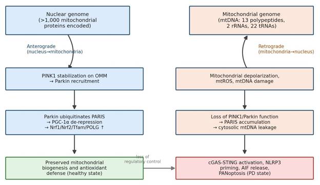
It helps to begin with what a healthy mitochondrion does when it senses trouble, because the failure modes in PD only make sense against that baseline. The nuclear-encoded kinase PINK1 is, under normal circumstances, imported into polarized mitochondria and rapidly cleaved by the presenilin-associated rhomboid-like protease (PARL); the cleaved fragment is then degraded by the ubiquitin-proteasome system, which keeps basal PINK1 levels deliberately low (Leites & Morais, 2021). The moment a mitochondrion depolarizes, however, that cleavage is blocked, full-length PINK1 accumulates on the outer mitochondrial membrane, and it phosphorylates both OMM substrates and ubiquitin to recruit Parkin — initiating selective mitophagy (Leites & Morais, 2021; Kamienieva et al., 2023), (Figure 1).

What is less widely appreciated, and arguably more consequential for PD, is that this same PINK1/Parkin axis reaches into the nucleus to control biogenesis, not merely clearance. Under basal conditions, Parkin ubiquitinates the transcriptional repressor PARIS (ZNF746), targeting it for proteasomal degradation (Panicker et al., 2022). When Parkin or PINK1 function is lost — through mutation, age, or chronic stress — PARIS is no longer cleared; it accumulates in the cytoplasm, translocates to the nucleus, and binds directly to the PGC-1α promoter, repressing its transcription (García-Yagüe et al., 2026; Shin et al., 2011; Figure 2).
PGC-1α is not a minor player here; it is the master coactivator of mitochondrial biogenesis, and its suppression drags down an entire downstream network —

Figure 1. Bidirectional mitochondrial-nuclear signaling under physiological versus Parkinson's-disease conditions. The left branch traces anterograde, nucleus-driven maintenance of mitochondrial biogenesis and antioxidant defense under healthy conditions; the right branch traces the retrograde consequences of mitochondrial depolarization, mtROS, and mtDNA damage when nuclear compensatory signaling fails in PD, culminating in cGAS-STING activation, NLRP3 priming, AIF release, and PANoptosis. Figure constructed from mechanistic evidence in Buneeva et al. (2020), Leites and Morais (2021), and He et al. (2026).
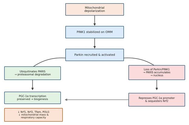
Figure 2. The PINK1/Parkin–PARIS–PGC-1α/Nrf2 axis governing nuclear-encoded mitochondrial biogenesis. Green boxes trace the physiological pathway, in which Parkin-mediated PARIS degradation preserves PGC-1α-driven transcription of Nrf1, Nrf2, Tfam, and POLG; red/orange boxes trace the pathological pathway activated when Parkin or PINK1 function is lost, in which nuclear PARIS accumulation represses PGC-1α and sequesters Nrf2, reducing mitochondrial mass and respiratory capacity. Figure constructed from Kamienieva et al. (2023), Shin et al. (2011), and García-Yagüe et al. (2026).

Nrf1, Nrf2, Tfam, and POLG among them (García-Yagüe et al., 2026; Kamienieva et al., 2023). Tfam in particular is essential for packaging, transcribing, and replicating mtDNA itself, so its depletion compromises the mitochondrial genome at the source, not merely its downstream products (García-Yagüe et al., 2026; Kamienieva et al., 2023). This is not a purely theoretical cascade: primary human fibroblasts carrying parkin mutations display smaller, less complex mitochondrial networks and consistent downregulation of PGC-1α, Nrf1, Nrf2, Tfam, and POLG together (Kamienieva et al., 2023) — which is about as close to a coherent molecular signature as this field currently has. Notably, accumulated PARIS also interferes with Nrf2 nuclear translocation independently of its effect on PGC-1α (García-Yagüe et al., 2026), meaning a single upstream lesion simultaneously cripples biogenesis and antioxidant defense — a dual insult rather than a single hit.

2.2 Non-Canonical Quality Control: The Geranylgeranylation-Dependent GBP2/NIX Axis

PINK1/Parkin is the pathway most textbooks emphasize, but it is not the only route to mitophagy, and this matters enormously when PINK1/Parkin itself is compromised. NIX (BNIP3L), an outer mitochondrial membrane protein, serves as an alternative mitophagy receptor capable of recruiting autophagic machinery independently (Cui et al., 2026). One might expect this redundancy to offer some protection in PD — and perhaps it once did — but recent work suggests this backup system is itself vulnerable to a specific, druggable point of failure.

That vulnerability centers on Guanylate-binding protein 2 (GBP2), a member of the dynamin superfamily of GTPases whose role in neurodegeneration has only recently come into focus (Cui et al., 2026). Under PD-relevant stress — MPP+ exposure or A53T α-synuclein expression, for instance — GBP2 is markedly upregulated in the substantia nigra of both transgenic mice and human patients, and its expression tracks inversely with tyrosine hydroxylase levels (Cui et al., 2026). Mechanistically, stress promotes geranylgeranylation of GBP2 at its C-terminal CAAX motif, increasing its hydrophobicity and driving accumulation on the mitochondrial membrane, where its large GTPase domain docks directly onto NIX (Cui et al., 2026). The consequence of that binding is not cooperative; it is destructive — GBP2 recruits E3 ligase machinery that hyper-ubiquitinates NIX and sends it for proteasomal degradation, halting mitophagic flux and trapping the cell with damaged, ROS-generating mitochondria it cannot clear (Cui et al., 2026; Figure 4).

The encouraging part of this story, and perhaps the reason it merits particular attention, is how cleanly reversible it appears in preclinical models. Genetic knockdown of GBP2, or pharmacological blockade of its geranylgeranylation with GGTI298, prevents its mitochondrial accumulation, restores NIX stability, reactivates mitophagy, and improves motor performance in MPTP-treated mice (Cui et al., 2026). Few nodes in this pathway offer such a tight mechanistic link between molecular correction and behavioral rescue, which is presumably why the GBP2/NIX axis is increasingly discussed as a genuinely tractable drug target.

2.3 Intrinsically Disordered Proteostasis: Nrf2 Misfolding and Cannabidiol Rescue

The cell's principal defense against the ROS bursts generated during dopaminergic degeneration is orchestrated by Nrf2, the master antioxidant transcription factor (Jurado-Coronel & Duennwald, 2026; García-Yagüe et al., 2026). Under basal conditions, Keap1 keeps Nrf2 levels deliberately low through constant ubiquitination; oxidative or electrophilic stress oxidizes reactive cysteines on Keap1, disrupting its hinge-and-latch mechanism and allowing Nrf2 to escape degradation, enter the nucleus, and drive transcription of cytoprotective genes such as HMOX1 and NQO1 (Jurado-Coronel & Duennwald, 2026; García-Yagüe et al., 2026).

In PD, though, this response is frequently paralyzed — and the reason turns out to be structural rather than purely regulatory. Nrf2 is largely an intrinsically disordered protein, lacking a fixed three-dimensional conformation under physiological conditions (Jurado-Coronel & Duennwald, 2026). That flexibility is normally an asset, letting Nrf2 interact promiscuously with transcriptional coactivators, but under the sustained oxidative assault characteristic of dopaminergic degeneration, it becomes a liability: overwhelming ROS and reactive quinones attack Nrf2's regulatory cysteines directly, driving the protein into insoluble cytoplasmic aggregates (Jurado-Coronel & Duennwald, 2026). The protein can still be phosphorylated at Serine 40, but it is physically stuck — unable to translocate despite receiving the correct activating signal. This is a subtle but important distinction, because it explains why conventional electrophilic Nrf2 activators such as sulforaphane, which act solely by modifying Keap1, are ineffective once Nrf2 itself has already misfolded (Jurado-Coronel & Duennwald, 2026).

Cannabidiol appears to address this proteostatic failure at its source rather than downstream of it. CBD's resorcinol moiety, bearing two phenolic hydroxyl groups, confers potent hydrogen-atom transfer and radical-scavenging capacity, allowing it to intercept reactive quinones and ROS before they can misfold Nrf2 (Jurado-Coronel & Duennwald, 2026). By preserving Nrf2 solubility, CBD permits normal Ser40 phosphorylation and nuclear translocation, restoring HMOX1 and NQO1 transcription, reducing global ROS, halting mitochondrial fragmentation, and normalizing mitophagic flux in both undifferentiated and mature dopaminergic neurons (Jurado-Coronel & Duennwald, 2026).

2.4 G-Protein Coupled Receptor Targets: Gentiopicroside, TGR5, and the cAMP/PKA/CREB Nexus

If covalent Keap1 modification carries off-target risk, an obvious alternative is to activate Nrf2 through a receptor-mediated route instead — and this is precisely the logic behind interest in TGR5, a membrane-bound bile acid receptor expressed on neurons, astrocytes, and microglia throughout the CNS (Gao et al., 2026). Gentiopicroside (GPS), a secoiridoid isolated from Gentiana manshurica Kitagawa, has emerged as a brain-permeable, non-bile-acid-like TGR5 agonist; molecular docking and dynamics simulations indicate that it stably occupies the bile acid-binding pocket of TGR5, behaving as a partial agonist with favorable safety and blood-brain barrier penetration (Gao et al., 2026).

Binding to TGR5 triggers a fairly classical Gs-mediated cascade: intracellular cAMP accumulates, PKA is activated, and PKA phosphorylates CREB at Serine 133 (Gao et al., 2026). Phosphorylated CREB then enters the nucleus and interacts directly with Nrf2, synergistically enhancing transcription of ARE-dependent genes (Gao et al., 2026). In practice, this TGR5/cAMP/PKA/CREB/Nrf2 cascade prevents Nrf2 downregulation, upregulates HO-1 and SOD in both MPP+-exposed SH-SY5Y cells and MPTP-treated mice, and substantially reduces mitochondrial ROS and membrane potential collapse (Gao et al., 2026). The dependency on TGR5 itself is unusually well demonstrated: the protective effect disappears entirely in Tgr5-knockout mice, in cells transfected with TGR5-targeting siRNA, and upon treatment with the TGR5 antagonist SBI-115 or the PKA inhibitor H89 (Gao et al., 2026) — a level of pharmacological rigor that strengthens confidence in the proposed mechanism considerably.

2.5 Systemic Axes, Chronobiology, and Retrograde Apoptotic Execution

Mitochondrial-nuclear crosstalk failure does not remain confined to a single neuron, or even to the brain, and this is perhaps the most conceptually important theme running through the recent literature. Within the CNS, microglia and astrocytes lacking functional PINK1 or Parkin accumulate damaged, depolarized mitochondria that leak mtROS and mtDNA into the cytosol (Leites & Morais, 2021). That cytosolic mtDNA functions as a damage-associated molecular pattern recognized by cGAS, activating the cGAS-STING pathway and driving interferon-mediated neuroinflammation (He et al., 2026). Simultaneously, mtROS leakage combines with extracellular α-synuclein aggregates to prime and activate the NLRP3 inflammasome, triggering caspase-1 cleavage and IL-1β/IL-18 release (Leites & Morais, 2021; Huang & Li, 2024; Panicker et al., 2022). Under sufficiently severe stress, this same environment can tip into PANoptosis — a recently described, highly inflammatory cell-death program integrating pyroptotic, apoptotic, and necroptotic signaling — producing rapid, non-cell-autonomous neuronal loss (He et al., 2026; Figure 4).

A parallel, more classically apoptotic route runs through Apoptosis-Inducing Factor. Under physiological conditions, Parkin physically binds AIF in the cytosol, restraining its nuclear translocation; under severe stress, AIF is released from the mitochondrial intermembrane space and redistributes to the nucleus, driving chromatin condensation and caspase-independent cell death (Pischedda et al., 2019). Parkin deficiency removes this restraint almost entirely, markedly accelerating nuclear AIF translocation and exacerbating apoptosis (Pischedda et al., 2019) — a reminder that Parkin's protective role extends well beyond mitophagy proper.

Beyond the CNS, PD is increasingly framed as a genuinely multi-organ disease. Gastrointestinal dysbiosis and intestinal barrier leakage allow lipopolysaccharide and inflammatory cytokines into systemic circulation, disrupting the blood-brain barrier and priming neuroinflammation via the gut-brain axis (Liu et al., 2026; Zhenxiong et al., 2025). This systemic inflammatory state also impairs hepatic cytochrome P450 detoxification, reducing clearance of environmental neurotoxins and promoting their
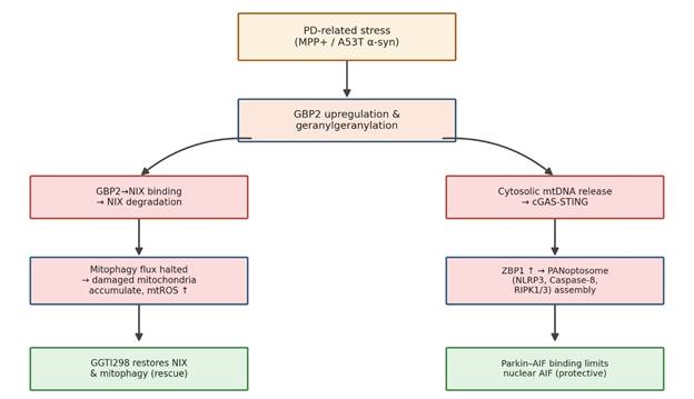
Figure 3. Convergent post-translational mitophagy failure (GBP2/NIX) and regulated cell-death execution (cGAS-STING/PANoptosis, AIF) in Parkinson's disease. The left branch shows how PD-related stress drives geranylgeranylation-dependent GBP2 accumulation, NIX degradation, and halted mitophagic flux; the right branch shows how cytosolic mtDNA release drives cGAS-STING and PANoptosome assembly. Green boxes indicate rescue points — GGTI298 restores NIX-dependent mitophagy, and Parkin-AIF binding restrains nuclear AIF translocation. Figure constructed from Cui et al. (2026), He et al. (2026), and Pischedda et al. (2019).
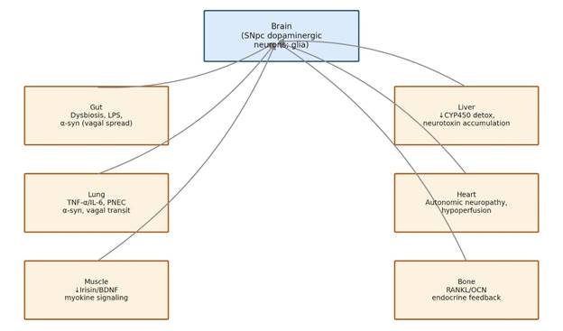
Figure 4. Peripheral organ-brain axes implicated in Parkinson's disease pathogenesis and their convergence on nigrostriatal dysfunction. Each peripheral organ box lists its representative molecular mediators and mechanism, with arrows indicating convergence of systemic signals onto central dopaminergic and glial dysfunction. Figure constructed from Liu et al. (2026).

accumulation in the substantia nigra via the liver-brain axis (Liu et al., 2026; Figure 3). Circadian disruption compounds this desynchronization further: loss of suprachiasmatic VIP neurons and accumulation of α-synuclein oligomers paralyze the rhythmic expression of clock genes such as PER2 and Bmal1, leaving dopaminergic neurons especially vulnerable to nighttime oxidative challenge (Huang & Li, 2024).

Taken as a whole, the literature converges on a fairly consistent picture: mitochondrial-nuclear crosstalk failure begins at discrete molecular nodes — PINK1/Parkin, PARIS/PGC-1α, GBP2/NIX, Nrf2 — but its consequences propagate outward through glial activation, innate immune signaling, and systemic organ-brain axes, ultimately converging back on the dopaminergic neuron from multiple directions simultaneously. This convergence is, arguably, the strongest rationale for pursuing multi-target rather than single-target therapeutics, a point taken up again in the Discussion

3. Methods

We readily admit that a narrative, mechanism-focused review does not carry the same evidentiary weight as a systematic review with meta-analysis — and we do not claim otherwise. Even so, we tried to make the process as transparent and reproducible as the format allows, following reporting conventions consistent with PubMed-indexed narrative and scoping review guidance so that another investigator could, in principle, retrace our steps.

3.1 Search Strategy and Information Sources

We searched PubMed/MEDLINE, Scopus, and Google Scholar for records addressing mitochondrial-nuclear crosstalk in Parkinson's disease, without restricting the search to a fixed calendar window, though the great majority of retrieved and cited primary literature falls between 1989 and 2026, reflecting both the foundational complex I studies (Schapira et al., 1989) and the most recent mechanistic work available at the time of writing. Search terms combined controlled vocabulary and free-text keywords using Boolean operators, structured broadly as: (“Parkinson's disease” OR “dopaminergic neurodegeneration”) AND (“mitochondrial-nuclear crosstalk” OR “retrograde signaling” OR “anterograde signaling” OR “PINK1” OR “Parkin” OR “PGC-1α” OR “PARIS” OR “Nrf2” OR “mitophagy” OR “GBP2” OR “NIX” OR “cGAS-STING” OR “PANoptosis” OR “AIF” OR “gut-brain axis” OR “organ-brain axis”). Reference lists of retrieved articles were hand-searched to identify additional mechanistically relevant studies, a practice consistent with standard snowballing methodology in narrative synthesis.

3.2 Eligibility Criteria

We included peer-reviewed original research articles, mechanistic studies, and reviews published in English that directly addressed nuclear-mitochondrial signaling pathways in the context of PD pathophysiology, whether investigated in cellular models (e.g., SH-SY5Y neuroblastoma cells, primary cortical neurons, patient-derived fibroblasts), animal models (principally MPTP- and 6-OHDA-induced rodent models), or human post-mortem/clinical tissue. We excluded conference abstracts, non-peer-reviewed preprints, and articles that referenced mitochondrial dysfunction only tangentially, without engaging the nuclear-genomic communication axis specifically. Where a given pathway (e.g., PARIS/PGC-1α) was described across multiple overlapping publications, we preferentially cited the study offering the most direct mechanistic or quantitative evidence, while retaining foundational or corroborating citations where appropriate.

3.3 Data Extraction and Synthesis

For each included study, we extracted, where reported: experimental model and species; the specific mitochondrial-nuclear signaling node investigated; the pharmacological or genetic intervention applied (if any); dosing regimen and route of administration; and the principal molecular, biochemical, and behavioral outcomes. This information underlies the quantitative synthesis presented in the Results (Section 4) and is compiled comprehensively in Table 1 (pharmacological interventions), Table 2 (systemic organ-brain axes), Table 3 (mitochondrial quality-control and cell-death pathways), and Table 4 (glial-neuronal and neuroendocrine signaling). Given the heterogeneity of experimental models, doses, and outcome measures across the evaluated literature, we did not attempt formal meta-analytic pooling of effect sizes; instead, we synthesized findings narratively, organized by mechanistic pathway rather than by publication, which we believe better serves a readership more interested in biological logic than in study-by-study cataloguing.

3.4 Figure and Diagram Construction

Figures 1–4 were constructed de novo to visually integrate mechanistic relationships described across the cited primary literature; they are original schematic syntheses rather than reproductions of any single source figure, and each is annotated with its principal supporting citations in the corresponding legend.

3.5 Risk of Bias and Limitations of the Approach

As a narrative synthesis rather than a registered systematic review, this manuscript was not preregistered and did not employ formal risk-of-bias instruments (e.g., SYRCLE for animal studies). We view this as a genuine limitation, not a minor caveat, and we return to it explicitly in the Discussion. Where preclinical findings derive predominantly from single research groups (as is the case for several of the more recently described pathways, including GBP2/NIX and TGR5/GPS signaling), we have tried to flag this in the text rather than presenting the findings as more broadly replicated than the current evidence base actually supports.

4. Molecular Pathomechanisms and Multitarget Therapeutic Landscapes

Pulled together, the evaluated literature sketches a genuinely multi-dimensional picture of PD pathogenesis — one that moves fairly deliberately away from a purely brain-centric account of dopaminergic loss and toward an integrated network of organelle stress, glial-neuronal feedback, and systemic organ-brain communication. What follows synthesizes the quantitative and mechanistic outcomes of this literature, organized around therapeutic intervention points rather than chronology.

4.1 Quantitative Efficacy of Novel Phytochemicals and Formulations

Across the preclinical models evaluated, targeting specific cellular stress pathways produced measurable, and in several cases substantial, cytoprotective and behavioral benefits (Table 1).

4.1.1 The CDNF–ER Stress Axis and Silybin Protection

In a subchronic MPTP-induced mouse model (30 mg/kg i.p. for 5 days), untreated animals exhibited severe motor coordination deficits, 30% mortality, and profound loss of tyrosine hydroxylase (TH) expression in the striatum and SNpc (Ramírez-Carreto et al., 2026). Daily oral silybin (100 mg/kg) significantly attenuated these deficits, lowering mortality to 15% and reducing latencies across pole, traction, and beam-walking assays by approximately 250% (Ramírez-Carreto et al., 2026; Table 1). This protective phenotype was accompanied by a selective, roughly twofold upregulation of cerebral dopamine neurotrophic factor (CDNF) in the striatum and SNpc, while the related factor MANF remained unchanged — an interesting dissociation suggesting genuine target specificity rather than a nonspecific trophic effect (Ramírez-Carreto et al., 2026). Mechanistically, silybin suppressed the pro-apoptotic PERK branch of the unfolded protein response (reducing p-PERK, p-eIF2α, and ATF4) while enhancing the adaptive IRE1α branch, shifting XBP1 splicing toward the pro-survival XBP1s isoform (Ramírez-Carreto et al., 2026). In silico docking confirmed stable engagement of silybin within the ATP-binding pockets of both PERK and IRE1α (Ramírez-Carreto et al., 2026).

4.1.2 Nrf2 Proteostasis and Redox Rescue by Gentiopicroside and Cannabidiol

Gentiopicroside (25 and 50 mg/kg i.p.), acting through TGR5 agonism, promoted Nrf2 nuclear translocation and upregulated HO-1 and SOD, preserving SNpc TH-positive neuron numbers and striatal fiber density in a dose-dependent manner in MPTP-treated mice, and reducing insoluble α-synuclein accumulation in primary cortical neurons exposed to preformed fibrils (Gao et al., 2026; Table 1). Cannabidiol (5 μM) conferred comparably robust, Nrf2-dependent neuroprotection in differentiated SH-SY5Y cells exposed to 6-OHDA, rotenone, or MPP+, rescuing cell viability to approximately 84% against 6-OHDA toxicity by preventing Nrf2 sequestration into insoluble aggregates and preserving mitochondrial network integrity (Jurado-Coronel & Duennwald, 2026; Table 1).

4.2 Post-Translational Control of Mitophagy and Programmed Cell Death

The GBP2–NIX Axis in Mitophagic Flux

Under PD-related toxic stress (MPTP/MPP+ or A53T α-synuclein overexpression), GBP2 was significantly upregulated in the substantia nigra of both patients and animal models, driven by stress-induced geranylgeranylation at its CAAX motif (Cui et al., 2026). Mitochondrially localized GBP2 bound NIX via its large GTPase domain, targeting NIX for polyubiquitination and proteasomal degradation and thereby halting mitophagic flux (Cui et al., 2026; Table 3). Genetic knockdown of GBP2, or pharmacological inhibition of its geranylgeranylation with GGTI298 (12 mg/kg), prevented NIX degradation, restored mitophagy, preserved

Table 1. Molecular targets, mechanistic actions, and preclinical outcomes of pharmacological agents modulating mitochondrial-nuclear crosstalk in Parkinson's disease. Each row summarizes the experimental model, primary molecular target(s), and key behavioral/biochemical outcomes reported for a given compound, allowing direct cross-compound comparison of mechanism and efficacy. Rows are drawn exclusively from the primary studies cited in the final column.

Compound

Model

Primary Target/Pathway

Key Outcomes

Reference

Silybin

Subchronic MPTP mouse (30 mg/kg i.p. ×5 d); silybin 100 mg/kg oral

↑CDNF; ↓PERK branch; modulates IRE1α/XBP1

↓Mortality 30%→15%; ↓motor latency ~250%; preserves striatal/nigral TH

Ramírez-Carreto et al. (2026)

Cannabidiol (CBD)

SH-SY5Y cells + 6-OHDA/rotenone/MPP+

Prevents Nrf2 aggregation; ↑Ser40 phosphorylation; ↑HMOX1/NQO1

~84% viability rescue vs 6-OHDA; ↓ROS; preserves mitochondrial network

Jurado-Coronel & Duennwald (2026)

Gentiopicroside (GPS)

MPTP mouse (25/50 mg/kg i.p.); MPP+ SH-SY5Y; α-Syn PFF neurons

TGR5 agonist → cAMP/PKA/CREB → Nrf2 nuclear entry

Preserves SNpc TH+ neurons; ↓α-Syn aggregates; abolished by TGR5-KO/SBI-115/H89

Gao et al. (2026)

GGTI298

MPTP mouse (12 mg/kg i.p. ×14 d); MPP+ SH-SY5Y

Inhibits GBP2 geranylgeranylation

Restores NIX; ↑2.4-fold TH; improves motor coordination

Cui et al. (2026)


Table 2. Peripheral organ-brain axes contributing to Parkinson's disease pathogenesis, with representative molecular mediators, pathophysiological mechanisms, and downstream effects on dopaminergic and cognitive systems. The table illustrates the shift from a brain-centric to a multisystem disease model of PD.

Axis

Key Mediators

Mechanism

Central Impact

Reference

Gut-Brain

SCFAs; LPS; TLR4/NF-κB; α-Syn

Dysbiosis → leaky gut → LPS translocation; vagal α-Syn spread

Microglial TLR4 activation; SNpc neuron loss

Liu et al. (2026)

Liver-Brain

CYP2D6; JAK/STAT; lipid peroxides

↓Hepatic detox → neurotoxin accumulation

Complex I inhibition; dopaminergic apoptosis

Liu et al. (2026)

Lung-Brain

TNF-α; IL-6; ROS

Pulmonary inflammation → systemic cytokines

BBB breakdown; microglial activation

Liu et al. (2026)

Heart-Brain

ANP; BNP; TNF-α

Cardiac autonomic neuropathy → hypoperfusion

Nigral oxidative stress; cognitive decline

Liu et al. (2026)

Muscle-Brain

Irisin; BDNF

Muscle atrophy → ↓myokine secretion

↓Hippocampal plasticity; ↓dopaminergic survival

Liu et al. (2026)

Bone-Brain

Osteocalcin; RANKL/OPG

Bone remodeling dysregulation

Microglial activation via RANKL; cognitive decline

Liu et al. (2026)

Table 3. Mitochondrial quality-control and regulated cell-death pathways in Parkinson's disease, detailing principal regulators, PD-associated alterations, and downstream pathological consequences for each pathway. The table integrates canonical (PINK1/Parkin) and non-canonical (GBP2/NIX) mitophagy routes alongside apoptotic (AIF), innate-immune (cGAS-STING), and inflammatory (PANoptosis) execution pathways.

Pathway

Regulators

Alteration in PD

Consequence

Reference

PINK1/Parkin Mitophagy

PINK1; Parkin; Miro1; Mfn2/Drp1

Mutated PINK1/Parkin fails to clear damaged organelles

ROS leakage; bioenergetic crisis

Kamienieva et al. (2023); Leites & Morais (2021)

PARIS/PGC-1α Biogenesis

PARIS; PGC-1α; Nrf1/2; TFAM; POLG

PARIS accumulates, represses PGC-1α promoter

↓mtDNA replication; ↓antioxidant defense

Kamienieva et al. (2023); Shin et al. (2011)

GBP2/NIX Alternative Mitophagy

GBP2; NIX/BNIP3L; GGTI298

Geranylgeranylated GBP2 degrades NIX

↑ROS; caspase-3 activation; apoptosis

Cui et al. (2026)

Parkin-AIF Translocation

AIF; Parkin

Cleaved AIF released, unrestrained by Parkin loss

Chromatin condensation; parthanatos

Pischedda et al. (2019)

cGAS-STING Activation

cGAS; STING; cytosolic mtDNA

mtDNA leakage activates cGAS

Interferon signaling; neuroinflammation

He et al. (2026)

Mitochondrial PANoptosis

ZBP1; NLRP3; ASC; Caspase-8; RIPK1/3

↓DRD2 → ↑ZBP1 → PANoptosome assembly

Inflammatory neuronal death

He et al. (2026)

Table 4. Multicellular, glial-neuronal, and neuroendocrine signaling disruptions contributing to chronic neuroinflammation and dopaminergic vulnerability in Parkinson's disease. Rows summarize cell types involved, the direction of pathological signaling shift, and the resulting impact on nigrostriatal integrity for each axis.

Axis

Cell Types

Signaling Shift

Impact

Reference

Astrocyte/Glutamate Excitotoxicity

Astrocytes; neurons; EAAT-1/2

↓KGDHC → ↓glutamate uptake

NMDA-mediated calcium overload

Wang & Xia (2023)

Microglial Complement Pruning

Microglia; hippocampal neurons; C3-C3aR

↑C3 → chronic C3aR overactivation

Excess synaptic pruning; comorbid depression

Huang & Li (2024)

HPA Axis Overactivation

HPA axis; microglia; adrenal glands

↑Glucocorticoids → ↓microglial GR

M1 polarization; ↑TNF-α/IL-1β

Xu et al. (2026)

Th17/Treg Imbalance

CD4+ T cells; Tregs; endothelium

Dysbiosis → Th17 polarization, ↓Tregs

IL-17-driven NF-κB/MAPK neurotoxicity

Liu et al. (2026)

mitochondrial membrane potential, increased SNpc TH expression by 2.4-fold, and rescued dopaminergic neurons from degeneration (Cui et al., 2026; Table 1).

4.2.1 Programmed Cell Death Modalities: AIF and PANoptosis

Parkin was shown to physically bind AIF in the cytosol, restraining its nuclear translocation under physiological conditions; in parkin-deficient neurons, this restraint was lost, accelerating nuclear AIF translocation and heightening apoptosis upon excitotoxic stress (Pischedda et al., 2019; Table 3). In parallel, cytosolic leakage of damaged mtDNA activated cGAS-STING signaling, and downregulated microglial DRD2 receptors permitted ZBP1 upregulation and assembly of the multi-protein PANoptosome (NLRP3, ASC, caspase-8, RIPK1/3), executing a highly inflammatory, non-cell-autonomous mode of dopaminergic neuron loss (He et al., 2026; Table 3).

4.3 The Systemic Pathology of Multi-Organ and Neuroendocrine Axes

4.3.1 Glial-Neuronal Dysregulation and Excitotoxicity

Astrocytic mitochondrial dysfunction impaired activity of the α-ketoglutarate dehydrogenase complex, reducing glutamate uptake via EAAT-1/2 transporters and triggering neuronal NMDA receptor-mediated calcium overload and excitotoxicity (Wang & Xia, 2023; Table 4). Loss of PINK1 or Parkin in astrocytes and microglia independently drove chronic neuroinflammation, with extracellular α-synuclein aggregates priming the NLRP3 inflammasome via microglial TLR/NF-κB signaling (Leites & Morais, 2021; Huang & Li, 2024). This activation was further amplified by HPA-axis overactivation: MPTP-induced stress elevated circulating glucocorticoids, which desensitized microglial glucocorticoid receptors, impaired immunosuppressive feedback, and shifted microglia toward a pro-inflammatory phenotype releasing iNOS, TNF-α, IL-1β, and IL-6 (Xu et al., 2026; Table 4).

4.3.2 Multi-Systemic Organ-Brain Communication Loops

Six distinct organ-brain axes were identified as dynamically modulating PD pathology over the disease course (Liu et al., 2026; Table 2). Early-stage pathology was associated with gut microbiota dysbiosis (decreased Prevotellaceae, increased Enterobacteriaceae) and compromised intestinal barrier integrity, permitting LPS translocation and microglial TLR4/MyD88 activation (Liu et al., 2026; Zhenxiong et al., 2025). Mid-term pathology implicated the liver-brain axis (impaired CYP2D6-mediated detoxification, reducing clearance of environmental neurotoxins) and the lung-brain axis (pulmonary TNF-α/IL-6 release disrupting blood-brain barrier integrity) (Liu et al., 2026). Late-stage amplification involved the muscle-brain axis (reduced irisin/BDNF myokine secretion compromising hippocampal plasticity) and the bone-brain axis (RANKL pathway overactivation and depleted osteocalcin signaling, correlating with cognitive decline) (Liu et al., 2026). Collectively, these interconnected axes describe a self-perpetuating, multi-organ feedback network sustaining progressive PD neurodegeneration (Figure 4; Table 2).

5. Discussion: Reframing Parkinson's Disease as a Disorder of Genomic Dialogue

It is tempting, once the mechanistic details accumulate, to lose sight of the larger claim being made here — so it is worth restating plainly. What the evidence assembled in this review suggests, taken together, is that PD is best understood not as a disease of a single protein, a single organelle, or even a single cell type, but as a progressive failure of communication between the nuclear and mitochondrial genomes, a failure that then propagates outward through glial, immune, and systemic channels. This is a somewhat different framing than the one most clinical teaching still relies on, and we think it has real implications for how disease-modifying therapy should be pursued.

5.1 A Convergent, Rather Than Linear, Model of Pathogenesis

One striking feature of the literature synthesized here is how consistently independent pathways converge on the same downstream outcomes — bioenergetic collapse, oxidative overload, and neuroinflammation — despite starting from quite different molecular triggers. The PINK1/Parkin–PARIS–PGC-1α axis, the GBP2/NIX mitophagy pathway, and Nrf2 proteostasis are, on paper, distinct systems; in practice, disabling any one of them appears sufficient to tip a dopaminergic neuron toward the same general trajectory of decline (Table 3). This convergence is, we would argue, the single most important pattern to emerge from the past several years of mechanistic PD research, and it has a fairly direct clinical implication: single-target interventions, however elegant mechanistically, may struggle to achieve meaningful disease modification if the remaining convergent pathways are left untouched.

5.2 The Case for Multi-Target Pharmacology

The four compounds reviewed here — silybin, cannabidiol, gentiopicroside, and GGTI298 — each act at a genuinely distinct node (ER stress/CDNF, Nrf2 proteostasis, TGR5-mediated Nrf2 activation, and GBP2/NIX mitophagy, respectively; Table 1), yet each independently produced meaningful preclinical benefit. We think this is telling. It suggests, at minimum, that the field should not be searching for a single “master switch” so much as identifying combinations of agents that simultaneously restore biogenesis, mitophagy, and antioxidant capacity. Whether such combinations would produce additive or synergistic effects in vivo remains untested, as far as we can determine from the current literature — a gap that strikes us as an obvious and fairly urgent next step for the field, rather than a minor omission.

5.3 Systemic Axes: A Genuinely New Layer, or an Overextension of the Model?

We want to be careful here, because the multi-organ axis literature (Liu et al., 2026; Table 2) is, admittedly, the newest and least mechanistically resolved component of this review. The gut-brain and liver-brain axes rest on reasonably direct evidence — LPS translocation, CYP450 suppression, and their downstream neuroinflammatory consequences are well documented (Liu et al., 2026; Zhenxiong et al., 2025; Pirooznia et al., 2020). The heart-brain, muscle-brain, and bone-brain axes, by contrast, are supported by a thinner and more correlative evidence base at present, and we would caution readers against over-interpreting these as established causal pathways rather than plausible, biologically coherent hypotheses awaiting further mechanistic dissection. That caveat aside, even a partial validation of this systemic framework would meaningfully expand where therapeutic intervention might reasonably be aimed — beyond the CNS entirely, toward gut microbiota modulation or hepatic detoxification support, for instance.

5.4 Limitations

Several limitations deserve explicit acknowledgment. First, this is a narrative rather than a systematic review; we did not apply formal risk-of-bias instruments, and publication bias toward positive preclinical findings almost certainly inflates the apparent efficacy of the interventions discussed. Second, nearly all of the mechanistic and pharmacological evidence reviewed here derives from rodent or cell-culture models — MPTP, 6-OHDA, and MPP+ paradigms chief among them — and none of the four featured compounds has, to our knowledge, been validated in human PD clinical trials at the time of writing. Third, several of the more recently described pathways (GBP2/NIX, TGR5/GPS) currently rest on findings from a comparatively small number of independent research groups, and independent replication will be essential before these targets can be considered validated rather than promising. We have tried to flag these limitations at the relevant points in the text rather than only here, but they bear restating collectively.

5.5 Implications for Disease-Modifying Therapy

If the convergent model outlined above holds up, the practical implication is that disease-modifying trials in PD may need to move away from single-mechanism agents and toward rationally designed combination strategies — pairing, for instance, a mitophagy-restoring agent with an Nrf2-stabilizing compound, rather than testing either in isolation. This is a more complex trial design, with its own regulatory and dosing challenges, but the mechanistic convergence documented throughout this review makes a reasonably strong case that it may be necessary rather than merely preferable.

6. Conclusion

Across the molecular, cellular, and systemic levels examined in this review, a consistent picture emerges: Parkinson's disease progresses when the nucleus and the mitochondrion stop coordinating effectively, and the specific point at which that coordination fails — PINK1/Parkin-PARIS-PGC-1α, GBP2/NIX, Nrf2 proteostasis, or AIF/PANoptosis execution — appears to matter less than the fact that, once one node fails, the downstream consequences (bioenergetic collapse, oxidative injury, neuroinflammation) look remarkably similar. This convergence extends outward as well, recruiting glial cells and, apparently, peripheral organs into a self-sustaining pathogenic network rather than remaining confined to the substantia nigra. Encouragingly, each of these nodes has proven pharmacologically tractable in preclinical models: silybin, cannabidiol, gentiopicroside, and geranylgeranylation inhibitors like GGTI298 each restored a distinct arm of mitochondrial-nuclear signaling and produced measurable neuroprotection. None of this constitutes proof of clinical efficacy, and the translational gap from rodent models to human disease remains substantial and should not be understated. Still, the mechanistic coherence documented here — the same downstream endpoints reached from multiple independent starting points — offers a genuinely rational basis for pursuing multi-target, disease-modifying strategies in PD, rather than continuing to rely primarily on symptomatic dopamine-replacement approaches. Restoring the dialogue between these two genomes may, in the end, be less a metaphor than a fairly literal therapeutic target.

References


Bloem, B. R., Okun, M. S., & Klein, C. (2021). Parkinson's disease. The Lancet, 397(10291), 2284–2303. https://doi.org/10.1016/S0140-6736(21)00218-X

Buneeva, O., Fedchenko, V., Kopylov, A., & Medvedev, A. (2020). Mitochondrial dysfunction in Parkinson's disease: Focus on mitochondrial DNA. Biomedicines, 8(12), Article 591. https://doi.org/10.3390/biomedicines8120591

Cui, W., Wang, T., & Feng, J. (2026). Upregulated GBP2 exacerbates Parkinson's disease pathogenesis by impairing NIX-dependent mitophagy. Redox Biology, 90, Article 104029. https://doi.org/10.1016/j.redox.2026.104029

Gao, Y., Tang, X., Yao, J., Sun, T., Chen, Y., Cheng, C., Yang, J., Wang, B., Liu, A., Yang, L., & Zhenxiong, M. (2026). Targeting the bile acid receptor TGR5 with Gentiopicroside to activate Nrf2 antioxidant signaling and mitigate Parkinson's disease in an MPTP mouse model. Journal of Advanced Research, 80, 977–990. https://doi.org/10.1016/j.jare.2025.05.039

García-Yagüe, A. J., Esteras, N., Dinkova-Kostova, A. T., Rojo, A. I., Shiels, P. G., Dinnyes, A., Tamas, V., van Goor, H., & Lastres-Becker, I. (2026). Summary of NRF2 implication in PD disease mouse models. Free Radical Biology and Medicine, 246, 760–779. https://doi.org/10.1016/j.freeradbiomed.2026.02.023

He, Y., Guo, L., Wang, L., Li, Z., Lu, R., Zhang, Q., & Xiong, K. (2026). Mitochondrial oxidative stress to PANoptosis: Current evidence and therapeutic implications for neurological diseases. Journal of Advanced Research. Advance online publication. https://doi.org/10.1016/j.jare.2026.02.023

Huang, J., & Li, W. (2024). Molecular crosstalk between circadian clock and NLRP3 inflammasome signaling in Parkinson's disease. Heliyon, 10(1), Article e24752. https://doi.org/10.1016/j.heliyon.2024.e24752

Jurado-Coronel, J., & Duennwald, M. L. (2026). Cannabidiol confers neuroprotection against 6-OHDA toxicity by rescuing Nrf2 proteostasis and preserving mitochondrial integrity. Free Radical Biology and Medicine, 255, 774–790. https://doi.org/10.1016/j.freeradbiomed.2026.08.012

Kamienieva, I., Charzynska, A., Duszynski, J., Malinska, D., & Szczepanowska, J. (2023). In search for mitochondrial biomarkers of Parkinson's disease: Findings in parkin-mutant human fibroblasts. Biochimica et Biophysica Acta – Molecular Basis of Disease, 1869(8), Article 166787. https://doi.org/10.1016/j.bbadis.2023.166787

Leites, E. P., & Morais, V. A. (2021). The PINK1-mediated crosstalk between neural cells and the underlying link to Parkinson's disease. Cells, 10(6), Article 1395. https://doi.org/10.3390/cells10061395

Liu, T., Wu, H., & Wei, J. (2026). Beyond the brain: Exploring the multi-organ axes in Parkinson's disease pathogenesis. Journal of Advanced Research, 80, 451–474. https://doi.org/10.1016/j.jare.2025.05.034

Panicker, N., Kam, T. I., Wang, H., Neifert, S., Chou, S. C., Kumar, M., … Dawson, T. M. (2022). Neuronal NLRP3 is a parkin substrate that drives neurodegeneration in Parkinson's disease. Neuron, 110(15), 2422–2437. https://doi.org/10.1016/j.neuron.2022.05.009

Pirooznia, S. K., Yuan, C., Khan, M. R., Karuppagounder, S. S., Wang, L., Xiong, Y., … Dawson, T. M. (2020). PARIS induced defects in mitochondrial biogenesis drive dopamine neuron loss under conditions of parkin or PINK1 deficiency. Molecular Neurodegeneration, 15, Article 29. https://doi.org/10.1186/s13024-020-00378-x

Pischedda, F., Piccoli, G., Rakovic, A., Klein, C., & Pichler, I. (2019). Parkin deficiency increases stress-induced translocation of AIF to the nucleus. International Journal of Molecular Sciences, 20(3), Article 748. https://doi.org/10.3390/ijms20030748

Ramírez-Carreto, R. J., Salas-Venegas, V., De la Rosa-Hernández, V., Medina-Campos, O. N., Martínez-Ortega, U., Torres-Guerrero, H., Hernández-Luis, F., Pedraza-Chaverri, J., Königsberg, M., & Chavarría, A. (2026). Silybin attenuates PERK/IRE1α ER-stress signaling, neuroinflammation, and restores CDNF levels in an MPTP-induced Parkinson's disease model. European Journal of Pharmacology, 1030, Article 179126. https://doi.org/10.1016/j.ejphar.2026.179126

Schapira, A. H., Cooper, J. M., Dexter, D., Jenner, P., Clark, J. B., & Marsden, C. D. (1989). Mitochondrial complex I deficiency in Parkinson's disease. The Lancet, 1(8649), 1269–1271. https://doi.org/10.1016/S0140-6736(89)92696-1

Shin, J. H., Ko, H. S., Kang, H., Lee, Y., Lee, Y. I., Pletinkova, O., … Dawson, T. M. (2011). PARIS (ZNF746) repression of PGC-1α contributes to neurodegeneration in Parkinson's disease. Cell, 144(5), 689–702. https://doi.org/10.1016/j.cell.2011.02.010

Spinazzola, A., & Zeviani, M. (2009). Mitochondrial diseases: A cross-talk between mitochondrial and nuclear genomes. Advances in Experimental Medicine and Biology, 652, 69–84. https://doi.org/10.1007/978-90-481-2913-3_6

Wang, Y., & Xia, Y. (2023). Astrocytic mitochondrial dysfunction and neuroinflammation in Parkinson's disease. Neurobiology of Disease, 184, Article 106224. https://doi.org/10.1016/j.nbd.2023.106224

Xu, L., Xiao, K., Jiang, J., Tang, Y., Yang, L., Xiao, S., Jin, X., Xiao, B., Zhou, T., Hao, C., Fan, H., & Chai, Z. (2026). Mechanism analysis of MPTP-induced Parkinson's disease-like pathology via the HPA axis–microglial glucocorticoid receptor pathway. Ecotoxicology and Environmental Safety, 323, Article 120715. https://doi.org/10.1016/j.ecoenv.2026.120715

Zhenxiong, Z. H. A. O., Mengdie, D. O. N. G., & Zhen, M. U. (2025). Gut-Brain Axis Dysregulation in Parkinson’s Disease: Mechanisms Linking Microbiota to Neuroinflammation and α-Synuclein Pathology. Journal of Pharmaceutical Analysis, 101521.  


Article metrics
View details
0
Downloads
0
Citations
37
Views

View Dimensions


View Plumx


View Altmetric


0
Save
0
Citation
37
View
0
Share