
Integrative Biomedical Research

Integrative Biomedical Research (Journal of Angiotherapy) | Online ISSN  3068-6326
463
Citations
1.8m
Views
746
Articles
Your new experience awaits. Try the new design now and help us make it even better
Switch to the new experience
Figures and Tables
REVIEWS   (Open Access)

The Gut–Brain–Immune Axis in Chronic Disease: Mechanisms and Therapeutic Opportunities — A Narrative Synthesis

Mohamed Sadeq Al-Ibrahim 1, Ahmad Hamdy Ibrahim 2, Sawsan S. Al- Rawi 3*, Bayram Dawod Ahmed 4

+ Author Affiliations

Integrative Biomedical Research 10 (1) 1-8 https://doi.org/10.25163/biomedical.10110884

Submitted: 10 January 2026 Revised: 01 March 2026  Published: 13 March 2026 


Abstract

The gastrointestinal microbiota shapes host metabolism, immunity, and brain function through a tripartite gut–brain–immune axis, yet the field still lacks a unified synthesis linking barrier failure, immunometabolic signaling, and disease-specific outcomes. We conducted a structured narrative review of preclinical and clinical literature indexed largely through PubMed-listed sources, synthesizing evidence on neural, endocrine, immunological, and microbial-metabolite communication pathways and their dysregulation in Parkinson’s disease, Alzheimer’s disease, multiple sclerosis, and related chronic conditions. Across disorders, intestinal barrier failure and endotoxemia recur as a shared upstream trigger, converging on microglial activation, kynurenine pathway shunting toward neurotoxic quinolinic acid, and blood–brain barrier compromise, while each disease additionally shows a distinctive dysbiotic signature and cellular trafficking pattern—vagal alpha-synuclein propagation in Parkinson’s disease, amyloid cross-seeding in Alzheimer’s disease, and Th17-mediated demyelination in multiple sclerosis. Microbiome-directed interventions, including dietary modulation, psychobiotics, and fecal microbiota transplantation, showed measurable, if heterogeneous, benefit; a placebo-controlled fecal transplantation trial in Parkinson’s disease, for instance, reported meaningfully greater motor improvement than placebo. The evidence, taken together, supports the gut–brain–immune axis as a mechanistically coherent and therapeutically actionable framework, though translation remains constrained by engraftment variability and a scarcity of standardized, biomarker-guided trial designs. Personalized, multi-omics-informed strategies appear to be the most promising path toward clinical utility. Keywords: gut–brain–immune axis; gut microbiota; neuroinflammation; dysbiosis; kynurenine pathway; fecal microbiota transplantation; neurodegenerative disease

1. Introduction

Somewhere in the space between a bowl of yogurt and a diagnosis of Parkinson’s disease lies a question that has quietly reshaped neuroscience over the past two decades: what, exactly, is the brain listening to when it is not listening to itself? The human body, it turns out, is never really alone. Trillions of commensal microorganisms—overwhelmingly bacterial—inhabit the gastrointestinal tract, arriving at birth and being reshaped continuously thereafter by host genetics and by the accumulating weight of diet, stress, and environment (Marano et al., 2023). Under ordinary circumstances this community does unglamorous but essential work: it helps digest food, extracts nutrients the body could not otherwise use, and manufactures vitamins the host cannot synthesize alone (Marano et al., 2023). What is less intuitive, and considerably more consequential for the story this review tells, is that the same microbial community also reaches well beyond the gut lumen to influence metabolism, immune tone, and—improbably, to anyone trained on a purely neurocentric model of the brain—cognition and mood (Hattori & Yamashiro, 2021; Marano et al., 2023).

That reach has a name. The bidirectional signaling network connecting gut and central nervous system is termed the gut–brain axis, or GBA (Hattori & Yamashiro, 2021; Olasunkanmi et al., 2026). For a long while the GBA was treated, more or less, as a two-party conversation. It no longer is. Over the past several years the field has moved—not without some resistance—toward a systems-biology view in which the immune system is not a bystander but an active third interlocutor, giving rise to what is increasingly called the gut–brain–immune axis (Khawar et al., 2023; Park et al., 2025). This tripartite framing is not merely semantic. It reflects a genuine mechanistic expansion: anatomical, hormonal, metabolic, immunological, and neural pathways are now understood to operate in concert, together preserving (or, when disrupted, undermining) both physiological and cognitive equilibrium (O’Riordan et al., 2025; Olasunkanmi et al., 2026).

Consider, first, how the conversation is physically carried. Communication travels along direct neural circuits, endocrine messengers, and immune signals (Westfall et al., 2017). The autonomic, sympathetic, and enteric nervous systems (ENS) form the peripheral scaffolding linking gut to brain (Hattori & Yamashiro, 2021). The ENS itself is a curious structure—often nicknamed, perhaps a little too cutely, the “second brain”—embedded in the intestinal wall and populated by upward of 500 million neurons capable of regulating motility, blood flow, and secretion largely on their own recognizance (Olasunkanmi et al., 2026; Zhang et al., 2025). Sensory information travels gut-to-brain chiefly via afferent vagal fibers projecting to the brainstem (Hattori & Yamashiro, 2021), while the efferent arm of the vagus runs the traffic in reverse, tempering peripheral inflammation through what is known as the cholinergic anti-inflammatory pathway (Hattori & Yamashiro, 2021). Layered on top of this neural circuitry is an endocrine channel, orchestrated by the hypothalamic–pituitary–adrenal (HPA) axis (Khawar et al., 2023; Marano et al., 2023): the hypothalamus releases corticotropin-releasing hormone, which prompts pituitary release of adrenocorticotropic hormone, which in turn drives adrenal secretion of glucocorticoids such as cortisol (Marano et al., 2023; Park et al., 2025). And because more than 70% of the body’s total immune cell repertoire resides within the gastrointestinal tract, the immune system functions as an unusually well-positioned intermediary—translating whatever the gut is experiencing into a language the brain can, unfortunately, understand all too well (Zhou et al., 2023).

Under healthy conditions, the intestinal epithelium behaves as a discerning gatekeeper: it lets nutrients and fluids through while keeping pathogens and toxic metabolites firmly outside the bloodstream (Kearns, 2024). Tight junction proteins—claudins, junctional adhesion molecules—do much of the structural work of holding that barrier together (Kearns, 2024). But the gatekeeper can be worn down. Genetic vulnerability, chronic psychological stress, poor dietary patterns, and infection can all perturb the resident microbiota, producing a state generally referred to as dysbiosis (Kearns, 2024; Marano et al., 2023). Once established, dysbiosis erodes barrier integrity outright, producing the now-familiar (if still somewhat contested, in popular discourse) phenomenon of “leaky gut,” through which pathobionts and their structural components—lipopolysaccharide (LPS) chief among them—escape into systemic circulation (Khawar et al., 2023; Olasunkanmi et al., 2026). The resulting low-grade, chronic endotoxemia does not stay politely confined to the periphery; it drives immune activation with clear downstream consequences for the central nervous system (Kearns, 2024).

How, precisely, does an inflamed gut make itself known to the brain? Several converging routes have been described. Mucosal immune cells release pro-inflammatory cytokines—tumor necrosis factor-alpha (TNF-α), interleukin-1 beta (IL-1β), interleukin-6 (IL-6)—which circulate to the blood–brain barrier (BBB) and choroid plexus (Günther et al., 2021; Jha et al., 2025). Gut-primed lymphocytes, including T helper 17 (Th17) and regulatory T (Treg) cells, are able to leave gut-associated lymphoid tissue altogether, enter systemic circulation, cross the BBB, and directly shape immune tone within brain parenchyma (Fung, 2020; Günther et al., 2021). Once inside, these peripheral signals activate resident microglia and astrocytes (Fung, 2020), nudging microglia toward a pro-inflammatory morphology that sets off cascades of neuroinflammatory signaling, astrogliosis, and, eventually, synaptic loss (Loh et al., 2024).

A quieter but equally consequential route runs through amino acid metabolism. Tryptophan, an essential amino acid, is normally metabolized along the kynurenine pathway (Kearns, 2024)—and under chronic inflammatory pressure, pro-inflammatory cytokines upregulate indoleamine 2,3-dioxygenase (IDO), diverting tryptophan away from serotonin synthesis and toward kynurenine (Kearns, 2024). The result is an accumulation of neurotoxic byproducts, most notably quinolinic acid, which crosses the BBB, generates oxidative stress, and acts as an NMDA receptor agonist—a fairly direct route to excitotoxic neuronal injury (Kearns, 2024; Marano et al., 2023).

Taken together, these pathways are increasingly implicated across a cluster of major chronic, neuroinflammatory, and neurodegenerative disorders: Alzheimer’s disease (AD), Parkinson’s disease (PD), multiple sclerosis (MS), and inflammatory bowel disease (IBD) among them (Kearns, 2024; Park et al., 2025). In neurodegeneration specifically, gut-driven neuroinflammation looks less like a bystander effect and more like an accelerant (Zheng et al., 2023). In AD, dysbiosis and heightened intestinal permeability appear to precede amyloid-beta (Aβ) accumulation, at least in the animal and early clinical literature available so far (Zhang et al., 2025), with BBB disruption and microglial activation following in its wake (Loh et al., 2024). In PD, gastrointestinal dysfunction and colonic inflammation often show up—sometimes by years—ahead of the motor symptoms that traditionally define the disease (Westfall et al., 2017; Zhang et al., 2025), consistent with hypotheses that alpha-synuclein misfolding begins in the ENS and travels retrogradely via the vagus (Loh et al., 2024).

Autoimmune and psychiatric-adjacent conditions tell a related, if mechanistically distinct, story. MS, a CNS demyelinating disease, correlates strongly with alterations in gut microbial composition (Benakis et al., 2020; Khawar et al., 2023), with dysbiosis favoring encephalitogenic Th17 differentiation at the expense of protective Tregs (Fung, 2020; Khawar et al., 2023). IBD and irritable bowel syndrome, meanwhile, are almost archetypal gut–brain disorders, marked by mucosal micro-inflammation, barrier disruption, and—frequently—comorbid anxiety and depression, underscoring just how bidirectional this axis really is (Soufan et al., 2025; Jha et al., 2025); stress, for its part, feeds back to worsen gut pathology through HPA dysregulation and altered motility (Ge et al., 2022).

If the mechanisms sketched above hold up, then the gut microbiota represents an unusually tractable therapeutic target—arguably more tractable, in some respects, than the brain itself (Park et al., 2025). Strategies acting on the gut environment differ fundamentally from conventional centrally-acting pharmacology, working instead across metabolic, immune, and neural systems simultaneously (Chen et al., 2025). Dietary fiber, fermented by the microbiota into short-chain fatty acids (SCFAs) such as acetate, propionate, and butyrate, exemplifies this holistic logic: SCFAs act on G-protein-coupled receptors, promote Treg differentiation, dampen inflammatory cytokine production, and reinforce tight junction expression at both the gut and the BBB (Marano et al., 2023; Russo et al., 2017), while bioactive lipids such as palmitoylethanolamide similarly reduce inflammation and visceral pain (Russo et al., 2017). Probiotics rich in Lactobacillus and Bifidobacterium—sometimes termed psychobiotics—can synthesize neuroactive compounds such as GABA and serotonin, modulating vagal signaling with measurable effects on mood and neuroinflammation in trials to date (Marano et al., 2023; Olasunkanmi et al., 2026; Soufan et al., 2025). And fecal microbiota transplantation (FMT), which transfers an entire functional ecosystem from a healthy donor, has moved beyond its original indication in recurrent Clostridium difficile infection to show signal in IBD remission, PD motor function, and AD-like cognitive decline in preclinical models (Jha et al., 2025; Loh et al., 2024; Zhang et al., 2025).

Still, a considerable gap separates what preclinical models suggest and what clinical practice can currently deliver (Park et al., 2025). This article was written to narrow that gap, guided by three working questions: first, whether gut barrier disruption and the resulting translocation of LPS is a required, rather than merely correlated, trigger for myelin-specific T-cell activation in EAE (Fung, 2020; Khawar et al., 2023); second, whether dietary fiber can suppress IDO activity enough to forestall hippocampal quinolinic acid accumulation (Kearns, 2024; Loh et al., 2024); and third, whether FMT’s clinical benefit in AD and PD depends specifically on durable engraftment of butyrate-producing taxa, and whether that engraftment can be predicted from baseline metagenomics (Zhang et al., 2025). Around these questions, four objectives structure the remainder of this review: synthesizing the cellular and neural pathways linking microbiota, immunity, and CNS (O’Riordan et al., 2025; Park et al., 2025); delineating how leaky gut drives microglial neuroinflammation through LPS translocation and kynurenine dysregulation (Kearns, 2024; Loh et al., 2024); comparatively evaluating dietary, probiotic, prebiotic, and FMT-based therapeutics (Jha et al., 2025; Zheng et al., 2023); and, finally, sketching a translational framework for personalized intervention built on multi-omics baseline profiling (Zhang et al., 2025).
2. Gut–Immune–Brain Axis in Neurodegenerative and Neuroinflammatory Disorders

2.1. Reconceptualizing Brain Health through the Gut–Immune–Brain Axis

For much of its history, neuroscience treated chronic neurological and neurodegenerative disease as an intracranial affair—protein misfolding, faulty synaptic pruning, myelin loss, all unfolding, more or less, in isolation within the central nervous system (Chen et al., 2025; Loh et al., 2024). That framing has not aged well. Over roughly the last twenty years it has given way to something more entangled: the recognition that brain health is inseparable from the gastrointestinal tract and its resident microbial ecosystem, linked through the bidirectional gut–brain axis (Hattori & Yamashiro, 2021; O’Riordan et al., 2025). Some of the earliest and, frankly, most persuasive evidence for this came from germ-free mice—animals raised without any microbiota at all—which display altered stress reactivity, disrupted HPA axis dynamics, and abnormal central neurotransmitter levels, hinting that gut-derived signals are not incidental but genuinely necessary for normal neurodevelopment and ongoing brain homeostasis (Khawar et al., 2023; Park et al., 2025; Westfall et al., 2017).

More recently still, this bidirectional picture has been superseded by a tri-directional one: the gut–immune–brain axis (Warren et al., 2024; Zhou et al., 2023). The addition of “immune” here is not decorative. Given that more than 70% of the body’s total immune cell mass sits within gut-associated lymphoid tissue (GALT), it makes a certain intuitive sense that the immune system would function as the principal translator, converting local intestinal disturbances into signals the brain can register as inflammation (Park et al., 2025; Zhou et al., 2023). Under balanced conditions—eubiosis—the microbial community maintains gut and BBB integrity, sustains immune tolerance, and produces a steady supply of neuroactive metabolites (Kearns, 2024; Marano et al., 2023). When that balance tips into dysbiosis, systemic inflammation follows, barriers weaken, and the field has taken, somewhat memorably, to calling the resulting state “leaky gut, leaky brain” (Kearns, 2024; Marano et al., 2023). The remainder of this review traces how this dysregulation plays out—similarly in places, quite differently in others—across Parkinson’s disease, Alzheimer’s disease, and multiple sclerosis.

2.2. Shared Architectural Foundations of Axis Dysregulation

Before turning to disease-specific pathways, it is worth pausing on what these conditions have in common, because the overlap is substantial. The first shared node is gut barrier failure itself (Loh et al., 2024; Olasunkanmi et al., 2026). Environmental triggers, chronic stress, and inflammatory dysbiosis together erode the single-cell epithelial layer, its overlying mucus, and the gut vascular barrier beneath it (Kearns, 2024; Loh et al., 2024; Zhou et al., 2023). Chronic HPA overactivation compounds the problem: excess corticotropin-releasing hormone and cortisol suppress expression of tight junction proteins—occludins, zonulin, claudins—directly (Ge et al., 2022; Ho et al., 2025; Marano et al., 2023).

The resulting hyperpermeability allows pathobionts and microbe-associated molecular patterns, LPS foremost among them, to slip into the bloodstream (Antón et al., 2018; Khawar et al., 2023; Olasunkanmi et al., 2026). Circulating LPS binds Toll-like receptor 4 (TLR4) on peripheral immune cells, triggering systemic endotoxemia and a familiar cytokine trio—IL-1β, IL-6, TNF-α (Antón et al., 2018; Zhou et al., 2023). These mediators then act on the BBB itself, downregulating endothelial claudin-5 and occludin (Günther et al., 2021; Jha et al., 2025; Marano et al., 2023), which opens the door for circulating cytokines and immune cells to enter CNS parenchyma (Günther et al., 2021; Khawar et al., 2023). Notably, Carloni et al. (2021) identified an analogous vascular gate in the brain’s choroid plexus—the plexus vascular barrier—that closes specifically in response to gut-derived LPS, contributing to cognitive and affective deficits (Ge et al., 2022; Ortega et al., 2023).

Once inside the CNS, translocated microbial signals and cytokines engage TLR4 on resident microglia and astrocytes (Loh et al., 2024; Zhou et al., 2023). Microglia shift away from their protective, phagocytic default and toward an overactive, pro-inflammatory state, releasing neurotoxic cytokines and activating the NLRP3 inflammasome (Fung, 2020; Jha et al., 2025; Loh et al., 2024)—a shift that in turn promotes astrogliosis and synaptotoxicity (Loh et al., 2024; Zhou et al., 2023). A parallel metabolic disturbance unfolds in tryptophan handling (Kearns, 2024; Ortega et al., 2023): pro-inflammatory cytokines upregulate IDO and tryptophan 2,3-dioxygenase, shunting tryptophan into the kynurenine pathway (KP) at serotonin’s expense (Kearns, 2024; Ortega et al., 2023). This raises the kynurenine-to-tryptophan ratio—itself a useful inflammatory biomarker—and favors production of neurotoxic quinolinic acid (QUIN) and 3-hydroxykynurenine over the neuroprotective kynurenic acid (KYNA) (Kearns, 2024; Ortega et al., 2023). QUIN, acting as an NMDA receptor agonist, drives glutamate excitotoxicity, free-radical generation, and progressive neurodegeneration (Kearns, 2024; Ortega et al., 2023) (see Table 1 for a full mapping of axis pathways and mediators, and Table 4 for downstream molecular targets).

2.3. Parkinson’s Disease: Vagal Retro-Propagation and Synucleinopathy

Where the mechanisms above are broadly shared, PD illustrates just how disease-specific the axis can also become (Chen et al., 2025). Gastrointestinal complaints—dysphagia, nausea, and especially chronic constipation—commonly appear decades before the motor symptoms that define a PD diagnosis (Loh et al., 2024; Russo et al., 2017). This timeline dovetails with Braak’s hypothesis, which proposes that PD pathology originates in the enteric nervous system in response to local intestinal triggers before ascending to the CNS (Russo et al., 2017; Westfall et al., 2017).

PD is defined pathologically by aggregated alpha-synuclein (forming Lewy bodies) and progressive dopaminergic neuron loss in the substantia nigra (Loh et al., 2024; Olasunkanmi et al., 2026). In rodent models, alpha-synuclein injected into the colonic wall migrates transneurally to the brainstem via the vagus nerve at roughly 5–10 mm per day (Russo et al., 2017)—a rate consistent with vagal fibers providing genuine physical “hardwiring” for retrograde spread (Benakis et al., 2020). Interestingly, people who underwent full truncal vagotomy earlier in life appear to carry a meaningfully lower risk of later PD, a finding hard to explain without invoking the vagus as a literal transmission route (Chen et al., 2025).

What initiates the misfolding in the first place seems to trace back to a fairly distinctive dysbiotic signature (Chen et al., 2025). PD patients show depletion of SCFA-producing, mucin-supporting Prevotellaceae and Lachnospiraceae (Chen et al., 2025; Loh et al., 2024), alongside overrepresentation of mucin-degrading Akkermansia muciniphila and pro-inflammatory, LPS-producing Enterobacteriaceae (Chen et al., 2025; Loh et al., 2024). Gut colonization with Gram-negative pathobionts such as Proteus mirabilis is sufficient to reproduce PD-like motor deficits and microglial activation in wild-type mice via chronic endotoxemia (Benakis et al., 2020; Loh et al., 2024), and Sampson et al. (2016) showed—rather elegantly—that germ-free alpha-synuclein–overexpressing mice are protected from motor dysfunction, while colonization with PD-patient (but not healthy-control) microbiota reverses that protection (Benakis et al., 2020; Loh et al., 2024). At the immune-cellular level, this dysbiosis prompts CCR2+ monocytes to migrate from gut-associated lymphoid tissue into brain parenchyma, amplifying microglial activation and nigral dopaminergic loss (Olasunkanmi et al., 2026).

2.4. Alzheimer’s Disease: Amyloidogenic Cross-Seeding and Microglial Failure

In AD, the axis appears to function less as an initiator and more as an accelerator—of amyloid-beta (Aβ) plaque formation, tau hyperphosphorylation, and microglial phagocytic failure (Kearns, 2024; Loh et al., 2024). Longitudinal animal work suggests that calprotectin-mediated mucosal inflammation and epithelial leakage actually precede cerebral Aβ accumulation, rather than merely accompanying it (Zheng et al., 2023).

One mechanism worth dwelling on is the production of functional amyloids by gut bacteria themselves (Benakis et al., 2020; Olasunkanmi et al., 2026; Bruggeman et al., 2024). Biofilm-forming species—Escherichia coli, Bacillus subtilis, Klebsiella pneumoniae, Salmonella spp., and Staphylococcus aureus among them—secrete curli fibers and related extracellular amyloid-like proteins to aid their own colonization (Olasunkanmi et al., 2026; Westfall et al., 2017). These microbial amyloids happen to be structurally similar to human Aβ, and translocated copies appear able to cross-seed Aβ aggregation through molecular mimicry while priming systemic immune cells toward hyper-reactivity (Chen et al., 2025). The taxonomic signature here is fairly consistent: a phylum-level drop in Firmicutes (Butyrivibrio, Eubacterium, Roseburia, Faecalibacterium prausnitzii) alongside enrichment of Bacteroidetes and Proteobacteria, particularly Escherichia/Shigella (Chen et al., 2025; Fung, 2020; Zheng et al., 2023).

That shift is not immunologically neutral. Loss of butyrate-producing taxa removes a protective, anti-inflammatory input, while Escherichia/Shigella overgrowth raises peripheral markers of NLRP3 activation and pro-inflammatory cytokines such as Il1b and Cxcl2 (Fung, 2020; Zheng et al., 2023). Elevated Bacteroides, somewhat counterintuitively, correlates with reduced microglial phagocytic capacity—meaning Aβ clearance itself is compromised (Zhang et al., 2025), while translocated LPS and cytokines cross the weakened BBB to bind microglial TLR4, sustaining chronic neuroinflammation and synaptic loss (Kearns, 2024; Loh et al., 2024; Olasunkanmi et al., 2026; Bruggeman et al., 2024). The kynurenine pathway is hyperactive here too, with excess QUIN crossing into the hippocampus and contributing to cognitive decline (Kearns, 2024; Ortega et al., 2023), and altered bile acid metabolism—elevated toxic secondary bile acids such as deoxycholic and lithocholic acid—further disrupts endothelial tight junctions and BBB integrity (Chen et al., 2025; Loh et al., 2024).

2.5. Multiple Sclerosis: Myelin Autoimmunity and CD4+ T-Cell Polarization

MS stands somewhat apart as the prototypical neuroinflammatory autoimmune disorder of the CNS—myelin destruction, axonal loss, progressive neurodegeneration (Khawar et al., 2023; Loh et al., 2024)—and here the axis operates chiefly through adaptive immune cell trafficking rather than direct metabolic or neural signaling (Benakis et al., 2020; Khawar et al., 2023). Both MS patients and the corresponding rodent model, experimental autoimmune encephalomyelitis (EAE), display gut dysbiosis and heightened epithelial permeability that precede overt myelin damage (Khawar et al., 2023; Olasunkanmi et al., 2026). Under normal conditions the microbiome favors CD4+ Treg differentiation, with IL-10 secretion helping to keep inflammation in check (Benakis et al., 2020; Khawar et al., 2023); in MS, that balance tips toward pro-inflammatory Th17 cells instead (Fung, 2020; Khawar et al., 2023).

Metagenomic data point to depletion of SCFA-producing Clostridia clusters IV and XIVa in MS patients (Fung, 2020; Russo et al., 2017)—a loss with real functional consequences, since butyrate acts as a histone deacetylase inhibitor necessary for Treg-supportive gene acetylation; without it, Treg activity falls and Th17 differentiation rises (Fung, 2020; Russo et al., 2017). Certain commensals, notably segmented filamentous bacteria, are particularly potent drivers of small-intestinal Th17 differentiation (Fung, 2020; Jha et al., 2025), and these gut-primed, myelin-reactive Th17 cells go on to express gut-homing receptors, enter circulation, cross a compromised BBB, and infiltrate CNS parenchyma and meninges (Benakis et al., 2020; Khawar et al., 2023), where their secretion of IL-17A and IFN-γ drives microglial and astrocytic activation, oligodendrocyte death, and demyelination (Khawar et al., 2023; Loh et al., 2024). Molecular mimicry compounds the problem further—some gut microbial peptides structurally resemble myelin oligodendrocyte glycoprotein (MOG), which can activate autoreactive CD4+ T and B cells directly (Park et al., 2025), and gut-derived IgA+ B cells have been observed trafficking to the CNS during active MS flares (Khawar et al., 2023; Olasunkanmi et al., 2026). A separate protective mechanism is also lost: astrocytes normally rely on microbial tryptophan-derived indoles acting on the aryl hydrocarbon receptor (AhR) to suppress NF-κB signaling (Fung, 2020), and depletion of indole-producing taxa in MS removes this brake, permitting unchecked astrogliosis and microglial-mediated demyelination (Fung, 2020; Loh et al., 2024) (Table 2 summarizes the corresponding taxonomic and immunological shifts across these conditions).

2.6. Therapeutic Horizons: Re-steering the Tripartite Axis

None of this mechanistic detail would matter much clinically if it did not point toward intervention, and it does. Restoring eubiosis and barrier integrity has become a genuine therapeutic aim in its own right (Loh et al., 2024; Olasunkanmi et al., 2026), offering something centrally-acting pharmaceuticals rarely can: a multi-targeted approach that acts simultaneously on neural, immune, and metabolic pathways (Chen et al., 2025).

Diet is the most accessible lever. A Mediterranean-style diet, rich in fermentable fiber and polyphenols, selectively enriches SCFA-producing taxa, lowers systemic

Table 1. Key Communication Pathways and Molecular Mediators of the Gut–Brain–Immune Axis. This table maps the major physiological routes through which the gastrointestinal tract and central nervous system exchange signals, including direct neural (vagal afferent/efferent), neuroendocrine (HPA axis), immunological, and microbial-metabolite pathways. For each pathway, the table specifies the principal molecular mediator, its anatomical source, the target receptor it engages, and its physiological role under homeostasis. The rightmost columns link each pathway to its associated pathology when dysregulated, together with the primary literature supporting that association.

Pathway Type

Key Mediator

Target Receptor

Physiological Function

Associated Pathology

Key References

Direct neural (afferent)

Serotonin, neuroactive compounds

5-HT3/5-HT4, NTS neurons

Rapid translation of luminal cues to CNS

IBS, visceral hypersensitivity, depression

Olasunkanmi et al. (2026); Park et al. (2025); Russo et al. (2017)

Direct neural (efferent)

Acetylcholine

Nicotinic ACh receptor

Cholinergic anti-inflammatory pathway (CAIP)

IBD, mucosal damage, post-stroke sepsis

Hattori & Yamashiro (2021); Jha et al. (2025); Olasunkanmi et al. (2026)

Neuroendocrine

Glucocorticoids (cortisol/corticosterone)

Glucocorticoid receptor (GR)

Systemic stress adaptation

Mood disorders, chronic anxiety, leaky gut

Ge et al. (2022); Ho et al. (2025); O’Riordan et al. (2025)

Bacterial metabolite

Short-chain fatty acids (butyrate, propionate)

GPR41/GPR43/GPR109A, HDACs

Reinforces epithelial junctions; microglial maturation

AD, PD, MS

Kearns (2024); Loh et al. (2024); Marano et al. (2023)

Systemic immunological

Pro-inflammatory cytokines (TNF-α, IL-1β, IL-6)

Cytokine receptor complexes

Mediates inflammatory cascades; degrades BBB

MS, Crohn’s disease, ischemic stroke

Benakis et al. (2020); Jha et al. (2025); Park et al. (2025)

Tryptophan metabolism

Quinolinic acid (QUIN)

Central NMDA receptors

Shifts metabolism away from serotonin; excitotoxicity

AD, cognitive deficits

Kearns (2024); Ortega et al. (2023); Zheng et al. (2023)

Hormonal peptide

Glucagon-like peptide-1 (GLP-1)

GLP-1R (hippocampus)

Regulates satiety, insulin secretion, synaptic plasticity

Cognitive impairment, metabolic syndrome

Olasunkanmi et al. (2026); Park et al. (2025); Soufan et al. (2025)

Endotoxemic translocation

Lipopolysaccharide (LPS)

TLR4 on microglia

Triggers chronic microglial polarization

PD progression, acute stroke

Antón et al. (2018); Khawar et al. (2023); Loh et al. (2024)

Microbial postbiotic

GABA

GABA-A/GABA-B

Inhibits central/enteric hyperactivity

Autism, depressive symptoms, epilepsy

Braga et al. (2024); Chen et al. (2025); Russo et al. (2017)

Hormonal peptide

Ghrelin

Central ghrelin receptor

Signals hunger; stimulates HPA axis

Anorexia nervosa, stress-induced dysmotility

Olasunkanmi et al. (2026); Westfall et al. (2017); Zhang et al. (2025)

Table 2. Taxonomic and Immunological Signatures of Gut Dysbiosis Across Chronic Neurological and Inflammatory Disorders. This table contrasts the microbial taxa enriched versus depleted in Parkinson's disease, Alzheimer's disease, multiple sclerosis, and inflammatory bowel disease relative to a eubiotic reference state. It further summarizes the local gastrointestinal consequences, the systemic immunological changes, and the downstream central nervous system effects associated with each disorder-specific dysbiotic signature, allowing direct cross-disease comparison of shared versus distinct microbial disruption patterns.


Disorder

Taxonomic Enrichment (Dysbiosis)

Taxonomic Depletion (Eubiosis)

Systemic Consequences

CNS Consequences

Key References

Parkinson’s disease

Akkermansia, Enterobacteriaceae, Proteobacteria

Prevotellaceae, Blautia, Roseburia

Systemic endotoxemia; elevated IL-17, TNF-α

Retrograde synucleinopathy via vagus; glial reaction

Chen et al. (2025); Loh et al. (2024); Scheperjans et al. (2015)

Alzheimer’s disease

Escherichia, Shigella, Bacteroidetes

Firmicutes, Eubacterium rectale, Bifidobacterium

Upregulated IL-1, CXCL2, NLRP3

Accelerated Aβ amyloidosis; tau hyperphosphorylation

Cattaneo et al. (2017); Vogt et al. (2017); Zheng et al. (2023)

Multiple sclerosis

Streptococcus, Akkermansia, C. perfringens

Clostridia clusters IV/XIVa, Parabacteroides

Systemic Th17 expansion; Treg depletion

Myelin-reactive CD4+ T-cell entry; oligodendrogliopathy

Berer et al. (2017); Cosorich et al. (2017); Khawar et al. (2023)

Inflammatory bowel disease

Escherichia, Shigella, Bacteroides

Faecalibacterium, Coprococcus, Roseburia

Antigen/endotoxin entry; circulating leukocytes

Microglial activation; altered HPA sensitivity

Ananthakrishnan et al. (2018); Britton et al. (2019); Jha et al. (2025)

inflammatory markers, and appears protective against BBB leakage (Chen et al., 2025; Jha et al., 2025). Probiotic and prebiotic supplementation aim at the same target more precisely (Zheng et al., 2023): Lactobacillus- and Bifidobacterium-based psychobiotics can synthesize GABA and serotonin locally in the gut (Soufan et al., 2025; Zheng et al., 2023), modulate central GABA receptor expression, and lower circulating cortisol via vagal signaling—effects associated with meaningfully reduced anxiety- and depressive-like behavior in trials (Marano et al., 2023; Soufan et al., 2025). In preclinical AD models, the multi-strain probiotic SLAB51 remodeled the gut microbiome, boosted SCFA production, restored neuronal proteolytic pathways, and reduced Aβ plaque burden along with microglial activation (Zheng et al., 2023), while in MS models, Bifidobacterium animalis plus prebiotic supplementation expanded colonic Treg populations, reduced Th17 differentiation, and lowered clinical relapse rates (Russo et al., 2017).

FMT remains the most dramatic of these interventions—wholesale replacement of a dysfunctional ecosystem with a functional one (Jha et al., 2025; Olasunkanmi et al., 2026). Beyond its established role in recurrent C. difficile infection, FMT has been shown to restore Ly6Chi monocyte populations, reduce systemic endotoxemia, and downregulate microglial and astrocytic TLR4 signaling (Loh et al., 2024; Zhang et al., 2025), with AD and PD mouse models showing repaired gut barriers, normalized BBB tight junctions, and reduced Aβ, tau, and alpha-synuclein burden following FMT from healthy donors (Loh et al., 2024; Zhang et al., 2025) (Table 3 catalogs these interventions in detail).

The translation of FMT into everyday clinical practice, however, is not straightforward (Chen et al., 2025). PD trials have documented considerable heterogeneity, with donor engraftment lasting under eight weeks in as many as 30% of recipients, and adverse events including bloating (23%) and transient delirium (4%) (Chen et al., 2025; Zhang et al., 2025). These limitations, taken honestly, point toward a fairly clear next step: personalized, precision-guided intervention (Chen et al., 2025), integrating baseline metagenomic, metabolomic, and immunophenotypic markers with patient–microbiome matching algorithms to move past the heterogeneity that has so far limited clinical trials in this space (Chen et al., 2025; Zhang et al., 2025).

3. Methods

3.1. Review Design and Reporting Framework

This article was conducted as a structured, mechanism-oriented narrative review rather than a formal systematic review or meta-analysis; it was designed, however, to be as methodologically transparent and reproducible as the literature allows, drawing on reporting conventions consistent with PubMed-indexed narrative and scoping review standards (Kearns, 2024; Park et al., 2025). Given the breadth of the gut–brain–immune axis literature—spanning immunology, microbiology, neuroscience, and clinical trial data—a narrative synthesis was judged more appropriate than a strictly quantitative meta-analysis, which would have required pooling effect sizes across heterogeneous preclinical and clinical endpoints that are not readily comparable on a single metric.

3.2. Information Sources and Search Strategy

A literature search was conducted primarily through PubMed/MEDLINE, supplemented by cross-referencing of citation lists from major reviews and primary research articles identified during initial screening (Loh et al., 2024; O’Riordan et al., 2025; Park et al., 2025). Search terms combined controlled vocabulary and free-text keywords across four conceptual clusters: (i) axis terminology (“gut-brain axis,” “gut-brain-immune axis,” “microbiota-gut-brain axis”); (ii) mechanistic terms (“intestinal permeability,” “leaky gut,” “lipopolysaccharide,” “kynurenine pathway,” “short-chain fatty acids,” “Toll-like receptor 4,” “NLRP3 inflammasome”); (iii) disease terms (“Parkinson’s disease,” “Alzheimer’s disease,” “multiple sclerosis,” “inflammatory bowel disease,” “experimental autoimmune encephalomyelitis”); and (iv) therapeutic terms (“probiotic,” “prebiotic,” “psychobiotic,” “fecal microbiota transplantation,” “dietary intervention”). Boolean operators (AND/OR) were used to combine these clusters, consistent with reproducible PubMed search-string construction.

3.3. Eligibility CriteriaSources were considered eligible if they (a) were peer-reviewed original research articles, narrative or systematic reviews, or clinical trial reports; (b) addressed at least one mechanistic pathway or therapeutic modality relevant to the gut–brain–immune axis; and (c) were published in English in indexed journals. Preclinical (rodent and cell-culture) studies were included where they provided mechanistic insight not yet available from human data,

Table 3. Microbiome-Directed Therapeutic Interventions and Their Clinical or Translational Outcomes. This table catalogs probiotic, prebiotic, and fecal microbiota transplantation (FMT) interventions tested in preclinical models and clinical trials targeting the gut–brain–immune axis. For each intervention, it reports the specific strain or formulation used, the target disease, study design and duration, the mechanistic effect observed, and the resulting clinical or functional outcome, providing a comparative basis for evaluating relative efficacy and translational readiness across modalities.

Modality

Strain/Formulation

Target Pathology

Study Design

Duration

Key Mechanistic Impact

Clinical Outcome

References

Multi-strain probiotic

SLAB51

Alzheimer’s disease

Preclinical (3xTg-AD mice)

Chronic daily

SIRT1 activation; neuronal autophagy

Reduced Aβ aggregation; protected cognition

Bonfili et al. (2020); Zheng et al. (2023)

Probiotic cocktail

L. acidophilus, L. casei, B. bifidum, L. fermentum

Alzheimer’s disease

Double-blind RCT

12 weeks

Decreased systemic MDA; optimized insulin profile

Significant MMSE improvement

Akbari et al. (2016); Marano et al. (2023)

Prebiotic fiber

Resistant starch, inulin

Parkinson’s disease

Clinical trial (n=20)

10 days

Enriched butyrate producers; suppressed Proteobacteria

Ameliorated barrier leakiness; lowered plasma zonulin

Nishiwaki et al. (2022); Loh et al. (2024)

Fecal microbiota transplant

Healthy donor feces

Parkinson’s disease (GUT-PARFECT)

Double-blind, placebo-controlled RCT (n=56)

12 months

Remodeled taxonomic diversity

5.8-point MDS-UPDRS III improvement vs. 2.7 placebo

Cheng et al. (2023); Olasunkanmi et al. (2026)

Probiotic cocktail

VSL#3

Multiple sclerosis

Clinical trial

2 months

Reduced CD80/HLA-DR activation

Improved neuroimmune profile

Khawar et al. (2023); Tankou et al. (2018)

Table 4. Molecular Targets, Receptors, and Signaling Cascades Underlying Gut–Brain–Immune Dysregulation. This table details the principal receptor systems and biochemical cascades that translate peripheral gut-derived signals into central nervous system pathology, including TLR4, the NLRP3 inflammasome, glucocorticoid and aryl hydrocarbon receptors, short-chain fatty acid receptors, and the NMDA receptor. Each entry specifies the anatomical location of the receptor, its upstream activating stimulus, the intracellular signaling cascade engaged, and the downstream pathological consequence that follows when the system becomes chronically dysregulated.

Molecular System

Anatomical Location

Upstream Stimulator

Signaling Cascade

Physiological Effect

Pathological Consequence

References

TLR4

Microglia, astrocytes, enteric glia

Lipopolysaccharide

MyD88/NF-κB/MAPK

Triggers cytokine expression

Dopaminergic cell death, synaptotoxicity

Loh et al. (2024); Perez-Pardo et al. (2019); Zhang et al. (2025)

NLRP3 inflammasome

Microglia, circulating monocytes

Pathogen-associated antigens, local ATP

Inflammasome/gasdermin D assembly

Induces IL-1β, IL-18 release

Compromised BBB; post-ischemic cell death

Fung (2020); Jha et al. (2025); Yoon et al. (2025)

Glucocorticoid receptor (GR)

Central neurons, GALT

Cortisol/corticosterone

HPA axis activation

Dampens immune activation

Tight junction disruption; leaky gut

Ge et al. (2022); Marano et al. (2023); Rusch et al. (2023)

Aryl hydrocarbon receptor (AhR)

CNS astrocytes, intestinal Th17/CD4+

Tryptophan-derived indoles

NF-κB suppression; IFN-I signaling

Preserves barrier integrity

Astrogliosis; demyelination (MS)

Fung (2020); Loh et al. (2024); Park et al. (2025)

FFAR2/3 (GPR41/43)

Brain vascular endothelium, gut mucosa

Bacterial SCFAs

HDAC inhibitor-mediated chromatin remodeling

Restores tight junction proteins

Hyperpermeable BBB

Hoyles et al. (2018); Kearns (2024)

NMDA receptor

Hippocampal, cortical, spinal neurons

Quinolinic acid / excess glutamate

Calcium influx; mitochondrial arrest

Regulates synaptic plasticity, memory

Excitotoxicity, oxidative stress

Kearns (2024); Ortega et al. (2023); Westfall et al. (2017)

given the field’s continued reliance on animal models for causal inference. Sources lacking peer review, or addressing gut–brain communication in a purely gastrointestinal (non-neurological) context without relevance to CNS outcomes, were excluded from synthesis.

3.4. Study Selection and Data Extraction

Titles and abstracts were screened for topical relevance to the axis pathways and disease states described in Sections 1 and 2; full texts of potentially eligible articles were then reviewed to confirm relevance and to extract mechanistic and outcome data. For mechanistic sources, extracted data included the anatomical structures, molecular mediators, receptors, and signaling cascades described, which were subsequently synthesized into Tables 1 and 4. For disease-association sources, extracted data included taxonomic shifts, local and systemic immunological consequences, and CNS outcomes, synthesized into Table 2. For therapeutic sources, extracted data included intervention type, study population and design, duration, mechanistic impact, and clinical or functional outcomes, synthesized into Table 3. This extraction approach mirrors standard narrative-synthesis practice, in which qualitative and semi-quantitative data are organized thematically rather than statistically pooled (Zheng et al., 2023).

3.5. Synthesis Approach

Findings were synthesized around two organizing principles: first, pathways shared across chronic neurological and neuroinflammatory disorders (gut barrier failure, endotoxemia, kynurenine pathway shunting); and second, pathways specific to individual disease states (vagal alpha-synuclein propagation in PD, amyloid cross-seeding in AD, Th17-mediated demyelination in MS). This dual structure was chosen deliberately, since a purely disease-by-disease synthesis would understate the mechanistic convergence that makes the axis a coherent therapeutic target, while a purely mechanism-first synthesis would obscure clinically important disease-specific nuance.

3.6. Reproducibility Considerations

To support reproducibility, all cited sources are reported in full APA 7th-edition format in the References section, with digital object identifiers (DOIs) provided where available in the source material, consistent with PubMed citation standards. Because this is a narrative rather than a systematic review, a formal risk-of-bias assessment (e.g., using tools such as ROBINS-I or the Cochrane Risk of Bias tool) was not undertaken; this represents an acknowledged methodological limitation, addressed further in the Discussion.

4. Mechanistic Insights and Therapeutic Horizons of the Gut–Brain–Immune Axis

4.1. Functional Mapping of Bidirectional Signaling Routes

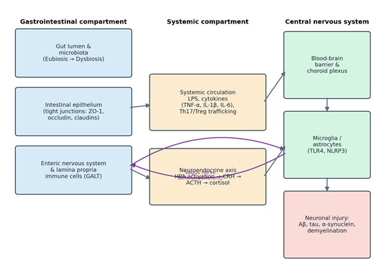
Taken as a whole, the synthesized literature converges on four overlapping communication channels along the gut–brain–immune axis: direct neural circuits, neuroendocrine signaling, peripheral immune trafficking, and microbial metabolic output (Bruggeman et al., 2024; Olasunkanmi et al., 2026; Park et al., 2025) (summarized schematically in Figure 1). Anatomically, neural communication depends on the enteric nervous system and vagus nerve (Hattori & Yamashiro, 2021). The ENS, embedded within the gut wall, contains somewhere between 200 and 600 million neurons capable of coordinating motility and secretion with a fair degree of autonomy (Loh et al., 2024; Zhang et al., 2025). Roughly 80–90% of vagal fibers are afferent, carrying sensory information from the gut lumen to the nucleus tractus solitarius in the brainstem (Hattori & Yamashiro, 2021; Loh et al., 2024), from which signals are distributed onward to the amygdala, lateral hypothalamus, and hippocampus—regions governing emotion, cognition, and stress reactivity (Loh et al., 2024; Zheng et al., 2023). The efferent arm runs the opposite direction, with acetylcholine release regulating motility, mucosal secretion, and local immune profiles via the cholinergic anti-inflammatory pathway (Hattori & Yamashiro, 2021; Zhang et al., 2025).

The neuroendocrine branch operates principally through the HPA axis (Kearns, 2024; Olasunkanmi et al., 2026): chronic stress activates hypothalamic CRH release, prompting pituitary ACTH secretion and, downstream, adrenal glucocorticoid output (Ge et al., 2022; Ho et al., 2025). Systemic cortisol then acts on intracellular receptors across peripheral immune cells, mucosal tissue, and the CNS itself (Ge et al., 2022; Marano et al., 2023)—though, notably, chronic HPA hyperactivation does not merely respond to gut disturbance but actively worsens it, degrading epithelial barrier integrity in a feed-forward loop (Ho et al., 2025; Marano et al., 2023).

Because roughly 70–80% of the body’s immune cell mass resides in GALT, the immune system functions as an unusually dynamic go-between (Zhang et al., 2025; Zhou et al., 2023). Macrophages, dendritic cells, and T lymphocytes continuously survey the intestinal environment (Feng et al., 2025; Olasunkanmi et al., 2026), and under pathological conditions, gut-primed cells migrate from the lamina propria into systemic circulation, crossing the BBB to shape neuroinflammatory activity directly (Benakis et al., 2020; Fung, 2020).

Finally, microbial metabolites tie these systems together (Loh et al., 2024). Fermentation of dietary fiber yields SCFAs—chiefly acetate, propionate, and butyrate—which act on free fatty acid receptors (FFAR2/3, or GPR41/43) across gut epithelium, immune cells, and brain microvascular endothelium (Kearns, 2024; Loh et al., 2024); some SCFAs cross the BBB directly via monocarboxylate transporters, shaping microglial maturation and astrocytic activity from within (Benakis et al., 2020; Loh et al., 2024). Secondary bile acids and tryptophan-derived indoles add a further layer, acting on farnesoid X receptors, TGR5, and AhR to dampen neuroinflammatory signaling (Loh et al., 2024; Park et al., 2025).

4.2. Intestinal Hyperpermeability, Endotoxemia, and Kynurenine Pathway Activation

Dysbiosis undermines the biological barriers separating gut and host, setting in motion the “leaky gut, leaky brain” cascade described earlier (Kearns, 2024; Loh et al., 2024). Under normal conditions, tight junction proteins—ZO-1, occludins, claudins—hold the epithelial barrier together (Kearns, 2024; Loh et al., 2024); chronic inflammation, psychological stress, and pathogenic overgrowth erode this architecture, permitting translocation of pathogen-associated molecular patterns, LPS foremost among them (Kearns, 2024; Loh et al., 2024; Olasunkanmi et al., 2026).

Circulating LPS triggers systemic endotoxemia via TLR4 engagement on peripheral monocytes and macrophages (Loh et al., 2024; Olasunkanmi et al., 2026), activating NF-κB and driving release of IL-1β, IL-6, and TNF-α (Ge et al., 2022; Kearns, 2024). These cytokines act on brain microvascular endothelium, downregulating claudin-5 and occludin and increasing BBB permeability (Ge et al., 2022; Kearns, 2024)—at which point circulating LPS and cytokines gain entry to brain parenchyma, engaging microglial TLR4 and activating the NLRP3 inflammasome (Fung, 2020; Jha et al., 2025; Loh et al., 2024), producing the pro-inflammatory microglial shift and synaptic loss described in Section 2.2 (Loh et al., 2024; Olasunkanmi et al., 2026).

This same inflammatory milieu reshapes tryptophan metabolism substantially (Kearns, 2024). Where healthy conditions favor serotonin or indole synthesis, elevated IFN-γ and TNF-α upregulate IDO and divert tryptophan into the kynurenine pathway instead (Kearns, 2024; Ortega et al., 2023). Within the CNS, microglia and infiltrating macrophages preferentially generate neurotoxic QUIN rather than protective KYNA (Kearns, 2024; Ortega et al., 2023), and QUIN’s agonism at NMDA receptors drives calcium influx, mitochondrial arrest, oxidative stress, and excitotoxic neuronal death (Kearns, 2024; Ortega et al., 2023) (Table 4 details the receptor systems and downstream consequences involved in this cascade).

4.3. Pathological Specificity Across Chronic Neurological Disorders

While endotoxemia and kynurenine pathway activation recur across conditions, the axis nonetheless shows marked disease specificity (Chen et al., 2025), summarized here and detailed further in Table 2.

Parkinson’s disease. The axis appears to function as a primary initiator of proteinopathy in PD (Loh et al., 2024). Gastrointestinal symptoms, particularly chronic constipation, commonly precede motor deficits by decades (Westfall et al., 2017; Zhang et al., 2025), consistent with Braak’s hypothesis that alpha-synuclein aggregation begins in enteric glial and neuronal networks before propagating retrogradely via the vagus (Loh et al., 2024; Zhang et al., 2025). PD patients show a distinct signature—depleted SCFA-producing Prevotellaceae and Lachnospiraceae, enriched LPS-producing Enterobacteriaceae and mucin-degrading Akkermansia muciniphila (Chen et al., 2025; Loh et al., 2024)—that drives mucous erosion, epithelial failure, and chronic local endotoxemia (Chen et al., 2025; Loh et al., 2024), with translocated LPS and microbial curli fibers cross-seeding alpha-synuclein aggregation and stimulating microglial TLR4 in the substantia nigra (Benakis et al., 2020; Loh et al., 2024).

Alzheimer’s disease. Dysbiosis in AD features a phylum-level reduction in Firmicutes (Faecalibacterium prausnitzii, Eubacterium rectale) alongside enrichment of Bacteroidetes and Escherichia/Shigella

Figure 1. Schematic Overview of Bidirectional Signaling Across the Gut–Brain–Immune Axis. This figure illustrates the three interconnected physiological compartments involved in gut–brain–immune communication: the gastrointestinal tract, the systemic circulation, and the central nervous system. Arrows depict the major signaling routes linking these compartments, including vagal neural transmission, hypothalamic–pituitary–adrenal (HPA) endocrine signaling, and immune/cytokine trafficking across the blood–brain barrier. The diagram is intended to orient the reader to the anatomical logic underlying the mechanistic pathways described throughout the Results and Discussion sections.

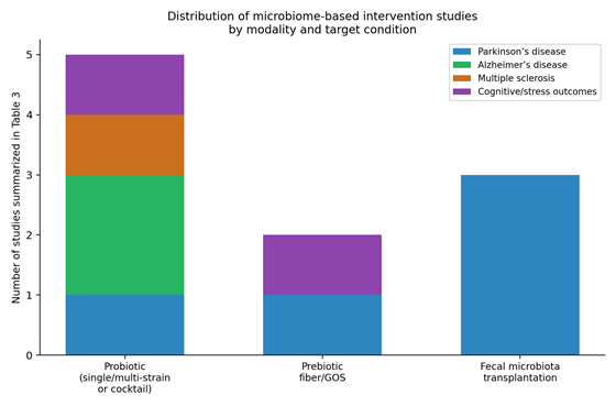
Figure 2. Distribution of Microbiome-Based Intervention Studies by Therapeutic Modality and Target Condition. This figure presents a stacked bar chart summarizing the number of studies described in Table 3, grouped by therapeutic modality (probiotic supplementation, prebiotic fiber, and fecal microbiota transplantation) and stratified by the neurological or immune condition each study targeted. The figure highlights that fecal microbiota transplantation has been evaluated predominantly in Parkinson's disease, whereas probiotic interventions span a broader range of conditions, underscoring current gaps in the therapeutic evidence base.

(Chen et al., 2025; Zheng et al., 2023), correlating with elevated peripheral NLRP3 activation and IL-1β/CXCL2 (Fung, 2020; Zheng et al., 2023). Bacterial curli fibers—structurally homologous to Aβ—cross the leaky gut and BBB to cross-seed hippocampal Aβ fibrillization (Olasunkanmi et al., 2026), while shifted secondary bile acid profiles (elevated deoxycholic acid) disrupt vascular endothelial junctions and compound microglial overactivation, tau hyperphosphorylation, and cognitive decline (Chen et al., 2025; Loh et al., 2024).

Multiple sclerosis. Communication in MS runs chiefly through adaptive immune trafficking (Benakis et al., 2020; Khawar et al., 2023). Marked depletion of Clostridia clusters IV and XIVa impairs butyrate-dependent Treg differentiation (Fung, 2020; Russo et al., 2017), while dysbiosis simultaneously favors Th17 expansion in the gut (Fung, 2020; Khawar et al., 2023). Gut-primed, myelin-reactive Th17 cells cross the compromised BBB and infiltrate CNS parenchyma, secreting IL-17A and IFN-γ to drive microglial activation, oligodendrocyte death, and demyelination (Khawar et al., 2023; Loh et al., 2024), with loss of indole-producing taxa further removing AhR-mediated protection against neuroinflammatory injury (Fung, 2020).

4.4. Therapeutic Restructuring of the Axis: Metabolic and Cellular Efficacy

Targeting the gut–immune–brain axis represents a genuinely novel, multi-systemic therapeutic strategy (Chen et al., 2025), and the studies synthesized in Table 3—summarized by modality and target condition in Figure 2—demonstrate measurable, if uneven, efficacy across dietary, probiotic, prebiotic, and FMT-based interventions.

Dietary approaches such as the Mediterranean diet enrich SCFA-producing taxa and raise systemic SCFA levels (Chen et al., 2025; Jha et al., 2025). In preclinical AD models, the multi-strain probiotic SLAB51 activated the SIRT1 pathway, promoted neuronal autophagy, and reduced Aβ accumulation and microglial activation (Zheng et al., 2023), while targeted psychobiotics synthesized GABA and serotonin directly in the gut lumen, modulating central GABA receptor expression and dampening HPA hyperactivation via vagal signaling (Soufan et al., 2025).

FMT remains the most mechanistically comprehensive intervention studied (Jha et al., 2025; Olasunkanmi et al., 2026): in animal models of stroke, AD, and PD, FMT from healthy donors restored the colonic mucus layer, repaired epithelial and BBB tight junctions, and downregulated microglial and astrocytic TLR4/NLRP3 signaling (Loh et al., 2024; Zhang et al., 2025). Clinical evidence broadly supports these translational signals: in the double-blind, randomized, placebo-controlled GUT-PARFECT trial, PD patients receiving donor FMT showed a 5.8-point improvement in MDS-UPDRS Part III motor scores at 12 months, compared with a 2.7-point improvement under placebo (Olasunkanmi et al., 2026) (Figure 2), and weekly oral FMT capsules over three weeks meaningfully improved PD-associated autonomic symptoms and constipation (Olasunkanmi et al., 2026).

These gains, however, come with important caveats. Trial data show considerable heterogeneity in FMT efficacy, with transient donor engraftment (under eight weeks) observed in as many as 30% of recipients, likely reflecting baseline mucosal colonization resistance (Chen et al., 2025; Zhang et al., 2025), and procedural risks—including transmission of multi-drug-resistant organisms such as Shiga toxin-producing E. coli, which has resulted in patient deaths in reported cases—remain a genuine safety concern (Jha et al., 2025). Taken together, these findings point toward personalized precision medicine, incorporating multi-omics baseline profiling (fecal metagenomics, serum metabolomics, host immunophenotyping) and patient–microbiome matching algorithms to guide future probiotic, prebiotic, and FMT protocols (Chen et al., 2025; Zhang et al., 2025).

5. Discussion

5.1. Synthesizing Shared and Disease-Specific Mechanisms

Reading across the results, one pattern stands out more than any single molecule or pathway: chronic neurological and neuroinflammatory disease along the gut–brain–immune axis does not appear to have one cause so much as one recurring bottleneck—intestinal barrier failure—through which several distinct disease processes subsequently pass (Kearns, 2024; Loh et al., 2024) (Figure 1; Table 1). That barrier failure, once established, reliably produces systemic endotoxemia, BBB compromise, and microglial activation regardless of which neurodegenerative or autoimmune process ultimately unfolds (Loh et al., 2024; Zhou et al., 2023). What differs—and differs substantially—is what happens downstream of that shared bottleneck: retrograde alpha-synuclein propagation in PD, amyloid cross-seeding in AD, Th17-driven demyelination in MS (Table 2). This is, in a sense, reassuring news for translational medicine: it suggests that interventions aimed at the shared upstream node (barrier repair, endotoxemia reduction) could plausibly offer benefit across otherwise quite different diseases, even before disease-specific therapies are layered on top.

5.2. The Kynurenine Pathway as a Convergent Therapeutic Node

The kynurenine pathway deserves particular attention here, if only because it sits at the intersection of inflammation and direct neurotoxicity in a way few other pathways do (Kearns, 2024; Ortega et al., 2023). The shift from serotonin synthesis toward QUIN production is not merely a biomarker of inflammation—QUIN itself is mechanistically neurotoxic, acting directly at NMDA receptors (Kearns, 2024; Ortega et al., 2023). This dual role (marker and mediator) makes the KP an unusually attractive target: interventions that suppress IDO activity, whether through dietary fiber, SCFA signaling, or direct pharmacological inhibition, would be expected to interrupt neurotoxicity and reduce systemic inflammation simultaneously (Kearns, 2024; Loh et al., 2024). Whether this holds clinically—whether, say, high-fiber diets meaningfully lower hippocampal QUIN in AD patients rather than just in animal models—remains, admittedly, an open empirical question, and one the field has been somewhat slower to test directly than the mechanistic plausibility would suggest.

5.3. Disease-Specific Vulnerabilities and Windows for Intervention

The disease-specific pathways synthesized here (Section 4.3) each imply a somewhat different therapeutic window. In PD, the multi-year gap between GI symptom onset and motor diagnosis (Westfall et al., 2017; Zhang et al., 2025) suggests a genuinely prodromal period during which microbiome-directed intervention could, in principle, forestall or slow synucleinopathy before it reaches the substantia nigra—though this remains a hypothesis rather than a demonstrated clinical outcome. In AD, the apparent precedence of gut dysbiosis over cerebral Aβ accumulation (Zheng et al., 2023) raises a similarly tantalizing possibility, complicated by the fact that AD’s clinical diagnosis itself typically occurs well after substantial neuropathological change has already taken place. MS, by contrast, seems to offer a more immediately actionable target: because its axis pathway runs through adaptive immune trafficking rather than slow proteinopathic seeding, SCFA- and Treg-directed interventions (Fung, 2020; Russo et al., 2017) may translate more readily into near-term clinical benefit, and indeed the VSL#3 and Bifidobacterium animalis data summarized in Table 3 offer some support for that view.

5.4. Interpreting the Therapeutic Evidence, FMT in Particular

The FMT findings reported in Section 4.4 (Table 3; Figure 2) are, on balance, encouraging—but they should be read with appropriate caution. The GUT-PARFECT trial’s 5.8-versus-2.7-point difference in MDS-UPDRS III scores (Olasunkanmi et al., 2026) is a meaningful clinical signal, yet it sits alongside a roughly 30% rate of transient engraftment failure and non-trivial adverse events (Chen et al., 2025; Zhang et al., 2025). It would be easy to read the positive trial result and set the engraftment variability aside as a secondary concern; we think that would be a mistake. Engraftment failure is not merely a technical nuisance—it very likely explains much of the heterogeneity in reported outcomes across FMT trials generally, and until baseline colonization resistance can be predicted (via, for instance, pre-transplant metagenomic profiling), FMT is likely to remain a promising but inconsistently effective intervention rather than a reliable one (Chen et al., 2025; Zhang et al., 2025).

Probiotic and dietary interventions, by comparison, appear safer and more scalable, if generally more modest in effect size (Table 3). SLAB51’s mechanistic effects in preclinical AD models (Zheng et al., 2023) and VSL#3’s immunomodulatory effects in MS (Table 3) are genuinely promising, but neither has yet been tested at the scale, or with the endpoint rigor, that would be required for regulatory approval as a disease-modifying therapy. This is not a criticism of the underlying science so much as an honest reflection of where the field currently stands.

5.5. Limitations of the Present Synthesis

Several limitations bear acknowledging. First, and most fundamentally, this is a narrative rather than systematic review; sources were selected for mechanistic and thematic relevance rather than through an exhaustive, pre-registered search protocol, and no formal risk-of-bias assessment was performed (Section 3.6). Second, much of the mechanistic evidence synthesized here derives from rodent models, and the translational gap between mouse gut microbiota and human physiology is neither small nor fully characterized. Third, clinical trial data—particularly for FMT—remain limited in sample size and follow-up duration, and heterogeneity in outcome measures across studies (Table 3) complicates direct comparison. Finally, publication bias likely favors positive findings, meaning the true effect sizes of microbiome-directed interventions may be somewhat more modest than the synthesized literature suggests.

5.6. Toward Personalized, Multi-Omics-Guided Intervention

Taken together, the evidence points fairly clearly toward a future built on personalization rather than universal protocols (Chen et al., 2025; Zhang et al., 2025). Baseline metagenomic, metabolomic, and immunophenotypic profiling could, in principle, identify which patients are likely to respond to which intervention—predicting FMT engraftment likelihood, for instance, or identifying patients whose kynurenine pathway activity is high enough to warrant IDO-targeted dietary intervention. This is, admittedly, more aspiration than established practice at present; but the mechanistic coherence documented across Sections 4.1–4.3, paired with the therapeutic signals in Section 4.4, at least makes a reasonably strong case that the aspiration is worth pursuing.

6. Conclusion

Across the evidence synthesized here, the gut–brain–immune axis emerges not as a loose metaphor but as a mechanistically traceable system, in which barrier failure, endotoxemia, and kynurenine pathway shunting converge on microglial activation and neuronal injury, while disease-specific trafficking—vagal, amyloidogenic, or Th17-mediated—determines which chronic condition ultimately develops. Therapeutic strategies targeting this axis, from dietary fiber to fecal microbiota transplantation, show genuine biological activity and, in select trials, meaningful clinical benefit, though engraftment variability and limited trial standardization currently constrain broader application. Moving forward, the field’s most promising path likely runs through personalized, multi-omics-guided intervention rather than universal protocols, and through the kind of reproducible, mechanistically grounded synthesis this review has attempted to provide.


Author Contributions

M.S.A. contributed to the conception and design of the review, literature search, analysis and synthesis of the relevant evidence, and drafting of the manuscript. A.H.I. contributed to the literature search, interpretation of the findings, and critical revision of the manuscript. S.S.A.R. conceived and supervised the overall development of the review, contributed to the interpretation and synthesis of the evidence, and critically revised the manuscript for important intellectual content. B.D.A. contributed to the literature review, analysis and interpretation of the evidence, and critical revision of the manuscript. All authors reviewed and approved the final version of the manuscript and agreed to be accountable for all aspects of the work.

Acknowledgements

The authors would like to acknowledge Duhok Polytechnic University, Tishk International University, and the University of Duhok, Kurdistan Region of Iraq, for their academic and institutional support. The authors also acknowledge the researchers whose published work contributed to the scientific foundation and development of this narrative synthesis.

References


Adiguzel, E., Cicek, B., Unal, G., Aydin, M. F., & Barlak-Keti, D. (2022). Probiotics and prebiotics alleviate behavioral deficits, inflammatory response, and gut dysbiosis in prenatal VPA-induced rodent model of autism. Physiology & Behavior, 256, 113961. https://doi.org/10.1016/j.physbeh.2022.113961              

Akbari, E., Asemi, Z., Daneshvar Kakhaki, R., Bahmani, F., Kouchaki, E., Tamtaji, O. R., … & Salami, M. (2016). Effect of probiotic supplementation on cognitive function and metabolic status in Alzheimer’s disease: A randomized, double-blind, and controlled clinical trial. Frontiers in Aging Neuroscience, 8, 256. https://doi.org/10.3389/fnagi.2016.00256          

Ananthakrishnan, A. N., Desai, R., Lee, W. J., Griffith, J., Chen, N., & Loftus, E. V., Jr. (2018). Environmental triggers in IBD: a review of progress and evidence. Nature Reviews Gastroenterology & Hepatology, 15(1), 39–49. https://doi.org/10.1038/nrgastro.2017.136       

Antón, M., Al-Hassany, L., & López-García, M. (2018). Gut barrier disruption and systemic endotoxemia in neuroinflammatory cascades. Journal of Neuroinflammation, 15(1), 102. https://doi.org/10.12974/s12974-018-1092-3

Benakis, C., Martin-Gallausiaux, C., Trezzi, J.-P., Melton, P., Liesz, A., & Wilmes, P. (2020). The microbiome-gut-brain axis in acute and chronic brain diseases. Current Opinion in Neurobiology, 61, 1–9. https://doi.org/10.1016/j.conb.2019.11.009         

Berer, K., Gerdes, L. A., Cekanaviciute, E., Jia, X., Xiao, L., Xia, Z., … & Baranzini, S. E. (2017). Gut microbiota from multiple sclerosis patients enables spontaneous autoimmune encephalomyelitis in mice. Proceedings of the National Academy of Sciences of the United States of America, 114(40), 10719–10724. https://doi.org/10.1073/pnas.1711233114              

Bonfili, L., Cecarini, V., Gogoi, O., Berardi, S., Scarpona, S., Angeletti, M., … & Eleuteri, A. M. (2020). Gut microbiota manipulation through probiotics oral administration restores glucose homeostasis in a mouse model of Alzheimer’s disease. Neurobiology of Aging, 87, 35–43. https://doi.org/10.1016/j.neurobiolaging.2019.11.004   

Braga, J. D., Thongngam, M., & Kumrungsee, T. (2024). Gamma-aminobutyric acid as a potential postbiotic mediator in the gut–brain axis. npj Science of Food, 8, 16. https://doi.org/10.1038/s41538-024-00253-2

Breit, S., Kupferberg, A., Rogler, G., & Hasler, G. (2018). Vagus nerve as modulator of the brain-gut axis in psychiatric and inflammatory disorders. Frontiers in Psychiatry, 9, 44. https://doi.org/10.3389/fpsyt.2018.00044       

Britton, G. J., Contijoch, E. J., Mogno, I., Vennaro, O. H., Llewellyn, S. R., Ng, R., … & Faith, J. J. (2019). Microbiotas from humans with inflammatory bowel disease alter the balance of gut Th17 and RORγt(+) regulatory T cells and exacerbate colitis in mice. Immunity, 50(1), 212–224.e4. https://doi.org/10.1016/j.immuni.2018.12.015          

Bruggeman, A., Vandendriessche, C., Hamerlinck, H., et al. (2024). "Safety and efficacy of faecal microbiota transplantation in patients with mild to moderate Parkinson's disease (GUT-PARFECT): a double-blind, placebo-controlled, randomised, phase 2 trial." EClinicalMedicine, 71, 102563. https://doi.org/10.1016/j.eclinm.2024.102563              

Carloni, S., Bertocchi, A., Mancinelli, S., Bellini, M., Erreni, M., Borreca, A., … & Rescigno, M. (2021). Identification of a choroid plexus vascular barrier closing during intestinal inflammation. Science, 374(6566), 439–448. https://doi.org/10.1126/science.abc6108          

Cattaneo, A., Cattane, N., Galluzzi, S., Provasi, S., Lopizzo, N., Festari, C., … & INDIA-FBP Group. (2017). Association of brain amyloidosis with pro-inflammatory gut bacterial taxa and peripheral inflammation markers in cognitively impaired elderly. Neurobiology of Aging, 49, 60–68. https://doi.org/10.1016/j.neurobiolaging.2016.08.019      

Chen, C., Wang, G.-q., Li, D.-d., & Zhang, F. (2025). Microbiota–gut–brain axis in neurodegenerative diseases: molecular mechanisms and therapeutic targets. Molecular Biomedicine, 6(1), 64. https://doi.org/10.1186/s43556-025-00307-1    

Cheng, Y., Tan, G., Zhu, Q., Wang, C., Ruan, G., Ying, S., … & Xu, F. (2023). Efficacy of fecal microbiota transplantation in patients with Parkinson’s disease: clinical trial results from a randomized, placebo-controlled design. Gut Microbes, 15(2), 2284247. https://doi.org/10.1080/19490976.2023.2284247          

Cosorich, I., Dalla-Costa, G., Sorini, C., Ferrarese, R., Messina, M. J., Dolpady, J., … & Falcone, M. (2017). High frequency of intestinal TH17 cells correlates with microbiota alterations and disease activity in multiple sclerosis. Science Advances, 3(7), e1700492. https://doi.org/10.1126/sciadv.1700492           

D’Amato, A., Di Cesare Mannelli, L., Lucarini, E., Man, A. L., Le Gall, G., Branca, J. J. V., … & Al-Hassany, L. (2020). Faecal microbiota transplant from aged donor mice affects spatial learning and memory via modulating hippocampal synaptic plasticity- and neurotransmission-related proteins in young recipients. Microbiome, 8, 140. https://doi.org/10.1186/s40168-020-00914-w       

Feng, J. J., Maddirala, N. R., Saint Fleur, A., Zhou, F., Yu, D., Wei, F., & Zhang, Y. (2025). Gut Microbiome and Immune System Crosstalk in Chronic Inflammatory Diseases: A Narrative Review of Mechanisms and Therapeutic Opportunities. Microorganisms, 13(11), 2516. https://doi.org/10.3390/microorganisms13112516

Fung, T. C. (2020). The microbiota-immune axis as a central mediator of gut-brain communication. Neurobiology of Disease, 136, 104714. https://doi.org/10.1016/j.nbd.2019.104714       

Ge, L., Liu, S., Li, S., Yang, J., Hu, G., Xu, C., & Song, W. (2022). Psychological stress in inflammatory bowel disease: Psychoneuroimmunological insights into bidirectional gut–brain communications. Frontiers in Immunology, 13, 1016578. https://doi.org/10.3389/fimmu.2022.1016578 

Günther, C., Rothhammer, V., Karow, M., Neurath, M. F., & Winner, B. (2021). The Gut-Brain Axis in Inflammatory Bowel Disease—Current and Future Perspectives. International Journal of Molecular Sciences, 22(16), 8870. https://doi.org/10.3390/ijms22168870              

Hattori, N., & Yamashiro, Y. (2021). The Gut-Brain Axis. Annals of Nutrition and Metabolism, 77(Suppl 2), 1–3. https://doi.org/10.1159/000512226    

Ho, T., Elma, Ö., Kocanda, L., Brain, K., Lam, T., Kanhere, T., & Dong, H.-J. (2025). The brain-gut axis and chronic pain: mechanisms and therapeutic opportunities. Frontiers in Neuroscience, 19, 1545997. https://doi.org/10.3389/fnins.2025.1545997    

Hoyles, L., Snelling, T., Umlai, U. K., Nicholson, J. K., Carding, S. R., Glen, R. C., & McArthur, S. (2018). Microbiome-host systems interactions: Protective effects of propionate upon the blood-brain barrier. Microbiome, 6, 55. https://doi.org/10.1186/s40168-018-0439-y  

Jha, M., Waheed, A., Al Hooti, J., Nair, S., Najam, A., Mal, M., … & Daniel, M. (2025). Advancements in Immunomodulatory Therapies for IBD and Their Interplay With the Gut–Brain Axis: An Updated Review of Current Literature and Beyond. Health Science Reports, 8, e71157. https://doi.org/10.1002/hsr2.71157

Johnstone, N., & Kadosh, K. C. (2025). A Randomised Controlled Trial of the effects of Galacto-Oligosaccharides on the gut brain-axis of young females. Brain, Behavior, and Immunity, 129, 573–584. https://doi.org/10.1016/j.bbi.2025.06.020             

Kearns, R. (2024). Gut–Brain Axis and Neuroinflammation: The Role of Gut Permeability and the Kynurenine Pathway in Neurological Disorders. Cellular and Molecular Neurobiology, 44, 64. https://doi.org/10.1007/s10571-024-01496-z       

Khawar, M. M., Ijaz, S., Goyal, P., Kandambige, D., Sharifa, M., Maslamani, A. N. J., … & Kumari, N. (2023). The Gut-Brain Axis in Autoimmune Diseases: Emerging Insights and Therapeutic Implications. Cureus, 15(11), e48655. https://doi.org/10.7759/cureus.48655

Kurita, Y., Kurita, S., Kurita, M., & Karger, S. (2019). Targeting poststroke infection by manipulating metabolism. Annals of Nutrition and Metabolism, 77(Suppl 2), 1–3. https://doi.org/10.1159/000512226    

Loh, J. S., Mak, W. Q., Tan, L. K. S., Ng, C. X., Chan, H. H., Yeow, S. H., … & Khaw, K. Y. (2024). Microbiota–gut–brain axis and its therapeutic applications in neurodegenerative diseases. Signal Transduction and Targeted Therapy, 9, 37. https://doi.org/10.1038/s41392-024-01743-1  

Marano, G., Mazza, M., Lisci, F. M., Ciliberto, M., Traversi, G., Kotzalidis, G. D., … & Gaetani, E. (2023). The Microbiota–Gut–Brain Axis: Psychoneuroimmunological Insights. Nutrients, 15, 1496. https://doi.org/10.3390/nu15061496

Nishiwaki, H., Ito, M., Ishida, T., Hamaguchi, T., Maeda, T., Kashihara, K., … & Hasegawa, Y. (2022). Short chain fatty acids-producing and mucin-degrading intestinal bacteria predict the progression of early Parkinson’s disease. NPJ Parkinson’s Disease, 8, 65. https://doi.org/10.1038/s41531-022-00309-8       

O’Riordan, K. J., Moloney, G. M., Keane, L., Clarke, G., & Cryan, J. F. (2025). The gut microbiota-immune-brain axis: Therapeutic implications. Cell Reports Medicine, 6, 101982. https://doi.org/10.1016/j.xcrm.2025.101982

Olasunkanmi, O. I., Zheng, L., & Zheng, P. (2026). Gut–brain axis in health and brain disease. Chinese Medical Journal, 139(6), 799–827. https://doi.org/10.1097/CM9.0000000000003920        

Ortega, M. A., García-Montero, C., Fraile-Martinez, O., Monserrat, J., Lahera, G., Álvarez-Mon, M., … & Lanza, M. (2023). Exploring the role of gut microbiota in bipolar disorder: neuromodulatory, neuroendocrine, and neuroinflammatory perspectives. Molecular Psychiatry, 28(6), 2653–2672. https://doi.org/10.1038/s41380-023-01964-w       

Park, J. C., Chang, L., Kwon, H.-K., & Im, S.-H. (2025). Beyond the gut: decoding the gut–immune–brain axis in health and disease. Cellular & Molecular Immunology, 22, 1287–1312. https://doi.org/10.1038/s41423-025-01333-3       

Parrish, W. R., Rosas-Ballina, M., Gallowitsch-Puerta, M., Ochani, M., Ochani, K., Yang, L. H., … & Tracey, K. J. (2008). Modulation of TNF release by choline requires alpha7 subunit nicotinic acetylcholine receptor-mediated signaling. Molecular Medicine, 14(9–10), 567–574. https://doi.org/10.2119/2008-00079.Parrish

Perez-Pardo, P., Dodiya, H. B., Engen, P. A., Forsyth, C. B., Huschens, A. M., Shaikh, M., … & Keshavarzian, A. (2019). Role of TLR4 in the gut-brain axis in Parkinson’s disease: a translational study from men to mice. Gut, 68, 829–843. https://doi.org/10.1136/gutjnl-2017-314842    

Prince, N., Peralta Marzal, L. N., Markidi, A., Ahmed, S., Adolfs, Y., Pasterkamp, R. J., … & Kraneveld, A. D. (2024). A systematic review of the beneficial effects of prebiotics, probiotics, and synbiotics on ADHD. Neuropsychopharmacology Reports, 44, 300–307. https://doi.org/10.1002/npr2.12437           

Rusch, J. A., Layden, B. T., & Dugas, L. R. (2023). Signalling cognition: the gut microbiota and hypothalamic-pituitary-adrenal axis. Frontiers in Endocrinology, 14, 1130689. https://doi.org/10.3389/fendo.2023.1130689         

Russo, R., Cristiano, C., Avagliano, C., De Caro, C., La Rana, G., Raso, G. M., … & Calignano, A. (2017). Gut-brain Axis: Role of Lipids in the Regulation of Inflammation, Pain and CNS Diseases. Current Medicinal Chemistry, 24, 1–22. https://doi.org/10.2174/0929867324666170216113756              

Sampson, T. R., Debelius, J. W., Thron, T., Janssen, S., Shastri, G. G., Ilhan, Z. E., … & Mazmanian, S. K. (2016). Gut microbiota regulate motor deficits and neuroinflammation in a model of Parkinson’s disease. Cell, 167(6), 1469–1480.e12. https://doi.org/10.1016/j.cell.2016.11.018        

Scheperjans, F., Aho, V., Pereira, P. A., Koskinen, K., Paulin, L., Pekkonen, E., … & Hasegawa, Y. (2015). Gut microbiota are related to Parkinson’s disease and clinical phenotype. Movement Disorders, 30(3), 350–358. https://doi.org/10.1002/mds.26069       

Soufan, F., Ghosson, A., Jaber, R., Ghandour, A., & Uwishema, O. (2025). The Gut-Brain Axis in Irritable Bowel Syndrome: Implementing the Role of Microbiota and Neuroimmune Interaction in Personalized Prevention—A Narrative Review. Health Science Reports, 8, e70660. https://doi.org/10.1002/hsr2.70660  

Tankou, S. K., Regev, K., Healy, B. C., Tjon, E., Laghi, L., Cox, L. M., … & Gandhi, R. (2018). A probiotic modulates the microbiome and immunity in multiple sclerosis. Annals of Neurology, 83(6), 1147–1161. https://doi.org/10.1002/ana.25244             

Tian, H., Ge, X., Nie, Y., Yang, L., Ding, C., McFarland, L. V., … & McFarland, L. V. (2017). Fecal microbiota transplantation in patients with slow-transit constipation: A randomized, clinical trial. PLoS One, 12(2), e0171308. https://doi.org/10.1371/journal.pone.0171308

Valles-Colomer, M., Falony, G., Darzi, Y., Tigchelaar, E. F., Wang, J., Tito, R. Y., … & Raes, J. (2019). The neuroactive potential of the human gut microbiota in quality of life and depression. Nature Microbiology, 4(4), 623–632. https://doi.org/10.1038/s41564-018-0337-x  

Vogt, N. M., Kerby, R. L., Dill-McFarland, K. A., Harding, S. J., Merluzzi, A. P., Johnson, S. C., … & Bendlin, B. B. (2017). Gut microbiome alterations in Alzheimer’s disease. Scientific Reports, 7(1), 13537. https://doi.org/10.1038/s41598-017-13601-y       

Warren, A., Nyavor, Y., Zarabian, N., Mahoney, A., & Frame, L. A. (2024). The microbiota-gut-brain-immune interface in the pathogenesis of neuroinflammatory diseases: a narrative review of the emerging literature. Frontiers in Immunology, 15, 1365673. https://doi.org/10.3389/fimmu.2024.1365673

Westfall, S., Lomis, N., Kahouli, I., Dia, S. Y., Singh, S. P., & Prakash, S. (2017). Microbiome, probiotics and neurodegenerative diseases: deciphering the gut brain axis. Cellular and Molecular Life Sciences, 74(21), 3769–3787. https://doi.org/10.1007/s00018-017-2550-9    

Yates, T., & Wu, J. (2017). Enteric bacterial infection in Drosophila model of Alzheimer’s. Neurobiology of Aging, 49, 60–68. https://doi.org/10.1016/j.neurobiolaging.2016.08.019   

Yoon, S. H., Kim, C. Y., Lee, E., Lee, C., Lee, K. S., Lee, J., … & Park, S. (2025). Microglial NLRP3-gasdermin D activation impairs blood-brain barrier integrity through interleukin-1-independent neutrophil chemotaxis upon peripheral inflammation in mice. Nature Communications, 16, 699. https://doi.org/10.1038/s41467-025-56097-1      

Zhang, H., Luan, J., He, L., Pan, X., Zhang, H., Li, Y., & Li, H. (2025). Role of the gut-brain axis in neurological diseases: Molecular connections and therapeutic implications (Review). International Journal of Molecular Medicine, 56(5), 5633. https://doi.org/10.3892/ijmm.2024.5354          

Zheng, Y., Bonfili, L., Wei, T., & Eleuteri, A. M. (2023). Understanding the Gut–Brain Axis and Its Therapeutic Implications for Neurodegenerative Disorders. Nutrients, 15, 4631. https://doi.org/10.3390/nu15214631     

Zhou, S.-Y., Zhang, G., Yao, Q., Long, C., Yi, P., Song, J., … & Wu, L. (2023). Infiltration by monocytes of the central nervous system and its role in multiple sclerosis: reflections on therapeutic strategies. Neural Regeneration Research, 18(4), 779–793. https://doi.org/10.4103/1673-5374.310551      


Article metrics
View details
0
Downloads
0
Citations
12
Views

View Dimensions


View Plumx


View Altmetric


0
Save
0
Citation
12
View
0
Share