1. Introduction
Ageing, when you strip away the sentiment attached to it, is really just accumulated failure — a slow, uneven erosion of the systems that once kept an organism resilient to insult (López-Otín et al., 2023; Lopez-Otin et al., 2013). It is also, inconveniently, the single largest risk factor for the chronic diseases that dominate modern medicine, and yet clinicians are still largely stuck measuring it with a calendar. Chronological ag
e is easy to record and almost impossible to act on. It tells a physician how many years have passed since a patient was born, but it says remarkably little about how that patient’s kidneys, arteries, or immune system are actually holding up (Li et al., 2022; Polidori, 2024; Satapathy et al., 2026). For decades, public-health guidelines, insurance actuaries, and epidemiological risk models have leaned on this single number anyway, not because it was ever a particularly good proxy, but because nothing more precise was available (Polidori, 2024; Satapathy et al., 2026).
The trouble becomes obvious the moment you compare two people born on the same day. One may still be running marathons at seventy; the other may already be managing three chronic conditions. Genetics, behaviour, socioeconomic circumstance, and sheer environmental luck interact in ways that pull these two trajectories apart, sometimes dramatically, and chronological age has no vocabulary for any of it (Horvath & Raj, 2018; Li et al., 2022; Polidori, 2024). Geroscience’s response to this problem has been to propose an alternative construct — biological age, sometimes called physiological or phenotypic age — intended to capture the actual functional integrity of an individual’s cells, tissues, and organ systems, rather than the number of times the earth has gone around the sun (Yamada, 2025). Decoupling biological age from the calendar matters clinically because it turns ageing into something fluid and, at least in principle, something that can be intervened upon before overt disease appears, rather than after (Li et al., 2022). High-throughput omics technologies have given researchers several candidate ways to estimate this hidden variable — transcriptomic clocks, proteomic clocks, metabolomic signatures — but among these, DNA methylation-based estimators have consistently proven the most robust and reproducible (Ibáñez-Cabellos et al., 2026; Satapathy et al., 2026).
It is worth pausing on why methylation, specifically, turned out to be such a useful signal. Epigenetics broadly refers to chemical modifications layered on top of DNA and chromatin that shape gene expression and cellular identity without altering the underlying sequence (Satapathy et al., 2026). DNA methylation is the best-characterized of these marks: DNA methyltransferases attach a methyl group to the 5′ carbon of cytosine bases that precede guanine, producing 5-methylcytosine at so-called CpG dinucleotides (Satapathy et al., 2026; Wang et al., 2022). The functional consequences depend heavily on location — hypermethylation within CpG islands near gene promoters tends to silence transcription by blocking transcription-factor binding and recruiting chromatin remodellers, whereas broad, genome-wide hypomethylation is more typical of transcriptionally active, open chromatin (Satapathy et al., 2026; Wang et al., 2022). Across the human lifespan, these marks do not simply drift randomly; they shift in a fairly predictable direction, a phenomenon researcher have taken to calling “epigenetic drift” — a genome-wide loss of methylation in late-replicating regions paired with more localized gains at bivalent chromatin domains and Polycomb-repressed promoters (Horvath & Raj, 2018; Galow & Peleg, 2022). Because the underlying DNA sequence itself stays essentially fixed while these methylation marks respond to physical inactivity, chronic stress, diet, smoking, and pollutant exposure, the methylome ends up functioning almost like a running ledger of a person’s lifetime exposures — one that can, remarkably, be mathematically decoded (Galow & Peleg, 2022; Satapathy et al., 2026).
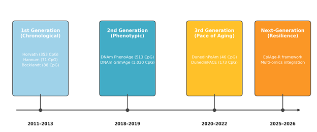
That decoding effort is what gave rise to “epigenetic clocks” — regularized regression models, typically elastic net, that assign predictive weights to a defined panel of CpG sites in order to output an estimated age (Horvath & Raj, 2018; Li et al., 2022). These tools have moved through several distinct generations since their introduction in the early 2010s, and the arc of that evolution is itself instructive. First-generation clocks were trained to predict chronological age directly, using blood or multi-tissue DNAm data; Bocklandt et al. (2011) built the earliest saliva-based estimator from 88 CpG sites in monozygotic twins, Hannum et al. (2013) followed with a 71-CpG blood clock, and Horvath’s (2013) 353-CpG pan-tissue model became the field’s benchmark, validated across fifty-one different human tissue and cell types. These models correlated with chronological age astonishingly well — often above r = 0.90 — but that very success turned out to conceal a limitation. Because the algorithms were optimized purely to track calendar time, they preferentially selected the most temporally stable CpGs, which meant they systematically discarded the more variable, environmentally responsive sites that actually carry information about health (Horvath & Raj, 2018; Levine et al., 2018; Yamada, 2025). Levine and colleagues later described this as the “paradox of chronological ageing”: accuracy against the calendar and biological relevance turned out to be, to some extent, competing objectives (Levine et al., 2018; Satapathy et al., 2026).
Second-generation clocks were built explicitly to correct for this. Rather than training on calendar age, Levine et al. (2018) trained DNAm PhenoAge on a composite “phenotypic age” derived from calendar age plus nine clinical markers spanning immune, metabolic, and renal function. Shortly after, Lu et al. (2019) constructed DNAm GrimAge by regressing methylation data against smoking pack-years and a panel of plasma proteins linked to mortality, including cystatin C, GDF-15, and PAI-1. Both models outperform their first-generation predecessors substantially in predicting all-cause mortality, cardiovascular disease, cancer incidence, and physical and cognitive decline (Li et al., 2022; Satapathy et al., 2026; Yamada, 2025). Third-generation clocks pushed the concept further still, aiming not to estimate a static age at all but to capture the instantaneous rate, or “pace,” of biological ageing. Drawing on repeated biomarker measurements from the Dunedin longitudinal cohort, Belsky and colleagues (2020) introduced DunedinPoAm, later refined into DunedinPACE (Belsky et al., 2022) — a model that behaves less like an odometer and more like a speedometer, and one that appears unusually sensitive to short-term lifestyle change.
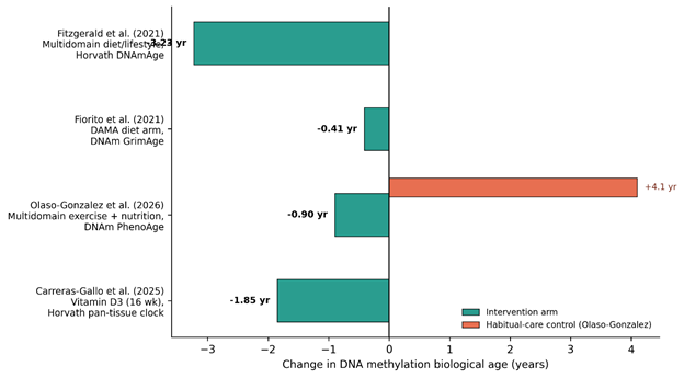
Despite this rapid conceptual progress, moving epigenetic clocks from population-level research tools into individual-level clinical instruments remains genuinely difficult. Technical noise across array batches can shift an individual’s estimated age by several years on its own (Apsley et al., 2025; Ibáñez-Cabellos et al., 2026); the methylome is tissue-specific, so a blood-based estimate may say little about pathology unfolding in cortical or cartilage tissue (Li et al., 2022; Satapathy et al., 2026; Yamada, 2025); and most reference datasets remain overwhelmingly drawn from European-ancestry cohorts, raising real concerns about equity when these tools are applied elsewhere (Apsley et al., 2025; Satapathy et al., 2026). What makes the field genuinely exciting, though — and this is really the premise of the present review — is the growing recognition that biological ageing is not a fixed, thermodynamically inevitable process but a plastic, biochemically reversible one (Satapathy et al., 2026). Randomized trials of multidomain lifestyle interventions (Fitzgerald et al., 2021; Olaso-Gonzalez et al., 2026), dietary and physical-activity programs (Fiorito et al., 2021), and even transient cellular reprogramming (Galow & Peleg, 2022) have each demonstrated measurable deceleration, and occasionally apparent reversal, of methylation age. That shift — from clocks as passive descriptors to clocks as therapeutic endpoints — motivates a newer conceptual framework, EpiAge-R (Epigenetic Age with Resilience), which attempts to separate degenerative methylation “noise” from adaptive, repair-associated signal by integrating chromatin topology, long-read nanopore methylation mapping, and mitochondrial dynamics into a single resilience-oriented model (Yamada, 2026).
Guided by this framework, the present review asks three linked questions that we believe should structure the next generation of longevity trials: first, a surrogacy question — can reversal of epigenetic age acceleration serve as a valid surrogate endpoint for hard clinical outcomes such as stroke or cardiovascular events; second, a synchronicity question — does localized tissue rejuvenation translate into systemic biological age deceleration; and third, a resilience question — can a composite resilience index outperform conventional clinical risk scores in predicting recovery from acute physiological stress. In pursuit of these questions, this review sets out four objectives: to compare the accuracy and clinical interpretability of first-, second-, and third-generation clocks across tissues and populations; to synthesize trial evidence on the mechanisms by which lifestyle and pharmacological interventions reverse DNAm age; to outline the EpiAge-R framework as a candidate next-generation vital sign; and, finally, to identify the methodological and ethical gaps that must be closed before biological age can be used responsibly at the level of the individual patient.