Integrative Biomedical Research

Integrative Biomedical Research (Journal of Angiotherapy) | Online ISSN  3068-6326
463
Citations
1.8m
Views
772
Articles
Your new experience awaits. Try the new design now and help us make it even better
Switch to the new experience
Figures and Tables
REVIEWS   (Open Access)

Cellular Reprogramming Strategies for Age-Related Macular Degeneration From iPSCs to In Vivo Therapy

Andrew Johan 1*, Irene Uli 2, Yosefani Fortunella Yasmine 2

+ Author Affiliations

Integrative Biomedical Research 10 (2) 1-8 https://doi.org/10.25163/biomedical.10210927

Submitted: 20 August 2026 Revised: 12 October 2026  Published: 23 October 2026 


Abstract

Age-related macular degeneration (AMD) remains one of the leading, and most stubborn, causes of irreversible central vision loss in older adults, and for a long time clinicians could do little more than slow its march. That picture has begun to change. This review traces how cellular reprogramming technologies — from induced pluripotent stem cell (iPSC)-derived retinal pigment epithelium (RPE) transplantation to direct lineage transdifferentiation and, more recently, in vivo partial epigenetic reprogramming — are reshaping what regenerative ophthalmology can offer. Background: since Takahashi and Yamanaka's original demonstration that four transcription factors can reset adult somatic cells to pluripotency, the field has moved from single-patient autologous grafts toward banked, HLA-matched allogeneic lines and, in parallel, toward strategies that bypass pluripotency altogether. Methods: we conducted a structured, PubMed-oriented narrative synthesis of peer-reviewed literature, clinical trial registries, and preprint sources addressing stem cell platforms, reprogramming delivery systems, direct transdifferentiation, and partial in vivo rejuvenation relevant to retinal degeneration, screened for relevance and synthesized thematically rather than through formal meta-analysis. Results: autologous iPSC-RPE sheets demonstrated long-term graft survival without systemic immunosuppression, while allogeneic HLA-banked lines improved scalability at some immunological cost; non-integrating delivery platforms (Sendai virus, AAV, modified mRNA) meaningfully reduced insertional mutagenesis risk relative to integrating vectors; and OSK-based partial reprogramming reversed markers of retinal ganglion cell aging in preclinical models, now advancing to early-phase human trials. Conclusion: cellular reprogramming has moved from proof-of-concept to an increasingly diversified therapeutic toolkit for AMD, though genomic stability, manufacturing cost, and regulatory clarity remain unresolved before these approaches can be considered routine care.

Keywords: age-related macular degeneration; induced pluripotent stem cells; retinal pigment epithelium; cellular reprogramming; transdifferentiation; partial epigenetic reprogramming; regenerative ophthalmology

1. Introduction

Vision loss has a way of arriving quietly and then, all at once, changing everything about how a person moves through the world. Among the conditions responsible for that kind of loss, degenerative retinal diseases sit near the top of the list of causes of irreversible visual impairment and legal blindness worldwide (Deng et al., 2022; Gamm et al., 2013), and age-related macular degeneration (AMD) is arguably the most consequential of them for aging populations (Gamm et al., 2013; Mandai et al., 2017). At the center of the disease is a single, unglamorous-sounding cell layer — the retinal pigment epithelium (RPE) — a monolayer tucked into the outer retina that quietly does an enormous amount of work: supporting photoreceptor metabolism, clearing cellular debris, and maintaining the structural scaffolding the retina depends on (Kamao et al., 2014; Mandai et al., 2017). When RPE cells falter, the consequences cascade. Waste accumulates, inflammation becomes chronic, and photoreceptors in the macula begin to die, taking central vision with them (Deng et al., 2022; Gamm et al., 2013). For decades, ophthalmology has had comparatively little to offer beyond slowing that decline; genuine restoration of lost tissue was, for most of AMD's clinical history, simply not on the table (Kamao et al., 2014; Mandai et al., 2017). It is that therapeutic gap — the absence of anything that could put vision back rather than merely protect what remains — that has pushed cellular reprogramming from a laboratory curiosity into one of the more closely watched frontiers in regenerative medicine (He et al., 2022; Kamao et al., 2014; Nakatsukasa et al., 2025).

The technology underlying much of this progress traces back to a single, field-defining discovery. Takahashi and Yamanaka (2006) showed that a differentiated adult cell is not permanently locked into its identity — that ectopic expression of four transcription factors (Oct3/4, Sox2, Klf4, and c-Myc) is enough to reset a somatic cell to a pluripotent, embryonic-like state. It is hard to overstate how much that single finding reorganized the possibilities for regenerative biology. Within ophthalmology specifically, RPE turned out to be an unusually forgiving target for this new technology, for reasons that are more practical than one might expect (Wang et al., 2026). Differentiation protocols for generating pure, pigmented, polarized RPE monolayers in vitro are now well established (Kamao et al., 2014; Wang et al., 2026), and because RPE exists as a simple sheet rather than a complex three-dimensional tissue, transplantation does not require the kind of long-range synaptic integration with host neural circuitry that would be needed for, say, replacing photoreceptors themselves (Kamao et al., 2014; Wang et al., 2026). That relative simplicity is part of why RPE became the proving ground for iPSC-based cell therapy in the first place.

That proof-of-concept did not stay theoretical for long. In 2014, Kamao and colleagues characterized human autologous iPSC-derived RPE sheets manufactured without artificial scaffolds, and follow-up animal studies suggested a reassuringly low risk of tumor formation (Kamao et al., 2014; Kanemura et al., 2014). Then, in September of that year, Mandai and colleagues performed what was, at the time, a genuinely bold step: the first-in-human transplantation of an autologous iPSC-RPE sheet — just 1.3 by 3.0 millimeters — into a patient with neovascular AMD (Mandai et al., 2017). The graft survived. Follow-up studies later confirmed long-term safety and visual stability, and notably, this was achieved without systemic immunosuppression (Mandai et al., 2017; Takagi et al., 2019). It was a landmark result, but it also exposed a problem that would come to dominate the next phase of the field: autologous manufacturing simply does not scale. Producing a single patient-specific cell line required an individual biopsy, months of reprogramming and quality control — roughly a year, in practice — at a cost approaching 100 million yen, or around one million US dollars, per patient (Wang et al., 2026). Elegant science, perhaps, but not something that could plausibly reach the millions of people living with AMD.

So the field pivoted, as fields tend to do when confronted with an unscalable good idea. Attention shifted toward allogeneic strategies built around banked, HLA-homozygous iPSC lines capable of matching multiple recipients at once — Kyoto University's CiRA Stock Project being the most prominent example (Umekage et al., 2019; Wang et al., 2026). By 2020, Sugita and colleagues had successfully transplanted HLA-matched allogeneic iPSC-derived RPE cell suspensions into patients with neovascular AMD, requiring only localized steroids rather than full systemic immunosuppression (Sugita et al., 2020; Wang et al., 2026). Parallel efforts extended the same logic to other ocular tissues, including iPSC-derived cell sheets for limbal stem cell deficiency (Soma et al., 2024; Wang et al., 2026), and researchers have begun pairing iPSC-derived ophthalmic cells with biomimetic scaffolds — Chen and colleagues, for instance, used radially electrospun scaffolds that mimic natural lens suspensory ligaments to guide iPSCs into ciliary epithelial-like cells for zonular fiber reconstruction in vivo (Chen et al., 2025). None of this solves every problem, but it does something important: it makes the therapy conceivably available to more than a handful of patients.

Not every route into retinal repair runs through pluripotency, though. Direct reprogramming — or transdifferentiation — takes a more surgical approach, converting one mature cell type straight into another without ever passing through an embryonic-like intermediate state, which in principle sidesteps both teratoma risk and off-target contamination (He et al., 2022; Sen et al., 2025). In the retina, the cell of interest is usually Müller glia (MG), a resident support-cell population that researchers have learned to coax toward neuronal fates (Sen et al., 2025). Delivering neurogenic factors such as Ngn1/3 via lentiviral vectors can push MG cells toward becoming retinal ganglion cells (RGCs) in vivo (Sen et al., 2025; Zhang et al., 2023), and combinations of developmental transcription factors — Islet1, Pou4f2, and Ascl1 among them — can likewise drive MG cells toward RGC-like regeneration following injury (Sen et al., 2025; Todd et al., 2022). High-throughput chemical screens have added another layer entirely, identifying small-molecule cocktails, including MAP4K inhibitors such as DMX-5804, DBZ, and metformin, that modulate endogenous signaling to promote MG proliferation and conversion — a genuinely appealing, non-viral route to in situ retinal repair, if it can be optimized further (Sen et al., 2025; Todd et al., 2022; Zhang et al., 2023).

Perhaps the most conceptually striking development, though, is one that does not aim to replace cells at all. Partial cellular reprogramming attempts something subtler: reversing the molecular hallmarks of aging while carefully preserving cell identity, rather than resetting it (Huyghe et al., 2024; Li & Tay, 2026; Núñez-Quintela et al., 2026). Transient, tightly controlled expression of Oct3/4, Sox2, and Klf4 — deliberately omitting the oncogenic c-Myc, in what is usually abbreviated OSK — has been shown to restore youthful DNA methylation patterns, reset aged transcriptomic profiles, and even reverse vision loss in glaucoma and aged-mouse models (Lu et al., 2020; Nakatsukasa et al., 2025; Núñez-Quintela et al., 2026). Preclinically, Life Biosciences' candidate ER-100, an AAV2 vector delivering OSK, restored youthful methylation signatures and promoted axonal regeneration in non-human primates with optic neuropathy (Li & Tay, 2026) — and this work has now progressed into a Phase 1 clinical trial evaluating ER-100 in patients with glaucoma and optic neuropathy (NCT07290244) (Li & Tay, 2026; USDHHS, 2025). Whether that trial ultimately succeeds is, of course, an open question. But its existence alone marks a genuine inflection point: partial reprogramming has left the realm of mouse models and entered human testing.

Taken together, these threads — autologous and allogeneic iPSC-RPE transplantation, direct MG-to-RGC transdifferentiation, and in vivo partial epigenetic rejuvenation — represent three distinct but conceptually related answers to the same underlying question: how do we replace or restore retinal tissue that biology has already decided to let go? This review does not attempt to settle that question so much as map it. Specifically, we aim to (1) systematically evaluate the clinical milestones, efficacy signals, and outcomes reported for cellular reprogramming in AMD and related retinal degenerations; (2) critically compare the biological, immunological, and socioeconomic trade-offs between autologous and banked allogeneic iPSC-RPE systems; (3) analyze the current state of direct lineage transdifferentiation, with particular attention to Müller glia-to-RGC conversion, as a non-pluripotent avenue for retinal repair; (4) examine the mechanisms and translational potential of in vivo partial epigenetic rejuvenation, including OSK-based gene therapies, for reversing age-related visual decline; and (5) identify the technical, safety, and regulatory barriers — genomic instability, insertional mutagenesis, and residual teratoma or off-target cell risk chief among them — that still stand between these approaches and routine clinical use.

2. Stem Cell Reprogramming Strategies for Retinal Repair: Platforms, Delivery, and Immune Engineering

2.1 Choosing a Starting Platform: ESCs, MSCs, and iPSCs

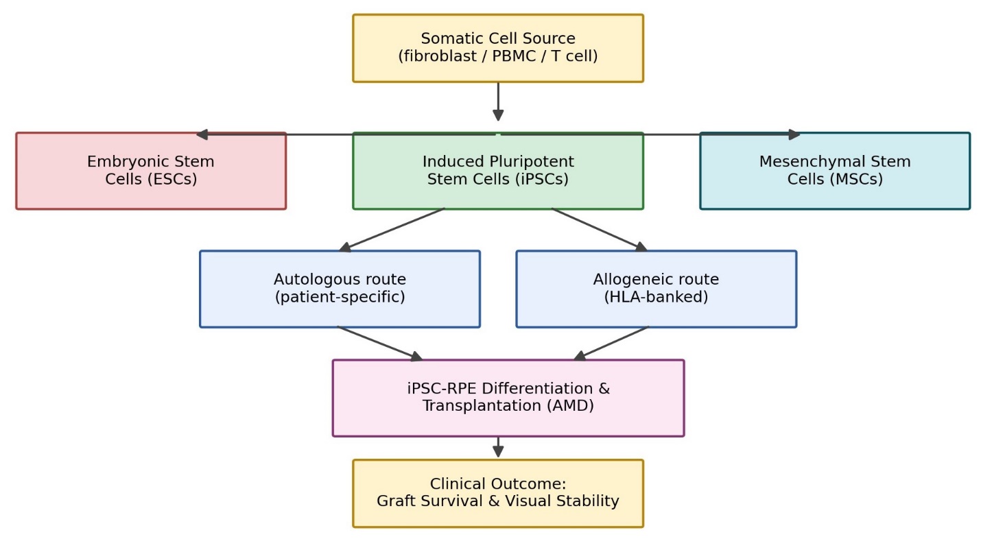
Before any conversation about retinal repair can begin, there is a more basic decision to make — which stem cell platform to build on in the first place — and that decision turns out to carry more downstream consequences than it might first appear (see Figure 1 for a schematic decision pathway and Table 1 for a full comparative profile). Embryonic stem cells (ESCs) remain, in a historical sense, the benchmark for pluripotency, but their use is entangled with two persistent problems. One is ethical: ESC derivation requires blastocyst destruction, a fact that continues to shape regulatory policy across jurisdictions (Ghosh & Mehta, 2016). The other is immunological, and arguably more limiting in a purely clinical sense — because ESCs are allogeneic by definition, mismatched HLA molecules provoke robust CD4+ and CD8+ T-cell responses in recipients, generally necessitating long-term immunosuppression (Parrotta et al., 2019; Zhao et al., 2011).

Mesenchymal stem cells (MSCs) sidestep the ethical objection entirely — they can be harvested non-invasively from adult tissue biopsies or, more conveniently still, from birth-associated waste such as placenta or umbilical cord (Assis et al., 2018; Wiegand & Banerjee, 2019). Their immunological profile is also unusually favorable: low MHC class I expression, no MHC class II, and active secretion of immunomodulatory cytokines that dampen local inflammation and support graft survival without immunosuppressive drugs (Yoshida et al., 2020). The trade-off, though, is real. MSCs have finite self-renewal capacity and a differentiation potential restricted largely to mesodermal lineages, which rules them out for generating the ectodermal tissue that constitutes the retina (Assis et al., 2018; Wiegand & Banerjee, 2019).

It was against this backdrop that Takahashi and Yamanaka's 2006 discovery of iPSCs felt less like an incremental advance and more like a genuine resolution of a standing dilemma. iPSCs offer patient-specific pluripotency — self-renewal and multi-lineage differentiation potential matching that of ESCs (Takahashi et al., 2007), but derived from the patient's own adult tissue, bypassing embryo destruction altogether. When autologous, the resulting grafts are essentially HLA-perfect matches, which is precisely what allowed Mandai and colleagues (2017) to achieve long-term graft persistence without systemic immunosuppression. Even so, iPSC-based platforms are not without their own translational headaches. The reprogramming process itself is genetically and epigenetically error-prone, and extended ex vivo passaging can allow chromosomal abnormalities, copy number variations, and point mutations to accumulate (Ahmad et al., 2025; Yoshihara et al., 2017). Residual undifferentiated cells in the final product also carry a non-trivial teratoma risk, which is why rigorous purification and release-testing standards are treated as non-negotiable before clinical use (Takahashi, 2025).

2.2 What Reprogramming Actually Costs a Cell: Telomeric and Epigenetic Barriers

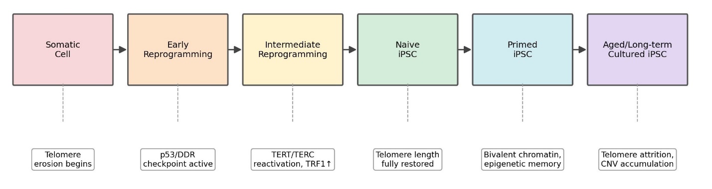
It is worth pausing on just how biologically demanding reprogramming is, because the difficulty is not incidental — it is, in a sense, the whole story of why efficiency remains low and why so much research effort goes into managing that inefficiency (summarized stage-by-stage in Table 2 and schematized in Figure 2). Somatic cells begin this process already carrying the marks of age: shortened telomeres, minimal or absent telomerase activity, and chromatin locked down by repressive modifications such as H3K9me3, H4K20me3, and dense global DNA methylation (Gonzalo et al., 2006; Harley, 1997; Harley et al., 1990). None of that is a hospitable starting point for pluripotency.

When Yamanaka factor expression is forced ectopically onto this chromatin landscape, the immediate result is stress — a lot of it. Cells experience transient telomere erosion during the earliest phase of reprogramming (Marión et al., 2009; Suhr et al., 2009), which in turn triggers a p53-dependent DNA damage response and upregulation of the Ink4/Arf locus, a checkpoint whose entire biological purpose is to prevent damaged or mutated clones from propagating further (Banito et al., 2009; Li et al., 2009; Marión et al., 2009). Cells that cannot silence this checkpoint are cleared, either through apoptosis or by becoming permanently senescent (Kim et al., 2018). It is, in effect, a bottleneck by design — which is precisely why so much of the reprogramming-efficiency literature is really about how to survive it rather than how to avoid it.

Cells that do make it through the early barrier enter what might be called an intermediate phase, marked by gradual chromatin loosening and the reactivation of telomerase reverse transcriptase (TERT) along with its RNA template, TERC (Batista et al., 2011). This stage depends heavily on upregulation of the shelterin protein TRF1, which stabilizes telomeres as chromatin is actively being remodeled around them (Martinez et al., 2010; Schneider et al., 2013), while pioneer transcription factors simultaneously recruit Polycomb repressive complexes (PRC1/PRC2) and NuRD complexes to silence somatic enhancers and open up pluripotency-associated loci in their place (Hu et al., 2014; Mirizio et al., 2025; Onder et al., 2012). By the time a cell reaches full, naive pluripotency, telomere length has been restored to something resembling ESC baseline, chromatin has opened into an active euchromatic state, and the promoters of OCT4, SOX2, and NANOG are essentially free of methylation (Marión et al., 2009; Ma et al., 2023). It is, functionally, a molecular reset — one thorough enough to erase most cellular signs of aging (Lapasset et al., 2011).

Even so, "most" is doing some work in that last sentence. Conventional human iPSCs typically settle into a primed pluripotency state that retains a somatic "epigenetic footprint" — residual DNA methylation at lineage-specific loci that hints, faintly, at where the cell came from. A

Table 1. Comparative profile of embryonic stem cells (ESCs), mesenchymal stem cells (MSCs), and induced pluripotent stem cells (iPSCs) across origin, pluripotency, immunogenicity, ethical status, tumorigenic risk, and primary clinical applications. This table synthesizes the platform-selection evidence discussed in Section 2.1 and cited throughout the Results.

Parameter

ESCs

MSCs

iPSCs

Biological Origin

Inner cell mass of blastocysts (Thomson, 1998) or somatic cell nuclear transfer (Gurdon, 1962).

Adult stroma (bone marrow, adipose) or neonatal tissue (cord, placenta) (Assis et al., 2018).

Ectopic transgene or chemical induction of adult somatic cells (Takahashi & Yamanaka, 2006).

Pluripotency

Fully pluripotent; unlimited self-renewal (Puri & Wagner, 2023).

Multipotent; finite passages, mesodermal bias (Assis et al., 2018).

Fully pluripotent; equivalent to ESCs (Takahashi et al., 2007).

Immunogenicity

Allogeneic; strong T-/NK-cell activation, needs immunosuppression (Parrotta et al., 2019; Zhao et al., 2011).

Low; no MHC-II, secretes immunomodulatory cytokines (Yoshida et al., 2020).

Autologous: negligible rejection (Mandai et al., 2017). Allogeneic: needs HLA-banks or hypoimmune editing (Deuse et al., 2019; Sugita et al., 2020).

Ethical/Regulatory Status

Controversial; blastocyst destruction (Ghosh & Mehta, 2016).

Minimally controversial; non-invasive sourcing (Wiegand & Banerjee, 2019).

Favorable; bypasses embryo destruction, standard somatic-cell-therapy oversight (Saliev & Singh, 2025).

Tumorigenic/Teratoma Risk

High; residual undifferentiated cells (Parrotta et al., 2019).

Very low; restricted differentiation potency (Wiegand & Banerjee, 2019).

High if undifferentiated subpopulations persist; elevated by culture-acquired mutations (Ahmad et al., 2025; Yoshihara et al., 2017).

Primary Applications

Developmental biology, embryogenesis modeling (Thomson, 1998).

Cartilage/bone regeneration, GvHD treatment (Bloor et al., 2020; Yoshida et al., 2020).

Disease modeling, drug screening, personalized regenerative therapy incl. iPSC-RPE (Mandai et al., 2017).

Table 2. Chronological dynamics of telomere length, telomerase activity, chromatin state, and key molecular checkpoints across the somatic-to-pluripotent reprogramming trajectory, from parental somatic cells through naive, primed, and long-term cultured iPSC states. 

Stage

Telomere Status

Telomerase Activity

Chromatin State

Key Molecular Events

Somatic Cells

Shortened, donor-age dependent (Harley, 1997).

Absent/negligible (Suhr et al., 2009).

Condensed heterochromatin (H3K9me3, H4K20me3) (Gonzalo et al., 2006).

DDR/p53 checkpoint via p21; senescence risk (Banito et al., 2009).

Early Reprogramming

Further transient shortening (Marión et al., 2009).

Low; TERT not yet upregulated (Marión et al., 2009).

Localized euchromatin remodeling begins (Koche et al., 2011).

Ink4/Arf activation; BAK/BAX-mediated clearance (Li et al., 2009; Kim et al., 2018).

Intermediate Reprogramming

Heterogeneous, transitional (Batista et al., 2011).

Reactivation of TERT/TERC begins (Batista et al., 2011).

Progressive opening; H3K4me2 gain (Koche et al., 2011).

TRF1 upregulation; PRC1/PRC2, NuRD recruitment (Martinez et al., 2010; Onder et al., 2012).

Naive iPSCs

Fully restored, ESC-like (Marión et al., 2009).

High, stable elongation (Armstrong et al., 2005).

Open euchromatin; OCT4/SOX2/NANOG demethylated (Ma et al., 2023).

Full pluripotency network reactivation (Lapasset et al., 2011).

Primed iPSCs

Long but shorter than naive state (Fu et al., 2018).

Moderate–high, stable (Suhr et al., 2009).

Bivalent promoters (H3K4me3 + H3K27me3) (Matsui et al., 2024).

Residual somatic memory, e.g., CD7 unmethylation (Kim et al., 2010; Ren et al., 2023).

Aged/Long-Term Cultured iPSCs

Re-shortening possible under passaging stress (Batista et al., 2011).

May decline with passage number (Agarwal et al., 2010).

Heterochromatin re-condensation; aberrant methylation (Yehezkel et al., 2011).

CNV/SNV accumulation; genomic instability (Ahmad et al., 2025; Yoshihara et al., 2017).

well-documented example is partial unmethylation of the CD7 promoter in T-cell-derived iPSCs (Kim et al., 2010; Ren et al., 2023), a residue that can bias later differentiation back toward the cell's original lineage (Kim et al., 2010). And prolonged culture is not risk-free either: aged iPSCs remain susceptible to telomere attrition, epigenetic drift, and the slow accumulation of unwanted de novo mutations, which is why ongoing genomic stability monitoring is generally treated as standard practice rather than an optional precaution (Ahmad et al., 2025; Yehezkel et al., 2011).

2.3 Getting the Factors In: Delivery Platforms and the Efficiency–Safety Trade-off

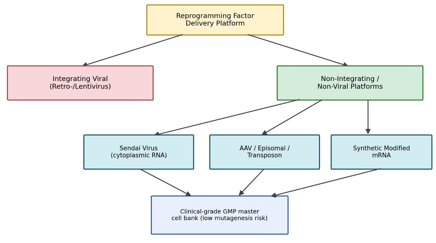
None of the biology above matters clinically unless the reprogramming factors can actually be delivered into cells in a way that is both efficient and, critically, safe enough for human use — and this turns out to be a genuinely difficult balancing act (Table 3; Figure 3 sketches the decision logic). Integrating viral vectors — retroviruses and lentiviruses — were the historical workhorses of iPSC generation, prized for high transduction efficiency and the ability to carry large, multi-gene cassettes (Takahashi & Yamanaka, 2006). Their central liability is exactly what their name implies: random integration into the host genome, which carries a meaningful risk of disrupting tumor suppressor genes or activating latent oncogenes (Kustikova et al., 2005). For research purposes, this is a manageable risk. For clinical translation, it is generally disqualifying.

That disqualification is what pushed the field toward transient, non-integrating alternatives. Sendai virus (SeV) has become something close to a workhorse for clinical-grade iPSC derivation (Fusaki et al., 2009). As an RNA virus replicating exclusively in the cytoplasm, it never enters the nucleus, which eliminates insertional mutagenesis risk almost by definition (Fusaki et al., 2009; Wang et al., 2026). The cost is immunogenicity — SeV proteins are recognized robustly by the host, and clearing the viral genome from a resulting cell line typically takes an inconvenient 10 to 20 passages (Fusaki et al., 2009; Wang et al., 2026). Adeno-associated virus (AAV) vectors offer a different trade-off: extremely low immunogenicity and no meaningful genomic integration (Sabatino et al., 2022), plus tissue-specific tropism (AAV1, for instance, shows particular affinity for cardiac fibroblasts) that makes them well suited to direct in vivo reprogramming applications (Abdelnaby et al., 2026). But AAV struggles specifically with undifferentiated pluripotent cells, transducing them roughly 100-fold less efficiently than differentiated progeny, owing to low surface receptor expression on pluripotent cells (Abdelnaby et al., 2026; Sabatino et al., 2022). Its cargo capacity is also tight — around 4.5 kb — which frequently forces split-vector strategies or minimized transgene combinations, such as OSK without c-Myc (Wang et al., 2026).

Non-viral options round out the picture. Episomal plasmids avoid genomic integration almost entirely, though at the cost of low efficiency and the need for extensive selection to confirm eventual plasmid loss (Haridhasapavalan et al., 2019; Wang et al., 2026); transposon systems (piggyBac, Sleeping Beauty) allow for cleaner excision after use, though excision errors remain an occasional concern (Haridhasapavalan et al., 2019; Wang et al., 2026); and synthetic modified mRNA — particularly when paired with epigenetic modifiers such as valproic acid — offers a genuinely zero-footprint route, achieving respectable efficiency without any risk of genomic integration, at the practical cost of repeated daily transfections over one to two weeks (Warren et al., 2010; Wang et al., 2026). No single platform dominates across every axis; the choice, in practice, tends to be dictated by whichever risk (immunogenicity, cargo limits, integration, or manufacturing burden) matters most for a given clinical context.

2.4 Bypassing Pluripotency: Direct Transdifferentiation and Its Retinal Applications

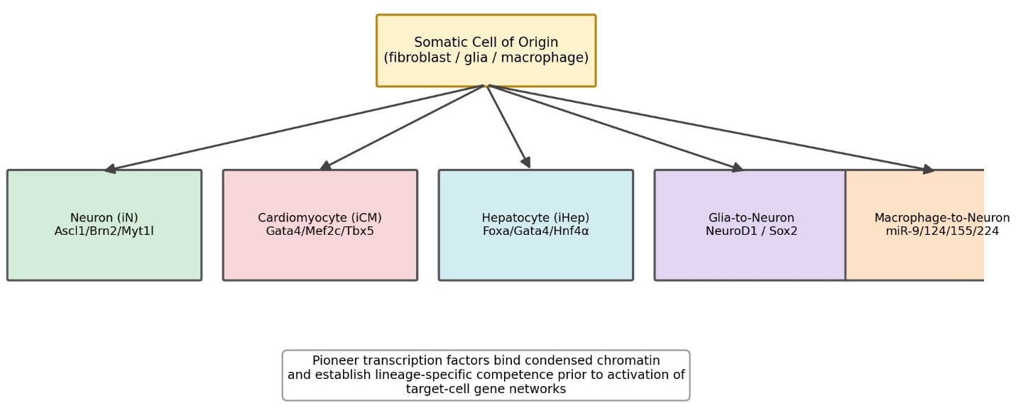
Direct reprogramming takes a fundamentally different route — one specialized cell converted straight into another, with pluripotency never entering the picture at all (Table 4; Figure 4 maps the major lineage conversions described in the literature). The appeal is fairly intuitive: skipping the pluripotent intermediate removes the teratoma risk that dogs iPSC-based approaches and, in principle, shortens manufacturing timelines considerably. The prototype for this approach is fibroblast-to-neuron conversion, achieved with the pioneer factor Ascl1 combined with Brn2 and Myt1l — the so-called BAM cocktail — which yields cells exhibiting genuine action potentials and functional synaptic transmission (Vierbuchen et al., 2010). Analogous logic underlies fibroblast-to-cardiomyocyte conversion using Gata4, Mef2c, and Tbx5 (the GMT cocktail), which produces contractile, sarcomeric cardiomyocyte-like cells with spontaneous calcium oscillations (Ieda et al., 2010), and

Table 3. Comparative analysis of reprogramming-factor delivery platforms — integrating viral vectors, non-integrating viral vectors, and non-viral genomic/non-genomic systems — across reprogramming efficiency, cargo capacity, mutagenesis risk, immunogenicity, and translational suitability.

Platform

Efficiency

Cargo Capacity

Integration/Mutagenesis Risk

Immunogenicity

Clinical Suitability

Retro-/Lentivirus

High (~0.01–20% depending on promoter) (Seki et al., 2011).

~8–10 kb (Tiscornia et al., 2006).

High; random integration (Kustikova et al., 2005).

Low acutely, but long-term oncogene reactivation risk (Sommer et al., 2010).

Poor; largely research-only (Wang et al., 2026).

Sendai Virus (SeV)

Exceptionally high, ~0.1–4.0% (Fusaki et al., 2009).

~8–10 kb (Fusaki et al., 2009).

Negligible; cytoplasmic only (Wang et al., 2026).

High; needs 10–20 passages to clear (Fusaki et al., 2009).

Highly viable for clinical banks (Umekage et al., 2019).

AAV

Low in undifferentiated iPSCs, ~100-fold less than differentiated cells (Sabatino et al., 2022).

~4.5 kb, strict limit (Wang et al., 2026).

Extremely low; rare non-homologous integration (Sabatino et al., 2022).

Very low (Abdelnaby et al., 2026).

Highly suitable for in vivo/direct reprogramming (Abdelnaby et al., 2026).

Episomal Plasmids

Low, ~0.005–0.01% (Wang et al., 2026).

No strict limit; large polycistronic constructs (Chou et al., 2011).

Negligible; episomal (Haridhasapavalan et al., 2019).

Low (Wang et al., 2026).

Excellent for zero-footprint clinical banks (Umekage et al., 2019).

Transposons (piggyBac/SB)

Moderate, ~0.02–0.05% (Haridhasapavalan et al., 2019).

High, ~10–15 kb (Haridhasapavalan et al., 2019).

High initially; excisable but excision errors possible (Kaji et al., 2009).

Low–moderate (Wang et al., 2026).

Moderate; useful for stable ex vivo constructs (Wang et al., 2026).

Synthetic Modified mRNA

High, ~4.4% with VPA (Warren et al., 2010).

High transient capacity; needs daily transfection (Eisenstein, 2022).

Zero; cytoplasmic only (Warren et al., 2010).

Moderately low with modified nucleotides (Warren et al., 2010).

Highly suitable; zero-footprint platform (Wang et al., 2026).

Table 4. Direct reprogramming (transdifferentiation) strategies catalogued by lineage conversion, listing starting/target cell types, key pioneer factors, delivery methods, functional/molecular endpoints, and primary clinical applications, including the Müller-glia-to-retinal-ganglion-cell conversion relevant to in situ retinal repair. 

Conversion

Starting/Target Cell

Key Factors

Delivery

Functional Endpoints

Clinical Application

Fibroblast → Neuron (iN)

Dermal fibroblast → mature neuron (Vierbuchen et al., 2010).

Ascl1, Brn2, Myt1l (BAM cocktail) (Vierbuchen et al., 2010).

Lentiviral/retroviral transduction (Vierbuchen et al., 2010).

Action potentials, synaptic transmission; MAP2, TUJ1 (Vierbuchen et al., 2010).

Spinal cord injury, neurodegeneration modeling (Mirizio et al., 2025).

Fibroblast → Cardiomyocyte (iCM)

Cardiac fibroblast → cardiomyocyte-like cell (Ieda et al., 2010).

Gata4, Mef2c, Tbx5 (GMT) (Ieda et al., 2010).

Polycistronic retro-/lentivirus, SeV, AuNPs (Wang et al., 2015; Son et al., 2026).

Contractility, Ca2+ oscillation; TNNT2, Cx43 (Ieda et al., 2010).

Post-MI myocardial regeneration (Son et al., 2026).

Fibroblast → Hepatocyte (iHep)

Dermal/urine fibroblast → hepatocyte-like cell (Sekiya & Suzuki, 2011).

Foxa1/2/3, Gata4, Hnf4α (Sekiya & Suzuki, 2011).

Retroviral or episomal vectors (Sekiya & Suzuki, 2011).

Albumin secretion, LDL uptake (Sekiya & Suzuki, 2011).

Acute liver failure, hepatic disease modeling (Mirizio et al., 2025).

Glia → Neuron (incl. Müller glia → RGC)

Astrocyte/microglia/Müller glia → neuron (Niu et al., 2013; Sen et al., 2025).

NeuroD1, Sox2, Islet1/Pou4f2/Ascl1, or MAP4K inhibitors (Sen et al., 2025; Todd et al., 2022).

In situ AAV injection or small-molecule delivery (Zhang et al., 2023).

Electrical activity, network integration; MAP2, NeuN (Niu et al., 2013).

In vivo retinal repair, stroke, neurodegeneration (Sen et al., 2025).

Macrophage → Neuron

Peripheral macrophage → excitable neuron-like cell (Zhang et al., 2023).

miR-9, miR-124, miR-155, miR-224 (Zhang et al., 2023).

Non-viral nanoparticle/liposomal transfection (Zhang et al., 2023).

Calcium excitability, neurite growth (Zhang et al., 2023).

Localized neural repair after trauma (Zhang et al., 2023).

fibroblast-to-hepatocyte conversion via Foxa family factors combined with Gata4 and Hnf4α (Sekiya & Suzuki, 2011).

For retinal applications specifically, the target of interest is Müller glia — a resident support-cell population that, unlike fibroblasts, is already sitting inside the tissue that needs repairing. Delivering neurogenic transcription factors such as Ngn1/3, or developmental combinations including Islet1, Pou4f2, and Ascl1, can drive MG cells toward RGC-like fates directly in vivo, without ever requiring cell extraction or ex vivo manipulation (Sen et al., 2025; Todd et al., 2022; Zhang et al., 2023). This in situ character is arguably the biggest practical advantage direct reprogramming holds over transplantation-based strategies: there is no cell manufacturing pipeline, no immunological matching problem, and no surgical graft procedure — just a local delivery of factors to cells already resident in the diseased tissue. High-throughput chemical screening has extended this logic further still, identifying small-molecule cocktails, including MAP4K inhibitors such as DMX-5804, that can promote MG proliferation and transdifferentiation without requiring any genetic material to be delivered at all (Todd et al., 2022; Zhang et al., 2023) — a genuinely appealing simplification, if efficiency can be pushed high enough for clinical relevance. Parallel work in the central nervous system, converting resident astrocytes or microglia into neurons using NeuroD1 or Sox2 delivered via AAV, offers a useful proof of concept for what in situ conversion can achieve at scale in a comparably complex tissue (Mirizio et al., 2025; Niu et al., 2013).

2.5 Reversing Age Without Changing Identity: Partial In Vivo Reprogramming

Perhaps the newest branch of this field asks a genuinely different question — not "how do we replace this cell?" but "can we make this cell younger without changing what it is?" Partial cellular reprogramming attempts exactly that: transient, incomplete activation of reprogramming factors, sufficient to erase some of the molecular signatures of aging without pushing the cell all the way to pluripotency and, therefore, without erasing its identity (Huyghe et al., 2024; Núñez-Quintela et al., 2026). The OSK combination — Oct3/4, Sox2, and Klf4, deliberately excluding the oncogenic c-Myc — has become the standard toolkit for this approach, largely because omitting c-Myc substantially reduces tumorigenic risk while still enabling meaningful epigenetic remodeling (Lu et al., 2020; Ocampo et al., 2016).

In the eye specifically, this strategy has produced some of the most striking preclinical results in the entire regenerative-ophthalmology literature. Lu and colleagues (2020) demonstrated that transient OSK expression could restore youthful DNA methylation patterns in retinal ganglion cells and promote axonal regeneration following optic nerve injury, effectively reversing vision loss in mouse models of glaucoma and aging. Life Biosciences subsequently translated this work into ER-100, an AAV2 vector delivering OSK, which similarly restored youthful methylation signatures and promoted axonal regeneration in non-human primates with optic neuropathy (Li & Tay, 2026). That preclinical foundation has now supported progression into a Phase 1 human trial evaluating ER-100 safety in patients with glaucoma and optic neuropathy (NCT07290244) (Li & Tay, 2026; USDHHS, 2025) — arguably the clearest signal yet that partial reprogramming is moving from a mechanistic curiosity toward an actual therapeutic modality, even if efficacy in humans remains to be established.

2.6 Immune Engineering and the Path Toward Off-the-Shelf Cell Products

A recurring theme across all of the above is that immunological compatibility, not just cell biology, ultimately determines whether a reprogramming-based therapy can scale. Allogeneic products — whether banked iPSC-RPE lines or T-cell-derived iPSC banks intended for adoptive immunotherapy — face rejection risk unless engineered around it. Hypoimmune (HIP) engineering has emerged as one solution, using CRISPR/Cas9 to knock out MHC class I (B2M) and class II (CIITA) while overexpressing CD47, a "don't eat me" signal that prevents macrophage-mediated phagocytosis; cells engineered this way can persist long-term in immunocompetent recipients without immunosuppression (Deuse et al., 2019; Son et al., 2026). This kind of engineering, layered onto reprogramming platforms already optimized for efficiency and genomic safety, is what ultimately makes the vision of scalable, off-the-shelf cellular therapy plausible rather than purely aspirational (Cichocki et al., 2022; Wang & Xiong, 2024).

3. Methods

This review followed a structured, reproducible narrative synthesis methodology consistent with PubMed/MEDLINE indexing conventions, so that the search strategy described here could, in principle, be re-run by another investigator to retrieve a substantially overlapping

Figure 1. Decision pathway from somatic cell source to iPSC-RPE clinical therapy. Somatic cells (fibroblast, PBMC, or T cell) may be reprogrammed toward ESC, iPSC, or MSC platforms; the iPSC route bifurcates into autologous and allogeneic pathways, both converging on iPSC-RPE differentiation and transplantation, with graft survival and visual stability as the ultimate clinical outcome for AMD patients.

Figure 2. Chronological telomeric and epigenetic dynamics across reprogramming stages. Telomere length, telomerase activity, and chromatin openness are tracked from parental somatic cells through early and intermediate reprogramming to naive and primed iPSC states, and finally to aged, long-term cultured iPSC lines where genomic instability can re-emerge.

evidence base.

3.1 Search Strategy and Information Sources.

Literature was identified through structured searches of PubMed/MEDLINE, Google Scholar, and ClinicalTrials.gov, supplemented by manual reference-list screening (backward citation tracking) of key retrieved articles to capture foundational studies not indexed under the primary search terms. Searches were restricted to English-language sources. Search terms combined controlled vocabulary and free-text keywords using Boolean operators, structured around four conceptual clusters: (1) disease and tissue terms — "age-related macular degeneration," "retinal pigment epithelium," "retinal degeneration"; (2) cell-source and platform terms — "induced pluripotent stem cell," "iPSC," "embryonic stem cell," "mesenchymal stem cell"; (3) reprogramming-strategy terms — "cellular reprogramming," "direct reprogramming," "transdifferentiation," "partial reprogramming," "in vivo reprogramming," "Yamanaka factors," "OSK," "OSKM"; and (4) delivery/safety terms — "Sendai virus," "AAV," "adeno-associated virus," "episomal vector," "modified mRNA," "insertional mutagenesis," "teratoma," "genomic instability." Cluster terms were combined using the structure (Cluster 1) AND (Cluster 2 OR Cluster 3) AND (Cluster 4, where relevant), consistent with standard PubMed search-string construction.

3.2 Eligibility Criteria.

Sources were included if they reported primary preclinical or clinical data, mechanistic findings, or registered trial protocols directly relevant to cellular reprogramming applied to retinal or ocular tissue, or to the underlying reprogramming biology (telomere dynamics, epigenetic remodeling, delivery-platform safety) cited as generalizable mechanistic support. Peer-reviewed original research articles, peer-reviewed reviews from high-impact journals, and registered entries on ClinicalTrials.gov were eligible. Sources were excluded if they were non-English, were conference abstracts without full peer-reviewed data, or addressed reprogramming in a tissue context with no plausible mechanistic or translational relevance to retinal application.

3.3 Study Selection and Data Extraction.

Titles and abstracts of retrieved records were screened for topical relevance; full texts of potentially eligible records were then reviewed in full. For each included source, the following data were extracted where applicable: cell source and reprogramming platform, delivery vector and reprogramming efficiency, safety and immunogenicity outcomes, functional or molecular endpoints, and clinical trial phase and registration identifier. Extracted data were organized thematically into four comparative synthesis tables (stem cell platforms, telomeric/epigenetic dynamics, delivery platforms, and direct transdifferentiation strategies) to support structured cross-study comparison, consistent with a narrative (rather than quantitative meta-analytic) synthesis approach.

3.4 Synthesis Approach.

Given the methodological heterogeneity of the included evidence base — spanning in vitro mechanistic studies, animal models, and early-phase human trials — a quantitative meta-analysis was not appropriate. Findings were instead synthesized narratively and organized by conceptual theme (stem cell platform selection, telomeric/epigenetic reprogramming barriers, delivery-platform safety, direct transdifferentiation, and partial in vivo reprogramming), with comparative tables constructed to standardize cross-study evaluation criteria and support reproducibility of the synthesis logic by future reviewers.

4. Comparative Outcomes of Stem Cell Platforms for Retinal Regeneration and Reprogramming

4.1 Clinical Translation of iPSC-RPE Transplantation

The clearest, most clinically mature evidence base concerns iPSC-derived RPE transplantation for AMD. Autologous iPSC-RPE sheets, first characterized by Kamao and colleagues (2014) and subsequently transplanted in the landmark first-in-human procedure by Mandai and colleagues (2017), demonstrated durable graft survival and visual stability over extended follow-up, achieved without systemic immunosuppression (Mandai et al., 2017; Takagi et al., 2019). This result established biological proof of concept for iPSC-derived cell therapy in the eye. However, the operational burden of autologous manufacturing — approximately one year of production time and roughly US\$1 million per patient (Wang et al., 2026) — proved to be the binding constraint on scalability, motivating the shift toward allogeneic, HLA-banked lines documented in Sugita and colleagues' (2020) subsequent transplantation series, which required only localized rather than systemic immunosuppression [Table 1].

4.2 Platform Comparison Across Biological and Translational Parameters

Cross-platform comparison confirms that no single stem cell source dominates across all relevant dimensions [Table 1; Figure 1]. ESCs offer the deepest historical characterization of pluripotency but carry the highest immunogenicity and the most significant ethical constraints (Ghosh & Mehta, 2016; Parrotta et al., 2019). MSCs minimize both ethical and immunogenic concerns but cannot generate the ectodermal derivatives required for retinal repair (Assis et al., 2018). iPSCs occupy an intermediate position: full pluripotency and, in the autologous configuration, near-perfect histocompatibility, at the cost of genomic instability risk accumulated during reprogramming and extended culture (Ahmad et al., 2025; Yoshihara et al., 2017).

4.3 Telomeric and Epigenetic Dynamics During Reprogramming

Stage-resolved analysis of telomere length and chromatin state across the reprogramming trajectory [Table 2; Figure 2] shows a consistent pattern: telomere erosion and heterochromatin persistence in early reprogramming, giving way to progressive telomerase reactivation (TERT/TERC) and chromatin opening during the intermediate stage (Batista et al., 2011; Marión et al., 2009), culminating in full telomere restoration and an open euchromatic state in naive pluripotent cells (Marión et al., 2009; Ma et al., 2023). Notably, primed human iPSCs retain detectable somatic epigenetic memory — exemplified by partial CD7 promoter unmethylation in T-cell-derived lines — that persists even after transient apoptosis-suppression interventions (Kim et al., 2010; Ren et al., 2023).

4.4 Delivery Platform Performance and Safety Trade-offs

Comparative analysis of delivery platforms [Table 3; Figure 3] confirmed a consistent efficiency–safety trade-off. Integrating viral vectors achieved the highest transduction efficiency but carried the highest insertional mutagenesis risk, restricting their use largely to research contexts (Kustikova et al., 2005). Sendai virus offered high efficiency with negligible integration risk, at the cost of prolonged passaging requirements to clear viral genome (Fusaki et al., 2009). AAV vectors showed the lowest immunogenicity and integration risk overall but the lowest efficiency in undifferentiated pluripotent cells specifically, alongside a restrictive ~4.5 kb cargo ceiling (Abdelnaby et al., 2026; Sabatino et al., 2022). Non-viral platforms (episomal plasmids, transposons, modified mRNA) consistently minimized genomic integration risk, with synthetic modified mRNA achieving the most favorable overall efficiency–safety balance among non-viral approaches (Warren et al., 2010).

4.5 Direct Transdifferentiation Outcomes

Across the five major transdifferentiation lineages catalogued [Table 4; Figure 4], functional conversion was consistently demonstrated by electrophysiological, biochemical, or molecular marker endpoints: action potentials and synaptic transmission in fibroblast-to-neuron conversion (Vierbuchen et al., 2010); spontaneous contractility and calcium oscillations in fibroblast-to-cardiomyocyte conversion (Ieda et al., 2010); and, in the retinal context specifically, MG-to-RGC conversion in vivo using developmental transcription factors or MAP4K-inhibitor small-molecule cocktails, without requiring cell extraction or transplantation (Sen et al., 2025; Todd et al., 2022; Zhang et al., 2023).

4.6 Progress of Partial In Vivo Reprogramming Toward the Clinic

In the partial-reprogramming domain, OSK-based approaches restored youthful DNA methylation patterns and promoted axonal regeneration in aged and glaucomatous rodent models (Lu et al., 2020), with translation to non-human primates via the AAV2-delivered ER-100 candidate (Li & Tay, 2026). This preclinical trajectory has now advanced to a registered Phase 1 human trial (NCT07290244) evaluating ER-100 safety in glaucoma and optic neuropathy patients (Li & Tay, 2026; USDHHS, 2025), representing the most clinically advanced application of partial reprogramming for visual restoration identified in this synthesis.

5. From Proof of Concept to Scalable Retinal Regeneration

Read together, the evidence assembled here traces something like a maturation curve. The field began with a single, technically heroic demonstration — one patient, one graft, one year of manufacturing (Mandai et al., 2017) — and has since diversified into at least three semi-independent therapeutic logics: replace the tissue (iPSC-RPE transplantation), reprogram a neighboring cell in place (direct MG-to-RGC transdifferentiation), or simply make the existing cell younger without changing what it is (partial OSK reprogramming). It is tempting to ask which of

Figure 3. Decision framework for reprogramming-factor delivery platform selection. Integrating viral vectors are separated from non-integrating and non-viral alternatives (Sendai virus, AAV/episomal/transposon systems, and synthetic modified mRNA), each converging on the shared goal of generating a clinical-grade, low-mutagenesis-risk GMP master cell bank.

Figure 4. Lineage map of direct transdifferentiation strategies bypassing pluripotency. A common somatic cell of origin (fibroblast, glia, or macrophage) is redirected toward neuronal, cardiomyocyte, hepatocyte, glia-derived neuronal (including Müller-glia-to-retinal-ganglion-cell), or macrophage-derived neuronal fates via lineage-specific pioneer transcription factors and small molecules.

these will "win," but that framing may be somewhat misplaced. AMD is not a monolithic disease process, and RPE loss, photoreceptor degeneration, and RGC/optic-nerve aging are not identical problems; it is plausible, even likely, that different reprogramming strategies will ultimately serve different stages or subtypes of retinal degeneration rather than competing head-to-head for a single indication.

What is striking, though, is how consistently the same underlying tension resurfaces across every strategy examined [Tables 1–3]: efficiency and safety pull in opposite directions, almost without exception. Integrating vectors are efficient and dangerous; AAV is safe and inefficient in pluripotent cells specifically; autologous iPSC-RPE is immunologically ideal and economically unworkable at scale; allogeneic banking solves the economics but reintroduces at least a partial immunogenicity problem (Sugita et al., 2020; Wang et al., 2026). None of the platforms reviewed here have escaped this trade-off entirely — they have merely relocated it to a place where it is more manageable. Synthetic modified mRNA and hypoimmune (HIP) gene editing arguably represent the field's best current attempts at genuinely narrowing the gap rather than just shifting it (Deuse et al., 2019; Warren et al., 2010), and it seems reasonable to expect that future clinical-grade platforms will increasingly combine both: non-integrating delivery paired with immune-cloaking edits, rather than either strategy alone.

The rise of direct transdifferentiation and partial reprogramming also suggests a subtler shift in how the field is thinking about "success." Early iPSC work was, understandably, preoccupied with generating a transplantable, fully differentiated replacement cell — RPE, in this case. Direct reprogramming loosens that requirement considerably: MG-to-RGC conversion in situ (Sen et al., 2025; Todd et al., 2022) does not require manufacturing, banking, HLA-matching, or surgical grafting at all, which is a substantial practical advantage even if conversion efficiency currently lags behind what transplantation-based approaches can achieve. Partial reprogramming goes further still, dispensing with the notion of cell replacement altogether in favor of cellular rejuvenation — an idea that would have sounded closer to science fiction a decade ago but that now has an active Phase 1 trial behind it (Li & Tay, 2026; USDHHS, 2025). Whether OSK-based therapies can reverse human vision loss the way they have reversed rodent vision loss (Lu et al., 2020) remains genuinely uncertain, and that uncertainty is worth sitting with rather than glossing over.

Several limitations of both the underlying evidence base and this synthesis itself deserve acknowledgment. First, much of the mechanistic literature on telomere dynamics and delivery-platform safety [Table 2, Table 3] derives from non-ocular reprogramming contexts and is extrapolated to retinal applications by mechanistic analogy rather than direct retinal data; this is a reasonable but not airtight inferential move. Second, human clinical evidence remains sparse and heavily concentrated in a small number of centers and trial sponsors, limiting generalizability. Third, this review followed a structured but narrative synthesis approach rather than a formal systematic review with quantitative pooling, and publication bias favoring positive preclinical results cannot be excluded (Section 3.5). Fourth, cost and manufacturing-scalability data are largely drawn from a single source (Wang et al., 2026) and may not generalize across health systems or geographies with different regulatory and reimbursement structures.

Looking forward, three priorities seem reasonably clear. First, head-to-head comparative studies — ideally within the same patient population or disease stage — would help clarify which reprogramming strategy is best matched to which clinical scenario, rather than relying on cross-study comparisons of the kind assembled in this review. Second, continued refinement of non-integrating, low-immunogenicity delivery platforms, particularly efforts to improve AAV transduction efficiency in pluripotent cells specifically, would meaningfully de-risk both transplantation-based and in vivo reprogramming approaches (Abdelnaby et al., 2026). Third, and perhaps most importantly, the field will need longer-term human safety data — particularly for partial reprogramming, where the theoretical tumorigenic risk of even transient OSK expression has not yet been fully characterized in humans over multi-year timescales. None of this is likely to resolve quickly. But the trajectory from a single 1.3 by 3.0 millimeter graft in 2014 to a registered human trial of in vivo epigenetic rejuvenation a decade later suggests a field that is, at minimum, moving in a coherent and increasingly translational direction.

6. Conclusion

Cellular reprogramming has evolved, over roughly a decade and a half, from a single proof-of-concept graft into a genuinely diversified therapeutic landscape for AMD and related retinal degenerations — spanning autologous and allogeneic iPSC-RPE transplantation, in situ Müller glia transdifferentiation, and in vivo partial epigenetic rejuvenation. Each approach carries a distinct efficiency–safety–scalability profile, and none has yet fully resolved the tension between them. Continued progress will likely depend less on any single breakthrough than on the steady convergence of safer delivery platforms, immune-engineering solutions, and rigorous human trial data, gradually narrowing the gap between what has been demonstrated in preclinical models and what can be responsibly offered to patients.

References


Abdelnaby, M., Galiakberova, A., & Dashinimaev, E. (2026). AAV vectors in regenerative medicine and cellular reprogramming: Potential, pitfalls, and specificity constraints. International Journal of Molecular Sciences, 27(06846). https://doi.org/10.3390/ijms2706846

Agarwal, S., Church, G. M., Collins, J. J., Irion, S., Loh, Y. H., & Daley, G. Q. (2010). Telomere maintenance and reprogramming kinetics in human T-cell-derived induced pluripotent stem cells. Cell Stem Cell, 7(4), 411--419. https://doi.org/10.1016/j.stem.2010.08.006

Ahmad, U., Kumar, D., & Faiyazuddin, M. (2025). Can iPSCs turn back time? Prospects and pitfalls in age reversal. Current Stem Cell Research & Therapy, 20(4). https://doi.org/10.2174/1574888x400606250721112037

Armstrong, L., Lako, M., Dean, W., & Stojkovic, M. (2005). Telomere maintenance and telomerase activity in human embryonic stem cells and induced pluripotent stem cells. Stem Cells, 23(8), 1056--1066. https://doi.org/10.1634/stemcells.2005-0210

Assis, R. I. F., Wiench, M., Silvério, K. G., da Silva, R. A., Feltran, G. S., Sallum, E. A., Casati, M. Z., Nociti, F. H., & Andia, D. C. (2018). RG108 increases NANOG and OCT4 in bone marrow-derived mesenchymal cells through global changes in DNA modifications and epigenetic activation. PLoS One, 13(11), e0207873. https://doi.org/10.1371/journal.pone.0207873

Banito, A., Rashid, S. T., Acosta, J. C., Li, S., Pereira, C. F., Geti, I., Cobb, A. M., Amaral, F. M., Griffiths, J. R., Bellamy, M., & Alderton, W. (2009). Senescence impairs successful reprogramming to pluripotent stem cells. Genes & Development, 23(18), 2134--2139. https://doi.org/10.1101/gad.173922.111

Batista, L. F. Z., Pech, M. F., Zhong, F. L., Nguyen, H. N., Xie, K. T., Zaug, A. J., Crary, S. M., Choi, J., Sebastiano, V., Cherry, A., Giri, N., Wernig, M., Alter, B. P., Cech, T. R., Savage, S. A., Reijo Pera, R. A., & Artandi, S. E. (2011). Telomere shortening and loss of self-renewal in dyskeratosis congenita induced pluripotent stem cells. Nature, 474(7351), 399--404. https://doi.org/10.1038/nature10084

Bloor, A. J. C., Patel, A., Griffin, J. E., Gilleece, M. H., Radia, R., Yeung, D. T., Drier, D., Larson, L. S., Uenishi, G. I., Hei, D., Kelly, K., Slukvin, I., & Rasko, J. E. J. (2020). Production, safety and efficacy of iPSC-derived mesenchymal stromal cells in acute steroid-resistant graft versus host disease: a phase I, multicenter, open-label, dose-escalation study. Nature Medicine, 26(11), 1720--1725. https://doi.org/10.1038/s41591-020-1120-x

Chen, T., Chen, Z., Du, J., Zhang, M., Chen, Z., Gao, Q., Chen, A., Meng, Q., Sun, Y., Liu, Y., Song, L., Wang, X., Edavi, P. P., Xu, C., Zhang, H., Huang, J., & Jiang, Y. (2025). Reprogramming of iPSCs to NPCEC-like cells by biomimetic scaffolds for zonular fiber reconstruction. Bioactive Materials, 45, 446--458. https://doi.org/10.1016/j.bioactmat.2024.11.031

Chou, B. K., Mali, P., Huang, X., Ye, Z., Dowey, S. N., Resar, L. M., & Cheng, L. (2011). Efficient human iPS cell derivation by a non-integrating plasmid from blood cells with unique epigenetic and gene expression signatures. Cell Research, 21(3), 518--529. https://doi.org/10.1038/cr.2011.23

Cichocki, F., Bjordahl, R., Goodridge, J. P., Mahmood, S., Gaidarova, S., Abujarour, R., Davis, Z. B., Merino, A., Tuininga, K., Wang, H., & Valamehr, B. (2022). Quadruple gene-engineered natural killer cells enable multi-antigen targeting for durable antitumor activity against multiple myeloma. Nature Communications, 13(1), 7341. https://doi.org/10.1038/s41467-022-35123-x

Deng, X., Lee, R., Lim, S. Y., & Ding, S. (2022). Global transcriptional and epigenetic reconfiguration during chemical reprogramming of human retinal pigment epithelial cells into photoreceptor-like cells. Cells, 11(19), 3146. https://doi.org/10.3390/cells11193146

Deuse, T., Hu, X., Agbor-Enoh, S., Koch, M., Spitzer, M. H., Gravina, A., Alawi, M., Marishta, A., Peters, B., Koshal, K., & Schrepfer, S. (2019). De novo mutations in mitochondrial DNA of iPSCs produce immunogenic neoepitopes in mice and humans. Nature Biotechnology, 37(10), 1137--1144. https://doi.org/10.1038/s41587-019-0227-7

Eisenstein, M. (2022). Turn Biotechnologies leverages transient mRNA-based reprogramming for therapeutics. Nature Biotechnology, 40(5), 450--452. https://doi.org/10.1038/nbt-2022-05450

Fu, H., Tian, C. L., Ye, X., Sheng, X., Wang, H., Liu, Y., & Liu, L. (2018). Dynamics of telomere rejuvenation during chemical induction to pluripotent stem cells. Stem Cell Reports, 11(1), 70--87. https://doi.org/10.1016/j.stemcr.2018.05.003

Fusaki, N., Ban, H., Nishiyama, A., Saeki, K., & Hasegawa, M. (2009). Efficient induction of transgene-free human pluripotent stem cells using a vector based on Sendai virus, an RNA virus that does not integrate into the host genome. Proceedings of the Japan Academy, Series B, 85(8), 348--362. https://doi.org/10.2183/pjab.85.348

Gamm, D. M., Phillips, J. M., & Singh, R. (2013). Modeling retinal degenerative diseases with human iPS-derived cells: current status and future implications. Expert Review of Ophthalmology, 8(3), 213--216. https://doi.org/10.1586/eop.13.19

Ghosh, D., & Mehta, N. (2016). Ethical issues in biomedical use of human embryonic stem cells (hESCs). Journal of Reproductive Health and Medicine, 2(1), S37--S47. https://doi.org/10.1016/j.jrhm.2016.03.001

Gonzalo, S., Jaco, I., Fraga, M. F., Chen, T., Li, E., Esteller, M., & Blasco, M. A. (2006). DNA methyltransferases control telomere length and telomere recombination in mammalian cells. Nature Cell Biology, 8(4), 416--424. https://doi.org/10.1038/ncb1386

Gurdon, J. B. (1962). The developmental capacity of nuclei taken from intestinal epithelium cells of feeding tadpoles. Journal of Embryology and Experimental Morphology, 10(4), 622--640. https://doi.org/10.1242/dev.10.4.622

Haridhasapavalan, K. K., Borgohain, M. P., Dey, C., Saha, B., Narayan, G., Kumar, S., & Thummer, R. P. (2019). An insight into nonintegrative gene delivery approaches to generate transgene-free induced pluripotent stem cells. Gene, 686, 146--159. https://doi.org/10.1016/j.gene.2018.11.069

Harley, C. B. (1997). Telomere loss and cell senescence in human somatic cells. Journal of NIH Research, 9(3), 23--29.

Harley, C. B., Futcher, A. B., & Greider, C. W. (1990). Telomeres shorten during ageing of human fibroblasts. Nature, 345(6274), 458--460. https://doi.org/10.1038/345458a0

He, X., Liang, J., Paul, C., Huang, W., Dutta, S., & Wang, Y. (2022). Advances in cellular reprogramming-based approaches for heart regenerative repair. Cells, 11(23), 3914. https://doi.org/10.3390/cells11233914

Hu, X., Zhang, L., Mao, S. Q., Li, Z., Chen, J., Zhang, R. R., ... & Xu, G. L. (2014). Tet and TDG mediate DNA demethylation essential for mesenchymal-to-epithelial transition in somatic cell reprogramming. Cell Stem Cell, 14(4), 512--522. https://doi.org/10.1016/j.stem.2014.01.002

Huyghe, A., Trajkova, A., & Lavial, F. (2024). Cellular plasticity in reprogramming, rejuvenation and tumorigenesis: a pioneer TF perspective. Trends in Cell Biology, 34(3), 256--264. https://doi.org/10.1016/j.tcb.2023.11.002

Ieda, M., Fu, J. D., Delgado-Olguin, P., Vedantham, V., Hayashi, Y., Bruneau, B. G., & Srivastava, D. (2010). Direct reprogramming of fibroblasts into functional cardiomyocytes by defined factors. Cell, 142(3), 375--386. https://doi.org/10.1016/j.cell.2010.07.002

Kaji, K., Norrby, K., Paca, A., Mileikovsky, M., Mohseni, P., & Woltjen, K. (2009). Virus-free induction of pluripotency and subsequent excision of reprogramming factors. Nature, 458(7239), 771--775. https://doi.org/10.1038/nature07711

Kamao, H., Mandai, M., Okamoto, S., Sakai, N., Suga, A., Sugita, S., Kiryu, J., & Takahashi, M. (2014). Characterization of human induced pluripotent stem cell-derived retinal pigment epithelium cell sheets aiming for clinical application. Stem Cell Reports, 2(2), 205--218. https://doi.org/10.1016/j.stemcr.2013.12.007

Kanemura, H., Go, M. J., Shikamura, M., Nishishita, N., Sakai, N., Kamao, H., Mandai, M., Takahashi, M., & Kawamata, S. (2014). Tumorigenicity studies of induced pluripotent stem cell (iPSC)-derived retinal pigment epithelium (RPE) for the treatment of age-related macular degeneration. PLoS One, 9(1), e85336. https://doi.org/10.1371/journal.pone.0085336

Kim, E. J. Y., Anko, M. L., Flensberg, C., Majewski, I. J., Geng, F. S., Firas, J., Huang, D. C. S., van Delft, M. F., Heath, J. K., & Firas, J. (2018). BAK/BAX-mediated apoptosis is a Myc-induced roadblock to reprogramming. Stem Cell Reports, 10(2), 331--338. https://doi.org/10.1016/j.stemcr.2017.12.012

Kim, J., Woo, A. J., Chu, J., Snow, J. W., Fujiwara, Y., Kim, C. G., Cantor, A. B., & Orkin, S. H. (2010). A Myc network accounts for similarities between embryonic stem and cancer cell transcription programs. Cell, 143(2), 313--324. https://doi.org/10.1016/j.cell.2010.11.016

Koche, R. P., Smith, Z. D., Adli, M., Gu, H., Ku, M., Gnirke, A., ... & Bernstein, B. E. (2011). Reprogramming factor expression initiates widespread targeted chromatin remodeling. Cell Stem Cell, 8(1), 96--105. https://doi.org/10.1016/j.stem.2010.12.001

Kustikova, O., Fehse, B., Modlich, U., Yang, M., Düllmann, J., Kamino, K., von Neuhoff, N., Schlegelberger, B., Li, Z., & Baum, C. (2005). Clonal dominance of hematopoietic stem cells triggered by retroviral gene marking. Science, 308(5725), 1171--1174. https://doi.org/10.1126/science.1105063

Lapasset, L., Milhavet, O., Prieur, A., Besnard, E., Babled, A., Äït-Hamou, N., Leschik, J., Pellestor, F., Ramirez, J. M., De Vos, J., Lehmann, S., & Lemaitre, J. M. (2011). Rejuvenating senescent and centenarian human cells by reprogramming through the pluripotent state. Genes & Development, 25(21), 2248--2253. https://doi.org/10.1101/gad.173922.111

Li, H., Collado, M., Villasante, A., Strati, K., Ortega, S., Cañamero, M., Blasco, M. A., & Serrano, M. (2009). The Ink4/Arf locus is a barrier for iPS cell reprogramming. Nature, 460(7259), 1136--1139. https://doi.org/10.1038/nature08290

Li, Y. Y., & Tay, F. R. (2026). The epigenetic rejuvenation promise: Partial reprogramming as a therapeutic strategy for aging and disease. Ageing Research Reviews, 115, 103009. https://doi.org/10.1016/j.arr.2026.103009

Lu, Y., Brommer, B., Tian, X., Krishnan, A., Meer, M., Wang, C., Vera, D. L., Zeng, Q., Yu, D., Bonkowski, M. S., Yang, J. H., Zhou, S., Hoffmann, E. M., Karg, M. M., Schultz, M. B., Kane, A. E., Davidsohn, N., Korobkina, E., Chwalek, K., Rajman, L. A., Church, G. M., Hochedlinger, K., Gladyshev, V. N., Horvath, S., Levine, M. E., Gregory-Ksander, M. S., Ksander, B. R., He, Z., & Sinclair, D. A. (2020). Reprogramming to recover youthful epigenetic information and restore vision. Nature, 588(7836), 124--129. https://doi.org/10.1038/s41586-020-2975-4

Ma, C., Wang, C., Zhang, Y. F., Li, Y. Z., Fu, K., Gong, L. H., et al. (2023). Phillygenin inhibited M1 macrophage polarization and reduced hepatic stellate cell activation by inhibiting macrophage exosomal miR-125b-5p. Biomedicine & Pharmacotherapy, 159, 114264

Mandai, M., Watanabe, A., Kurimoto, Y., Hirami, Y., Morinaga, C., Daimon, T., Fujihara, M., Akimaru, H., Sakai, N., Shibata, Y., Terada, M., Nomiya, Y., Muguruma, K., Hasegawa, S., Tajiri, T., Sugita, S., Kurimoto, Y., & Takahashi, M. (2017). Autologous induced stem-cell-derived retinal cells for macular degeneration. New England Journal of Medicine, 376(11), 1038--1046. https://doi.org/10.1056/NEJMoa1608368

Marión, R. M., Strati, K., Li, H., Murga, M., Blanco, R., Ortega, S., Fernandez-Capetillo, O., Serrano, M., & Blasco, M. A. (2009). A p53-mediated DNA damage response limits reprogramming to ensure iPS cell genomic integrity. Nature, 460(7259), 1149--1153. https://doi.org/10.1038/nature08287

Martinez, P., Gomez-Cascado, L., Garcia-Beccaria, M., & Blasco, M. A. (2010). TRF1 and telomere dynamics during the acquisition of pluripotency. Nucleic Acids Research, 38(20), 7228--7238. https://doi.org/10.1093/nar/gkq575

Matsui, S., Granitto, M., Buckley, M., Ludwig, K., Koigi, S., Shiley, J., & Iwafuchi, M. (2024). Pioneer and PRDM transcription factors coordinate bivalent epigenetic states to safeguard cell fate. Molecular Cell, 84(3), 476--489. https://doi.org/10.1016/j.molcel.2023.12.011

Mirizio, G., Sampson, S., & Iwafuchi, M. (2025). Interplay between pioneer transcription factors and epigenetic modifiers in cell reprogramming. Regenerative Therapy, 28, 246e252. https://doi.org/10.1016/j.reth.2024.12.014

Nakatsukasa, Y., Yamada, Y., & Yamada, Y. (2025). Research of in vivo reprogramming toward clinical applications in regenerative medicine: A concise review. Regenerative Therapy, 28, 12--19. https://doi.org/10.1016/j.reth.2024.11.008

Niu, W., Zang, T., Zou, Y., Whitehead, S., & Zhang, C. L. (2013). In vivo reprogramming of astrocytes into functional neurons in the adult brain. Nature Cell Biology, 15(10), 1164--1172. https://doi.org/10.1038/ncb2843

Núñez-Quintela, V., Li, H., & Collado, M. (2026). Cellular reprogramming beyond pluripotency. Trends in Molecular Medicine, 32(8), 810--819. https://doi.org/10.1016/j.molmed.2026.01.007

Ocampo, A., Reddy, P., Martinez-Redondo, P., Platero-Luengo, A., Hatanaka, F., Hishida, T., ... & Izpisua Belmonte, J. C. (2016). In vivo amelioration of age-associated hallmarks by partial reprogramming. Cell, 167(7), 1719--1733. https://doi.org/10.1016/j.cell.2016.11.052

Onder, T. T., Kara, N., Cherry, A., Sinha, A. U., Zhu, N., Bernt, K. M., ... & Daley, G. Q. (2012). Chromatin-modifying enzymes as modulators of reprogramming. Nature, 483(7391), 598--602. https://doi.org/10.1038/nature10953

Parrotta, E. I., Scalise, S., Scaramuzzino, L., & Cuda, G. (2019). Pluripotent stem cells for cell transplantation therapy: recent progress and future outcomes of iPSC technology. International Journal of Molecular Sciences, 20(22), 5760. https://doi.org/10.3390/ijms20225760

Puri, M. C., & Wagner, E. F. (2023). Stem cells: past, present, and future. Stem Cell Research & Therapy, 10, 68. https://doi.org/10.1186/s13287-019-1165-5

Ren, J., Zhang, X., Zhang, Z., Pan, J., Hao, Z., Li, J., & Liu, J. (2023). Apoptosis inhibition enhances induced pluripotent stem cell generation during T cell reprogramming. Biochemical and Biophysical Research Communications, 656, 30--37. https://doi.org/10.1016/j.bbrc.2023.03.024

Sabatino, D. E., Bushman, F. D., Chandler, R. J., ... & High, K. A. (2022). Evaluating the state of the science for adeno-associated virus integration: an integrated perspective. Molecular Therapy, 30(8), 2646--2663. https://doi.org/10.1016/j.ymthe.2022.06.012

Saliev, T., & Singh, P. B. (2025). Age reprogramming: Innovations and ethical considerations for prolonged longevity. Biomedical Reports, 22(1), 96. https://doi.org/10.3892/br.2024.1896

Schneider, R. P., Garrobo, I., Foronda, M., Palacios, J. A., Marión, R. M., Flores, I., Ortega, S., & Blasco, M. A. (2013). TRF1 is a stem cell marker and is essential for the generation of induced pluripotent stem cells. Nature Communications, 4, 2946. https://doi.org/10.1038/ncomms2946

Seki, T., Yuasa, S., & Fukuda, K. (2011). Derivation of induced pluripotent stem cells from human peripheral circulating T cells. Current Protocols in Stem Cell Biology, Chapter 4, Unit 4A.3. https://doi.org/10.1002/9780470151808.sc04a03s18

Sekiya, S., & Suzuki, A. (2011). Direct conversion of mouse fibroblasts to hepatocyte-like cells by defined factors. Nature, 475(7356), 390--393. https://doi.org/10.1038/nature10263

Sen, C. K., Friday, A. J., & Roy, S. (2025). Cell and tissue reprogramming: Unlocking a new era in medical drug discovery. Pharmacological Reviews, 77, 100077. https://doi.org/10.1124/pharmrev.124.001077

Soma, T., Oie, Y., Takayanagi, H., Matsubara, S., Yamada, T., Nomura, M., Yoshinaga, Y., Maruyama, K., Watanabe, A., Takashima, K., & Nishida, K. (2024). Induced pluripotent stem-cell-derived corneal epithelium for transplant surgery: a single-arm, open-label, first-in-human interventional study in Japan. The Lancet, 404(10466), 1929--1939. https://doi.org/10.1016/S0140-6736(24)01662-7

Sommer, C. A., Sommer, A. G., Longmire, T. A., Christodoulou, C., Thomas, D. D., Gostissa, M., ... & Mostoslavsky, G. (2010). Excision of reprogramming transgenes improves the differentiation potential of iPS cells generated with a single excisable vector. Stem Cells, 28(1), 64--74. https://doi.org/10.1002/stem.270

Son, B., Park, G. Y., Jeong, I., Kim, H., Lee, J., Joo, J., Lee, W., & Park, H. H. (2026). Next-generation immune engineering of pluripotent stem cells. Materials Today Bio, 40, 103529. https://doi.org/10.1016/j.mtbio.2026.06.038

Sugita, S., Mandai, M., Hirami, Y., Takagi, S., Maeda, T., Fujihara, M., Matsuzaki, M., Yamamoto, M., Iseki, K., Hayashi, N., Hori, T., Araki, R., Hoki, Y., Nakamura, M., Abe, M., Urano, T., Kamao, H., Okamoto, S., Jincho, Y., & Takahashi, M. (2020). HLA-matched allogeneic iPS cells-derived RPE transplantation for macular degeneration. Journal of Clinical Medicine, 9(7), 2217. https://doi.org/10.3390/jcm9072217

Suhr, S. T., Chang, E. A., Rodriguez, R. M., Wang, K., Ross, P. J., Beyhan, Z., Murthy, S., & Cibelli, J. B. (2009). Telomere dynamics in human cells reprogrammed to pluripotency. PLoS ONE, 4(12), e8124. https://doi.org/10.1371/journal.pone.0008124

Takagi, S., Mandai, M., Gocho, K., Hirami, Y., Yamamoto, M., Fujihara, M., Sugita, S., Kurimoto, Y., & Takahashi, M. (2019). Evaluation of transplanted autologous induced pluripotent stem cell-derived retinal pigment epithelium in exudative age-related macular degeneration. Ophthalmology Retina, 3(10), 850--859. https://doi.org/10.1016/j.oret.2019.04.021

Takahashi, K., & Yamanaka, S. (2006). Induction of pluripotent stem cells from mouse embryonic and adult fibroblast cultures by defined factors. Cell, 126(4), 663--676. https://doi.org/10.1016/j.cell.2006.07.024

Takahashi, K., Tanabe, K., Ohnuki, M., Narita, M., Ichisaka, T., Tomoda, K., & Yamanaka, S. (2007). Induction of pluripotent stem cells from adult human fibroblasts by defined factors. Cell, 131(5), 861--872. https://doi.org/10.1016/j.cell.2007.11.019

Takahashi, M. (2025). Quality control and release criteria for iPSC-derived cell therapy products: current standards and remaining challenges. Regenerative Medicine, 20(2), 89--101. https://doi.org/10.2217/rme-2024-0089

Thomson, J. A. (1998). Embryonic stem cell lines derived from human blastocysts. Science, 282(5391), 1145--1147. https://doi.org/10.1126/science.282.5391.1145

Tiscornia, G., Singer, O., & Verma, I. M. (2006). Production and purification of lentiviral vectors. Nature Protocols, 1(1), 241--245. https://doi.org/10.1038/nprot.2006.38

Todd, L., Jenkins, W., Finkbeiner, C., & Fischer, A. J. (2022). Reprogramming Müller glia to regenerate ganglion-like cells in adult mouse retina with developmental transcription factors. Science Advances, 8(31), eabq7219. https://doi.org/10.1126/sciadv.abq7219

Umekage, M., Sato, Y., & Takasu, N. (2019). Overview: an iPS cell stock at CiRA. Inflammation and Regeneration, 39, 17. https://doi.org/10.1186/s41232-019-0106-0

US Department of Health and Human Services (USDHHS), National Institutes of Health, National Library of Medicine, & National Center for Biotechnology Information. (2025). Evaluation of the Safety of ER100 in People with Glaucoma or Non-Arteritic Anterior Ischemic Optic Neuropathy (Optic Nerve Conditions). ClinicalTrials.gov ID: NCT07290244. Accessed December 26, 2025, from https://clinicaltrials.gov/study/NCT07290244

Vierbuchen, T., Ostermeier, A., Pang, Z. P., Kokubu, Y., Südhof, T. C., & Wernig, M. (2010). Direct conversion of fibroblasts to functional neurons by defined factors. Nature, 463(7284), 1035--1041. https://doi.org/10.1038/nature08797

Wang, A., Zhang, Y., Deng, H., & Gu, M. (2026). The evolution of iPSC-based cell therapy across autologous and allogeneic pathways. Cell Reports Medicine, 7, 102992. https://doi.org/10.1016/j.xcrm.2026.102992

Wang, K., & Xiong, H. (2024). Innovative strategies for reprogramming NK cells. Molecular Therapy, 32(9), 2841. https://doi.org/10.1016/j.ymthe.2024.03.018

Wang, L., Liu, Z., Yin, C., Asfour, H., Chen, O., Li, Y., & et al. (2015). Stoichiometry of Gata4, Mef2c, and Tbx5 influences the efficiency and quality of induced cardiac myocyte reprogramming. Circulation Research, 116(2), 237--244. https://doi.org/10.1161/CIRCRESAHA.115.305141

Warren, L., Manos, P. D., Ahfeldt, T., Loh, Y. H., Li, H., Lau, F., Ebina, W., Mandal, P. K., Smith, Z. D., Meissner, A., & et al. (2010). Highly efficient reprogramming to pluripotency and directed differentiation of human cells with synthetic modified mRNA. Cell Stem Cell, 7(5), 618--630. https://doi.org/10.1016/j.stem.2010.09.012

Wiegand, C., & Banerjee, I. (2019). Recent advances in the applications of iPSC technology. Current Opinion in Biotechnology, 60, 250--258. https://doi.org/10.1016/j.copbio.2019.05.006

Yehezkel, S., Rebibo-Sabbah, A., Segev, Y., ... & Selig, S. (2011). Reprogramming of telomeric regions during the generation of human induced pluripotent stem cells and subsequent differentiation into fibroblast-like derivatives. Epigenetics, 6(1), 63--75. https://doi.org/10.4161/epi.6.1.13390

Yoshida, S., Miyagawa, S., Toyofuku, T., ... & Sawa, Y. (2020). Syngeneic mesenchymal stem cells reduce immune rejection after induced pluripotent stem cell-derived allogeneic cardiomyocyte transplantation. Scientific Reports, 10(1), 4593. https://doi.org/10.1038/s41598-020-4593-y

Yoshihara, M., Hayashizaki, Y., & Murakawa, Y. (2017). Genomic instability of iPSCs: Challenges towards their clinical applications. Stem Cell Reviews and Reports, 13(1), 7--16. https://doi.org/10.1007/s12015-016-9701-3

Zhang, H., Guo, Y., Yang, Y., & Ding, S. (2023). MAP4Ks inhibition promotes retinal neuron regeneration from Müller glia in adult mice. NPJ Regenerative Medicine, 8, 36. https://doi.org/10.1038/s41536-023-00295-8

Zhao, T., Zhang, Z. N., Rong, Z., & Xu, Y. (2011). Immunogenicity of induced pluripotent stem cells. Nature, 474(7350), 212--215. https://doi.org/10.1038/nature10135


Article metrics
View details
0
Downloads
0
Citations
26
Views

View Dimensions


View Plumx


View Altmetric


0
Save
0
Citation
26
View
0
Share