Integrative Biomedical Research

Integrative Biomedical Research (Journal of Angiotherapy) | Online ISSN  3068-6326
463
Citations
1.8m
Views
749
Articles
Your new experience awaits. Try the new design now and help us make it even better
Switch to the new experience
Figures and Tables
REVIEWS   (Open Access)

Extracellular Vesicles as Diagnostic and Therapeutic Tools: Closing the Translational Gap

Betty Fitriyasti 1, Siska Ferilda 2, Widia Sari 3*

+ Author Affiliations

Integrative Biomedical Research 10 (1) 1-8 https://doi.org/10.25163/biomedical.10110886

Submitted: 17 March 2026 Revised: 03 May 2026  Published: 16 May 2026 


Abstract

Extracellular vesicles (EVs) have moved from being dismissed as inert cellular debris to being recognized as active, evolutionarily conserved carriers of intercellular information — a shift that now underpins two overlapping clinical ambitions: non-invasive liquid biopsy diagnostics and engineered, cell-free drug delivery. Yet, despite an expanding preclinical literature, few EV products have reached routine practice. We conducted a narrative, informed synthesis of peer-reviewed literature, supplemented by registry data from ClinicalTrials.gov, focusing on EV biology, isolation methodology, single-vesicle diagnostics, bioengineering, and regulatory frameworks. Synthesis of more than one hundred registered interventional trials, together with comparative isolation and bioengineering data, shows that mesenchymal stem cell-derived EVs dominate the therapeutic pipeline, that single-EV digital platforms (eSimoa, ddSEE, nFCM) now resolve rare oncogenic subpopulations once masked by bulk assays, and that tangential flow filtration and emerging passive-concentration platforms are gradually displacing ultracentrifugation as the scalable isolation standard. Closing the translational gap will depend less on further proof-of-concept biology and more on harmonized potency assays, cGMP-compatible bioprocessing, and converging regulatory pathways across major jurisdictions.

Keywords: extracellular vesicles; exosomes; liquid biopsy; drug delivery; translational medicine; single-vesicle profiling; regulatory science

1. Introduction

Medicine, it seems, keeps rediscovering that the smallest things carry the largest consequences. Over roughly the last decade and a half, regenerative medicine and molecular oncology have quietly pivoted away from whole-cell transplantation and toward something more modest in size but arguably more ambitious in scope: cell-free therapeutic and diagnostic platforms built around extracellular vesicles, or EVs (Ghodasara et al., 2023; Rajendran & Gangadaran, 2026). These are lipid bilayer-delimited, nanoscale particles that essentially every living cell releases, whether it wants to communicate something or simply needs to get rid of it (Guzowska et al., 2026; Lundy et al., 2026). It is a strange history, really. When EVs were first glimpsed in the mid-twentieth century, nobody thought much of them; they were written off as “platelet dust,” an unglamorous by-product of cell turnover (Chargaff & West, 1946; Wolf, 1967). It took decades — and a fair amount of scientific stubbornness — before the field came around to a very different view: that these vesicles function as highly regulated “communicasomes,” purpose-built vehicles that ferry proteins, membrane receptors, lipids, and coding or non-coding RNA between cells, shaping both ordinary physiology and disease (Bernad et al., 2025; Choi et al., 2026; Guzowska et al., 2026; Lundy et al., 2026).

Part of what makes EVs difficult to talk about cleanly is that the word itself is something of a catch-all. Beneath the umbrella term sits a genuinely heterogeneous population, distinguished less by a single defining feature than by size, biogenesis route, and biophysical behavior (Ghodasara et al., 2023; Greening et al., 2025). Exosomes, typically in the 30–150 nm range, arise from the endosomal system — late endosomal membranes bud inward to form intraluminal vesicles inside multivesicular bodies, which are then released by exocytosis (Choi et al., 2026; Guzowska et al., 2026; Zhang et al., 2026). Microvesicles, sometimes called microparticles, are larger (roughly 100–1,000 nm) and form differently, budding directly outward from the plasma membrane (Choi et al., 2026; Greening et al., 2025). Apoptotic bodies, the largest of the three (500–5,000 nm), appear during programmed cell death and help clear cellular remains without triggering inflammation (Choi et al., 2026; Guzowska et al., 2026). Because these categories overlap in size and can be difficult to separate cleanly in practice, the International Society for Extracellular Vesicles has pushed the field toward operational definitions — grounded in source material, isolation method, and molecular markers rather than biogenesis alone — through its widely cited MISEV2023 guidance (Limongi et al., 2026; Lundy et al., 2026).

What makes this more than a biological curiosity is the clinical promise sitting on either side of it. On one side is diagnostics. Because EVs are shed abundantly into blood, urine, saliva, cerebrospinal fluid, and even breast milk, they offer something close to a real-time molecular snapshot of tissues that would otherwise be inaccessible without a biopsy (Cheng, 2024; Ghodasara et al., 2023; Morales & Ko, 2022). Their lipid bilayer does real protective work here, shielding encapsulated microRNAs, long non-coding RNAs, circular RNAs, and proteins from the enzymatic chaos of blood or urine (Cheng, 2024; Choi et al., 2026; Guzowska et al., 2026). In pancreatic cancer specifically, glypican-1-positive EVs, alongside exosomal ALPPL2 and THBS2, can separate early-stage disease from healthy tissue with striking accuracy — area-under-curve values above 0.98, well beyond what conventional blood markers manage (Cheng, 2024; Kong et al., 2026).

On the other side sits therapeutics, where EVs are being explored both as native biological drugs in their own right and as engineered delivery vehicles (Burnouf et al., 2020; Lundy et al., 2026). Mesenchymal stem cell-derived EVs, in particular, seem to retain much of the regenerative, immunomodulatory, and pro-angiogenic character of their parent cells, which makes them attractive for inflammatory, metabolic, and neurodegenerative conditions (Abolhasani et al., 2026; Burnouf et al., 2020; Rajendran & Gangadaran, 2026). And because they are cell-free, they sidestep several of the safety concerns that dog live-cell therapies — uncontrolled differentiation, tumor formation, vascular blockage, poor post-transplant survival (Abolhasani et al., 2026; Rajendran & Gangadaran, 2026). Engineering adds another layer still: donor cells can be modified to display targeting peptides — RVG fused to Lamp2b is a familiar example — or vesicles can be altered after isolation through click chemistry or electroporation, producing nanocarriers capable of crossing barriers as formidable as the blood–brain barrier (Burnouf et al., 2020; Luisotti et al., 2025; Yang et al., 2022). In principle, this opens the door to delivering siRNAs, small-molecule chemotherapeutics, or even CRISPR/Cas9 machinery directly into target cells (Burnouf et al., 2020; Luisotti et al., 2025).

And yet — this is where the story gets less triumphant — none of this has translated cleanly into approved products. As of early 2026, despite hundreds of registered clinical trials, no eukaryotic EV-based therapeutic or diagnostic has secured broad regulatory approval from either the US Food and Drug Administration or the European Medicines Agency (Ghodasara et al., 2023; Lundy et al., 2026; Luisotti et al., 2025). This gap between preclinical promise and clinical reality is often called the “translational valley of death” (Cheng, 2024; Chin et al., 2021), and it is not owed to any single failure so much as a cluster of compounding ones: isolation techniques that trade purity for yield or vice versa (Guzowska et al., 2026; Morales & Ko, 2022; Su et al., 2025); a heterogeneity problem that bulk assays simply cannot resolve (Lundy et al., 2026; Su et al., 2025); a scale-up problem in moving from flask to bioreactor without losing batch consistency (Kong et al., 2026; Limongi et al., 2026); an almost complete absence of validated, quantitative potency assays (Abolhasani et al., 2026; Lundy et al., 2026); and a regulatory landscape that treats EVs inconsistently across jurisdictions (Limongi et al., 2026; Lundy et al., 2026).

There is, however, reason for cautious optimism. Automated bioreactor platforms, single-EV digital profiling tools such as Simoa, droplet digital PCR, and nano-flow cytometry, and — increasingly — machine learning models trained to classify vesicle subpopulations or optimize cargo loading are beginning to chip away at these obstacles (Bernad et al., 2025; Cheng, 2024; Choi et al., 2026; Zhang et al., 2026). This review attempts to take stock of where that effort currently stands: to synthesize the biological groundwork, weigh the diagnostic and therapeutic evidence, examine isolation and bioengineering methodology side by side, and ask, as concretely as the literature allows, what it would actually take to close the gap between what EVs can do in a dish and what they can do in a patient.

2. Extracellular Vesicles in Cancer: From Communication to Clinical Translation

2.1. Deconstructing the “Communicasome”: Biological Foundations

It is worth pausing on how much the framing of EVs has changed. For a long time they were treated almost as an afterthought — membrane debris shed during ordinary cell turnover, not worth much attention (Guzowska et al., 2026). The current view is nearly the opposite: EVs are now understood as evolutionarily conserved, functionally organized carriers of bidirectional intercellular signaling, sometimes described, a little poetically but not inaccurately, as “communicasomes” (Guzowska et al., 2026; Yang et al., 2022).

Biologically, they form a continuum rather than a single class. Exosomes (30–150 nm) originate through the endosomal sorting pathway, where intraluminal vesicles accumulate within multivesicular bodies before their eventual release, a process governed by either ESCRT-dependent or ESCRT-independent machinery (Guzowska et al., 2026). Microvesicles, or ectosomes (100–1,000 nm), instead bud directly outward from the plasma membrane under cytoskeletal control (Guzowska et al., 2026; Tanaka, 2025). At the larger end, oncosomes (roughly 1–10 µm) emerge from aggressive cancer cells through a more dramatic membrane-blebbing process (Guzowska et al., 2026). Because conventional isolation workflows rarely separate these overlapping populations cleanly, much of the field has, somewhat pragmatically, shifted toward treating the “EV preparation” — rather than an idealized pure isolate — as the meaningful unit of analysis for both diagnostics and therapeutics (Lundy et al., 2026). Whatever their exact composition, the protective phospholipid bilayer does consistent work: nucleic acids, proteins, and lipids carried within EVs remain notably stable in circulation, resisting the rapid degradation that would otherwise destroy them (Zhang, Shen, & Zhang, 2025).

2.2. Onco-EVs as Systemic Orchestrators of Cancer Progression

In cancer biology, tumor-derived EVs — often shorthanded as “onco-EVs” — do considerably more than pass along static information; they actively reshape both the local tumor microenvironment and distant tissues (Greening et al., 2025; Guzowska et al., 2026). Locally, hypoxic tumor cells tend to ramp up EV secretion and shift its cargo toward pro-angiogenic factors, including VEGF and various matrix metalloproteinases, which push nearby endothelial cells toward new vessel formation (Guzowska et al., 2026). At the same time, tumor EVs deliver factors such as tissue transglutaminase and specific microRNAs, including miR-1247-3p, that convert ordinary fibroblasts into tumor-supportive, cancer-associated fibroblasts (Greening et al., 2025; Guzowska et al., 2026).

The reach extends further than the tumor’s immediate neighborhood. Circulating onco-EVs travel to distant organs and appear to prime “pre-metastatic niches” — essentially preparing tissue for the arrival of metastatic cells before those cells ever get there (Greening et al., 2025). This homing is not random; it is guided by surface integrins on the vesicles themselves. EVs bearing integrin αvβ5, for instance, tend to bind Kupffer cells and favor liver tropism, while α6β4 and α6β1 integrins steer vesicles toward lung fibroblasts and lung colonization instead (Guzowska et al., 2026). Once settled, these EVs release cytokines such as macrophage migration inhibitory factor or S100A4, recruiting bone-marrow-derived cells and setting the stage for metastatic outgrowth (Guzowska et al., 2026). There is also a coagulation angle worth noting: tumor EVs displaying integrin β2 can interact with host platelets to trigger systemic aggregation, contributing to the well-documented association between cancer and thrombosis (Greening et al., 2025).

Immune evasion is perhaps the most clinically consequential of these mechanisms. Exosomal PD-L1 binds directly to PD-1 on cytotoxic T cells, dampening their activity much as the membrane-bound form does (Guzowska et al., 2026). More cleverly still, circulating exosomal PD-L1 can act as a molecular decoy — soaking up therapeutic anti-PD-L1 antibodies before they reach the tumor and hauling them off toward the liver for degradation, which may partly explain why some patients fail to respond to checkpoint immunotherapy (Guzowska et al., 2026). Onco-EVs also work more indirectly, delivering miR-21-5p and miR-200a to nudge nearby macrophages toward an immunosuppressive M2 phenotype (Guzowska et al., 2026).

2.3. Liquid Biopsies and the Shift Toward Single-EV Biosensing

Because circulating EVs are abundant and comparatively stable, they have become an appealing reservoir for liquid biopsy — a way to screen, stage, and monitor treatment response without repeated invasive sampling (Guzowska et al., 2026; Tanaka, 2025). Several exosomal biomarkers have already accumulated meaningful clinical validation. In pancreatic cancer, the combined detection of exosomal GPC1 mRNA and protein enables notably early detection (Guzowska et al., 2026; Zhang et al., 2026). In prostate cancer, the urinary ExoDx Prostate IntelliScore test evaluates three exosomal mRNA markers to flag high-grade disease at the point of initial biopsy, potentially sparing patients from unnecessary invasive procedures (Guzowska et al., 2026; Tanaka, 2025). Colorectal cancer has its own candidate: fecal EVs expressing CD147 and CD33 achieve roughly 89% sensitivity, outperforming serum CEA in head-to-head comparisons (Zhang, Shen, & Zhang, 2025). For hepatocellular carcinoma, serum exosomal LINC00853 and plasma miR-10b-5p both show strong discriminatory performance even at early stages (Li et al., 2026). And in ovarian cancer, the Mercy Halo™ assay uses a five-marker exosomal surface panel — BST-2, FOLR1, MUC1, MUC16, and sTn — to screen asymptomatic postmenopausal women with high specificity (Greening et al., 2025).

None of this would matter much, though, if the underlying assays couldn’t actually find the relevant signal. And this is where bulk methods tend to fall short: because tumor-derived onco-EVs typically represent well under 0.1% of the total circulating EV pool, conventional techniques such as Western blotting, standard ELISA, or bulk qPCR average that rare signal into background noise — what researchers have started calling the “bulk average trap” (Greening et al., 2025; Tanaka, 2025). The response has been a move toward single-particle, digital resolution. Platforms like eSimoa pair magnetic bead capture with digital ELISA to achieve femtomolar quantification of individual surface markers such as CD81 and CD63, alongside luminal mutant proteins like KRAS G12D (Cheng et al., 2024). Digital dual-CRISPR-Cas arrays extend this further, profiling surface proteins and luminal microRNAs from single vesicles simultaneously (Zhang et al., 2026). And label-free approaches — nanoplasmonic exosome assays, surface-enhanced Raman spectroscopy — allow phenotypic profiling directly from clinical samples, tracking treatment response essentially in real time (Cheng et al., 2024).

2.4. Bioengineered EVs as Next-Generation Nanomedicines

The same features that make EVs attractive diagnostically — low immunogenicity, native biocompatibility, an apparent capacity to cross the blood–brain barrier — also make them appealing as delivery vehicles, provided they can be engineered with enough precision (Guzowska et al., 2026; Yang et al., 2022). Broadly, cargo loading happens either endogenously, during biogenesis, or exogenously, after the vesicle has already formed (Yang et al., 2022).

Endogenous loading leverages the donor cell’s own sorting machinery: transfecting parent cells with engineered plasmids allows therapeutic proteins or non-coding RNAs to be swept up naturally as vesicles bud inward (Yang et al., 2022). A particularly elegant example is the EXPLOR system, which uses light-responsive cryptochrome 2 and CIBN proteins to actively drive cargo into exosomes on demand (Luisotti et al., 2025; Yang et al., 2022). Exogenous loading, by contrast, relies on physically opening the membrane — electroporation, sonication, extrusion — to let chemotherapeutics or synthetic nucleic acids in from the outside (Yang et al., 2022). For payloads too large for these methods, such as full CRISPR/Cas9 constructs, researchers have begun fusing natural EV membranes with synthetic liposomes to create semi-synthetic “hybrid EVs,” trading a little biological purity for substantially greater carrying capacity (Tanaka, 2025; Yang et al., 2022).

Circulation time is its own engineering problem. Left unmodified, EVs are cleared from the bloodstream within minutes by macrophages in the liver and spleen. Displaying CD47 on the vesicle surface — a “don’t eat me” signal recognized by SIRP-α on macrophages — meaningfully extends this window (Guzowska et al., 2026; Yang et al., 2022). Targeting can be layered on top by engineering exosomal scaffold proteins such as LAMP2B, CD63, or PTGFRN to display homing peptides: the heptapeptide PTHTRWA, for instance, directs EVs toward lung cancer cells expressing α5β1 integrin, while RYYRITY targets activated cancer-associated fibroblasts within the tumor stroma (Luisotti et al., 2025; Yang et al., 2022).

Some of this has already reached patients. In the Phase I iEXPLORE trial (NCT03608631), MSC-derived exosomes loaded with KRAS G12D-targeting siRNA — marketed as iExoKrasG12D — were well tolerated in advanced pancreatic cancer and showed measurable tumor regression (Guzowska et al., 2026; Tanaka, 2025). Dendritic cell-derived exosomal vaccines pulsed with MAGE antigens have completed Phase I/II testing in non-small cell lung cancer (Guzowska et al., 2026; Tanaka, 2025). And exoSTING, an exosomal STING agonist formulation, demonstrated over one-hundred-fold greater potency than free agonist in preclinical and early clinical testing, largely by confining inflammatory signaling to the tumor microenvironment rather than triggering systemic cytokine release (Guzowska et al., 2026; Tanaka, 2025).

2.5. Navigating the “Translational Valley of Death”: Manufacturing, Storage, and Regulation

Even with this preclinical momentum, moving EV products into routine clinical use remains genuinely difficult — technically, industrially, and regulatorily (Guzowska et al., 2026; Tanaka, 2025). Manufacturing scalability is arguably the most immediate bottleneck. Differential ultracentrifugation remains the laboratory default, but it is slow, equipment-dependent, and prone to co-isolating protein and lipoprotein contaminants that muddy downstream analysis (Tanaka, 2025). Scalable alternatives — tangential flow filtration, size-exclusion chromatography, various chromatography matrices — are under active development, though maintaining batch-to-batch consistency under current Good Manufacturing Practice remains a persistent challenge (Limongi et al., 2026; Tanaka, 2025). Producers are also expected to confirm parent-cell stability, rely on chemically defined serum-free media, and validate that biomarkers are genuinely encapsulated within the vesicle lumen using nuclease- and protease-protection assays (Limongi et al., 2026; Tanaka, 2025). Storage adds yet another layer of difficulty: freezing at −80°C or lyophilizing EVs can damage membrane structure and degrade nucleic acid cargo unless cryoprotectants such as trehalose are used (Limongi et al., 2026; Tanaka, 2025).

Regulatory ambiguity compounds all of this. EV products simply do not map cleanly onto existing pharmaceutical categories (Limongi et al., 2026). In Europe, the Committee for Advanced Therapies has clarified that EVs which are “not substantially modified” fall outside the Advanced Therapy Medicinal Products classification, leaving them to be evaluated case by case under other frameworks (Limongi et al., 2026). The US FDA, by contrast, regulates therapeutic exosomes as biological drugs outright, requiring the full weight of Investigational New Drug and Biologics License Application pathways (Limongi et al., 2026; Lundy et al., 2026). This transatlantic divergence has real consequences — it raises commercialization risk and has already contributed to the financial collapse or clinical suspension of several EV-focused biotechnology companies (Gurriaran-Rodriguez et al., 2026; Lundy et al., 2026). Meanwhile, unregulated “exosome” products sold by wellness clinics outside any clinical framework continue to erode public and institutional trust in a field that is, underneath all the marketing noise, still trying to establish its scientific legitimacy (Limongi et al., 2026; Lundy et al., 2026).

3. Methods

3.1. Review Design

This article was constructed as a narrative literature synthesis, informed — though not formally registered — by the reporting logic of the Preferred Reporting Items for Systematic Reviews and Meta-Analyses (PRISMA) framework, to allow the search-and-selection process to be independently reproduced. Given the deliberately broad scope of the topic (spanning basic EV biology, isolation engineering, diagnostic biosensing, therapeutic bioengineering, and regulatory science), a fully systematic meta-analytic design was judged inappropriate; a structured narrative approach was instead chosen to permit qualitative synthesis across these several, only partially overlapping literatures (Ghodasara et al., 2023; Lundy et al., 2026).

3.2. Data Sources and Search Strategy

Peer-reviewed literature was identified through structured searches of PubMed/MEDLINE, Scopus, and Web of Science, supplemented by manual screening of the Journal of Extracellular Vesicles, Theranostics, and Advanced Science tables of contents for the same interval, given their disproportionate share of high-quality EV-specific publications. Registry-level trial data were obtained directly from ClinicalTrials.gov (accessed February 2026). Search terms combined the controlled vocabulary term “extracellular vesicles” (and its principal synonyms — exosomes, microvesicles, ectosomes) with secondary terms capturing each of the review’s four analytic domains: (i) isolation and purification (e.g., “ultracentrifugation,” “size-exclusion chromatography,” “tangential flow filtration,” “GMP bioprocessing”); (ii) diagnostics (e.g., “liquid biopsy,” “single-vesicle profiling,” “digital ELISA,” “nano-flow cytometry”); (iii) therapeutics and bioengineering (e.g., “cargo loading,” “surface functionalization,” “CD47,” “targeting peptide,” “drug delivery”); and (iv) regulatory science (e.g., “MISEV2023,” “advanced therapy medicinal product,” “IND,” “cGMP compliance”). Boolean operators (AND/OR) were used to combine these term clusters, and searches were restricted to articles published between January 2019 and February 2026 to preserve currency in a fast-moving field, with select foundational or historically significant citations retained outside this window where relevant to establishing biological or historical context (e.g., Chargaff & West, 1946; Wolf, 1967).

3.3. Eligibility Criteria

Records were considered eligible if they (a) were published in English in a peer-reviewed journal; (b) reported primary data, a systematic or narrative review, or registry-level clinical trial information directly pertaining to extracellular vesicle biology, isolation, diagnostics, or therapeutics; and (c) provided sufficient methodological detail to support extraction of at least one of the following: isolation methodology, diagnostic performance metric (e.g., sensitivity, specificity, area under the curve), bioengineering strategy, or regulatory classification. Records were excluded if they were conference abstracts without full-text availability, non-peer-reviewed preprints, or opinion pieces lacking a primary data or reference basis. Where multiple publications reported overlapping trial data, the most recent and most comprehensive source was retained.

3.4. Study Selection and Data Extraction

Titles and abstracts were first screened for topical relevance, followed by full-text review of records meeting initial inclusion criteria. Data extraction was organized around four pre-specified domains corresponding to the review’s analytic structure: (1) registered interventional clinical trials of EV-based therapeutics, extracted with trial identifier, EV cellular source, modification type, targeted indication, phase, and reported outcome; (2) validated or experimental EV-based diagnostic biomarkers, extracted with cancer type, biofluid source, molecular marker class, and analytical performance; (3) comparative isolation and purification methodologies, extracted with purity, yield, scalability, GMP compatibility, and structural integrity outcomes as reported in the primary or review literature; and (4) bioengineering and cargo-loading strategies, extracted with mechanism, targeting ligand, and translational status. Extracted data were cross-checked against at least one secondary source where available to reduce single-source transcription error.

3.5. Synthesis Approach

Given the heterogeneity of outcome metrics across the diagnostic, manufacturing, and therapeutic literatures — which precluded meaningful quantitative pooling — data were synthesized narratively and organized into comparative tables (Tables 1–4) and summary figures (Figures 1–2) to permit visual as well as textual comparison across methodologies and biomarker panels. Quantitative performance metrics reported in the original sources (e.g., AUC, sensitivity, specificity) were reproduced as originally published without re-calculation or meta-analytic pooling, consistent with the narrative-synthesis design.

3.6. Reproducibility Statement

To support reproducibility in line with standard PubMed-indexed reporting expectations, the full search string architecture, database access dates, and inclusion/exclusion logic are described above in sufficient detail to permit independent replication of the search; the complete reference list (Section 7) documents every source contributing extracted data to Tables 1–4 and the accompanying narrative synthesis.

4. Synthesizing the Clinical, Diagnostic, and Methodological Landscape of Extracellular Vesicles

Pulling together the literature this way — trials, biomarkers, isolation methods, and engineering strategies side by side — makes something clear that is easy to miss when these topics are read separately: EVs have already moved well past the stage of laboratory curiosity. What follows organizes that synthesis across four dimensions, each of which speaks, in its own way, to how the field is working through the translational valley of

Table 1. Registered Clinical Trials of Extracellular Vesicle (EV) Therapeutics. This table provides a comprehensive overview of the translational landscape of EV-based therapies currently or recently evaluated in clinical trials globally, drawn from ClinicalTrials.gov registries. Trials span Phase I safety studies through Phase III efficacy evaluations, illustrating the dominance of unmodified mesenchymal stem cell-derived EV products in the current pipeline alongside a smaller cohort of precisely engineered candidates (Lundy et al., 2026; Limongi et al., 2026).

Trial ID / NCT No.

Product / Formulation

EV Cellular Source

Modification Type

Targeted Indication

Phase

Sponsor / Institution

Key Observations / Status / Outcomes

NCT05354141

ExoFlo™

Bone marrow-derived MSCs

Natural (unmodified)

Severe COVID-19-associated ARDS; Crohn's disease; ulcerative colitis

Phase III (EXTINGUISH-ARDS)

Direct Biologics, LLC (USA)

Active study. Confirmed safety and demonstrated a preliminary mortality benefit in specific subgroups; criticized for limited transparency in dosing, purity, and CQA reporting (Lundy et al., 2026; Limongi et al., 2026).

NCT04493242

ExoFlo™

Bone marrow-derived MSCs

Natural (unmodified)

Severe COVID-19-associated ARDS; systemic inflammatory disorders

Phase II/III

Direct Biologics, LLC (USA)

Part of the broader evaluation of allogeneic unmodified MSC secretome products; evaluated for safety, tolerability, and immunomodulatory effects (Lundy et al., 2026; Limongi et al., 2026).

NCT04276987

MEXCOVID

Human adipose-derived MSCs

Natural (unmodified)

COVID-19 pneumonia / acute respiratory distress

Phase I

Adipose MSC Investigators (China)

Completed. Aerosol inhalation well tolerated in 7 patients with no dose-limiting toxicities; concurrent antiviral use confounded efficacy analysis (Ghodasara et al., 2023; Limongi et al., 2026).

NCT04327635

Allogeneic MSC sEVs

Bone marrow-derived MSCs

Natural (unmodified)

Ischemic stroke and neurological deficits

Phase I

Ischemic Stroke Research Group

Completed. Evaluated safety, tolerability, and preliminary efficacy of IV allogeneic MSC small EVs in 15 stroke patients (Abolhasani et al., 2026).

NCT03608631

iExoKrasG12D (iExosomes)

Bone marrow-derived MSCs

Genetically loaded with KRASG12D siRNA

Advanced PDAC with G12D mutation

Phase I (iEXPLORE)

MD Anderson Cancer Center / PranaX

Active. Favorable safety, no dose-limiting toxicities, direct target engagement, increased CD8+ T-cell tumor infiltration (Lundy et al., 2026; Luisotti et al., 2025).

NCT04592484

exoSTING

HEK293 cells

Engineered with cGAMP STING agonists and PTGFRN scaffold

Solid tumors, metastatic melanoma, pancreatic cancer

Phase I

Codiak BioSciences / Lonza

Terminated/suspended after Codiak's 2023 bankruptcy. Early data showed >100-fold greater potency than free agonist, tumor-localized inflammation, no systemic toxicity (Lundy et al., 2026; Limongi et al., 2026).

NCT01159288

Mature DC-derived EVs

Autologous dendritic cells

Pulsed with MAGE peptides

NSCLC maintenance immunotherapy

Phase II

University Hospital of Bordeaux (France)

Completed. Excellent biocompatibility/tolerability but no detectable tumor-specific T-cell response or broad efficacy (Ghodasara et al., 2023).

NCT04747574

EXO-CD24

Engineered source cells

Surface display of CD24 protein

COVID-19-associated moderate-to-severe ARDS

Phase IIb

Tel Aviv Sourasky Medical Center (Israel)

Completed Phase IIb. CD24 exosomes bind/downregulate TLR signaling, suppressing cytokine storm (Limongi et al., 2026).

NCT05774509

SECRET-HF

iPSC-derived cardiac progenitor cells

Natural (unmodified)

Chronic heart failure, dilated cardiomyopathy

Phase I

Academic Clinical Sponsor (Europe)

Recruiting. Investigating safety and baseline functional cardiac recovery of iPSC-progenitor EVs (Lundy et al., 2026; Limongi et al., 2026).

NCT05127122

Umbilical Cord MSC-EVs

Human umbilical cord-derived MSCs

Natural (unmodified)

Acute respiratory distress syndrome (ARDS)

Phase I

Clinical Trial Investigator Group

Active/recruiting. Exploring safety and immunomodulatory pharmacodynamics of allogeneic cord MSC-EVs (Ghodasara et al., 2023).

Table 2. Clinically Validated and Experimental EV-Based Biomarkers in Liquid Biopsy. This table synthesizes major diagnostic, prognostic, and therapeutic-response biomarkers isolated from biofluids, detailing molecular cargo, analytical performance, and detection platforms. The comparison spans single-analyte assays to multiplexed single-vesicle digital platforms across five malignancies (Cheng et al., 2024; Greening et al., 2025).

Biomarker Signature

Cancer Type

Biofluid Source

Vesicle Subpopulation / Capture

Marker Class

Molecules Evaluated

Diagnostic Performance

Analytical Platform

ExoDx Prostate IntelliScore (EPI)

Prostate cancer

Urine

Urinary small EVs

mRNA

ERG, PCA3 lncRNA, SPDEF (ref. gene)

Outperformed standard clinical indicators predicting high-grade cancer risk at initial biopsy (Lundy et al., 2026; Ghodasara et al., 2023).

Non-invasive qRT-PCR (CLIA-validated, NCCN-endorsed) (Lundy et al., 2026; Ghodasara et al., 2023).

Mercy Halo™ Screening Assay

Ovarian cancer

Blood (plasma/serum)

EpCAM / single EVs

Surface protein panel

BST-2, FOLR1, MUC1, MUC16, sTn

High specificity/sensitivity distinguishing ovarian cancer from benign masses in asymptomatic women (Greening et al., 2025; Lundy et al., 2026).

Multiplexed single-EV immunoassay, 5-marker colocalization (Greening et al., 2025; Lundy et al., 2026).

fEV Glycoprotein Panel

Colorectal cancer

Feces

Faecal-derived EVs

Transmembrane glycoproteins

CD147, A33 (GPA33)

CD147 AUC = 0.903; A33 AUC = 0.904; combined 89% sensitivity, outperforming CEA (Zhang et al., 2025).

Solid-phase ELISA + targeted TMT-LC-MS/MS proteomics (Zhang et al., 2025).

Exosomal GPC1 Signature

Pancreatic cancer (PDAC)

Serum

Circulating onco-EVs

Proteoglycan + mRNA

Glypican-1 (GPC1) protein and mRNA

100% sensitivity/specificity vs. pancreatitis; GPC1 + CA19-9 raised accuracy to 85% (Guzowska et al., 2026; Greening et al., 2025).

Single-particle flow cytometry, digital scoring chips (Guzowska et al., 2026; Greening et al., 2025).

eSimoa Multiplexed Panel

Colorectal cancer / PDAC

Plasma

EpCAM / CD63 sEVs

Luminal + surface proteins

CD81, CD63 (surface); RAS, KRASG12D (luminal)

Femtomolar quantification from 100 µL biofluid; distinguished tumor subsets with high accuracy (Cheng et al., 2024; Lundy et al., 2026).

Simoa integrated with immunomagnetic capture beads (Cheng et al., 2024; Lundy et al., 2026).

sEV-derived LINC00853

Hepatocellular carcinoma

Serum

Circulating sEVs

lncRNA

LINC00853

93.75% sensitivity, 89.77% specificity for early AFP-negative Stage I HCC (Li et al., 2026).

qRT-PCR normalized to HMBS reference gene (Li et al., 2026).

sEV-derived miR-10b-5p

Hepatocellular carcinoma

Plasma

Circulating sEVs

miRNA

miR-10b-5p

AUC = 0.968 vs. chronic liver disease controls (Li et al., 2026).

Small RNA sequencing + ddPCR validation (Li et al., 2026).

ddSEE Digital Dual-CRISPR-Cas

Breast cancer

Plasma

Single circulating sEVs

Protein + miRNA

CD81, CD63 (surface); miR-21 (intravesicular)

92% accuracy distinguishing metastatic patients from healthy donors, single-vesicle level (Limongi et al., 2026).

Microfluidic dual-CRISPR array (Cas12a + Cas13a) (Limongi et al., 2026).

Plasma sEV LINC00161

Hepatocellular carcinoma

Plasma

Circulating sEVs

lncRNA

LINC00161

AUC = 0.794 vs. at-risk cirrhotic patients (Li et al., 2026).

Microarray RNA profiling + qRT-PCR validation (Li et al., 2026).

death (Tanaka, 2025).

4.1. Evolving Clinical Realities: Therapeutic Indications and Trial Horizons

More than one hundred interventional trials are currently registered on ClinicalTrials.gov, ranging from early Phase I safety work through to Phase III efficacy testing (Table 1). Mesenchymal stem/stromal cell-derived EVs dominate this landscape by a wide margin, largely because they retain much of the regenerative and immunomodulatory character of their parent cells without carrying the same biosafety risks as live-cell therapy (Abolhasani et al., 2026).

Direct Biologics’ ExoFlo™ is, in a sense, the field’s clearest proof of concept so far. Derived from bone-marrow MSCs and left biologically unmodified, it has progressed to a multicenter Phase III trial (EXTINGUISH-ARDS, NCT05354141) in severe COVID-19-associated acute respiratory distress syndrome, building on Phase I/II data that suggested a mortality benefit in certain patient subgroups (Lundy et al., 2026). A related but earlier-stage program, Exo Biologics’ EXOB-001, has entered Phase I/II testing to prevent bronchopulmonary dysplasia in premature infants — notably, the first exosome-based Investigational New Drug application to gain EMA clearance (Lundy et al., 2026).

Beyond these largely unmodified regenerative products, the pipeline increasingly includes precisely engineered EVs. PranaX’s iExoKrasG12D (NCT03608631), an MSC-derived exosome loaded with siRNA against oncogenic KRAS G12D, produced Phase I safety data with no dose-limiting toxicities in advanced pancreatic ductal adenocarcinoma, alongside evidence of direct target engagement and increased CD8+ T-cell infiltration into tumor tissue (Lundy et al., 2026). Codiak BioSciences’ exoSTING (NCT04592484) achieved more than one-hundred-fold greater therapeutic index than free STING agonist in early testing, confining inflammatory signaling to the tumor microenvironment while avoiding systemic toxicity (Lundy et al., 2026).

But the picture is not uniformly encouraging. Codiak BioSciences filed for bankruptcy in 2023, and several IND-cleared programs have since stalled — Aruna Bio’s neural stem cell-derived AB126, intended for acute ischemic stroke, remains without funding to begin first-in-human dosing (Lundy et al., 2026). This contrast — genuine preclinical strength paired with fragile commercial footing — is, if anything, the single clearest pattern across the trial data in Table 1 (Lundy et al., 2026).

4.2. The Liquid Biopsy Paradigm: Single-EV Resolution and Diagnostic Panels

The diagnostic case for EVs rests on two related facts: they are abundant in accessible biofluids, and their bilayer genuinely protects the molecular cargo inside them (Guzowska et al., 2026). What the literature shows most clearly, though, is a transition — away from bulk-average assays, which tend to miss rare, early oncogenic signals, and toward single-particle digital platforms capable of resolving them (Table 2) (Lundy et al., 2026; Tanaka, 2025).

Conventional ELISA and Western blot methods typically require an input of 10⁴–10⁶ vesicles, which is simply too coarse a resolution for small, localized lesions (Lundy et al., 2026). Modeling work by Ferguson and colleagues suggests that while bulk assays are effectively limited to detecting large, late-stage tumors on the order of 10 cm³, single-EV digital assays could in principle detect micro-lesions as small as 10⁻⁵ cm³ — a population of perhaps ten thousand active tumor cells (Lundy et al., 2026). This is not purely theoretical; the CLIA-validated ExoDx Prostate IntelliScore, which profiles three urinary exosomal mRNA transcripts (ERG, PCA3, SPDEF), already applies something close to this logic in routine prostate cancer risk stratification (Guzowska et al., 2026; Lundy et al., 2026).

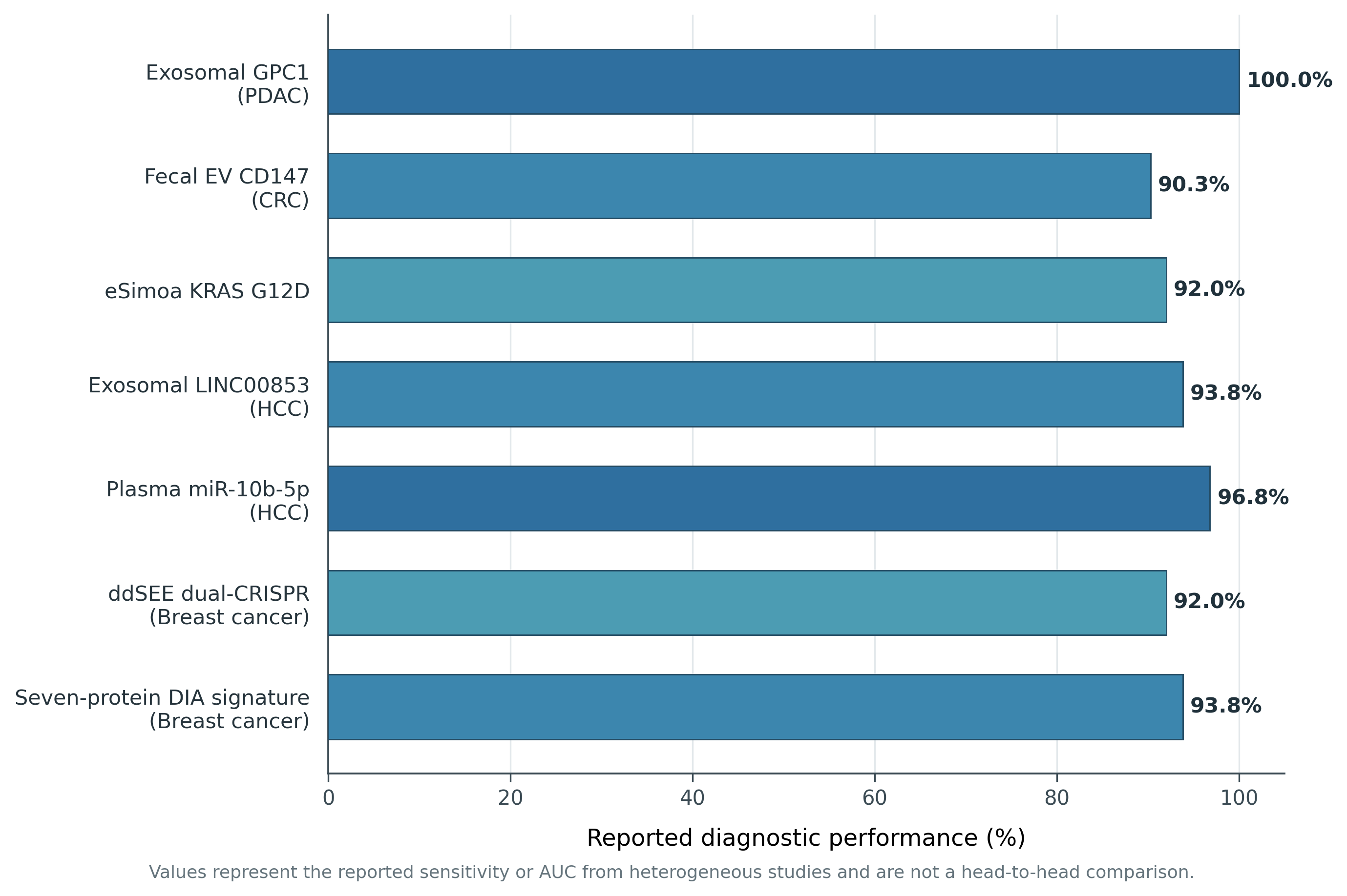
Comparable performance appears across other cancers. Glypican-1-positive circulating onco-EVs distinguish early pancreatic ductal adenocarcinoma from pancreatitis with high specificity (Guzowska et al., 2026), while the Mercy Halo™ assay’s five-marker panel (BST-2, FOLR1, MUC1, MUC16, sTn) achieves high-specificity ovarian cancer detection in asymptomatic women (Lundy et al., 2026). In hepatocellular carcinoma, exosomal LINC00853 reaches 93.75% sensitivity and 89.77% specificity for early-stage disease (Li et al., 2026), and plasma miR-10b-5p achieves an AUC of 0.968 in distinguishing early HCC from chronic liver disease controls (Li et al., 2026) — both summarized alongside comparable panels in Figure 1, which plots representative diagnostic performance metrics side by side.
To resolve rare onco-EV signal against what can be a roughly 350-fold excess of background host-derived vesicles, single-particle platforms have matured quickly (Lundy et al., 2026). The eSimoa framework

Figure 1. Analytical performance of representative single-EV and bulk liquid-biopsy biomarker panels across malignancies. Bar chart summarizing reported sensitivity or area-under-curve values (expressed as a percentage) for seven representative EV-based diagnostic panels spanning pancreatic, colorectal, hepatocellular, and breast cancers, including the exosomal GPC1 signature, fecal EV CD147 panel, eSimoa KRAS G12D assay, exosomal LINC00853, plasma miR-10b-5p, the ddSEE dual-CRISPR platform, and a seven-protein DIA proteomic signature. The figure visually demonstrates that single-vesicle digital platforms now achieve diagnostic performance broadly comparable to, and in several cases exceeding, that of established bulk molecular assays (Cheng et al., 2024; Li et al., 2026; Greening et al., 2025).
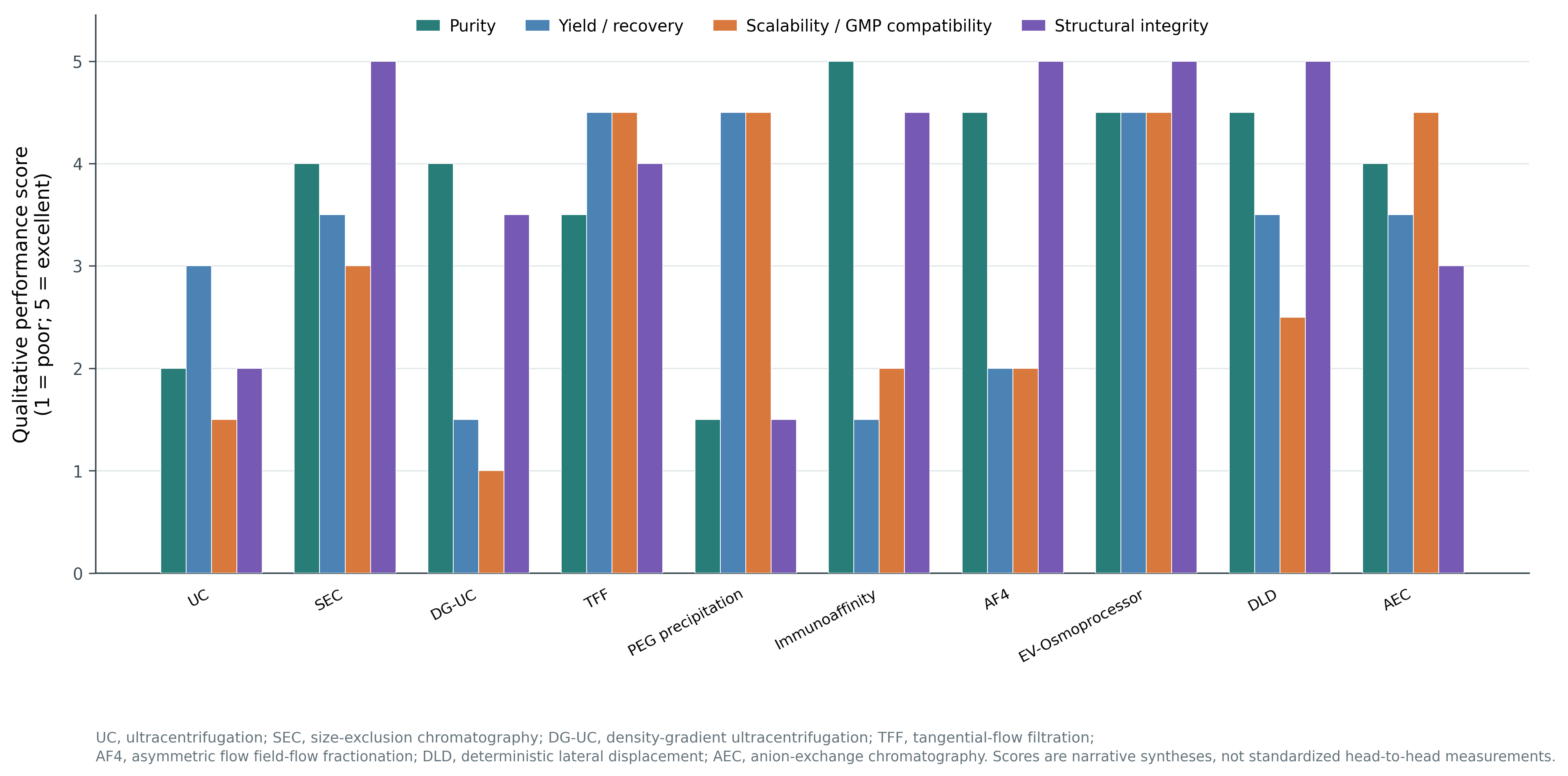
Figure 2. Comparative qualitative benchmarking of extracellular vesicle isolation and purification platforms. Grouped bar chart scoring ten isolation methodologies (1 = poor, 5 = excellent) across four performance dimensions—purity, yield/recovery, scalability/GMP compatibility, and structural integrity—derived narratively from the comparative data presented in Table 3. The figure illustrates the persistent trade-off structure across isolation platforms, showing that gentler, lower-throughput techniques such as size-exclusion chromatography and immunoaffinity capture generally preserve higher structural integrity, while pressure-driven and passive-concentration platforms such as tangential flow filtration and the EV-Osmoprocessor better satisfy industrial scalability requirements (Lundy et al., 2026; Abolhasani et al., 2026).


immunomagnetically captures target EVs via CD81/CD63, then isolates individual bead–EV complexes within femtoliter microwells to quantify luminal oncoproteins such as mutant KRAS G12D at sub-femtomolar concentrations (Cheng et al., 2024). The digital dual-CRISPR-Cas (ddSEE) system goes a step further, combining Cas12a-based surface-protein detection with Cas13a-based intravesicular microRNA profiling to reach 92% diagnostic accuracy in breast cancer (Zhang et al., 2026).

4.3. Resolving Methodological Bottlenecks: A Comparative Evaluation of Isolation Protocols

A large part of why EV preparations remain difficult to standardize traces back to the isolation step itself, and the literature makes clear that no single method wins across every dimension (Table 3) (Lundy et al., 2026; Tanaka, 2025). This trade-off structure is visualized in Figure 2, which scores the major platforms across purity, yield, scalability, and structural integrity.

Differential ultracentrifugation remains the most widely used approach in academic research — largely a function of history and its ability to handle large starting volumes — but it is slow, equipment-intensive, and mechanically harsh; the high gravitational forces involved (100,000–200,000 × g) can aggregate vesicles and damage membranes, and the technique co-sediments non-vesicular contaminants including lipoproteins that, in human plasma, can outnumber EVs by roughly six orders of magnitude (Lundy et al., 2026). That level of background noise is enough to confound both biomarker validation and potency testing.

Size-exclusion chromatography and tangential flow filtration have emerged as gentler, more industrially compatible alternatives (Limongi et al., 2026). SEC preserves vesicle structure and bioactivity effectively, though its relatively low throughput and tendency to dilute the sample mean downstream concentration is usually still required (Lundy et al., 2026). TFF, by contrast, achieves meaningfully higher yield and purity than ultracentrifugation while maintaining membrane integrity through continuous, low-shear tangential flow — a profile that fits reasonably well with scalable cGMP-compliant manufacturing (Abolhasani et al., 2026; Lundy et al., 2026).

Newer platforms push further still. Asymmetric Flow Field-Flow Fractionation, Deterministic Lateral Displacement, and passive EV-Osmoprocessor (EVOs) concentration each offer distinct advantages (Lundy et al., 2026). EVOs, in particular, uses osmotic pressure across a semipermeable membrane to achieve roughly fifty-fold volume reduction of conditioned media while removing about 99.7% of contaminating albumin within two hours — and when paired with downstream SEC, raises the particle-to-protein ratio to approximately 1 × 10⁹ particles per microgram, a genuinely scalable, high-purity upstream workflow (Lundy et al., 2026).

4.4. Advanced Surface Engineering and Cargo Loading: The Bioengineering Toolbox

Maximizing the therapeutic index of an EV formulation generally comes down to three overlapping problems: getting the right cargo in, keeping the vesicle in circulation long enough to matter, and directing it to the right tissue once it gets there (Table 4) (Lundy et al., 2026). Cargo loading itself splits along two broad lines — endogenous, “top-down” packaging during biogenesis, and exogenous, “bottom-up” loading after isolation (Lundy et al., 2026).

Endogenous approaches rely on the donor cell’s native sorting machinery: genetically fusing a target cargo to abundant tetraspanins such as CD63, CD9, or PTGFRN allows it to be actively packaged during vesicle formation (Lundy et al., 2026). The EXPLOR system represents perhaps the most refined version of this idea, using a light-reversible cryptochrome 2–CIB1 interaction to drive highly efficient, on-demand loading of therapeutic proteins into the exosome lumen (Lundy et al., 2026). Exogenous loading instead relies on physically or chemically permeabilizing an already-formed vesicle; electroporation remains the most common route for siRNA, microRNA, or larger gene-editing payloads, though cargo aggregation and membrane damage remain persistent, protocol-dependent risks (Luisotti et al., 2025; Lundy et al., 2026).

Circulation half-life is a separate, equally important variable. Unmodified EVs are typically cleared from the bloodstream within tens of minutes by the mononuclear phagocyte system in the liver and spleen (Lundy et al., 2026). Engineering vesicles to overexpress CD47 — the well-characterized “don’t eat me” signal recognized by SIRP-α on macrophages — substantially extends this window (Lundy et al., 2026). Targeting adds a further layer: fusing homing peptides to scaffold proteins like LAMP2B or CD63 enables tissue-specific binding, with the RVG peptide directing EVs across the blood–brain barrier and other engineered ligands supporting delivery to

Table 3. Comparative Assessment of Extracellular Vesicle Isolation and Purification Methods. This table compares eleven isolation methodologies used in laboratory-scale research and industrial cGMP manufacturing, detailing trade-offs in yield, purity, cost, and translational readiness. No single method optimizes every performance dimension simultaneously (Abolhasani et al., 2026; Lundy et al., 2026).

Isolation Method

Physical / Chemical Principle

Vesicle Purity

Vesicle Yield / Recovery

Scalability / Throughput

GMP Compatibility

Operational Cost

Structural & Functional Integrity

Differential Ultracentrifugation (UC)

Sedimentation via high-speed gravitational fields (100,000–120,000 × g)

Low-to-moderate; co-isolates protein aggregates and similar-density lipoproteins (Abolhasani et al., 2026).

Moderate; repeated pelleting causes sample loss/disruption (Abolhasani et al., 2026).

Low; limited by tube volume, manual batches (Lundy et al., 2026).

Low; labor-intensive, hard to automate, batch variation (Stella et al., 2026).

Low capital-intensive; minimal consumable cost (Abolhasani et al., 2026).

Variable; shear stress causes aggregation and membrane damage (Stella et al., 2026).

Size-Exclusion Chromatography (SEC)

Size-based separation via porous gel filtration matrix

High; separates from soluble proteins, not fully from chylomicrons/VLDLs (Stella et al., 2026).

Moderate-to-high; gentle, but sample dilution occurs (Lundy et al., 2026).

Moderate; scaled by column volume, needs downstream concentration (Lundy et al., 2026).

Moderate-to-high; integrable into automated fluidics (Lundy et al., 2026).

Moderate (column/resin cost) (Abolhasani et al., 2026).

Excellent; gentlest technique, preserves native proteins and receptors (Abolhasani et al., 2026).

Density Gradient Ultracentrifugation (DG-UC)

Equilibrium buoyant density separation in sucrose/iodixanol gradients

High; separates by density despite overlapping size (Abolhasani et al., 2026).

Low; selective harvesting causes severe sample loss (Abolhasani et al., 2026).

Low; labor-intensive, unsuited to high throughput (Abolhasani et al., 2026).

Low; complex manual harvesting resists standardization (Stella et al., 2026).

High (materials, ultracentrifuges, labor) (Abolhasani et al., 2026).

Good; gradient cushions protect vesicles, but osmotic pressure must be managed (Stella et al., 2026).

Tangential Flow Filtration (TFF)

Pressure-driven membrane filtration, parallel feed flow

Moderate-to-high; removes small contaminants, co-isolates similar-size lipoproteins (Stella et al., 2026).

High; continuous recirculation optimizes recovery (Stella et al., 2026).

High; suited to liter-scale clinical batches (Lundy et al., 2026).

High; standard automated closed systems in commercial bioprocessing (Lundy et al., 2026).

Moderate; automation cost offset by disposable cartridges (Abolhasani et al., 2026).

Good; low-shear filtration preserves membrane structure and cargo (Abolhasani et al., 2026).

Polymer Precipitation (e.g., PEG)

Solubility modulation via water-excluding polymers

Low; co-precipitates albumin, immunoglobulins, polymer residues (Limongi et al., 2026).

High; precipitates broad particle range at low speed (Abolhasani et al., 2026).

High; simple protocol enables rapid parallel processing (Abolhasani et al., 2026).

Low; polymer contaminants toxic, hard to remove (Stella et al., 2026).

Low (inexpensive reagents, standard centrifuges) (Abolhasani et al., 2026).

Poor-to-variable; causes irreversible aggregation, membrane fusion (Abolhasani et al., 2026).

Immunoaffinity Capture

Antigen-antibody binding on solid-phase substrates

High; exceptional purity via marker-defined selective isolation (Limongi et al., 2026).

Low; isolates only the targeted antigen-positive fraction (Limongi et al., 2026).

Low-to-moderate; suited to diagnostics, not clinical-scale manufacturing (Stella et al., 2026).

Moderate; costly antibodies, but standardized chips (Abolhasani et al., 2026).

High (monoclonal antibodies, magnetic substrates) (Abolhasani et al., 2026).

Excellent; gentle capture, though low-pH elution may affect integrity (Stella et al., 2026).

Asymmetric Flow Field-Flow Fractionation (AF4)

Fluid flow with perpendicular cross-flow, no stationary phase

High; separates from chylomicrons and overlapping lipoproteins (Lundy et al., 2026).

Low-to-moderate; precise fractionation yields moderate recovery (Lundy et al., 2026).

Low-to-moderate; requires customized instrumentation (Lundy et al., 2026).

Moderate; emerging automated QC/analytical tool (Lundy et al., 2026).

High (specialized instrumentation, trained personnel) (Lundy et al., 2026).

Excellent; continuous, label-free, fully preserves structure (Lundy et al., 2026).

EV-Osmoprocessor (EVOs)

Passive concentration via high-osmolarity polymer solution across permeable membrane

High; concentrates 50-fold, removes 99.7% albumin (Lundy et al., 2026).

High; passive, low-shear, prevents sample loss (Lundy et al., 2026).

High; scalable, fast (~2h), minimal intervention (Lundy et al., 2026).

High; simple, single-use, GMP-compatible (Lundy et al., 2026).

Low-to-moderate (simple polymer setup) (Lundy et al., 2026).

Excellent; passive concentration, preserves membrane markers (Lundy et al., 2026).

Deterministic Lateral Displacement (DLD)

Microfluidic post arrays redirecting particles by bifurcation angle/size

High; sorts subpopulations with precise cutoffs below 100 nm (Lundy et al., 2026).

Moderate-to-high; continuous sorting ensures high recovery (Lundy et al., 2026).

Low-to-moderate; limited by channel dimensions, but parallelizable (Lundy et al., 2026).

Moderate-to-high; standardized, integrable into lab-on-chip pipelines (Lundy et al., 2026).

High (chip fabrication, micro-pump equipment) (Lundy et al., 2026).

Excellent; continuous, label-free, low-shear sorting (Lundy et al., 2026).

Anion-Exchange Chromatography (AEC)

Electrostatic adsorption to positive stationary phases

High; separates negatively charged sEVs from non-EV contaminants (Limongi et al., 2026).

Moderate-to-high; strong binding can cause incomplete elution (Limongi et al., 2026).

High; scalable to liter-scale supernatant volumes (Limongi et al., 2026).

High; closed, automated, standard in protein/antibody manufacturing (Limongi et al., 2026).

Moderate (commercially standardized resins) (Limongi et al., 2026).

Variable; high-salt elution or pH shifts can impact stability (Limongi et al., 2026).

Table 4. EV Bioengineering, Cargo Loading, and Surface Modification Strategies. This table details eleven genetic, chemical, physical, and enzymatic strategies used to load therapeutic cargo and display targeting moieties on EVs, specifying mechanism, targeting ligand, and translational status. The table shows a progression from simple electroporation-based loading toward increasingly precision-targeted platforms now entering early clinical evaluation (Lundy et al., 2026; Limongi et al., 2026).

Engineering Category

Cargo / Payload Type

Loading Stage

Biochemical / Biophysical Mechanism

Surface Modification / Ligand

Targeted Cell / Tissue Site

Biocompatibility / Safety

Translation Status

Active Optically Induced Sorting (EXPLOR)

Regulatory proteins, transcription factors, enzymes

Pre-isolation (endogenous)

Light-inducible, reversible protein-protein interaction using CRY2/CIBN fused to exosomal CD9 scaffolds (Lundy et al., 2026).

CD9 exosomal surface scaffold (no additional peptide)

Cytosol of targeted host recipient cells

Highly biocompatible; avoids chemical residues or membrane disruption (Lundy et al., 2026).

Preclinical development and in vivo target validation (Lundy et al., 2026).

Electroporation

Therapeutic nucleic acids (siRNA, miRNA, shRNA)

Post-isolation (exogenous)

Electrical field creates transient nanoscale membrane pores, allowing passive cargo diffusion (Lundy et al., 2026).

Fused target-specific peptides (e.g., RVG, iRGD)

Receptors of recipient tissues (e.g., brain, tumor sites)

Risk of aggregation, cargo loss, altered pharmacokinetics without tight control (Lundy et al., 2026).

Active Phase I (e.g., iExoKrasG12D, NCT03608631) (Lundy et al., 2026).

CD47 Display ("Don't Eat Me" Cloaking)

Chemotherapeutics, siRNAs, gene-editing vectors

Pre-isolation (endogenous)

CD47 plasmid overexpression displays CD47 on exosomal membranes, signaling macrophages not to phagocytose (Lundy et al., 2026).

Surface display of CD47 fusion protein

Evades MPS clearance, prolonging circulation

Highly biocompatible; mimics self-recognition, reduces immunogenicity (Lundy et al., 2026).

Preclinical models of chronic inflammatory disease and oncology (Lundy et al., 2026).

Copper-Free Click Chemistry

Peptides, monoclonal antibodies, SERS tags, imaging agents

Post-isolation (exogenous)

Strain-promoted alkyne-azide cycloaddition (SPAAC) conjugates ligands to outer membrane proteins (Limongi et al., 2026).

PTHTRWA (lung-targeting) or RYYRITY (CAF-targeting) peptides

Lung cancer cells (α5β1 integrin) or CAFs in TME

Potential immunogenic risk from non-natural linkers; needs rigorous testing (Limongi et al., 2026).

Preclinical in vivo target homing/imaging studies (Limongi et al., 2026).

Membrane Hybridization (Hybrid EVs)

Large CRISPR/Cas9 plasmids, hydrophobic drugs

Post-isolation (exogenous)

Natural EV membranes fused with synthetic liposomes via extrusion/freeze-thaw, forming chimeric nanovesicles (Lundy et al., 2026).

Retains natural tetraspanins (CD9, CD63, CD81)

Tumors via passive EPR effect and active targeting

Combines natural biocompatibility with synthetic carrying capacity; low immunogenicity (Lundy et al., 2026).

Advanced preclinical multi-drug-resistant cancer models (Lundy et al., 2026).

Lonza Xcite EV Platform

Cytokines, therapeutic proteins, antibodies

Pre-isolation (endogenous)

Target proteins expressed as fusions to enriched EV scaffold proteins (PTGFRN or BASP1) (Lundy et al., 2026).

PTGFRN surface display or BASP1 luminal sorting

Solid tumor microenvironments (e.g., IL-12 display)

Highly biocompatible; localizes therapy, preventing systemic cytokine toxicity (Lundy et al., 2026).

Phase I evaluation of Exo-IL-12 (NCT04592484) (Lundy et al., 2026).

Dopamine Membrane Conjugation

Autophagy-inducing proteins, nucleic acids

Post-isolation (exogenous)

Covalent conjugation of dopamine moieties to surface lipids of isolated ADSC-derived EVs (Lundy et al., 2026).

Dopamine targeting moieties

Dopaminergic neurons in the brain (BBB traversal)

Crosses BBB and safely targets neurons, reducing α-synuclein pathology (Lundy et al., 2026).

Preclinical Parkinson's disease models (Lundy et al., 2026).

Cellular Nanoporation

Large mRNA transcripts encoding targeting peptides

Pre-isolation (endogenous)

Donor cells passed over a silicon micro-channel array under electronic stimulation, driving mRNA entry (Lundy et al., 2026).

CDX or CREKA peptides fused to CD47

EGFR-expressing cancer cells or bone marrow

Pure biological biogenesis avoids chemical toxicity; preserves stability (Lundy et al., 2026).

Preclinical tumor-targeting models (Lundy et al., 2026).

Aptamer Surface Display

CpG oligonucleotides (ODNs), siRNA cargo

Post-isolation (exogenous)

Covalent conjugation or lipid-tail insertion of synthetic aptamers into the phospholipid bilayer (Limongi et al., 2026).

CD63 or tumor-specific aptamers

Receptor-positive cancer or activated immune cells

Low immunogenic risk vs. monoclonal antibodies; stable and versatile (Limongi et al., 2026).

Preclinical melanoma postsurgical immunotherapy models (Limongi et al., 2026).

Ubiquitination-Targeted Luminal Sorting

Luminal target proteins, Cas9 protein

Pre-isolation (endogenous)

Cargo fused to Nedd4-mediated ubiquitination domains, triggering selective packaging during biogenesis (Lundy et al., 2026).

Retains native membrane receptor profiles

Cytoplasm of targeted host recipient cells

Exploits natural sorting machinery; avoids cargo degradation/leakage (Lundy et al., 2026).

Preclinical gene-editing and cell-reprogramming studies (Lundy et al., 2026).

muscle or tumor tissue (Lundy et al., 2026).

Finally, where cargo capacity outstrips what natural EVs can carry — full CRISPR/Cas9 expression systems, for instance — semi-synthetic “hybrid EVs,” formed by fusing natural EV membranes with synthetic liposomes or polymers, offer a practical middle ground: retaining much of the low immunogenicity of natural membranes while approaching the cargo capacity and manufacturability of fully synthetic nanocarriers (Bernad et al., 2025; Lundy et al., 2026).

5. Discussion

5.1. From Biological Promise to Clinical Signal

Taken together, the evidence assembled here — spanning trial registries, diagnostic validation studies, and comparative methodology — suggests the EV field has largely cleared its first hurdle: demonstrating that these vesicles carry genuine, exploitable biological information (Table 1; Table 2). What remains far less settled is whether that information can be captured, manufactured, and delivered with the consistency regulators and clinicians actually require. The dichotomy in Section 4.1 is telling in this respect: ExoFlo™‘s advance into Phase III trials (Lundy et al., 2026) sits uncomfortably alongside Codiak BioSciences’ 2023 bankruptcy and the stalled AB126 program (Lundy et al., 2026), which together suggest that scientific validity alone has rarely been the limiting factor — commercial and manufacturing fragility have mattered just as much, if not more.

5.2. Diagnostics: Closing the Sensitivity Gap, Opening a Standardization One

The shift from bulk to single-vesicle diagnostics represents, arguably, the most unambiguous technical success documented in this review (Figure 1; Table 2). Platforms such as eSimoa and ddSEE have solved a real problem — the “bulk average trap” that historically buried early, rare oncogenic signal within an overwhelming background of normal host-derived EVs (Cheng et al., 2024; Zhang et al., 2026). Yet solving the sensitivity problem has, in some ways, exposed a new one: cross-platform standardization. Each biomarker panel summarized in Table 2 and Figure 1 was validated using a distinct capture chemistry, reference gene, and cutoff methodology, which makes direct performance comparison across studies harder than the headline AUC and sensitivity figures suggest. Before tools like the ExoDx Prostate IntelliScore or Mercy Halo™ assay can be adopted more broadly outside their originating centers, the field likely needs the kind of inter-laboratory harmonization efforts that other liquid biopsy modalities, such as circulating tumor DNA, have already undergone.

5.3. Manufacturing: Why Isolation Method Choice Is Not a Neutral Decision

The comparative data in Table 3 and Figure 2 make an important point that is easy to lose in individual method papers: there is no universally superior isolation technique, only trade-offs suited to different downstream goals. Differential ultracentrifugation’s continued dominance in the academic literature — despite its well-documented purity and reproducibility limitations (Lundy et al., 2026) — looks, in this light, less like a scientific endorsement and more like inertia born of historical familiarity and low capital cost. For clinical-grade manufacturing specifically, the data assembled here point fairly consistently toward tangential flow filtration and passive concentration platforms such as EVOs as more GMP-compatible alternatives (Abolhasani et al., 2026; Limongi et al., 2026; Lundy et al., 2026), a conclusion with direct implications for how future trial sponsors should be selecting their bioprocessing pipelines rather than defaulting to whatever protocol a given laboratory has historically used.

5.4. Bioengineering: Sophistication Has Outpaced Regulatory Language

The engineering strategies catalogued in Table 4 — CD47 cloaking, click-chemistry conjugation, hybrid EV fusion, light-inducible cargo loading — represent a genuinely impressive expansion of what is technically possible. But it is worth being honest that this sophistication has, if anything, widened the translational gap rather than narrowed it, because current regulatory frameworks were not built with these hybrid, multi-component products in mind (Limongi et al., 2026). The EMA’s 2025 clarification that “not substantially modified” EVs fall outside the Advanced Therapy Medicinal Products classification only partially resolves this; a heavily engineered, CD47-cloaked, peptide-targeted, cargo-loaded EV plainly does not fit that description, yet no equally clear alternative pathway currently exists either in Europe or under the FDA’s biologics framework (Limongi et al., 2026; Lundy et al., 2026).

5.5. Toward a Realistic Roadmap

If there is a single unifying implication across Sections 4.1–4.4, it is this: further proof-of-concept biology is not what is holding the field back. What appears to be missing is convergence — standardized potency assays that regulators across jurisdictions will accept, isolation and manufacturing protocols validated specifically for cGMP contexts rather than academic convenience, and diagnostic panels harmonized enough to be compared meaningfully across centers. None of this is a purely technical problem; it requires sustained coordination between academic researchers, industry sponsors, and regulatory bodies of the kind organizations like the ISEV Translation, Regulation and Advocacy Committee are beginning to attempt (Gurriaran-Rodriguez et al., 2026). Whether that coordination happens quickly enough to prevent further commercial casualties like Codiak’s remains, honestly, an open question.

5.6. Limitations

This synthesis has the limitations inherent to any narrative review: source selection, while structured, was not exhaustive in the manner of a formal systematic review, and quantitative performance metrics were reproduced as originally reported rather than independently re-analyzed or meta-analytically pooled. Several cited trials remain active or unpublished in full, meaning some outcome data reflect interim rather than final results.

6. Conclusion

Extracellular vesicles have earned their place as a genuinely promising cell-free platform for both precision diagnostics and targeted therapeutics, supported now by a substantial and maturing evidence base. Yet the translational valley of death persists — not for lack of biological validation, but because isolation standardization, GMP-scale manufacturing, potency assay development, and regulatory harmonization have not kept pace with laboratory innovation. Closing this gap will require coordinated, multi-institutional effort rather than further incremental proof-of-concept studies. With deliberate investment in reproducible bioprocessing and converging regulatory science, EVs are well positioned to transition from an actively promising research frontier into a validated pillar of precision, cell-free clinical medicine within the coming decade.

Author Contributions

B.F. contributed to the conception and design of the review, literature search, analysis and synthesis of the relevant evidence, and drafting of the manuscript. S.F. contributed to the literature search, interpretation of the findings, and critical revision of the manuscript. W.S. supervised the overall development of the review, contributed to the conceptual framework, interpretation and synthesis of the evidence, and critically revised the manuscript for important intellectual content. All authors reviewed and approved the final version of the manuscript and agreed to be accountable for all aspects of the work.

Acknowledgements

The authors would like to acknowledge the Faculty of Medicine and Medical Sciences, Universitas Baiturrahmah, Padang, Indonesia, for providing academic and institutional support during the preparation of this review. The authors also acknowledge the researchers whose published studies and clinical trial data contributed to the scientific foundation of this work.

References


Abolhasani, S., Ahmadi, Y., & Jalizih, A. (2026). Mesenchymal stem cell-derived exosomes for cardiac repair: Challenges, standardization gaps and a realistic path to clinical translation. Clinical and Translational Discovery, 6, e70185. https://doi.org/10.1002/ctd2.70185            

Bernad, E. S., Vasilache, I. A., Bernad, R. L., Hogea, L., Ene, D., Duica, F., Tudora, B., Bernad, S. I., Craina, M. L., & Mateiovici, L. (2025). Extracellular vesicles and nanoparticles in regenerative and personalised medicine: Diagnostic and therapeutic roles—A narrative review. Pharmaceutics, 17(10), 1331. https://doi.org/10.3390/pharmaceutics17101331   

Burnouf, T., Agrahari, V., & Agrahari, V. (2020). Extracellular vesicles as nanomedicine: Hopes and hurdles in clinical translation. International Journal of Nanomedicine, 15, 125–154. https://doi.org/10.2147/IJN.S221458

Chargaff, E., & West, R. (1946). The biological significance of the thromboplastic protein of blood. Journal of Biological Chemistry, 166(1), 189–197.           

Cheng, C. A. (2024). Before translating extracellular vesicles into personalized diagnostics and therapeutics: What we could do. Molecular Pharmaceutics, 21(6), 2625–2636. https://doi.org/10.1021/acs.molpharmaceut.4c00185    

Cheng, C.-A., Hou, K.-C., Hsu, C.-W., & Chiang, L.-C. (2024). Ultrasensitive and high-resolution protein spatially decoding framework for tumor extracellular vesicles. Advanced Science, 11(3), 2304926. https://doi.org/10.1002/advs.202304926  

Chin, A., Saing, H., et al. (2021). Bridging bench to bedside: Extracellular vesicles in clinical diagnostics. ACS Nano, 15(9), 113–128. https://doi.org/10.1021/acsnano.1c03562

Choi, S. J., Choi, J., Kim, J., Kim, S. H., Cho, H. G., Lim, M. Y., Chae, S., Lim, K. S., Ha, S. J., & Kim, H. O. (2026). Advanced technologies in extracellular vesicle biosensing: Platforms, standardization, and clinical translation. Molecules, 31(1), 227. https://doi.org/10.3390/molecules31010227           

Ghodasara, A., Raza, A., Wolfram, J., Salomon, C., & Popat, A. (2023). Clinical translation of extracellular vesicles. Advanced Healthcare Materials, 12(28), 2301010. https://doi.org/10.1002/adhm.202301010        

Greening, D. W., Xu, R., Rai, A., Suwakulsiri, W., Chen, M., & Simpson, R. J. (2025). Clinical relevance of extracellular vesicles in cancer—Therapeutic and diagnostic potential. Nature Reviews Clinical Oncology, 22(12), 924–952. https://doi.org/10.1038/s41571-025-01795-7          

Gurriaran-Rodriguez, U., Bussolati, B., Gimona, M., Glebov, K., Fujita, Y., Marcilla, A., Neri, C., Fu, Q. L., Das, S., Pluchino, S., Zarovni, N., Salomon, C., Witwer, K., & Falcon-Perez, J. M. (2026). From lab to industry: Advancing the translational potential of extracellular vesicles through the ISEV Translation, Regulation and Advocacy Committee (ISEV-TRA). Journal of Extracellular Vesicles, 15(1), 70220. https://doi.org/10.1002/jev2.70220         

Guzowska, J., Kowalski, S., Schachta, I., Piekus-Slomka, N., & Slomka, A. (2026). Connecting the dots: Milestones in the history of extracellular vesicle research. International Journal of Molecular Sciences, 27(5), 2470. https://doi.org/10.3390/ijms27052470      

Kong, L., Zhao, G., Wu, X., & Ma, S. (2026). Extracellular vesicles in cancer diagnosis and therapy: Advances, challenges, and prospects for clinical translation. International Journal of Molecular Sciences, 27(22), 2280. https://doi.org/10.3390/ijms27222280      

Li, K., Zuo, Z., Ou, X., Yang, M., Qin, Y., Zhang, B., et al. (2026). Unlocking the future of hepatocellular carcinoma early diagnosis: The promise of extracellular vesicle biomarkers. Journal of Clinical and Translational Hepatology, 14(4), 462–477. https://doi.org/10.14218/JCTH.2025.00589            

Limongi, T., Milla, P., Stella, B., & Arpicco, S. (2026). Regulatory challenges and opportunities for cell-derived extracellular vesicles in pharmaceutical development: A European and global perspective. Journal of Extracellular Vesicles, 15(4), 70332. https://doi.org/10.1002/jev2.70332   

Luisotti, L., Germelli, L., Piccarducci, R., Giacomelli, C., Marchetti, L., & Martini, C. (2025). Extracellular vesicles as vehicles for small non-coding RNA therapeutics: Standardization challenges for clinical translation. Extracellular Vesicles and Circulating Nucleic Acids, 6, 403–432. https://dx.doi.org/10.20517/evcna.2025.33   

Lundy, D. J., Chau, Z. L., Chen, S. Y., & Lai, J. J. (2026). Beyond extracellular vesicle (EV) hype: Practical solutions and remaining hurdles in EV research, manufacturing, and clinical translation. Advanced Science, 13(26), e21913. https://doi.org/10.1002/advs.202521913  

Morales, D. B., & Ko, J. (2022). Digital assays for extracellular vesicles: Heterogeneity, liquid biopsies, and clinical translation. ACS Nano, 16(8), 11619–11645. https://doi.org/10.1021/acsnano.2c04337      

Rajendran, R. L., & Gangadaran, P. (2026). Mesenchymal stem cells and extracellular vesicles: Bridging the translational gap in regenerative medicine. International Journal of Molecular Sciences, 27(10), 5423. https://doi.org/10.3390/ijms27105423      

Stella, B., Milla, P., Arpicco, S., & Limongi, T. (2026). Regulatory challenges and opportunities for cell-derived extracellular vesicles in pharmaceutical development: A European and global perspective. Journal of Extracellular Vesicles, 15(1), 70332. https://doi.org/10.1002/jev2.70332   

Su, Y., He, W., Zheng, L., Fan, X., & Tony, Y. H. (2025). Toward clarity in single extracellular vesicle research: Defining the field and correcting missteps. ACS Nano, 19(17), 16193–16203. https://doi.org/10.1021/acsnano.5c00705

Tanaka, M. (2025). Parkinson’s disease: Bridging gaps, building biomarkers, and reimagining clinical translation. Cells, 14(15), 1161. https://doi.org/10.3390/cells14151161            

Welsh, J. A., Goberdhan, D. C. I., O’Driscoll, L., & MISEV Consortium. (2024). Minimal information for studies of extracellular vesicles (MISEV2023): From basic to advanced approaches. Journal of Extracellular Vesicles, 13(2), e12404. https://doi.org/10.1002/jev2.12404            

Wolf, P. (1967). The nature and significance of platelet products in human plasma. British Journal of Haematology, 13(3), 269–288. https://doi.org/10.1111/j.1365-2141.1967.tb08741.x           

Yang, G., Yuan, L., & Song, B. (2022). Advances in extracellular vesicle engineering for clinical translation. Theranostics, 12(15), 6740–6759. https://doi.org/10.7150/thno.2022.12.6740         

Zhang, J., Pan, Y., She, P., & Rao, L. (2026). From bench to bedside: The promise and roadblocks of extracellular vesicle therapeutics. Theranostics, 16(9), 5044–5064. https://doi.org/10.7150/thno.131621     

Zhang, L., Shen, H., & Zhang, N. (2025). Advances in extracellular vesicle (EV) biomarkers for precision diagnosis and therapeutic in colorectal cancer. Frontiers in Oncology, 15, 1581015. https://doi.org/10.3389/fonc.2025.1581015


Article metrics
View details
0
Downloads
0
Citations
1
Views

View Dimensions


View Plumx


View Altmetric


0
Save
0
Citation
1
View
0
Share