Integrative Biomedical Research

Integrative Biomedical Research (Journal of Angiotherapy) | Online ISSN  3068-6326
463
Citations
1.8m
Views
746
Articles
Your new experience awaits. Try the new design now and help us make it even better
Switch to the new experience
Figures and Tables
REVIEWS   (Open Access)

Mitochondrial Transfer Between Cells: Physiological Roles and Therapeutic Potential

Jonathan Lim Chee Woei1, Ummi Nadira Daut1, How Soon Hin2, Johnson Stanslas1,*

+ Author Affiliations

Integrative Biomedical Research 10 (1) 1-8 https://doi.org/10.25163/biomedical.10110882

Submitted: 12 November 2025 Revised: 01 January 2026  Published: 15 January 2026 


Abstract

Mitochondria were long regarded as strictly intracellular organelles, inherited only vertically and confined within the cytoplasm of a single cell. That assumption no longer holds. Over the last two decades, a growing body of work has shown that mitochondria, and fragments of their genome, move between cells through tunneling nanotubes, gap junctions, extracellular vesicles, and even as free organelles ejected into the extracellular space — a phenomenon now termed horizontal mitochondrial transfer (HMT). This review synthesizes the physiological logic of HMT and evaluates the translational progress of artificial mitochondrial transplantation (MTT). A structured narrative review was conducted of peer-reviewed literature indexed and supplemented by hand-searching of reference lists, focusing on molecular mechanisms of HMT, in vivo and in vitro transplantation models, and extracellular-vesicle biology, following a reproducible, informed search and eligibility strategy. Contact-dependent transfer relies on Miro1/2-TRAK-KIF5-driven trafficking along tunneling nanotubes and on connexin-43 gap junctions, while contact-independent transfer is carried out chiefly by extracellular vesicles — exosomes, microvesicles, exophers, and migrasomes — each with distinct cargo and destination. Preclinical models across cardiac, cerebral, renal, and musculoskeletal injury consistently show that exogenous mitochondria restore bioenergetics and reduce apoptosis, and early-phase clinical experience in pediatric cardiac surgery and stroke supports feasibility and safety. HMT is not a laboratory curiosity but a conserved physiological communication system that, once dysregulated, can be hijacked by tumors and, once harnessed deliberately, offers a genuinely novel therapeutic modality. Its clinical maturation will depend on resolving questions of mitonuclear compatibility, immunogenicity, and manufacturing scale.

Keywords: mitochondrial transfer; tunneling nanotubes; extracellular vesicles; mitochondrial transplantation; intercellular communication; mitochondrial quality control; regenerative medicine

1. Introduction

For most of the twentieth century, the mitochondrion occupied a fairly settled place in the textbooks: a double-membraned organelle, the cell's own private power plant, generating adenosine triphosphate (ATP) through oxidative phosphorylation and otherwise minding its own business inside the cytoplasm that made it. That picture traces back, of course, to the endosymbiotic origin of the organelle — an alphaproteobacterium engulfed by an ancestral eukaryotic host, a relationship so old that mitochondria still carry their own circular genome and a faint echo of bacterial machinery (Borcherding & Brestoff, 2023). Inheritance, everyone assumed, was vertical and strictly intracellular: mitochondria divided when the cell divided, and that was that.

It turns out that assumption was incomplete, and the correction has been one of the more quietly radical developments in cell biology of the last two decades. The first hard demonstration came in 2006, when Spees and colleagues showed something that should not, by the old rules, have been possible: mtDNA-depleted, respiration-deficient A549 lung carcinoma cells regained the ability to breathe aerobically simply by being co-cultured with mesenchymal stem cells (MSCs), which had apparently donated intact, functioning mitochondria across the gap between them (Spees et al., 2006). A few years later, Islam et al. (2012) extended this from a dish to a living lung, showing that bone marrow-derived stromal cells could actively ferry mitochondria into damaged alveolar epithelial cells and rescue animals from lipopolysaccharide-induced acute lung injury. Findings like these did not just add a footnote to mitochondrial biology; they forced a rewrite. The field now has a name for this traffic — horizontal mitochondrial transfer (HMT) — and, rather charmingly, a name for the whole mobile signaling ecosystem of extracellular mitochondria and their genomes: the “momioma” (Torralba et al., 2016).

What is perhaps most striking, once you start looking, is how many different physical routes cells have evolved to move organelles between one another. The best-studied is the tunneling nanotube (TNT), a thin, F-actin-based cytoplasmic bridge — anywhere from about 50 to 1500 nm across — that can span distances well over 100 μm and functions almost like a corridor between two otherwise separate rooms (Liu et al., 2021). Cargo does not simply drift along these bridges; it is actively driven. The outer-membrane, calcium-sensitive Rho-GTPase Miro1 (and its relative Miro2) links mitochondria to Milton/TRAK adaptor proteins, which in turn hook the organelle onto kinesin-1 (KIF5) motors for directed transport along microtubules, while myosin XIX and myosin X handle the actin-based legs of the journey (Ahmad et al., 2014). Alongside TNTs sit gap junction channels built from connexin 43 (Cx43); these pores are far too narrow to let an entire mitochondrion through, but they are not irrelevant — Cx43 hemichannels stabilize cell-cell contacts, help TNTs form in the first place, and let calcium and reactive oxygen species pass between cells, which in turn seems to steer mitochondrial traffic toward the cell that needs it (Yao et al., 2018). Cell fusion, whether complete (yielding stable hybrid polyploid cells) or partial and transient, offers a third, less appreciated contact-dependent route, one that can even nudge somatic cells toward a more progenitor-like state (Liu & Mao, 2024).

Not every transfer requires direct contact, though. Cells also package mitochondria and mitochondrial fragments into extracellular vesicles — exosomes, microvesicles, and the larger, more specialized exophers — typically marked by tetraspanins such as CD9, CD63, and CD81, and released under stress through a CD38/cyclic-ADP-ribose-dependent calcium pathway (Wang et al., 2026). These vesicles can then travel considerable distances through extracellular fluid or blood before being taken up by a recipient cell. And in what is perhaps the most surprising variant, cells can simply eject free, “naked” mitochondria into the extracellular matrix; these organelles somehow retain their membrane potential and circular genome outside any protective vesicle and are picked up by neighboring cells through clathrin-mediated endocytosis, macropinocytosis, or heparan-sulfate-proteoglycan-assisted capture (Al Amir Dache et al., 2020).

Under ordinary, non-pathological conditions, this whole apparatus seems to function as a kind of quiet housekeeping system. In “licensed mitophagy,” or transmitophagy, cells that cannot easily replace themselves — neurons, for instance — hand off their damaged mitochondria to neighboring astrocytes for disposal rather than trying to degrade everything internally (Davis et al., 2014); retinal photoreceptors do something similar with Müller glia, and cardiomyocytes eject exopher-like packages of dysfunctional mitochondria that resident macrophages then clear (Nicolas-Avila et al., 2020). The same basic mechanism turns out to underlie metabolic tuning in adipose and bone tissue — adipocytes transferring mitochondria to macrophages to keep inflammation and insulin sensitivity in check (Borcherding et al., 2022), osteoblasts nudging progenitor differentiation — and it moonlights as an immunological dial, dampening inflammation when stem cells hand mitochondria to macrophages, but working in exactly the opposite, malignant direction when tumor cells strip mitochondria away from T lymphocytes to disable the very immune cells meant to be attacking them (Guan et al., 2024).

Given how central mitochondrial dysfunction is to cardiovascular, neurodegenerative, and metabolic disease, it was probably inevitable that someone would ask whether this natural transfer machinery could be redirected on purpose. That question has produced mitochondrial transplantation therapy (MTT), sometimes called artificial mitochondrial transfer (AMT): isolate healthy, respiration-competent mitochondria — usually from a patient's own skeletal muscle or from stem cells — and deliver them into damaged tissue. Because passive co-incubation is inefficient, several engineered delivery platforms have emerged, including MitoCeption (centrifugation plus mild thermal shock), MitoPunch (a pressure-driven mechanical plunger), magnetomitotransfer (magnetic bead-coupled uptake), photothermal nanoblades (laser-triggered microinjection), and peptide- or liposome-based surface coatings that bypass endolysosomal degradation (Caicedo et al., 2017). Clinical translation, remarkably, has already begun: McCully and colleagues pioneered intramyocardial injection of autologous mitochondria in pediatric cardiac surgery patients with encouraging functional recovery and no pro-arrhythmic complications (McCully et al., 2016), children with large-scale mtDNA deletion syndromes have undergone CD34+ stem-cell augmentation with maternally derived mitochondria, and early-phase human trials in acute cerebral ischemia have reported acceptable safety.

And yet, for all this progress, several fundamental questions remain genuinely open — not rhetorically open, but open in the sense that we do not yet have good answers. What signals tell a cell, in real time, to send a “rescue-me” call for mitochondria, and do those signals differ between acute and chronic injury? How does an internalized, foreign mitochondrion avoid being degraded by the very lysosomal system built to destroy such things? Does mismatch between donor mtDNA and recipient nuclear DNA — mitonuclear incompatibility — quietly undermine the long-term stability of transplanted cells? And how much do donor age, tissue origin, and sex shape the bioenergetic value and immunogenicity of the transferred organelles? This review is organized around exactly these questions, with four linked objectives: to map the molecular pathways of contact-dependent and contact-independent transfer; to evaluate HMT's physiological roles in quality control and immune signaling; to assess the preclinical and clinical evidence for artificial mitochondrial transplantation; and to sketch the safety, immunogenic, and bioethical groundwork that standardized clinical translation will eventually require.

2. Horizontal Mitochondrial Transfer: Mechanisms, Biological Functions, and Translational Potential

2.1. The Paradigm Shift of Horizontal Mitochondrial Transfer

For more than a century, textbook biology treated the mitochondrion as an intracellular fixture — a permanent resident of the cell that made it, descended from an ancient alphaproteobacterial endosymbiont and inherited strictly along the vertical, parent-to-daughter line (Borcherding & Brestoff, 2023). That description was not wrong, exactly, but it was, we now know, badly incomplete. Over the past two decades this assumption of absolute cytoplasmic segregation has been dismantled piece by piece, and what has replaced it is a rather more interesting picture: functional mitochondria, along with mtDNA, mitochondrial RNAs, and respiratory-chain proteins, cross cell boundaries routinely, not as an aberration but as an active, regulated physiological program (Torralba et al., 2016). Spees et al. (2006) were first to document this cleanly, in co-cultured cells, and the phenomenon — horizontal mitochondrial transfer, or HMT — has since been confirmed across a striking range of animal tissues and disease contexts (Liu et al., 2021).

Under baseline conditions HMT seems to work quietly as a homeostatic coordinator, something closer to routine tissue maintenance than emergency response. Under acute stress — hypoxia, ischemia-reperfusion, chronic inflammation — the behavior shifts: damaged recipient cells actively recruit healthy organelles from donor populations such as mesenchymal stem/stromal cells (MSCs) and astrocytes, apparently as a way of buying time before metabolic collapse (Hayakawa et al., 2016). Cells accomplish this transfer through several distinct physical strategies, ranging from direct cytoplasmic bridges to long-distance vesicle trafficking, and this collective communication network has earned its own name, the “momioma” (Torralba et al., 2016), a term that captures how mobile and genetically consequential extracellular mitochondria really are. What follows works through that machinery in more detail: intracellular quality control first, then the classification and biogenesis of mitochondria-carrying vesicles, then the sorting and delivery logic that gets cargo to the right destination, and finally the dual-edged pathophysiology — protective in most contexts, exploitable in cancer — before turning to the translational and diagnostic frontier.

2.2. Intracellular Mitochondrial Dynamics: The Quality-Control Rheostat

Mitochondrial health inside a single cell is maintained through continual cycles of fusion and fission, together referred to as mitochondrial dynamics (Anand et al., 2014). Fusion knits separate organelles into interconnected tubular networks, which raises oxidative phosphorylation capacity and, importantly, dilutes out mutated mtDNA before it can accumulate to pathogenic levels. Outer-membrane fusion depends on Mitofusin 1 and Mitofusin 2 (MFN1/MFN2), while inner-membrane fusion is carried out by the dynamin-related GTPase Optic Atrophy 1 (OPA1) — itself regulated by alternative splicing and by proteolytic cleavage from the metalloproteases OMA1 and YME1L, which convert the long, membrane-anchored form (L-OPA1) into a short, soluble form (S-OPA1); the balance between these two isoforms turns out to matter a great deal for preserving network architecture (Anand et al., 2014).

Fission runs in the opposite direction, breaking larger networks into smaller, more manageable segments — useful both for distributing organelles evenly during cell division and for isolating damaged sections so they can be marked for removal. The central actor here is the cytosolic GTPase Dynamin-related protein 1 (DRP1, also called DNM1L), recruited to constriction sites on the outer membrane by receptor proteins including FIS1, mitochondrial fission factor (MFF), and MiD49/MiD51. DRP1's activity is switched by phosphorylation state: modification at Serine 616 by ERK or AKT promotes fission, while phosphorylation at Serine 637 by protein kinase A restrains it. Fragments that emerge from this process with low membrane potential and excessive reactive oxygen species are then recognized by the PINK1-Parkin selective autophagy (mitophagy) pathway and routed to lysosomes for degradation — the cell's internal, single-organism version of quality control, and the necessary backdrop against which the intercellular version, HMT, needs to be understood.

2.3. Classification and Biogenesis of Mitochondria-Containing Extracellular Vesicles (mitoEVs)

When intracellular quality-control capacity is overwhelmed — when there is simply more damage than the fusion-fission-mitophagy system can absorb — cells appear to fall back on an export strategy, packaging mitochondrial material into extracellular vesicles (EVs) for disposal or, sometimes, for deliberate delivery elsewhere. Following the MISEV (Minimal Information for Studies of Extracellular Vesicles) framework, these mitochondria-containing EVs, or mitoEVs, are broadly split into small EVs (under roughly 200 nm) and large EVs (above 200 nm) (Wang et al., 2026).

Small EVs, typically exosomes generated from multivesicular bodies, are, quite simply, too small to carry an intact organelle. What they carry instead is mitochondrial debris in a useful sense — fragments, respiratory-chain proteins, circular mtDNA, and mitochondrial RNA (Sansone et al., 2017). A distinct subset of brain-derived small EVs, termed mitovesicles, carries its own characteristic mitochondrial protein signature and appears to be worth treating as a separate category rather than lumping in with conventional exosomes. Large EVs — mainly microvesicles or ectosomes that bud directly from the plasma membrane through a calcium-dependent process — are the ones actually capable of encapsulating and protecting a structurally intact, respiratory-competent mitochondrion during transit (Phinney et al., 2015).

Beyond these two general classes, cells produce specialized vesicle populations under particular conditions. Exophers — large, membrane-bound structures roughly 3.5 to 4.0 μm across — are extruded preferentially by cardiomyocytes and brown adipocytes when autophagic capacity is compromised; they bundle up damaged, depolarized mitochondria and misfolded protein and release the whole package into the extracellular matrix, where cardiac-resident macrophages engulf and degrade it in what has been called “licensed mitophagy” (Nicolas-Avila et al., 2020). Migrasomes are a different animal altogether: they form on the retraction fibers trailing behind migrating cells and support a clearance pathway termed mitocytosis, which protects motile cells from accumulating mitochondrial damage as they crawl (Jiao et al., 2021).

2.4. Sorting and Molecular Packaging of Mitochondrial Cargo

How does a cell decide what mitochondrial material to sort into which vesicle? Several coordinated mechanisms appear to be involved. At the level of the organelle itself, mitochondrial-derived vesicles (MDVs) — small, 70–150 nm carriers budding directly off the mitochondrial membrane under oxidative stress — are shaped by SNX9 and OPA1 and regulated through the PINK1/Parkin pathway; these MDVs then fuse with late endosomes, enriching the mtDNA and protein content of the exosomes that eventually form (Liang et al., 2023).

Packaging whole mitochondria into microvesicles, by contrast, leans on cytoskeletal transport machinery. Miro1 and Miro2, the calcium-sensing Rho-GTPases on the outer mitochondrial membrane, recruit TRAK1/2 adaptors that hook the organelle onto KIF5B kinesin motors for movement along microtubules toward the cell periphery, while an interaction with myosin XIX anchors mitochondria to cortical actin near the membrane itself (Ahmad et al., 2014). The cargo protein arrestin domain-containing protein 1 (ARRDC1) has also been implicated as a regulator of mitochondrial incorporation into shedding microvesicles, and the small GTPase Rab7 functions almost like a switch: active, GTP-bound Rab7 favors lysosomal degradation of damaged mitochondria under normal conditions, but when lysosomal function is compromised, Rab7 flips to its inactive GDP-bound state, lysosomal trafficking halts, and whole mitochondria are redirected instead into multivesicular bodies destined for extracellular release (Liang et al., 2023).

2.5. Targeted Delivery, Internalization, and Recipient Cell Fate

Once released, mitoEVs are not simply drifting debris — they appear to find specific targets through surface receptor-ligand interactions. In the peripheral nervous system, for instance, macrophage-derived EVs deliver mitochondria to dorsal root ganglion neurons through an interaction between CD200 receptor (CD200R) on the vesicle and iSEC1 ligand on the neuron, resolving neuroinflammation in the process (van der Vlist et al., 2022). Internalization itself proceeds through clathrin-dependent endocytosis, macropinocytosis, or direct membrane fusion, and in the central nervous system this is orchestrated largely by an NAD+/CD38/cADPR signaling cascade: ischemic neurons release extracellular NAD+, astrocytic CD38 converts this into cyclic ADP-ribose, calcium is released from the endoplasmic reticulum, and the resulting actin remodeling drives membrane invagination and uptake of astrocyte-derived mitochondria (Hayakawa et al., 2016).

What happens after uptake is, if anything, even more interesting. Donor mitochondria that manage to escape the endo-lysosomal pathway can integrate directly into the host's own mitochondrial network, a process that depends on the recipient cell's own fusion machinery — MFN1/MFN2 and OPA1 again (Picone et al., 2021). But Lin et al. (2024) offered a genuinely useful correction to how this benefit should be understood: the therapeutic effect of HMT does not appear to hinge simply on the donor organelle surviving indefinitely inside the recipient. Rather, the arrival of exogenous mitochondria seems to function as a priming signal, triggering the host's own mitophagy machinery to clear out damaged endogenous organelles while simultaneously switching on the SIRT1-PGC-1α axis to stimulate biogenesis of new, healthy mitochondria (Figure 2). In other words, the transplanted organelle may matter less as a permanent replacement part than as a catalyst that restarts the recipient cell's own repair program.

2.6. Physiological Rescue versus Maladaptive Hijacking

The consequences of this transfer system are genuinely two-sided, and that duality is, in a sense, the central tension of the whole field. In acute injury, EV-mediated transfer clearly functions as a rescue mechanism. During ischemic stroke, astrocytes release microvesicles carrying polarized, functional mitochondria that home to hypoxic neurons, raise ATP levels, and reduce post-stroke apoptosis (Hayakawa et al., 2016). In the heart and lungs, damaged cardiomyocytes and endothelial cells appear to send out distress signals that draw MSCs to the site, where they release mitochondria-rich EVs that restore oxidative phosphorylation and stabilize the alveolar-capillary barrier in models of acute respiratory distress syndrome and myocardial infarction. Crewe et al. (2021) went further, describing an inter-organ signaling loop in which metabolically stressed adipocytes package oxidatively damaged but still respiration-competent mitochondria into circulating EVs that travel to the heart and precondition cardiomyocytes against future ischemic insult — a genuinely elegant piece of systemic physiology.

Cancer biology tells a darker version of the same story. Glioma cells under nutrient stress secrete vesicles that pull in healthy mitochondria from neighboring astrocytes, boosting oxidative phosphorylation, tumorigenicity, and radioresistance in the process (Guan et al., 2024). In breast and colorectal cancer, cancer-associated fibroblasts and stromal cells package complete mitochondrial genomes into exosomes and deliver them to chemoresistant, glycolytic tumor cells, restoring respiratory activity, sidestepping the Warburg effect, and, ultimately, driving multidrug resistance and metastatic spread (Sansone et al., 2017). And under conditions of severe mitochondrial damage, mitoEVs can turn actively pathogenic: EVs carrying fragmented or oxidized mtDNA lack the usual histone protection, get recognized by host pattern-recognition receptors as something bacterial-like, and, once internalized, trigger cGAS-STING and Toll-like receptor 9 signaling — fueling the release of IL-1β and TNF-α and contributing to sterile neuroinflammation, hepatic steatohepatitis, and systemic inflammatory syndromes.

2.7. Diagnostic Biomarkers and Translational Hurdles

The diagnostic and therapeutic promise of mitoEVs is real, but so are the obstacles standing between the current evidence base and routine clinical use. On the diagnostic side, because brain tissue is not something one can biopsy casually, circulating CNS-derived mitoEVs — identifiable through surface markers such as CD46 and CD200, and through the mtDNA mutations they carry — offer an appealing, minimally invasive biomarker platform for tracking neurodegenerative disease progression in blood or cerebrospinal fluid.

On the therapeutic side, at least three practical hurdles recur across the literature (Table 3, Table 4). Production and scalability remain limited, since typical cell cultures secrete relatively few large, mitochondria-containing microvesicles; preconditioning strategies, such as treating donor endothelial cells with resveratrol to activate PGC-1α, have shown promise for boosting yield. Product heterogeneity is a second problem — EVs inevitably co-purify with non-mitochondrial cargo, which muddies mechanistic attribution unless studies include proper inactivated controls, such as mitochondria-depleted EVs or respiratory-chain inhibitors like antimycin A. And targeting specificity is a third: systemically injected mitoEVs are cleared rapidly by the mononuclear phagocyte system in the liver and spleen, which has pushed researchers toward engineering homing peptides or co-administering chaperones such as heat shock protein 27 to improve delivery to brain and heart. None of these problems looks unsolvable, but none of them is solved yet either, and resolving them is really what stands between HMT as an intriguing biological phenomenon and HMT as a dependable clinical tool.

3. Methods

This article was prepared as a structured narrative review rather than a primary experimental study, and the methodology below describes how the underlying literature was identified, screened, and synthesized, so that another reviewer could reasonably reproduce the search and arrive at a comparable evidence base.

3.1. Search Strategy

A systematic literature search was conducted across PubMed/MEDLINE, Scopus, and Web of Science for records published between January 2006 (the year Spees et al. first formally described HMT) and mid-2026, with no language restriction applied at the search stage. The search strategy combined controlled vocabulary (MeSH terms such as “Mitochondria/physiology,” “Cell Communication,” and “Extracellular Vesicles”) with free-text keywords, connected using Boolean operators in the general form: (“mitochondrial transfer” OR “horizontal mitochondrial transfer” OR “mitochondrial transplantation” OR “tunneling nanotube” OR “mitoEV” OR “artificial mitochondrial transfer”) AND (“mechanism” OR “therapeutic” OR “in vivo” OR “in vitro” OR “clinical trial”). Reference lists of key reviews (e.g., Borcherding & Brestoff, 2023; Torralba et al., 2016; Kubat et al., 2025) were hand-searched to capture additional primary studies not returned by the database search, a snowballing step included specifically to guard against keyword-related omission bias.

3.2. Eligibility Criteria

Studies were included if they (a) reported original in vitro, in vivo, or first-in-human data on intercellular or artificial mitochondrial transfer, or (b) were peer-reviewed reviews providing mechanistic synthesis directly relevant to the objectives stated in the Introduction. Conference abstracts without full-text availability, non-peer-reviewed preprints, and studies focused exclusively on maternal mtDNA inheritance (a vertical, non-horizontal process) were excluded. Where multiple publications reported overlapping datasets from the same research group (for example, sequential porcine cardiac transplantation studies from the McCully laboratory), the most complete or most recent report was prioritized for quantitative detail, while earlier reports were retained for methodological context.

3.3. Data Extraction

For each eligible study, the following data were extracted where available: species/model system, donor mitochondrial source, isolation and delivery method, dose or concentration, recipient tissue or cell type, analytical/validation techniques used to confirm transfer (e.g., MitoTracker labeling, Seahorse extracellular flux analysis, transmission electron microscopy, mtDNA genotyping), and the reported functional or bioenergetic outcome. These extracted parameters form the basis of the preclinical in vivo and in vitro summary tables

Table 1. Preclinical in vivo models demonstrating the therapeutic efficacy of mitochondrial transplantation and horizontal transfer across acute and chronic disease systems. This table summarizes twelve animal studies spanning cardiac, cerebral, renal, ocular, and corneal injury models, detailing the species used, the source and route of mitochondrial administration, and the dosage or concentration delivered. For each model, the resulting functional and physiological outcomes are reported alongside the recipient tissue or cell type targeted, together with the corresponding APA-style reference citation, allowing direct cross-model comparison of transplantation efficacy.

Disease/Injury Model

Species/System

Donor Source

Route

Key Outcome

Reference

Cardiac ischemia-reperfusion

Rabbit

Autologous skeletal muscle

Intramyocardial injection

↑ O2 consumption, ↓ infarct size, no arrhythmia

Masuzawa et al. (2013); McCully et al. (2016)

Myocardial I/R (prophylactic)

Porcine

Autologous skeletal muscle

Intracoronary injection

↓ infarct size, preserved contractility

Guariento et al. (2018)

Acute ischemic stroke

Rat/mouse

Autologous skeletal muscle

IV / intracerebroventricular

↓ infarct volume, reversed deficits

Zhang et al. (2019)

Parkinson's disease (6-OHDA)

Rat

Pep-1-labeled allo/xenogeneic

Stereotactic / intranasal

Restored locomotion, ↑ striatal ATP

Chang et al. (2016)

Alzheimer's disease (Aβ1-42)

Mouse

Human-isolated mitochondria

IV (tail vein)

Improved cognition, ↓ neuronal loss

Nitzan et al. (2019)

Diabetic nephropathy

Mouse

BM-MSCs

IV administration

PGC-1α activation, M2 polarization

Yuan et al. (2021)

Diabetic proximal tubule injury

Rat

BM-MSCs

Sub-renal capsule

Restored PTEC structure, ↑ SGLT2

Konari et al. (2019)

Acute kidney injury

Porcine

Allo/autologous skeletal muscle

Intra-arterial

↑ ATP, ↓ tubular necrosis

Doulamis et al. (2020)

Polymicrobial sepsis (CLP)

Mouse

Pectoral muscle tissue

IV (tail vein)

↑ bacterial clearance, ↑ survival

Miao et al. (2025)

Ischemic cardiomyopathy

Rat

AD-MSCs

Epicardial injection

Improved ventricular function

Mori et al. (2023)

Optic nerve injury

Rat

Liver-derived allogeneic

Intravitreal

Neuroprotection of RGCs

Huang et al. (2016)

Corneal wound healing

Mouse

iPSC-MSCs

Topical/subconjunctival

↑ re-epithelialization, ↓ ROS

Jiang et al. (2016); Paliwal et al. (2018)

Table 2. In vitro experimental models used to characterize the cellular kinetics, bioenergetic recovery, and survival effects of mitochondrial transfer and transplantation. This table compiles eleven cell-culture-based injury models — including hypoxia-reoxygenation, oxygen-glucose deprivation, and drug-induced toxicity paradigms — describing the recipient cell type, donor mitochondrial source, and delivery method used in each study. 

Injury Model

Recipient Cell

Donor Source

Method

Key Outcome

Reference

H/R injury

Human neuroblastoma

Rat brain synaptosomes

Co-incubation

↑ ΔΨm, ↑ ATP, ↓ apoptosis

Picone et al. (2021)

MPP+ toxin exposure

SH-SY5Y

HepG2 cells

Co-culture

↑ complex I activity, ↑ OCR

Miao et al. (2025)

H/R damage

SH-SY5Y

Platelet mitochondria

Co-cultivation

↓ apoptosis via FUNDC2/PIP3/Akt

Shi et al. (2021)

OGD ischemia

Cortical neurons

Astrocyte vesicles

Co-incubation

Restored ATP, axonal regrowth

Hayakawa et al. (2016)

OGD/reoxygenation

Cortical neurons

BHK fibroblasts

Co-incubation

Restored respiration

Huang et al. (2016)

Rotenone toxicity

RA fibroblasts/PC12

HEK293T cells

Photothermal nanoblade

Metabolic rescue, bypassed lysosome

Wu et al. (2016)

Dexamethasone atrophy

C2C12/L6 myotubes

Skeletal muscle

MitoCeption variant

Blocked AMPK/FoxO3 catabolism

Kim et al. (2018)

Cisplatin nephrotoxicity

Renal PTECs

Kidney tissue

Co-incubation

↑ Bcl-2, ↓ Bax, ↓ apoptosis

Miao et al. (2025)

Doxorubicin cardiotoxicity

iPSC cardiomyocytes

MSCs

Defined physical stress

↑ PGC-1α, protected contractility

O'Brien et al. (2021)

Okadaic acid neurotoxicity

SH-SY5Y

MSCs (EV-mediated)

Conditioned medium

↓ tau phosphorylation, ↓ ROS

Zhang et al. (2020)

RA / chondrocytic injury

Articular chondrocytes

MSCs (EV-mediated)

EV transfer

↓ MMP-13, cartilage regeneration

Wang et al. (2026); Luo et al. (2024)

presented in the Results (Table 1, Table 2), while EV-specific biogenesis, cargo, and signaling data were tabulated separately (Table 3), and pharmacological/genetic modulators of transfer pathways were compiled into a fourth summary table (Table 4).

3.4. Data Synthesis

Given the mechanistic and methodological heterogeneity across the included studies — spanning cell lines, animal species, injury models, and delivery platforms — a formal meta-analytic pooling of effect sizes was not appropriate, and findings are instead synthesized narratively, organized by physiological system and by mechanism of transfer. Where quantitative outcomes were reported consistently across multiple studies (e.g., reduction in infarct size, changes in ATP production, apoptotic marker expression), these are described comparatively in the Results and Discussion sections but are not statistically combined. This approach follows established conventions for narrative and scoping reviews in the mitochondrial biology literature (Iorio et al., 2024; Kubat et al., 2025) and is intended to make the review's evidentiary basis transparent and its search strategy reproducible for future updates.

4. Results

4.1. Preclinical In Vivo Efficacy of Mitochondrial Transplantation and Transfer

Across the preclinical animal literature compiled here (Table 1), mitochondrial transplantation therapy (MTT) and horizontal mitochondrial transfer (HMT) produce consistent, and in several cases quite substantial, therapeutic benefit across acute ischemic and chronic degenerative disease models. In cardiovascular medicine, Masuzawa et al. (2013) showed that direct intramyocardial microinjection of autologous, skeletal-muscle-derived mitochondria into the ischemic zone of rabbit hearts during early reperfusion preserved myocardial energetics — evidenced by higher post-ischemic oxygen consumption, faster high-energy phosphate synthesis, and a favorable shift in the myocardial proteome, all without triggering arrhythmia. Guariento et al. (2018) extended this logic to a larger, more clinically representative porcine model, in which preischemic autologous mitochondrial transplantation via serial intracoronary injection reduced infarct size and preserved contractile function; a broadly similar pattern of post-ischemic recovery was subsequently reported in rodent hindlimb ischemia-reperfusion, where intramuscular injection of syngeneic mitochondria reduced infarct area and improved functional recovery on gait analysis (Orfany et al., 2020).

In the central nervous system, where bioenergetic demand is unusually high and injury tolerance correspondingly low, mitochondrial transplantation has produced some of the most striking neuroprotective results in the dataset. In rodent middle cerebral artery occlusion models of ischemic stroke, intravenous administration of muscle-derived mitochondria significantly reduced infarct volume and reversed sensorimotor deficits, with systemically injected organelles apparently able to cross the blood-brain barrier and integrate into the injured cerebral parenchyma (Zhang et al., 2019; Wang et al., 2026). In Alzheimer's disease mouse models, intravenous infusion of isolated human mitochondria improved performance on water-maze cognitive testing, alongside reduced hippocampal neuronal loss, attenuated reactive gliosis, and restored citrate synthase and cytochrome c oxidase activity (Nitzan et al., 2019). In 6-hydroxydopamine-lesioned Parkinson's disease rats, targeted microinjection of Pep-1-labeled mitochondria into the medial forebrain bundle — or, notably, non-invasive intranasal delivery — partially rescued dopaminergic neuron loss and restored locomotor activity over a three-month observation period (Chang et al., 2016).

Outside the heart and brain, MTT has shown comparable tissue-specific benefit. In diabetic nephropathy models, transfer of healthy mitochondria from bone marrow-derived MSCs to renal macrophages restricted local inflammation and reduced kidney injury through activation of the PGC-1α pathway (Yuan et al., 2021), while restoring expression of key tubular transporters, including megalin and SGLT2 (Konari et al., 2019). In a porcine model of acute kidney injury mimicking donation-after-circulatory-death conditions, direct intra-arterial injection of psoas-derived mitochondria provided measurable renal protection and mitigated ischemia-induced tubular necrosis (Doulamis et al., 2020). Taken together, the in vivo dataset (Table 1) spans a genuinely broad set of organ systems and injury types, yet converges on a fairly consistent mechanistic story: exogenous mitochondria restore local bioenergetics and, through that restoration, blunt downstream apoptotic and inflammatory cascades.

4.2. In Vitro Mechanisms of Internalization, Bioenergetics, and Viability

Figure 1. Contact-dependent and contact-independent routes of horizontal mitochondrial transfer (HMT) between donor and recipient cells. Donor cells (e.g., mesenchymal stem cells, astrocytes) deliver mitochondria to injured or metabolically stressed recipient cells through two broad classes of pathway: direct cytoplasmic bridging via tunneling nanotubes (Miro1/2–TRAK–KIF5-driven trafficking) and gap junctions (connexin-43-mediated Ca²⁺/ROS signaling), or long-distance, contact-independent transport via extracellular vesicles and free/naked mitochondria taken up by endocytosis. Under physiological conditions this network maintains organelle quality control, immune balance, and tissue repair, whereas in pathological microenvironments such as tumors, the same machinery can be hijacked to strip mitochondria from immune cells.

Figure 2. Proposed intracellular fate of internalized exogenous mitochondria following transplantation or transfer. After entry into the recipient cell, donor mitochondria must escape endo-lysosomal degradation before fusing with the host's endogenous network through MFN1/MFN2 and OPA1. Rather than acting solely as a permanent replacement organelle, the internalized mitochondrion appears to function as a priming signal that triggers host mitophagy of damaged endogenous organelles while activating the SIRT1–PGC-1α axis to stimulate new mitochondrial biogenesis. This dual action culminates in restored ATP production, a lowered Bax/Bcl-2 ratio, reduced caspase-3/9 activation, and improved recipient cell survival.

In vitro experimental systems have been essential for isolating the precise cellular kinetics of mitochondrial internalization, bioenergetic recovery, and viability, at a resolution that in vivo work cannot easily provide (Table 2). When recipient cells are subjected to injuries such as hypoxia-reoxygenation or oxygen-glucose deprivation, their endogenous mitochondrial networks depolarize rapidly and apoptotic signaling begins; co-culture experiments show that healthy donor cells, most often MSCs or astrocytes, can interrupt this cascade by donating functional mitochondria to the stressed recipient. Primary cortical neurons subjected to oxygen-glucose deprivation, for example, show marked increases in ATP and survival once they internalize astrocyte-derived mitochondria (Hayakawa et al., 2016), and this transfer is further enhanced under mild hypothermia, which upregulates Miro1 expression in the donor astrocytes (Li et al., 2021).

Once internalized, donor mitochondria have to escape endo-lysosomal degradation in order to functionally integrate into the host's own network, and in cardiomyocytes and fibroblasts this survival step depends on the coordinated activity of the host's own fusion machinery — MFN1, MFN2, and OPA1 (Picone et al., 2021) (Figure 2). Successful integration of respiration-competent mitochondria correspondingly lowers the cellular Bax/Bcl-2 ratio, prevents cytosolic release of cytochrome c, and suppresses caspase-3 and caspase-9 activation, effectively steering the recipient cell away from a programmed-death trajectory (Jin et al., 2024).

Several delivery methods have been developed specifically to raise the otherwise low efficiency of passive co-incubation. Centrifugation-assisted MitoCeption forces rapid, actin-dependent macropinocytic uptake of isolated organelles, producing dose-dependent increases in oxygen consumption rate and ATP production (Caicedo et al., 2015). More precise biophysical platforms — the photothermal nanoblade and the mechanically driven MitoPunch — achieve stable, long-term retention of donor mtDNA in replication-limited recipient cells, generating so-called stable isolated mitochondrial recipient (SIMR) clones that rescue the metabolic profile of mtDNA-depleted lines (Patananan et al., 2020; Sercel et al., 2021).

4.3. Biophysical Diversity and Molecular Regulation of EV-Mediated Transfer

Extracellular vesicles serve as the principal contact-independent vehicle for mitochondrial trafficking, protecting fragile cargo from enzymatic degradation and immune surveillance during transit (Table 3; Figure 1). Following MISEV classification, mitoEVs separate into small EVs — typically exosomes, physically too small to carry an entire organelle and instead enriched in fragmented mtDNA, mitochondrial protein, and mitochondrial RNA (Sansone et al., 2017) — and large EVs, chiefly microvesicles, which are large enough to encapsulate an intact, polarized, respiration-competent mitochondrion and deliver it over distance to a bioenergetically compromised recipient tissue (Phinney et al., 2015).

Packaging into these large vesicles is governed by at least three distinguishable molecular switches. The Rab7 GTPase acts as a decision point: active, GTP-bound Rab7 favors ordinary lysosomal degradation of damaged mitochondria, whereas impaired lysosomal function drives Rab7 into its inactive, GDP-bound state, which halts lysosomal routing and redirects whole organelles into microvesicles for extracellular release instead (Liang et al., 2023). Under severe proteotoxic or autophagic stress, cardiomyocytes and brown adipocytes eject exophers — large, 3.5–4.0 μm vesicles that bundle damaged, depolarized mitochondria and misfolded protein for clearance by resident macrophages via Mertk-mediated endocytosis, thereby preserving cardiac and adipose metabolic stability (Nicolas-Avila et al., 2020). Migrating cells, meanwhile, rely on a distinct pathway — mitocytosis — in which damaged mitochondria are packaged into migrasomes that form on retraction fibers, clearing dysfunctional organelles to protect the migrating cell's own systemic energetics (Jiao et al., 2021).

4.4. Pharmacological and Genetic Modulation of Transfer Pathways

The pharmacological and genetic tools compiled in Table 4 further clarify how tightly regulated these transfer pathways actually are. TNT-mediated transfer depends heavily on connexin-43 gap junctional hemichannels, and the selective peptide inhibitors Gap19 and Gap26 essentially shut down TNT-mediated mitochondrial rescue in models of airway inflammation and myocardial ischemia (Yao et al., 2018). Cytoskeletal inhibitors — cytochalasin B and cytochalasin D, both of which depolymerize F-actin — confirm that organelle movement along nanotubes genuinely requires active actin remodeling rather than

Table 3. Molecular and biogenetic characteristics of mitochondria-containing extracellular vesicles (mitoEVs) classified according to MISEV guidelines. This table maps eleven distinct EV subtypes — including exosomes, microvesicles, exophers, and migrasomes — by diameter, mitochondrial cargo composition, and the molecular regulators governing their packaging and secretion (e.g., Rab7, ARRDC1, CD38/cADPR). Each entry specifies the donor and recipient cell or tissue of origin and the downstream physiological or signaling response elicited upon delivery, illustrating how vesicle class dictates cargo capacity and biological function.

EV Class

Size

Cargo

Regulator

Donor → Recipient

Reference

Small EVs (exosomes)

30–150 nm

mtDNA fragments, mtRNA

ESCRT, SNX9, OPA1, Rab9

Ovarian cancer → plasma

Wang et al. (2026)

Small EVs (exosomes)

30–150 nm

Full mtDNA genome

Endocytic/MVB, Rab7-GDP

CAFs → breast cancer stem cells

Sansone et al. (2017)

Small EVs (exosomes)

30–150 nm

mtRNA transcripts

YBX1 binding

DPSCs → alveolar epithelium

Chen et al. (2025)

Microvesicles

100–1000 nm

Intact whole mitochondria

ARRDC1, Rab7-GDP

MSCs → macrophages

Phinney et al. (2015)

Microvesicles

100–1000 nm

Intact mitochondria

CD200R

Macrophages → DRG neurons

van der Vlist et al. (2022)

Microvesicles

100–1000 nm

Polarized mitochondria

CD38/cADPR

Astrocytes → cortical neurons

Hayakawa et al. (2016)

Microvesicles

100–1000 nm

Intact mitochondria

Plasma membrane budding

Platelets → MSCs

Levoux et al. (2021)

Large EVs (MVs)

200–1000 nm

Intact mitochondria, ATP5A

CD38, actin remodeling

Brain endothelial → ischemic BEC

D'Souza et al. (2021)

Exophers

3.5–4.0 μm

Damaged mitochondria

Autophagy/lysosomal dysfunction

Cardiomyocytes → macrophages

Nicolas-Avila et al. (2020)

Exophers

3.5–4.0 μm

Damaged mitochondria

Parkin-mediated

Brown adipocytes → ATMs

Rosina et al. (2022)

Migrasomes

0.5–3.0 μm

Damaged mitochondria

Kif5b, Myosin19, Drp1

Migrating cells → extracellular space

Jiao et al. (2021)

Table 4. Pharmacological inhibitors, genetic activators, and biophysical delivery technologies used to experimentally modulate horizontal mitochondrial transfer. This table catalogs twelve natural and synthetic agents or engineered platforms — spanning gap-junction blockers (Gap19, Gap26), cytoskeletal and endocytosis inhibitors, genetic overexpression constructs (Miro1, Cx43), and artificial transplantation technologies (MitoCeption, MitoPunch, peptide surface coating) — specifying each agent's chemical identity, precise mechanism of action, and resulting functional consequence on transfer efficiency, together with their in vitro and/or in vivo applicability and source citation.

Target

Agent

Mechanism

Effect

Reference

Cx43 gap junctions

Gap19

Cx43 hemichannel peptide blocker

Inhibits GJIC and Ca2+/ROS exchange

Guan et al. (2024); Zuo et al. (2024)

Cx43 gap junctions

Gap26

Extracellular-loop mimetic peptide

Blocks TNT-mediated Cx43 signaling

Guan et al. (2024); Zuo et al. (2024)

TNT F-actin

Cytochalasin B

F-actin depolymerization

Blocks TNT biogenesis

Guan et al. (2024); Zuo et al. (2024)

TNT F-actin

Cytochalasin D

G-/F-actin binding toxin

Prevents TNT extension/stability

Guan et al. (2024); Zuo et al. (2024)

Microtubules

Nocodazole

β-tubulin polymerization inhibitor

Blocks kinesin-based transport

Guan et al. (2024); Zuo et al. (2024)

Clathrin endocytosis

Dynasore

Dynamin GTPase inhibitor

Prevents mitoEV/mitochondria uptake

Guan et al. (2024); Zuo et al. (2024)

Exosome biogenesis

GW4869

N-SMase inhibitor

Blocks MVB formation/exosome release

Guan et al. (2024); Zuo et al. (2024)

Miro1 transport

Miro1 overexpression

RHOT1 transfection

Accelerates Miro/TRAK/KIF5 transport

Ahmad et al. (2014)

Cx43 communication

Cx43 overexpression

GJA1 transfection

Stabilizes donor-recipient contact

Islam et al. (2012); Yao et al. (2018)

Biophysical delivery

MitoCeption

Centrifugation + thermal shock

Forces macropinocytic uptake

Caicedo et al. (2015); Cabrera et al. (2019)

Biophysical delivery

MitoPunch

Pressure-driven mechanical plunger

High-throughput stable mtDNA integration

Sercel et al. (2021); Patananan et al. (2020)

Surface modification

Pep-1/TAT peptide coating

Cell-penetrating peptide coating

Bypasses endocytosis, ↑2× uptake

Chang et al. (2016); Maeda et al. (2020)

passive diffusion (Bukoreshtliev et al., 2009), while the endocytosis inhibitor dynasore blocks dynamin-dependent clathrin-mediated uptake, preventing internalization of both free mitochondria and vesicle-encapsulated cargo by recipient cells (Paliwal et al., 2023).

At the genetic level, overexpressing Miro1 — the calcium-sensitive adaptor that couples mitochondria to KIF5B motors — in donor MSCs substantially increases TNT-mediated transfer to injured airway epithelium, reducing apoptosis and accelerating repair in vivo (Ahmad et al., 2014). And to bypass the low efficiency of passive co-incubation altogether, chemical surface modification of isolated mitochondria with cell-penetrating peptides such as Pep-1 (Chang et al., 2016) or cationic TAT-dextran (Maeda et al., 2020) neutralizes the organelle's negative surface charge, allowing it to bypass conventional endocytic uptake and roughly double cellular internalization efficiency.

5. Discussion

5.1. A Coordinated, Multi-Route Physiological System

Read together, the mechanistic and preclinical evidence gathered here (Tables 1–4; Figures 1–2) supports a fairly coherent overall picture: horizontal mitochondrial transfer is not an occasional or accidental event but a coordinated, multi-route physiological system, one whose contact-dependent and contact-independent arms (Figure 1) appear to be selected situationally, according to distance, urgency, and tissue architecture, rather than functioning as redundant backups of one another. Direct, TNT- and gap-junction-mediated transfer seems best suited to acute, localized rescue between adjacent cells, whereas EV-mediated transfer allows the same basic rescue logic to operate at a distance — as in the adipocyte-to-heart preconditioning circuit described by Crewe et al. (2021) — or across an anatomical barrier such as the blood-brain barrier (Wang et al., 2026).

5.2. The Dual Nature of Mitochondrial Transfer: Rescue versus Hijacking

What is harder to reconcile comfortably, and what the evidence does not let us look away from, is the dual-use nature of this system (Table 3). The very machinery that allows an astrocyte to save a dying neuron after stroke (Hayakawa et al., 2016) is, mechanistically, close kin to the machinery a glioma cell uses to steal mitochondria from a neighboring astrocyte in order to fuel its own growth and radioresistance (Guan et al., 2024), or that a breast cancer stem-like cell uses to import mitochondrial genomes from cancer-associated fibroblasts and escape therapy-induced dormancy (Sansone et al., 2017). This is not, we would argue, a contradiction so much as a reminder that intercellular organelle transfer is context-dependent machinery — its physiological valence is set by the tissue microenvironment, not by anything intrinsic to the transfer process itself. That has a direct clinical implication: any therapeutic strategy built on amplifying HMT (through Miro1 overexpression, for instance, or Cx43 upregulation) will need tissue- and disease-specific safeguards, because the same intervention that rescues an ischemic neuron could, in principle, provision a nearby tumor.

5.3. Translational Gaps in Mitochondrial Transplantation Therapy

On the translational side, the preclinical evidence in Table 1 and Table 2 is genuinely encouraging — the consistency of benefit across cardiac, cerebral, renal, and musculoskeletal models is not something one sees often with a mechanistically novel therapeutic class — but it should not be mistaken for proof of durable clinical efficacy. Several practical and biological gaps remain. First, the field still leans heavily on relatively short observation windows; the Parkinson's disease model summarized in Table 1 (Chang et al., 2016), for instance, extended to three months, which is a meaningful start but far short of what chronic neurodegenerative disease requires for confidence. Second, and more fundamentally, the mechanistic reframing offered by Lin et al. (2024) — that transplanted mitochondria may act principally as a priming signal for host mitophagy and SIRT1-PGC-1α-driven biogenesis, rather than as a permanent replacement organelle (Figure 2) — raises an open question about dosing and repeat administration that the current literature has not yet systematically addressed. If the therapeutic mechanism is catalytic rather than substitutive, then optimal dosing regimens likely look quite different from what current single-bolus protocols assume.

Delivery efficiency remains a genuine bottleneck as well. Passive co-incubation is inefficient enough that a whole cottage industry of biophysical platforms — MitoCeption, MitoPunch, photothermal nanoblades, peptide and TAT-dextran coatings (Table 4) — has emerged specifically to force the issue (Caicedo et al., 2015; Sercel et al., 2021; Maeda et al., 2020). These methods work, in the sense that they demonstrably raise internalization rates, but each brings its own translational complication: mechanical and laser-based methods are difficult to scale beyond single-cell or small-batch applications, and chemical surface modification introduces an additional regulatory variable (the coating itself) that will need its own safety characterization before clinical use expands.

5.4. Immunogenicity and Mitonuclear Compatibility

Immunogenicity and mitonuclear compatibility deserve particular emphasis, because they sit at the center of the questions posed in the Introduction and remain, honestly, under-addressed by the current evidence base. Fragmented or oxidized mtDNA released outside a protective vesicle is recognized by host pattern-recognition receptors as a bacterial-like danger signal, triggering cGAS-STING and TLR9 signaling and, in susceptible contexts, sterile inflammation (Table 3). This raises a legitimate safety question for allogeneic or xenogeneic mitochondrial transplantation protocols, several of which appear in Table 1 (e.g., Chang et al., 2016, using xenogeneic donor mitochondria in Parkinson's disease models), where donor-recipient mismatch could plausibly provoke exactly this kind of innate immune activation. Long-term heteroplasmy dynamics — what happens, over months or years, to a cell carrying two genetically distinct mitochondrial populations — are similarly under-characterized in the human clinical literature so far, largely because follow-up periods in first-in-human studies have understandably been short.

5.5. Future Directions

Looking forward, we think three priorities stand out. First, the field needs longer-term, ideally allogeneic-matched, in vivo studies that track heteroplasmy, immunogenicity, and functional outcome well beyond the three- to six-month windows typical of current work. Second, manufacturing and delivery standardization — including the “inactivated control” step recommended for EV studies (mitochondria-depleted EVs or respiratory-chain inhibitor controls) — will be essential for regulatory bodies to compare therapeutic claims across laboratories on equal footing. Third, and perhaps most importantly, dose-finding and repeat-administration studies should be designed around the priming/catalytic mechanism proposed by Lin et al. (2024) rather than assuming a simple replacement model, since these two mechanistic pictures imply quite different optimal regimens. None of this diminishes what has already been demonstrated — a genuinely novel therapeutic modality, grounded in a real and previously unrecognized physiological system — but it does mean that the gap between “biologically plausible” and “clinically standardized” is still real, and closing it will take exactly the kind of mechanistic and longitudinal work this review has tried to point toward.

6. Conclusion

Mitochondrial transfer between cells has moved, in the space of about twenty years, from an unexpected laboratory observation to a recognized physiological communication system with genuine clinical application. Contact-dependent and contact-independent pathways together maintain organelle quality control, metabolic homeostasis, and immune balance across tissues, while the same machinery can be hijacked by tumors when the microenvironment shifts. Preclinical transplantation studies across cardiac, cerebral, and renal injury consistently demonstrate restored bioenergetics and improved survival, and early clinical experience supports feasibility. What remains is the harder, slower work of establishing long-term safety, resolving mitonuclear and immunogenic uncertainties, and standardizing manufacturing — the necessary bridge between a compelling biological story and a dependable therapy.


Author Contributions

J.L.C.W. contributed to the conception and design of the review, literature search, analysis and interpretation of the relevant literature, and drafting of the manuscript. U.N.D. contributed to the literature review, synthesis of the evidence, and critical revision of the manuscript. H.S.H. contributed to the interpretation of the literature and critical review of the manuscript. J.S. supervised the overall development of the review, contributed to the conceptual framework, and critically revised the manuscript for important intellectual content. All authors reviewed and approved the final version of the manuscript and agreed to be accountable for all aspects of the work.

Acknowledgements

The authors would like to acknowledge the Department of Medicine, Faculty of Medicine and Health Sciences, Universiti Putra, Malaysia, and the Kulliyyah of Medicine, International Islamic University Malaysia, for their academic and institutional support. The authors also thank the researchers whose published studies contributed to the scientific foundation of this review.

References


Ahmad, T., Mukherjee, S., Pattnaik, B., Kumar, M., Singh, S., Kumar, M., Rehman, R., Tiwari, B. K., Jha, K. A., Barhanpurkar, A. P., Wani, M. R., Roy, S., & Dixit, A. (2014). Miro1 regulates intercellular mitochondrial transport & enhances mesenchymal stem cell rescue efficacy. The EMBO Journal, 33(9), 994–1010. https://doi.org/10.1002/embj.201386030          

Al Amir Dache, Z., Otandault, A., Tanos, R., Pastor, B., Meddeb, R., Sanchez, C., Arena, G., Lasorsa, L., Bennett, A., Grange, T., El Messaoudi, S., Mazard, T., Prevostel, C., & Thierry, A. R. (2020). Blood contains circulating cell-free respiratory competent mitochondria. FASEB Journal, 34(3), 3616–3630. https://doi.org/10.1096/fj.201901917RR  

Anand, R., Wai, T., Baker, M. J., Kladt, N., Schauss, A. C., Rugarli, E., & Langer, T. (2014). The i-AAA protease YME1L and OMA1 cleave OPA1 to balance mitochondrial fusion and fission. The Journal of Cell Biology, 204(6), 919–929. https://doi.org/10.1083/jcb.201308006             

Bao, Y., Hu, C., Wang, B., Liu, X., Wu, Q., Xu, D., Shi, Z., & Sun, C. (2025). Mitochondrial reverse electron transport: Mechanisms, pathophysiological roles, and therapeutic potential. Biology, 14(9), 1140. https://doi.org/10.3390/biology14091140    

Blitzer, D., Guariento, A., Doulamis, I. P., Shin, B., Moskowitzova, K., Barbieri, G. R., Orfany, A., del Nido, P. J., & McCully, J. D. (2020). Delayed transplantation of autologous mitochondria for cardioprotection in a porcine model. The Annals of Thoracic Surgery, 109(3), 711–719. https://doi.org/10.1016/j.athoracsur.2019.08.007           

Borcherding, N., & Brestoff, J. R. (2023). The power and potential of mitochondria transfer. Nature, 623(7986), 283–291. https://doi.org/10.1038/s41586-023-06537-z  

Borcherding, N., Jia, W., Giwa, R., Field, L. R., Moley, J. R., Kopecky, B. J., Chan, M., Yoshida, A. J., Nyachieo, K., Zeng, X., Zeng, H., Bagchi, S., Wu, D., & Brestoff, J. R. (2022). Dietary lipids inhibit mitochondria transfer to macrophages to divert adipocyte-derived mitochondria into the blood. Cell Metabolism, 34(10), 1499–1513.e8. https://doi.org/10.1016/j.cmet.2022.08.002         

Bukoreshtliev, N. V., Wang, X., Hodneland, E., Gurke, S., Barroso, J. F., & Gerdes, H.-H. (2009). Selective block of tunneling nanotube (TNT) formation inhibits intercellular organelle transfer between PC12 cells. FEBS Letters, 583(9), 1481–1488. https://doi.org/10.1016/j.febslet.2009.03.065  

Cabrera, F., Ortega, M., Velarde, F., Parra, E., Gallardo, S., Cabezas, J., Aleman, J., Trujillo, D., Chavez, C., Rojas, D., & Caicedo, A. (2019). Primary allogeneic mitochondrial mix (PAMM) transfer/transplant by MitoCeption to address damage in PBMCs caused by ultraviolet radiation. BMC Biotechnology, 19(1), 42. https://doi.org/10.1186/s12896-019-0534-1  

Caicedo, A., Aponte, P. M., Cabrera, F., Hidalgo, C., & Khoury, M. (2017). Artificial mitochondria transfer: Current challenges, advances, and future applications. Stem Cells International, 2017, Article 7610414. https://doi.org/10.1155/2017/7610414       

Caicedo, A., Fritz, V., Brondello, J.-M., Ayala, M., Dennemont, I., Abdellaoui, N., de Fraipont, F., Moisan, A., Prouteau, C. A., Boukhaddaoui, H., & Jorgensen, C. (2015). MitoCeption as a new tool to assess the effects of mesenchymal stem/stromal cell mitochondria on cancer cell metabolism and function. Scientific Reports, 5, Article 9073. https://doi.org/10.1038/srep09073       

Chang, J.-C., Wu, S.-L., Liu, K.-H., Chen, Y.-H., Chuang, C.-S., Cheng, F.-C., Su, H.-L., Wei, Y.-H., Kuo, S.-J., & Liu, C.-S. (2016). Allogeneic/xenogeneic transplantation of peptide-labeled mitochondria in Parkinson's disease: Restoration of mitochondria functions and attenuation of 6-hydroxydopamine-induced neurotoxicity. Translational Research, 170, 40–56.e13. https://doi.org/10.1016/j.trsl.2016.01.005        

Chen, H., Chen, X., Zhou, Z., Zheng, J., Lu, Y., Lin, P., & Zhao, X. (2025). Mesenchymal stromal cell-mediated mitochondrial transfer unveils new frontiers in disease therapy. Stem Cell Research & Therapy, 16, Article 546. https://doi.org/10.1186/s13287-025-04675-x        

Chen, J., Xie, Z., Zhou, H., Ou, Y., Tan, W., Zhang, A., & Fan, X. (2025). Mitochondria transfer in mesenchymal stem cells: Unraveling the mechanism and therapeutic potential. Current Stem Cell Research & Therapy, 20(11), 1153–1173. https://doi.org/10.2174/1573396320666240214115421              

Chen, R., & Chen, J. (2024). Mitochondrial transfer – a novel promising approach for the treatment of metabolic diseases. Frontiers in Endocrinology, 14, Article 1346441. https://doi.org/10.3389/fendo.2023.1346441            

Clemente-Suárez, V. J., Martín-Rodríguez, A., Yáñez-Sepúlveda, R., & Tornero-Aguilera, J. F. (2023). Mitochondrial transfer as a novel therapeutic approach in disease diagnosis and treatment. International Journal of Molecular Sciences, 24(10), 8848. https://doi.org/10.3390/ijms24108848              

Cowan, D. B., Yao, R., Akurathi, V., Dinardo, J. A., Melo, E., Zurakowski, D., Del Nido, P. J., & McCully, J. D. (2017). Transit and integration of extracellular mitochondria in human heart cells. Scientific Reports, 7, Article 17450. https://doi.org/10.1038/s41598-017-17813-0  

Crewe, C., Funcke, J.-B., Li, S., Joffin, N., Gliniak, C. M., Ghaben, A. L., An, Y. A., Vishvanath, L., Nelson, A. T., Deng, Y., Zhu, Q., Fan, K. H., Gordillo, R., & Scherer, P. E. (2021). Extracellular vesicle-based interorgan transport of mitochondria from energetically stressed adipocytes. Cell Metabolism, 33(9), 1853–1868.e11. https://doi.org/10.1016/j.cmet.2021.08.002        

Dave, K. M., Stolz, D. B., Venna, V. R., Quaicoe, V. A., Maniskas, M. E., Reynolds, M. J., Rizvi, F. F., Kofler, J., Reddy, R. C., Chun, M., Kagan, V. E., & Manickam, D. S. (2023). Mitochondria-containing extracellular vesicles (EV) reduce mouse brain infarct sizes and EV/HSP27 protect ischemic brain endothelial cultures. Journal of Controlled Release, 354, 368–393. https://doi.org/10.1016/j.jconrel.2023.01.003  

Davis, C. H. O., Kim, K. Y., Bushong, E. A., Mills, E. A., Boassa, D., Shih, T., Kinebuchi, M., Phan, S., Zhou, Y., Bihlmeyer, N. A., Nguyen, J. V., Jin, Y., Ellisman, M. H., & Marsh-Armstrong, N. (2014). Transcellular degradation of axonal mitochondria. Proceedings of the National Academy of Sciences, 111(26), 9633–9638. https://doi.org/10.1073/pnas.1401140111          

Doulamis, I. P., Guariento, A., Duignan, T., Orfany, A., Kido, T., Zurakowski, D., Del Nido, P. J., & McCully, J. D. (2020). Mitochondrial transplantation for myocardial protection in diabetic hearts. European Journal of Cardio-Thoracic Surgery, 57(5), 836–845. https://doi.org/10.1093/ejcts/ezz326 

D'Souza, A., Burch, A., Dave, K. M., Sreeram, A., Reynolds, M. J., Dobbins, D. X., Kamte, Y. S., Zhao, W., Sabatelle, C., Joy, G. M., Soman, V., Chandra, P. K., Cheng, M. H., Bahar, I., & Manickam, D. S. (2021). Microvesicles transfer mitochondria and increase mitochondrial function in brain endothelial cells. Journal of Controlled Release, 338, 505–526. https://doi.org/10.1016/j.jconrel.2021.08.038  

Guan, F., Wu, X., Zhou, J., Lin, Y., He, Y., Fan, C., Zeng, Z., & Xiong, W. (2024). Mitochondrial transfer in tunneling nanotubes—a new target for cancer therapy. Journal of Experimental & Clinical Cancer Research, 43, Article 147. https://doi.org/10.1186/s13046-024-03069-w 

Guariento, A., Blitzer, D., Doulamis, I. P., Shin, B., Moskowitzova, K., Barbieri, G. R., Orfany, A., del Nido, P. J., & McCully, J. D. (2018). Preischemic autologous mitochondrial transplantation by intracoronary injection for myocardial protection. The Journal of Thoracic and Cardiovascular Surgery, 156(6), 1160–1168. https://doi.org/10.1016/j.jtcvs.2018.06.010              

Han, H., Hu, J., Yan, Q., Zhu, J., Zhu, Z., & Chen, Y. (2020). Mesenchymal stem/stromal cell-mediated mitochondrial transfer and the therapeutic potential in treatment of neurological diseases. Stem Cells International, 2020, Article 8838046. https://doi.org/10.1155/2020/8838046             

Hayakawa, K., Esposito, E., Wang, X., Terasaki, Y., Liu, Y., Xing, C., Ji, X., & Lo, E. H. (2016). Transfer of mitochondria from astrocytes to neurons after stroke. Nature, 535(7613), 551–555. https://doi.org/10.1038/nature18928

Huang, P. J., Kuo, C. C., Lee, H. C., Shen, C. I., Cheng, F. C., Wu, S. F., Chang, C. H., Pan, H. C., Lin, S. Z., Liu, C. S., & Su, H. L. (2016). Transferring xenogenic mitochondria provides neural protection against ischemic stress in ischemic rat brains. Cell Transplantation, 25(5), 913–927. https://doi.org/10.3727/096368915X689785      

Iorio, R., Petricca, S., Mattei, V., & Delle Monache, S. (2024). Horizontal mitochondrial transfer as a novel bioenergetic tool for mesenchymal stromal/stem cells: Molecular mechanisms and therapeutic potential in a variety of diseases. Journal of Translational Medicine, 22, Article 491. https://doi.org/10.1186/s12967-024-05047-4           

Islam, M. N., Das, S. R., Emin, M. T., Wei, M., Sun, L., Westphalen, K., Rowlands, D. J., Quadri, S. K., Bhattacharya, S., & Bhattacharya, J. (2012). Mitochondrial transfer from bone-marrow–derived stromal cells to pulmonary alveoli protects against acute lung injury. Nature Medicine, 18(5), 759–765. https://doi.org/10.1038/nm.2736

Jiang, D., Gao, F., Zhang, Y., Wong, D. S. H., Li, Q., Tse, H.-F., Xu, G., Yu, Z., & Lian, Q. (2016). Mitochondrial transfer of mesenchymal stem cells effectively protects corneal epithelial cells from mitochondrial damage and promotes corneal wound healing. Cell Death & Disease, 7(11), e2467. https://doi.org/10.1038/cddis.2016.358     

Jiao, H., Jiang, D., Hu, X., Du, W., Ji, L., Yang, Y., Li, X., Sho, T., Wang, X., Li, Y., Wu, Y., Wei, Y., Wang, X., & Yu, L. (2021). Mitocytosis, a migrasome-mediated mitochondrial quality-control process. Cell, 184(11), 2896–2910.e13. https://doi.org/10.1016/j.cell.2021.04.027        

Jin, N., Zhang, M., Zhou, L., Jin, S., Cheng, H., Li, X., & Zhang, Y. (2024). Mitochondria transplantation alleviates cardiomyocytes apoptosis through inhibiting AMPK-mTOR mediated excessive autophagy. The FASEB Journal, 38(10), e23655. https://doi.org/10.1096/fj.202401077R              

Kaza, A. K., Wamala, I., Friehs, I., Kuebler, J. D., Rathod, R. H., Berra, I., Ericsson, M., Yao, R., Thedsanamoorthy, J. K., Zurakowski, D., Levitsky, S., del Nido, P. J., Cowan, D. B., & McCully, J. D. (2017). Myocardial rescue with autologous mitochondrial transplantation in a porcine model of ischemia/reperfusion. The Journal of Thoracic and Cardiovascular Surgery, 153(4), 934–943. https://doi.org/10.1016/j.jtcvs.2016.10.024             

Khan, M. M., Paez, H. G., Pitzer, C. R., & Alway, S. E. (2023). The therapeutic potential of mitochondria transplantation therapy in neurodegenerative and neurovascular disorders. Current Neuropharmacology, 21(5), 1100–1116. https://doi.org/10.2174/1567205020666230316110015              

Kim, M. J., Hwang, J. W., Yun, C.-K., Lee, Y., & Choi, Y.-S. (2018). Delivery of exogenous mitochondria via centrifugation enhances cellular metabolic function. Scientific Reports, 8(1), Article 3330. https://doi.org/10.1038/s41598-018-21539-y             

Konari, N., Nagaishi, K., Kikuchi, S., & Fujimiya, M. (2019). Mitochondria transfer from mesenchymal stem cells structurally and functionally repairs renal proximal tubular epithelial cells in diabetic nephropathy in vivo. Scientific Reports, 9(1), Article 5184. https://doi.org/10.1038/s41598-019-41524-7  

Kubat, G. B., Picone, P., Tuncay, E., Aryan, L., Girgenti, A., Palumbo, L., Akat, F., Turkel, I., Singh, K. K., & Nuzzo, D. (2025). Biotechnological approaches and therapeutic potential of mitochondria transfer and transplantation. Nature Communications, 16, Article 2839. https://doi.org/10.1038/s41467-025-61239-6    

Levoux, J., Prola, A., Lafuste, P., Gervais, M., Chevallier, N., Koumaiha, Z., Kefi, K., Braud, L., Schmitt, A., Yacia, A., Schirmann, A., Hivelin, M., Dussiot, M., Cambot, M., Auffret, M., & Vignais, M. L. (2021). Platelets facilitate the wound-healing capability of mesenchymal stem cells by mitochondrial transfer and metabolic reprogramming. Cell Metabolism, 33(2), 283–299.e9. https://doi.org/10.1016/j.cmet.2020.12.006      

Li, X., Li, Y., Zhang, Z., Bian, Q., Gao, Z., & Zhang, S. (2021). Mild hypothermia facilitates mitochondrial transfer from astrocytes to injured neurons during oxygen-glucose deprivation/reoxygenation. Neuroscience Letters, 756, Article 135940. https://doi.org/10.1016/j.neulet.2021.135940  

Liang, W., Sagar, S., Ravindran, R., Najor, R. H., Quiles, J. M., Chi, L., & Gustafsson, Å. B. (2023). Mitochondria are secreted in extracellular vesicles when lysosomal function is impaired. Nature Communications, 14(1), Article 5031. https://doi.org/10.1038/s41467-023-40680-5  

Lin, R. Z., Im, G. B., Luo, A. C., Zhu, Y., Hong, X., Neumeyer, J., Tang, H. W., Perrimon, N., & Melero-Martin, J. M. (2024). Mitochondrial transfer mediates endothelial cell engraftment through mitophagy. Nature, 629, 660–668. https://doi.org/10.1038/s41586-024-07340-0

Liu, D., Gao, Y., Liu, J., Huang, Y., Yin, J., Feng, Y., Shi, L., Meloni, B. P., Zhang, C., Zheng, M., & Gao, J. (2021). Intercellular mitochondrial transfer as a means of tissue revitalization. Signal Transduction and Targeted Therapy, 6(1), Article 65. https://doi.org/10.1038/s41392-020-00440-z  

Liu, H., & Mao, H. (2024). Intercellular mitochondrial transfer: The novel therapeutic mechanism for diseases. Traffic, 25(9), e12951. https://doi.org/10.1111/tra.12951       

Liu, Y., Dissanayaka, W. L., & Yiu, C. (2025). Therapeutic implications of mitochondrial transfer on stem cell fate in regenerative medicine. Journal of Translational Medicine, 23, Article 568. https://doi.org/10.1186/s12967-025-06472-9      

Luo, H., Li, X., Zhang, W., Chen, Y., Wang, S., Liu, F., Zhao, Y., & Zhang, B. (2024). Mitochondrial transplantation: A promising strategy for treating degenerative joint diseases. Journal of Translational Medicine, 22, Article 941. https://doi.org/10.1186/s12967-024-05979-x  

Maeda, H., Kami, D., Maeda, R., Murata, Y., Jo, J.-I., Kitani, T., & Yamamoto, M. (2020). TAT-dextran-mediated mitochondrial transfer enhances recovery from models of reperfusion injury in cultured cardiomyocytes. Journal of Cellular and Molecular Medicine, 24(9), 5007–5020. https://doi.org/10.1111/jcmm.15118             

Masuzawa, A., Black, K. M., Pacak, C. A., Ericsson, M., Barnett, R. J., Drumm, C., Seth, P., Bloch, D. B., Levitsky, S., Cowan, D. B., & McCully, J. D. (2013). Transplantation of autologously derived mitochondria protects the heart from ischemia-reperfusion injury. American Journal of Physiology-Heart and Circulatory Physiology, 304(7), H966–H982. https://doi.org/10.1152/ajpheart.00805.2012   

McCully, J. D., Levitsky, S., del Nido, P. J., & Cowan, D. B. (2016). Mitochondrial transplantation for therapeutic use. Clinical and Translational Medicine, 5, Article 16. https://doi.org/10.1186/s40169-016-0095-4  

Meng, S., Bai, M., Ma, C., Han, B., Duan, M., Zhang, L., Guo, J., Shi, C., Li, K., & He, M. (2025). Mitochondrial transfer as a novel therapeutic approach in ischemic stroke treatment: Current challenges and future perspectives. Neuroprotection, 3, 253–265. https://doi.org/10.1002/nep3.70004   

Miao, C., Jiang, P., Wang, Z., Kong, W., & Feng, L. (2025). Mitochondrial transplantation: A novel therapeutic strategy. MedComm, 6(1), e70253. https://doi.org/10.1002/mco2.70253         

Mohammadalipour, A., Dumbali, S. P., & Wenzel, P. L. (2020). Mitochondrial transfer and regulators of mesenchymal stromal cell function and therapeutic efficacy. Frontiers in Cell and Developmental Biology, 8, Article 603292. https://doi.org/10.3389/fcell.2020.603292       

Mori, D., Miyagawa, S., Kawamura, T., Yoshioka, D., Hata, H., Ueno, T., Toda, K., Kuratani, T., & Sawa, Y. (2023). Mitochondrial transfer induced by adipose-derived mesenchymal stem cell transplantation improves cardiac function in rat models of ischemic cardiomyopathy. Cell Transplantation, 32, 9636897221148456. https://doi.org/10.1177/09636897221148456              

Nicolas-Avila, J. A., Lechuga-Vieco, A. V., Esteban-Martinez, L., Sanchez-Diaz, M., Diaz-Garcia, E., Santiago, D. J., Rubio-Ponce, A., Li, J. L., Balachander, A., Quintana, J. A., Martinez-de-Mena, R., Castejon-Vega, B., Pun-Garcia, A., Traves, P. G., Bonzon-Kulichenko, E., Garcia-Marques, F., Cusso, L., Núñez, V., Gonzalez-Guerra, A., … Hidalgo, A. (2020). A network of macrophages supports mitochondrial homeostasis in the heart. Cell, 183(1), 94–109.e23. https://doi.org/10.1016/j.cell.2020.08.031  

Nitzan, K., Benhamron, S., Valitsky, M., Kesner, E. E., Lichtenstein, M., Ben-Zvi, A., Ella, E., Segalstein, Y., Nathan, A., Hoffman, S., & Nitzan, R. (2019). Mitochondrial transfer ameliorates cognitive deficits, neuronal loss, and gliosis in Alzheimer's disease mice. Journal of Alzheimer's Disease, 72(2), 587–604. https://doi.org/10.3233/JAD-190853             

O'Brien, C. G., Ozen, M. O., Ikeda, G., Vaskova, E., Jung, J. H., Bayardo, N., Santoso, M. R., Shi, L., Wahlquist, C., Jiang, Z., Nakamura, K., Yu, A., Rota, M., Shrestha, R., Yamaguchi, S., De Jesus Perez, V. A., Zamboni, C. G., Toma, I., Stayton, P. S., … Wu, J. C. (2021). Mitochondria-rich extracellular vesicles rescue patient-specific cardiomyocytes from doxorubicin injury: Insights into the SENECA trial. JACC: CardioOncology, 3(3), 428–440. https://doi.org/10.1016/j.jaccao.2021.05.006      

Orfany, A., Arriola, C. G., Doulamis, I. P., Guariento, A., Ramirez-Barbieri, G., Moskowitzova, K., Shin, B., Blitzer, D., Rogers, C., del Nido, P. J., & McCully, J. D. (2020). Mitochondrial transplantation ameliorates acute limb ischemia. Journal of Vascular Surgery, 71(3), 1014–1026. https://doi.org/10.1016/j.jvs.2019.03.041       

Paliwal, S., Chaudhuri, R., Agrawal, A., & Mohanty, S. (2018). Regenerative abilities of mesenchymal stem cells through mitochondrial transfer. Journal of Biomedical Science, 25(1), Article 31. https://doi.org/10.1186/s12929-018-0429-1    

Paliwal, S., Jain, S., Mudgal, P., Verma, K., Paliwal, S., & Sharma, S. (2023). Mitochondrial transfer restores impaired liver functions by AMPK/mTOR/PI3K-AKT pathways in metabolic syndrome. Life Sciences, 332, Article 122116. https://doi.org/10.1016/j.lfs.2023.122116         

Patananan, A. N., Sercel, A. J., Wu, T.-H., Ahsan, F. M., Torres, A., Jr., Kennedy, S. A. L., Vandiver, A. R., Collier, A. J., Mehrabi, A., Van Haute, L., Fasching, T., Minczuk, M., Rugg-Gunn, P., Chow, M. P. Y. E., & Teitell, M. A. (2020). Pressure-driven mitochondrial transfer pipeline generates mammalian cells of desired genetic combinations and fates. Cell Reports, 33(13), Article 108562. https://doi.org/10.1016/j.celrep.2020.108562  

Phinney, D. G., Di Giuseppe, M., Njah, J., Sala, E., Shiva, S., St. Croix, C. M., Stolz, D. B., Watkins, S. C., Di, Y. P., Leikauf, G. D., Kolls, J., Riches, D. W. H., Deiuliis, G., Kaminski, N., Boregowda, S. V., McKenna, D. H., & Ortiz, L. A. (2015). Mesenchymal stem cells use extracellular vesicles to outsource mitophagy and shuttle microRNAs. Nature Communications, 6, Article 8472. https://doi.org/10.1038/ncomms9472

Picone, P., Porcelli, G., Bavisotto, C. C., Nuzzo, D., Galizzi, G., Biagio, P. L. S., Bulone, D., & Di Carlo, M. (2021). Synaptosomes: New vesicles for neuronal mitochondrial transplantation. Journal of Nanobiotechnology, 19, Article 6. https://doi.org/10.1186/s12951-020-00748-6  

Rodrigues, T., & Ferraz, L. S. (2020). Therapeutic potential of targeting mitochondrial dynamics in cancer. Biochemical Pharmacology, 182, Article 114282. https://doi.org/10.1016/j.bcp.2020.114282      

Rosina, M., Ceci, V., Turchi, R., Chuan, Z., Borcherding, N., Sciarretta, F., Sanchez-Diaz, M., Tortolici, F., Karlinsey, K., Chiurchiù, V., Fenizia, C., Karlstaedt, A., D'Amico, D., Ricciardi, S., & Scherer, P. E. (2022). Ejection of damaged mitochondria and their removal by macrophages ensure efficient thermogenesis in brown adipose tissue. Cell Metabolism, 34(4), 533–548.e12. https://doi.org/10.1016/j.cmet.2022.02.016      

Sansone, P., Savini, C., Kurelac, I., Chang, Q., Amato, L. B., Strillacci, A., Stepanova, A., Iommarini, L., Mastroleo, C., Daly, L., Galké, B., Thakur, B. K., Soplop, N., Uryu, K., Hoshino, A., Norton, L., Bonafe, M., Cricca, M., Gasparre, G., … Bromberg, J. (2017). Packaging and transfer of mitochondrial DNA via exosomes regulate escape from dormancy in hormonal therapy-resistant breast cancer. Proceedings of the National Academy of Sciences, 114(43), E9066–E9075. https://doi.org/10.1073/pnas.1704862114    

Sercel, A. J., Patananan, A. N., Man, T., Wu, T.-H., Yu, A. K., Guyot, G. W., Rabizadeh, S., Niazi, K. R., Chiou, P.-Y., & Teitell, M. A. (2021). Stable transplantation of human mitochondrial DNA by high-throughput, pressurized isolated mitochondrial delivery. eLife, 10, e63102. https://doi.org/10.7554/eLife.63102   

Shi, C., Guo, H., & Liu, X. (2021). Platelet mitochondria transplantation rescues hypoxia/reoxygenation-induced mitochondrial dysfunction and neuronal cell death involving the FUNDC2/PIP3/Akt/FOXO3a axis. Cell Transplantation, 30, 9636897211024210. https://doi.org/10.1177/09636897211024210          

Shi, P., Gao, H., Cheng, Z., & Zhang, Y. (2024). Static magnetic field-modulated mesenchymal stem cell-derived mitochondria-containing microvesicles for enhanced intervertebral disc degeneration therapy. Journal of Nanobiotechnology, 22, Article 457. https://doi.org/10.1186/s12951-024-02716-1  

Spees, J. L., Olson, S. D., Whitney, M. J., & Prockop, D. J. (2006). Mitochondrial transfer between cells can rescue aerobic respiration. Proceedings of the National Academy of Sciences, 103(5), 1283–1288. https://doi.org/10.1073/pnas.0510511103       

Torralba, D., Baixauli, F., & Sánchez-Madrid, F. (2016). Mitochondria know no boundaries: Mechanisms and functions of intercellular mitochondrial transfer. Frontiers in Cell and Developmental Biology, 4, Article 107. https://doi.org/10.3389/fcell.2016.00107          

Tripathi, K., & Ben-Shachar, D. (2024). Mitochondria in the central nervous system in health and disease: The puzzle of the therapeutic potential of mitochondrial transplantation. Cells, 13, Article 410. https://doi.org/10.3390/cells13050410   

van der Vlist, M., Raoof, R., Willemen, H. L. D. M., Prado, J., Versteeg, S., Martin Gil, C., Vos, M., Lokhorst, R. E., Pasterkamp, R. J., Kojima, T., Karasuyama, H., Khoury-Hanold, W., Meyaard, L., & Eijkelkamp, N. (2022). Macrophages transfer mitochondria to sensory neurons to resolve inflammatory pain. Neuron, 110(4), 613–626.e9. https://doi.org/10.1016/j.neuron.2021.11.020       

Wang, L., Zhou, X., & Lu, T. (2025). Role of mitochondria in physiological activities, diseases, and therapy. Molecular Biomedicine, 6, Article 42. https://doi.org/10.1186/s43556-025-00284-5           

Wang, M., Wang, W., Chopp, M., Zhang, Z. G., & Zhang, Y. (2026). Therapeutic and diagnostic potential of extracellular vesicle (EV)-mediated intercellular transfer of mitochondria and mitochondrial components. Journal of Cerebral Blood Flow & Metabolism, 46(2), 289–305. https://doi.org/10.1177/0271678X251338971            

Wu, T.-H., Sagullo, E., Case, D., Zheng, X., Li, Y., Hong, J. S., TeSlaa, T., Patananan, A. N., McCaffery, J. M., Niazi, K., Braas, D., Koehler, C. M., Graeber, T. G., Chiou, P.-Y., & Teitell, M. A. (2016). Mitochondrial transfer by photothermal nanoblade restores metabolite profile in mammalian cells. Cell Metabolism, 23(5), 921–929. https://doi.org/10.1016/j.cmet.2016.04.008        

Xu, N., Yang, F., Ma, W., & Liu, G. (2026). Mechanisms and therapeutic potential of mitochondrial-targeted therapies in skeletal system diseases. Annals of Medicine, 58(1), 2641277. https://doi.org/10.1080/07853890.2026.2641277          

Yao, Y., Fan, X.-L., Jiang, D., Zhang, Y., Li, X., Xu, Z.-B., Fang, S. B., Tu, W., Chiu, K., Fu, Q.-L., & Gerdes, H.-H. (2018). Connexin 43-mediated mitochondrial transfer of iPSC-MSCs alleviates asthma inflammation. Stem Cell Reports, 11(5), 1120–1135. https://doi.org/10.1016/j.stemcr.2018.09.013  

Yuan, Y., Yuan, L., Li, L., Liu, F., Liu, J., Chen, Y., Cheng, J., & Zhang, B. (2021). Mitochondrial transfer from mesenchymal stem cells to macrophages restricts inflammation and alleviates kidney injury in diabetic nephropathy mice via PGC-1α activation. Stem Cells, 39(7), 913–928. https://doi.org/10.1002/stem.3375

Zhang, Z., Ma, Z., Yan, C., Pu, K., Wu, M., Bai, J., Li, Y., & Wang, Q. (2019). Muscle-derived autologous mitochondrial transplantation: A novel strategy for treating cerebral ischemic injury. Behavioural Brain Research, 356, 322–331. https://doi.org/10.1016/j.bbr.2018.09.005

Zhang, Z., Sheng, H., Liao, L., Xu, C., Zhang, A., Yang, Y., Zhao, L., Duan, L., Chen, H., & Yang, Y. (2020). Mesenchymal stem cell-conditioned medium improves mitochondrial dysfunction and suppresses apoptosis in okadaic acid-treated SH-SY5Y cells by extracellular vesicle mitochondrial transfer. Journal of Alzheimer's Disease, 78(3), 1161–1176. https://doi.org/10.3233/JAD-200788

Zuo, B., Li, X., Xu, D., Zhao, L., Yang, Y., Luan, Y., & Zhang, B. (2024). Targeting mitochondrial transfer: A new horizon in cardiovascular disease treatment. Journal of Translational Medicine, 22(1), Article 1160. https://doi.org/10.1186/s12967-024-05979-x

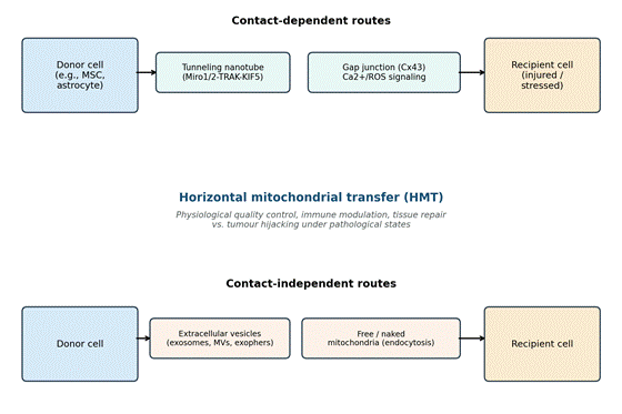
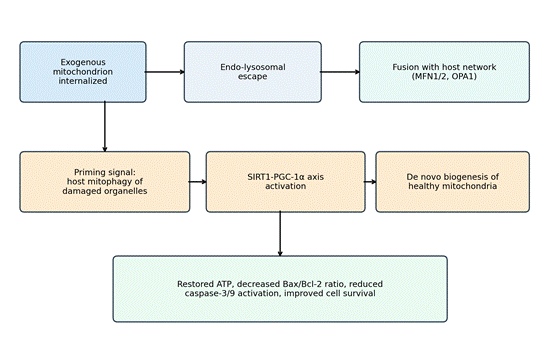
Article metrics
View details
0
Downloads
0
Citations
16
Views

View Dimensions


View Plumx


View Altmetric


0
Save
0
Citation
16
View
0
Share