Biosensors and Nanotheranostics

Bionanotechnology, Drug Delivery, Therapeutics | online ISSN 3064-7789
175
Citations
113k
Views
68
Articles
Your new experience awaits. Try the new design now and help us make it even better
Switch to the new experience
Figures and Tables
REVIEWS   (Open Access)

Stealth Nanocarriers with a Tunable Protein Corona to Prolong Circulation and Reduce Hepatic Clearance in Cancer Nanotheranostics 

Aya Elfiki 1*

+ Author Affiliations

Biosensors and Nanotheranostics 5 (1) 1-8 https://doi.org/10.25163/biosensors.5110971

Submitted: 12 June 2026 Revised: 02 August 2026  Published: 13 August 2026 


Abstract

Cancer nanotheranostics promises something clinically unusual: a single intravenous construct that reports where it has gone and treats what it finds. That promise is routinely defeated within minutes of injection. The liver, through Kupffer cell phagocytosis and sinusoidal filtration, removes 90–95% of a conventional nanocarrier dose, so the tumour sees only a residual fraction of what was administered. The decisive event occurs earlier than most design frameworks assume. On contact with plasma, a nanocarrier acquires a biomolecular corona that supersedes its synthetic surface and supplies the biological identity the mononuclear phagocyte system actually reads. This review examines how that corona can be governed rather than merely resisted. We first map the physicochemical determinants — hydrodynamic size, zeta potential, grafting conformation and interfacial elasticity — that set the balance between renal loss, tumour extravasation and hepatic capture. We then compare stealth architectures: PEGylation and its accelerated blood clearance liability, zwitterionic coatings that resist fouling without antibody induction, intrinsically disordered polypeptides, biomimetic membrane cloaking, and the emerging class of deliberately tuned coronas that recruit albumin, clusterin and apolipoproteins in place of opsonins. Evidence that protein adsorption is required for, not merely tolerated by, the stealth effect reframes the whole design objective. Reported gains are substantial — phagocytosis suppressed up to 2.6-fold, half-life extended from minutes to roughly 21 hours or, with erythrocyte membranes, several days — and translate into measurable theranostic performance across MRI-, photoacoustic- and ultrasound-guided platforms. Reproducibility, corona variability between donors and species, and manufacturing control remain the binding constraints on translation.

Keywords: protein corona; stealth nanocarrier; hepatic clearance; Kupffer cell; dysopsonin; cancer nanotheranostics; PEGylation; biomimetic membrane camouflage

1. Introduction

Cancer remains among the leading causes of death worldwide, and the reason is not usually that we lack cytotoxic chemistry. It is that we cannot deliver it where it is needed. Systemic chemotherapy for unresectable or metastatic disease is limited by short vascular residence, poor aqueous solubility, rapid metabolic degradation, indiscriminate biodistribution and dose-limiting toxicity in tissues that were never the target (Omidian et al., 2025; Wong et al., 2020). These are pharmacokinetic failures more than pharmacological ones, and that distinction matters, because it tells us where the engineering effort should go.

Cancer nanotheranostics emerged from precisely that reasoning. The concept is to integrate therapy — chemotherapy, photothermal therapy (PTT), photodynamic therapy (PDT), gene silencing, radiosensitisation — with diagnostic imaging — magnetic resonance imaging (MRI), computed tomography (CT), positron emission tomography (PET), photoacoustic imaging (PAI) and near-infrared fluorescence (NIR-FL) — inside one nanoscale platform (Sathuvan et al., 2026; Wong et al., 2020). A construct of this kind does more than carry a drug. It reports its own biodistribution, permits non-invasive monitoring of accumulation kinetics, and allows therapeutic response to be assessed while treatment is still modifiable (Omidian et al., 2025). Hepatocellular carcinoma, where imaging and intervention are tightly coupled in routine practice, has become a particularly instructive testing ground for this integration (Wang et al., 2026).

Translation depends on a condition that is easy to state and difficult to satisfy: the carrier must survive the journey. Once in the bloodstream, an unshielded nanocarrier enters an environment that is actively hostile to foreign particulates. Immune recognition is rapid, clearance is efficient, and the fraction of the injected dose that reaches tumour tissue is correspondingly small (Baek et al., 2026; Alexis et al., 2008). The liver dominates this process. Sadauskas and colleagues (2007) established that Kupffer cells are central to nanoparticle removal, and subsequent work has consistently placed hepatic sequestration of conventional carriers in the 90–95% range (Singh et al., 2025; El-Sayed Ellakwa & Ellakwa, 2026).

What makes this difficult rather than merely inconvenient is the mechanism. Clearance is not driven by the synthetic surface a chemist designs. It is driven by the layer of plasma protein that assembles on that surface within seconds of injection — the protein corona — which replaces the engineered identity with a biological one (Monopoli et al., 2012; Cedervall et al., 2007). The particle that macrophages encounter is therefore not the particle that was formulated. Any design strategy that ignores this substitution is, in effect, optimising a surface that no longer exists by the time it matters.

For roughly three decades the field’s answer was exclusion. Dense hydrophilic polymer brushes, polyethylene glycol above all, were grafted onto carriers to form a hydrated steric barrier against protein adsorption (Suk et al., 2016). The approach works, up to a point. Its limits are now well documented: repeated administration elicits anti-PEG IgM, which alters corona composition, activates complement and drives the accelerated blood clearance (ABC) phenomenon, so that the second dose behaves worse than the first (Grenier et al., 2018; Zhao et al., 2018).

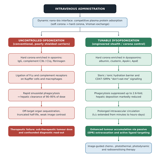
A more interesting result complicated the exclusion premise altogether. Schöttler et al. (2016) demonstrated that protein adsorption is required for the stealth effect of PEG- and poly(phosphoester)-coated carriers — specifically, enrichment of clusterin. Stealth, on this account, is not the absence of a corona but the presence of the right one. That inversion underwrites the strategies reviewed here: selective recruitment of dysopsonins such as albumin, clusterin, apolipoprotein A-I and apolipoprotein E, achieved through molecular imprinting (Takeuchi et al., 2017), recombinant protein shields (Oh et al., 2018), surfactant-mediated corona modulation (Müller et al., 2017) or CD47-mimetic peptide display. Figure 1 sets out the two trajectories a carrier may follow at the nano–bio interface and identifies the transition this field is attempting to engineer.

This review synthesises current strategies for engineering stealth nanocarriers with tunable protein coronas, with emphasis on mechanisms that extend systemic circulation and suppress hepatic clearance. Five objectives organise the work.

First, to analyse critically the physicochemical determinants of nano–bio interaction — size, geometry, surface charge and elasticity — and how each governs corona kinetics, composition and hard/soft layer evolution (Table 1). Second, to compare synthetic and biomimetic stealth strategies, including PEGylation, zwitterionic functionalisation, intrinsically disordered polypeptides, cell membrane camouflage and recombinant protein shields, against the specific benchmark of ABC mitigation (Table 1; Table 3). Third, to deconstruct hepatic elimination, mapping sinusoidal microvascular architecture against Kupffer cell phagocytosis, fenestral transit and hepatobiliary excretion (Figure 2). Fourth, to detail tunable corona strategies for dysopsonin enrichment and the quantitative gains they deliver (Table 2). Fifth, to examine clinical applications in multimodal imaging-guided therapy and the regulatory, manufacturing and safety obstacles that remain (Table 4; Figure 4).

2. Protein Corona Control and Stealth Engineering for Extended Circulation of Theranostic Nanocarriers

2.1 The biomolecular corona paradigm

A nanocarrier synthesised in vitro has one identity; the same object circulating in vivo has another. The transition is fast. Within seconds of entering plasma, thousands of circulating proteins compete for the high-energy nanoparticle surface through electrostatic, hydrophobic, van der Waals and hydrogen-bonding interactions, generating the dynamic shell termed the protein corona (Monopoli et al., 2012; Rampado et al., 2020).

Corona assembly is sequential rather than instantaneous in composition. Abundant but low-affinity plasma proteins arrive first, forming a loosely associated and rapidly exchanging soft corona. Over minutes to hours, higher-affinity species displace them — the Vroman effect — yielding a tightly bound, near-monolayer hard corona that persists and defines biological behaviour (Cedervall et al., 2007; Monopoli et al., 2012). Cedervall and colleagues (2007) quantified the exchange rates and affinities underlying this hierarchy, giving the model a thermodynamic rather than merely descriptive basis.

The consequence is that corona composition, not core chemistry, determines hydrodynamic diameter, effective surface charge, colloidal stability, immunogenicity and ultimate organ fate (Monopoli et al., 2012; Singh et al., 2025). Critically, the corona measured in buffer or diluted serum does not reliably predict the corona formed in a living animal. Hadjidemetriou et al. (2015) recovered clinically used and antibody-targeted liposomes from the circulation of live animals and found an in vivo biomolecule corona distinct in composition from its in vitro counterpart — a finding with direct implications for how preclinical screening should be conducted. Figure 1 summarises how corona composition partitions carriers into divergent fates.

2.2 Opsonins, dysopsonins and the functional dichotomy

Adsorbed proteins are not equivalent in effect. Two functional classes dominate the literature (Table 2).

Opsonins — immunoglobulin G, complement components C3b, C1q and C4BPA, and fibrinogen — act as recognition tags. Once deposited in the hard corona they ligate Fcγ, complement and scavenger receptors on Kupffer cells and sinusoidal endothelial cells, triggering endocytosis and directing the bulk of the administered dose to liver and spleen (Chinen et al., 2017; Rampado et al., 2020). Chinen et al. (2017) illustrated this dependence elegantly using spherical nucleic acids, where oligonucleotide sequence alone redirected corona composition and biodistribution: G-rich constructs accumulated complement factors and showed greater than 85% liver and spleen uptake within eight hours, whereas poly-T constructs recruited albumin and reached approximately 55%.

Dysopsonins — serum albumin, apolipoprotein A-I, apolipoprotein E, apolipoprotein C-III and clusterin (apolipoprotein J) — have the opposite effect. They mask recognition epitopes, suppress complement activation and extend intravascular residence (Schöttler et al., 2016; Müller et al., 2017). The practical insight is that these proteins are already present in plasma at high concentration. A carrier that recruits them preferentially does not need an exogenous shield; it borrows one.

2.3 Hepatic architecture as a size- and charge-selective filter

The mononuclear phagocyte system (MPS), formerly the reticuloendothelial system, is the principal sink for circulating carriers, and within it the liver is dominant (Alexis et al., 2008; Baek et al., 2026). The reason is structural (Figure 2).

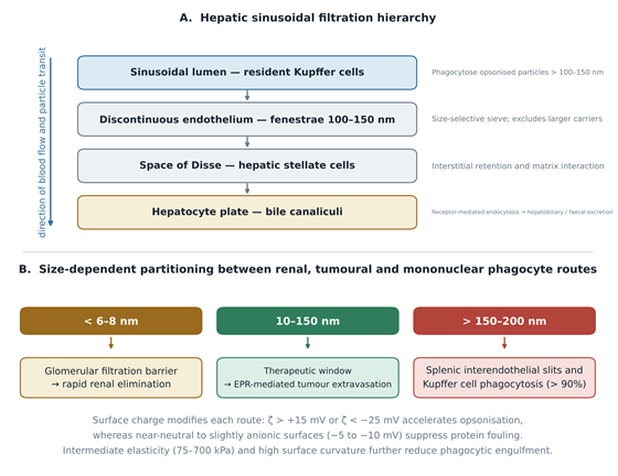
Portal and arterial blood traverses hepatic sinusoids lined by a discontinuous endothelium perforated by fenestrae of 100–150 nm. Kupffer cells sit within the sinusoidal lumen itself, screening flow directly rather than from behind a barrier (Sadauskas et al., 2007). Particles above roughly 100–150 nm, particularly when opsonised, are phagocytosed at this first encounter. Smaller or better-shielded constructs may pass the fenestrae into the space of Disse, interacting with hepatic stellate cells or undergoing hepatocyte endocytosis and subsequent hepatobiliary excretion into bile canaliculi (Baek et al., 2026). At the opposite extreme, ultrasmall carriers below 6–8 nm clear the glomerular filtration barrier and are lost renally before they can accumulate anywhere useful (Singh et al., 2025).

Three physicochemical parameters govern position within this hierarchy (Table 1). Hydrodynamic size. Carriers above 150–200 nm face mechanical entrapment in splenic interendothelial slits and rapid Kupffer capture; those below 6–8 nm are renally excreted; the operative window lies between, approximately 10–150 nm (Baek et al., 2026; Alexis et al., 2008). Surface charge. Strongly cationic (> +15 mV) and strongly anionic (< −25 mV)

Figure 1. Divergent biological trajectories determined by protein corona composition at the nano–bio interface. Following intravenous administration, competitive plasma protein adsorption converts the synthetic surface into a biological one through sequential soft-to-hard corona exchange. The left-hand pathway shows uncontrolled opsonisation, in which IgG, complement C3b/C1q and fibrinogen ligate Fcγ and complement receptors on Kupffer cells, producing hepatic clearance of 90–95% of the injected dose and therapeutic failure. The right-hand pathway shows tunable dysopsonisation, in which albumin, clusterin, ApoA-I and ApoE dominate the hard corona, phagocytosis is suppressed up to 2.6-fold, and circulation is extended sufficiently for EPR-mediated and ligand-directed tumour accumulation. Redirecting carriers from the left to the right trajectory is the central engineering objective reviewed here.

Figure 2. Hepatic sinusoidal filtration architecture and size-dependent partitioning of systemically administered nanocarriers. Panel A traces the sequential anatomical barriers encountered during hepatic transit: Kupffer cells within the sinusoidal lumen phagocytose opsonised particles above 100–150 nm; the discontinuous endothelium presents fenestrae of 100–150 nm acting as a size-selective sieve; carriers passing into the space of Disse interact with hepatic stellate cells; and hepatocytes mediate endocytosis with hepatobiliary and faecal excretion. Panel B shows how hydrodynamic diameter partitions carriers between renal elimination below 6–8 nm, the therapeutic window of 10–150 nm, and splenic and Kupffer capture above 150–200 nm. Surface charge, elasticity and curvature modulate each route, as indicated beneath the panel.

surfaces both accelerate non-specific protein binding and macrophage recognition. Near-neutral to slightly negative potentials, around −5 to −10 mV, minimise deposition (Abstiens et al., 2019; Singh et al., 2025). Abstiens et al. (2019) demonstrated this directly across PEG-PLA nanoparticles bearing amine, carboxy, methoxy and zwitterionic termini, where protein adsorption varied by more than twofold as a function of terminal group alone.

Mechanical elasticity and curvature. Intermediate elasticity in the 75–700 kPa range and high surface curvature reduce phagocytic engulfment by altering membrane wrapping energetics and protein adsorption geometry (Baek et al., 2026; El-Sayed Ellakwa & Ellakwa, 2026).

2.4 PEGylation: the benchmark and its ceiling

PEGylation has been the reference stealth strategy for good reason. Hydrophilic PEG chains bind water through hydrogen bonding, generating a hydrated cloud that imposes steric and osmotic penalties on approaching proteins (Suk et al., 2016). Performance scales with architecture rather than mere presence: raising molecular weight from 2 kDa to 5 kDa and increasing grafting density shifts chains from a loose mushroom to a dense brush conformation, and Zhou et al. (2018) showed that dense, dynamically fluctuating PEG shells cloak nanoparticles specifically from liver sinusoidal endothelial cell uptake, extending circulation half-life to approximately 21 hours (Table 1).

The ceiling is immunological. Repeated dosing elicits anti-PEG antibodies, predominantly IgM, which bind subsequent doses, activate the classical complement pathway and accelerate hepatic sequestration — the ABC phenomenon (Suk et al., 2016; Zhao et al., 2018). Grenier et al. (2018) went further, showing that anti-PEG antibodies do not simply opsonise; they alter the corona deposited on the particle and thereby reroute the physiological pathways governing its fate. Anti-PEG reactivity is also measurable in treatment-naive populations, which makes the problem one of baseline prevalence rather than cumulative exposure alone. Schöttler et al. (2016) supplied the complementary mechanistic point noted earlier: PEG works by recruiting clusterin, not by preventing adsorption outright.

2.5 Zwitterionic and disordered-polypeptide alternatives

Zwitterionic coatings address the ABC liability at its source by removing PEG from the interface. Polycarboxybetaine, sulfobetaine and zwitterionic peptides such as EK10 present equimolar positive and negative charges that bind water through ion–dipole interactions — stronger and more ordered than PEG’s hydrogen bonding — producing an ultra-stable hydration shell (Zhao et al., 2018; Loiola et al., 2019). Zhao and colleagues (2018) demonstrated that zwitterionic stealth peptide-protected gold nanoparticles achieve long circulation without inducing ABC on repeated administration, and Loiola et al. (2019) characterised the shielding contribution of zwitterionic moieties in double-functionalised silica (Table 1). Resistance to fouling in undiluted plasma for up to three days is a frequently reported benchmark.

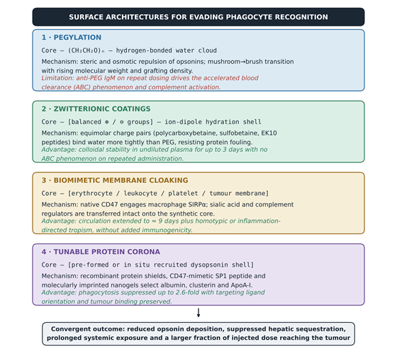
Intrinsically disordered polypeptides (IDPs) represent a different bet: that an unstructured, albumin-inspired hydrophilic domain can provide steric shielding genetically rather than synthetically. Zhang et al. (2025b) reported roughly fourfold half-life extension relative to PEGylated lipid nanoparticles alongside suppressed RAW 264.7 macrophage phagocytosis. A related peptide strategy — anionic, proline-rich sequences on liposomes — likewise prolonged circulation and evaded ABC after repeated administration (Zhang et al., 2025a). Figure 3 places these architectures alongside one another.

2.6 Biomimetic membrane camouflage

Rather than synthesising a stealth interface, biomimetic approaches borrow one. Hu et al. (2011) established the template by cloaking polymeric cores in intact erythrocyte membranes, transferring CD47, sialic acid residues and complement regulatory proteins onto a synthetic particle. CD47 engages SIRPα on macrophages, delivering an inhibitory “don’t eat me” signal that blocks actin-mediated engulfment; circulation extending to roughly nine days has been reported (Table 3).

The principle generalises across membrane sources. Leukocyte-derived proteolipid vesicles — leukosomes — retain LFA-1, Mac-1, ICAM-1 and chemokine receptors, directing active tropism to inflamed and tumour endothelium (Molinaro et al., 2016). Corbo et al. (2017) then did something methodologically important: they characterised the in vivo corona of circulating leukocyte-like carriers directly, finding reduced total protein adsorption with clusterin enrichment in the hard corona. Biomimetic cloaking, in other words, does not abolish the corona; it biases it toward dysopsonins. Platelet membranes contribute P-selectin and integrin αIIbβ3 for

Figure 3. Comparative surface architectures for evading mononuclear phagocyte recognition. Four stealth strategies are shown with their interfacial construction, governing mechanism, and principal advantage or limitation. PEGylation generates a hydrogen-bonded hydration cloud but elicits anti-PEG IgM on repeat dosing, driving the accelerated blood clearance phenomenon. Zwitterionic coatings bind water through stronger ion–dipole interactions, remaining stable in undiluted plasma for up to three days without inducing ABC. Biomimetic membrane cloaking transfers native CD47 and complement regulators onto the synthetic core, extending circulation to approximately nine days while adding homotypic tropism. Tunable protein corona strategies recruit endogenous dysopsonins directly. Despite differing chemistry, all four converge on reduced opsonin deposition and greater delivered dose.

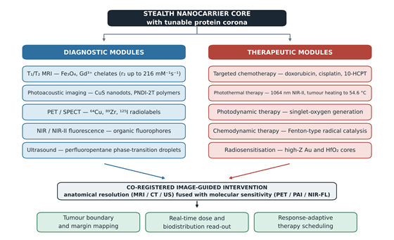
Figure 4. Integration of diagnostic and therapeutic modules on a stealth nanocarrier core with a tunable protein corona. A single long-circulating platform carries diagnostic reporters — T₁/T₂ MRI contrast agents, photoacoustic absorbers, PET/SPECT radiolabels, NIR/NIR-II fluorophores and ultrasound-active phase-transition droplets — alongside therapeutic modules spanning targeted chemotherapy, photothermal and photodynamic therapy, chemodynamic catalysis and high-Z radiosensitisation. Co-registration of anatomical resolution from MRI, CT or ultrasound with molecular sensitivity from PET, photoacoustic imaging or fluorescence enables tumour margin mapping, real-time dose read-out and response-adaptive scheduling. Every function depicted is contingent on circulation time, which is why corona control is the enabling step rather than an incremental refinement.

binding damaged tumour vasculature, while homologous cancer cell membranes exploit homotypic adhesion for self-recognition (Biswal et al., 2026; Sathuvan et al., 2026).

2.7 Deliberately tuned coronas

The newest strategies treat the corona as a design variable (Table 2). Three approaches recur. Pre-formed protein shields: Oh et al. (2018) constructed protein corona-shielded nanoparticles by decorating surfaces with recombinant fusion proteins — a HER2-binding affibody fused to glutathione-S-transferase — which suppressed complement and coagulation activation and reduced opsonisation while preserving targeting orientation. Combining an albumin shell with a CD47-mimetic minimal-self peptide (SP1) on mesoporous silica reduced RAW 264.7 phagocytosis 2.6-fold relative to aminated controls, against 1.6-fold for albumin alone (Table 2). In situ recruitment: Takeuchi et al. (2017) imprinted artificial recognition cavities for human serum albumin into polymeric nanogels, which then captured endogenous albumin on entering the circulation — stealth acquired after injection rather than before it, with prolonged half-life and reduced hepatic accumulation.

Corona composition steering: Müller et al. (2017) showed that amphiphilic polyphosphoester surfactants reshape the hard corona, stripping IgG and albumin while enriching ApoA-I and clusterin, with a corresponding fall in macrophage uptake. Targeted stealth constructs such as uPAR-directed small mesoporous silica for pancreatic cancer illustrate how these principles are being carried into specific disease contexts (Pontón et al., 2026).

2.8 Translation into theranostic function

Circulation time is not an end in itself; it is the precondition for everything a theranostic platform is supposed to do (Figure 4). Extended residence permits passive EPR-mediated extravasation and active ligand–receptor targeting, and both are prerequisites for adequate imaging contrast and therapeutic dose at the tumour (Wong et al., 2020; Sathuvan et al., 2026).

Reported platforms span the diagnostic spectrum (Table 4). Janus Fe₃O₄-Au(shell)-PEG nanostructures combine T₂-weighted MRI at r₂ = 216 mM⁻¹s⁻¹ with photoacoustic imaging and 1064 nm NIR-II photothermal ablation (Kang et al., 2021). CuS@mSiO₂-Pc(DOX)@HA systems integrate PAI and NIR fluorescence with triple PTT/PDT/chemotherapy under hyaluronidase-triggered release (Lv et al., 2022). Phase-transitional perfluoropentane nanodroplets add ultrasound to tri-modal MRI/PAI imaging with focused-ultrasound-triggered release (Liu et al., 2018). Metal-organic framework platforms enable T₂ MRI-monitored chemo-photothermal therapy in ovarian models (Qiang et al., 2022), and semiconducting polymer nanoparticles deliver deep-tissue NIR-II photoacoustic contrast with combined photothermal and photodynamic action (Wen et al., 2019).

3. Methods

This work is reported as a structured narrative review. The protocol below is specified at a level of detail sufficient for independent replication of the search, screening and synthesis, in line with the reporting expectations applied to reviews indexed in PubMed/MEDLINE.

3.1 Review design and guiding question

A narrative synthesis with systematic search architecture was selected in preference to a meta-analytic design. The justification is methodological rather than pragmatic: the included evidence spans heterogeneous nanomaterial classes, incompatible pharmacokinetic end points and non-standardised proteomic read-outs, so numerical pooling would impose a false commensurability on the data. The review was guided by a single structured question — in preclinical cancer nanotheranostic systems, which surface-engineering and protein corona modulation strategies measurably prolong systemic circulation and reduce hepatic clearance, and through what mechanisms? Population, intervention, comparator and outcome elements were defined as: engineered nanocarriers administered systemically (population); stealth surface modification or deliberate corona modulation (intervention); unmodified, bare or conventionally PEGylated carriers (comparator); circulation half-life, hepatic and splenic accumulation, macrophage uptake, corona composition and tumour accumulation (outcomes).

3.2 Information sources

Four bibliographic databases were interrogated: PubMed/MEDLINE, Scopus, Web of Science Core Collection and Embase. Searches covered the period from 1 January 2007 — the year in which the corona exchange-rate framework and the central role of Kupffer cells in nanoparticle removal were both established (Cedervall et al., 2007; Sadauskas et al., 2007) — to 30 June 2026. Reference lists of all included reviews were hand-searched (backward citation chaining), and forward citation tracking was performed in Google Scholar on the field-defining primary reports (Hu et al., 2011; Monopoli et al., 2012; Schöttler et al., 2016).

3.3 Search strategy

The PubMed strategy is reproduced in full; it was translated to the syntax of the remaining databases with field tags adjusted accordingly.

A structured PubMed search combined five concepts with Boolean logic. Concept 1 captured the corona phenomenon (protein corona, biomolecular corona, hard/soft corona, opsonin*, dysopsonin*); Concept 2 the platform (nanoparticle*, nanocarrier*, nanomedicine*, liposome*, micelle*, nanogel*, mesoporous silica, Nanoparticles[MeSH]); Concept 3 stealth engineering (PEGylat*, zwitterionic, cell membrane camouflag*, CD47, accelerated blood clearance); Concept 4 hepatic and mononuclear-phagocyte handling (Kupffer cell*, liver uptake, biodistribution, Liver[MeSH]); Concept 5 oncology and theranostics (theranostic*, tumor*, Neoplasms[MeSH]). Terms within concepts were combined with OR, truncated, and field-tagged [tiab]. Sets were combined as #1 AND #2 AND (#3 OR #4), then intersected with #5, limited to English-language records published 1 January 2007 to 30 June 2026.

3.4 Eligibility criteria

Records were eligible if they met all of the following: (i) reported original experimental data or a substantive synthesis concerning engineered nanocarriers intended for systemic administration; (ii) characterised at least one of protein corona composition, macrophage or Kupffer cell uptake, circulation pharmacokinetics, or organ-level biodistribution; (iii) employed a defined stealth or corona-modulating surface strategy with an explicit comparator; and (iv) were published in English in a peer-reviewed venue.

Records were excluded if they: (i) addressed exclusively non-systemic routes (topical, intratumoural, oral, pulmonary) in which hepatic first-pass clearance is not the governing constraint; (ii) reported corona characterisation in buffer or diluted serum alone with no cellular, in vivo or whole-plasma correlate; (iii) concerned non-oncological indications without transferable mechanistic content; (iv) were conference abstracts, editorials, patents or preprints lacking peer review; or (v) presented insufficient methodological reporting to permit extraction of the specified outcomes.

3.5 Study selection

Records were deduplicated in reference management software by digital object identifier and then by fuzzy title match. Two reviewers independently screened titles and abstracts against the criteria above, followed by independent full-text assessment of all records passing the first stage. Disagreements were resolved by discussion, with a third reviewer adjudicating where consensus was not reached; inter-reviewer agreement at full-text stage was recorded as a Cohen’s κ. Reasons for exclusion at full-text stage were logged by category. The selection process is reported as a PRISMA 2020 flow diagram; identification, screening, eligibility and inclusion counts should be re-verified against the final database re-run at the point of submission. Thirty-six sources constitute the final synthesis corpus underpinning Tables 1–4 and Figures 1–4.

3.6 Data extraction

A piloted extraction form was applied to each included record by one reviewer and independently verified by a second. Extracted fields comprised: nanocarrier core composition and geometry; surface modification chemistry, ligand density and grafting architecture; hydrodynamic diameter and polydispersity index (dynamic light scattering); zeta potential with the measurement medium and ionic strength stated; corona characterisation method (SDS-PAGE, LC-MS/MS label-free quantification, or both) and the plasma source, species and dilution used; macrophage model, cell line and uptake quantification method; animal species, strain, sex, tumour model and administered dose; pharmacokinetic parameters (t₁/₂, AUC, clearance) with the compartmental model specified; percentage injected dose per gram in liver, spleen and tumour with the time point stated; and therapeutic or imaging outcome measures. Where a value was reported only graphically, it was recorded as approximate and flagged.

3.7 Quality appraisal and risk of bias

Preclinical in vivo studies were appraised against SYRCLE risk-of-bias criteria, with particular attention to sequence generation, allocation concealment, blinded outcome assessment and completeness of reporting. Adherence to the ARRIVE 2.0 essential-ten items was recorded separately. In vitro and physicochemical characterisation studies were appraised against the Minimum Information Reporting in Bio–Nano Experimental Literature (MIRIBEL) framework, which proved the more discriminating instrument: incomplete reporting of plasma source, protein-to-particle ratio and corona isolation protocol was the most frequent deficiency encountered and is the principal reason cross-study corona comparisons should be treated cautiously. No study was excluded on quality grounds alone; appraisal outcomes instead weight the confidence attached to each claim in the synthesis.

3.8 Synthesis approach

Extracted data were organised into four thematic matrices corresponding to Tables 1–4: physicochemical determinants and surface engineering (Table 1); corona dynamics and opsonin–dysopsonin profiles (Table 2); biomimetic membrane interfaces (Table 3); and multimodal theranostic performance (Table 4). Within each matrix, findings were compared on mechanism, quantitative effect size where reported, and the comparator against which the effect was measured — a necessary discipline, since fold-reductions in phagocytosis are meaningless without knowing the control. Mechanistic relationships identified during synthesis were rendered as schematic flow diagrams (Figures 1–4) constructed directly from the extracted matrices rather than reproduced from source publications. Where studies disagreed, the discordance is reported rather than reconciled, and probable sources — species differences in complement activity, plasma dilution, corona isolation method — are identified in the Discussion.

4. Synthesis of Findings: Convergent Determinants of Circulation Time, Hepatic Escape and Theranostic Yield

4.1 Overview of the evidence base

Across the thirty-six included sources, a reasonably coherent picture assembles — though not a uniform one, and the inconsistencies are themselves informative. Four determinants recur: dimensional positioning within the filtration hierarchy, interfacial charge, the architecture of the shielding layer, and the compositional bias of the resulting corona. What follows organises the evidence around those four, then turns to therapeutic output.

4.2 Dimensional and electrokinetic positioning

The size evidence is the most consistent in the corpus. Carriers must thread a window bounded below by renal filtration and above by mechanical and phagocytic capture: below 6–8 nm, glomerular elimination is rapid; above 150–200 nm, splenic slit entrapment and Kupffer phagocytosis dominate; the usable range is approximately 10–150 nm (Baek et al., 2026; Alexis et al., 2008) (Figure 2). Unfunctionalised gold and silica particles exceeding 100 nm show greater than 90% hepatic sequestration within minutes of injection (Singh et al., 2025; Sadauskas et al., 2007), whereas sub-100 nm constructs retain measurable vascular half-lives and accumulate passively in solid tumours through the EPR effect (Wong et al., 2020).

Charge behaves similarly but with a subtlety worth noting. Both strongly cationic (> +15 mV) and strongly anionic (< −25 mV) surfaces accelerate opsonisation — the mechanisms differ, scavenger receptor recognition dominating at the anionic extreme and membrane disruption contributing at the cationic one, but the net effect converges (Singh et al., 2025; Abstiens et al., 2019). Near-neutral and slightly anionic surfaces (−5 to −10 mV) minimise deposition. The cleanest demonstration remains the terminal-group series of Abstiens et al. (2019), where amine-PEG-PLA particles at +20 mV adsorbed more than 1600 µg/m² of protein and destabilised in serum, while zwitterionic-PEG termini at −10 mV held adsorption below 700 µg/m² with maximal serum stability (Table 1). That is a greater than twofold difference attributable to a terminal group, on an otherwise identical particle.

One caveat applies throughout. Zeta potential is a medium-dependent measurement, and the included studies report it in water, in phosphate buffer and in diluted plasma more or less interchangeably. Comparisons across studies are therefore softer than the numbers suggest.

4.3 Shielding architecture: conformation matters more than identity

Steric passivation performance tracks architecture rather than chemical identity. For PEG, raising molecular weight from 2 kDa to 5 kDa and increasing grafting density shifts chains from mushroom to brush conformation and extends circulation half-life of gold nanocarriers to approximately 21 hours, against less than one hour for non-PEGylated controls (Zhou et al., 2018; Suk et al., 2016) (Table 1).

Table 1. Physicochemical determinants and surface-engineering strategies governing stealth behaviour of systemically administered nanocarriers. The table compares five coating architectures across the parameters that control nano–bio interaction: hydrodynamic size, zeta potential and interfacial conformation. For each system the dominant shielding mechanism is paired with the measured biological consequence, so that the relationship between a physicochemical choice and a pharmacokinetic outcome can be read directly. Values are reported as given in the source publications; because zeta potential is medium-dependent and was not measured in a common buffer across studies, absolute numbers should be compared with caution. The final column cites the supporting sources in APA 7th edition format.

Nanocarrier system and core

Surface modification / stealth strategy

Physicochemical parameters (size, charge, conformation)

Stealth and immune-evasion mechanism

Biological outcome (circulation and clearance)

Reference

PEGylated PLA polymeric nanoparticles

Terminal-group series: methoxy-, carboxy-, amino- and zwitterionic-PEG

Size ≈ 60–70 nm; ζ: amine +20 mV, carboxy −25 mV, zwitterionic −10 mV, methoxy −5 mV

Hydrophilic PEG binds water by hydrogen bonding, forming a hydration cloud and steric barrier; zwitterionic termini additionally suppress charge-mediated adsorption

Amine-PEG adsorbs > 1600 µg/m² protein with serum instability; zwitterionic-PEG gives lowest binding (< 700 µg/m²), maximal serum stability and least cargo leakage

Abstiens et al. (2019); El-Sayed Ellakwa & Ellakwa (2026)

Zwitterionic polycarboxybetaine / sulfobetaine silica and gold nanoparticles

Super-hydrophilic zwitterionic coatings (PCB, sulfobetaine, SBS silanes) with equimolar charge pairs

Size 10–50 nm; ζ near-neutral (0 to −10 mV); dense ionic solvation shell

Ion–dipole solvation forms a tightly bound, ultra-stable hydration layer resisting non-specific adsorption and complement activation

Antifouling stability in undiluted plasma for up to 3 days; no ABC phenomenon and no anti-PEG antibody response on repeat dosing

Zhao et al. (2018); Loiola et al. (2019); Biswal et al. (2026)

Dense linear and dynamic PEG shells on gold nanoparticles and SPIONs

High-density grafting of linear PEG (2–20 kDa) or cross-linked/branched architectures

Size 15–100 nm; ζ neutral to slightly negative (−5 to −15 mV); mushroom-to-brush transition

Steric hindrance plus dynamic chain fluctuation lowers protein binding affinity and prevents opsonin deposition on liver sinusoidal endothelium

Circulation half-life extended to ≈ 21 h (100% 5 kDa PEG); reduced Kupffer uptake; enhanced passive tumour extravasation via EPR

Zhou et al. (2018); Suk et al. (2016); Singh et al. (2025)

Intrinsically disordered polypeptide (IDP) coatings on lipid and carbon nanocarriers

Non-covalent modification with hydrophilic, albumin-inspired disordered polypeptides

Size ≈ 80–120 nm; hydrophilic, essentially uncharged interface

Steric shielding and anti-fouling hydration without triggering complement or immune recognition cascades

≈ 4-fold half-life extension over PEGylated lipid nanoparticles (t₁/₂ 150.5 min vs 17.8 min); suppressed RAW 264.7 uptake

Zhang et al. (2025b); Zhang et al. (2025a)

Polyphosphoester (PPE) surfactants on polymeric nanocarriers

Non-covalent adsorption of amphiphilic PPE surfactants with long hydrophilic blocks

Size ≈ 50–100 nm; hydrophilic, biodegradable polyphosphoester interface

Alters hard corona composition — strips IgG and albumin while selectively recruiting dysopsonic proteins

Enriches ApoA-I and clusterin, markedly decreasing macrophage phagocytosis and preventing serum-induced aggregation

Müller et al. (2017); Schöttler et al. (2016); Rampado et al. (2020)

Table 2. Protein corona dynamics, opsonin versus dysopsonin enrichment, and the resulting clearance phenotype. Five nanocarrier platforms are arranged from uncontrolled opsonisation through to deliberately engineered dysopsonin recruitment, illustrating the central argument of this review: that biological fate tracks corona composition rather than synthetic surface chemistry. For each platform the identified corona proteins are classified functionally, the interfacial mechanism driving that composition is described, and the measured consequence for macrophage uptake and biodistribution is given. Fold-reduction figures are reported against the comparator used in each original study, which differs between rows and limits direct cross-row comparison.

Nanocarrier platform

Corona layer and key adsorbed proteins

Functional classification

Interface interaction and corona dynamics

Impact on clearance, macrophage uptake and biodistribution

Reference

Unfunctionalised / anionic silica and gold nanoparticles

IgG, complement C3b, C1q and C4BPA, fibrinogen, acute-phase proteins

Predominantly opsonins

High surface free energy with hydrophobic and electrostatic interaction drives rapid formation of a dense, strongly bound hard corona

Opsonin tags ligate scavenger and complement receptors on Kupffer cells; > 90% hepatic sequestration, rapid MPS clearance, minimal tumour accumulation

Chinen et al. (2017); Singh et al. (2025); Rampado et al. (2020)

Albumin- and CD47-peptide-coated mesoporous silica (MSN-Ke-BSA/SP1)

Human/bovine serum albumin co-presented with CD47-mimicry minimal-self peptide SP1

Engineered dysopsonin shell

Amine–yne click anchoring of BSA and SP1 forms a biological stealth layer with a pH-responsive acetal gatekeeper

BSA coating alone reduces RAW 264.7 uptake 1.6-fold; BSA/SP1 co-functionalisation achieves 2.6-fold reduction versus aminated controls

Oh et al. (2018); El-Sayed Ellakwa & Ellakwa (2026)

Molecularly imprinted polymeric nanogels (MIP-NGs)

Native human serum albumin recruited from circulating plasma

Selective in situ dysopsonin recruitment

Artificial recognition cavities imprinted during synthesis capture endogenous HSA after intravenous administration (in situ cloaking)

Endogenous albumin shell masks synthetic surface features, significantly prolonging blood half-life and reducing hepatic accumulation

Takeuchi et al. (2017); Rampado et al. (2020)

Recombinant protein corona-shielded nanoparticles (PCSNs)

Recombinant fusion of HER2-binding affibody with glutathione-S-transferase

Controlled recombinant shield

Stable GSH–GST coordination preserves functional affibody orientation while forming a physical barrier against opsonins

Markedly decreases total plasma protein adsorption, inhibits complement and coagulation activation, reduces MPS uptake with target binding preserved

Oh et al. (2018); Rampado et al. (2020)

G-rich versus poly-T spherical nucleic acids (SNAs)

G-rich: ApoB100, complement factor H, complement C3. Poly-T: serum albumin

G-rich opsonin/complement-rich; poly-T dysopsonin-rich

Oligonucleotide sequence alters surface charge density and tertiary structure, directing differential protein recruitment

> 85% of G-rich SNAs accumulate in liver and spleen within 8 h through complement deposition, versus ≈ 55% for poly-T constructs

Chinen et al. (2017); Monopoli et al. (2012)

Zhou and colleagues (2018) also localised the benefit specifically to evasion of liver sinusoidal endothelial cells, which is a finer-grained claim than “reduced liver uptake” and a more useful one.

The limitation is equally well evidenced. Anti-PEG IgM induction on repeat dosing activates complement and precipitates the ABC phenomenon (Suk et al., 2016; Grenier et al., 2018). Grenier et al. (2018) demonstrated that these antibodies act by altering corona deposition itself — the particle’s biological identity is rewritten by prior exposure, which is a different and more troubling mechanism than simple antibody-mediated opsonisation.

Zwitterionic coatings circumvent this. Equimolar charge pairs bind water through ion–dipole interaction rather than hydrogen bonding, yielding denser and more persistent hydration shells; colloidal stability in undiluted plasma for up to three days without antibody-mediated clearance is reported (Zhao et al., 2018; Loiola et al., 2019) (Table 1). Intrinsically disordered polypeptides offer a third route, with roughly fourfold half-life extension over PEGylated lipid nanoparticles (t₁/₂ ≈ 150.5 min versus 17.8 min) and suppressed RAW 264.7 uptake (Zhang et al., 2025b); anionic proline-rich peptide coatings achieve comparable ABC evasion on liposomes (Zhang et al., 2025a).

4.4 Corona composition as the proximate determinant

Here the synthesis becomes genuinely interesting, because the data undercut the premise that organised the previous three decades. Schöttler et al. (2016) showed that the stealth effect of PEG- and poly(phosphoester)-coated carriers requires protein adsorption — specifically clusterin enrichment. Remove the protein and the stealth disappears. Shielding, on this reading, works by selecting a corona rather than by preventing one (Table 2).

Several lines of evidence converge on that conclusion. Müller et al. (2017) reshaped hard corona composition using amphiphilic polyphosphoester surfactants, stripping IgG and albumin while enriching ApoA-I and clusterin, with reduced macrophage phagocytosis following. Chinen et al. (2017) showed that oligonucleotide sequence alone can set corona class and thereby biodistribution: G-rich spherical nucleic acids recruited complement factors and showed greater than 85% liver and spleen accumulation at eight hours, against approximately 55% for albumin-recruiting poly-T constructs (Table 2). Corbo et al. (2017), working in vivo rather than in serum, found leukocyte-like carriers adsorbed less total protein and enriched clusterin in the hard corona. Hadjidemetriou et al. (2015) had already established why this matters methodologically — in vivo coronas differ compositionally from those formed in vitro, which casts doubt on screening cascades that stop at serum incubation.

Deliberate recruitment produces quantifiable gains. Albumin-coated mesoporous silica reduced RAW 264.7 phagocytosis 1.6-fold relative to aminated controls; adding the CD47-mimetic SP1 peptide took the reduction to 2.6-fold (Table 2). Molecularly imprinted nanogels bearing artificial albumin cavities recruit endogenous human serum albumin after injection, extending half-life and reducing hepatic deposition (Takeuchi et al., 2017). Recombinant affibody–GST protein shields suppress complement and coagulation activation while preserving target binding (Oh et al., 2018).

4.5 Biomimetic interfaces: borrowed identity

Membrane cloaking achieves through biology what synthetic chemistry approximates (Table 3). Erythrocyte membrane camouflage transfers CD47, which engages macrophage SIRPα and blocks actin-mediated engulfment; circulation approaching nine days and roughly 71% tumour growth inhibition in breast cancer models are reported (Hu et al., 2011; Biswal et al., 2026). Leukosomes retain LFA-1, Mac-1, ICAM-1 and chemokine receptors, adding active tropism to inflamed tumour endothelium on top of passive evasion (Molinaro et al., 2016; Corbo et al., 2017). Platelet membranes supply P-selectin and integrin αIIbβ3 for binding exposed subendothelial matrix at tumour vasculature, and homologous cancer membranes exploit homotypic adhesion (Biswal et al., 2026; Sathuvan et al., 2026; Omidian et al., 2025).

A reservation is warranted. The nine-day figure is an upper bound from favourable models, and membrane-cloaked systems carry source-material variability that synthetic coatings do not — donor-to-donor differences in membrane protein density are rarely reported, and batch definition for a membrane-derived product remains unresolved.

4.6 Theranostic output

Circulation gains translate into function (Table 4; Figure 4). Janus Fe₃O₄-Au(shell)-PEG constructs achieve T₂ relaxivity of 216 mM⁻¹s⁻¹ with photoacoustic co-registration, and under 1064 nm NIR-II irradiation reach 54.6 °C with complete

Table 3. Biomimetic cell membrane cloaking strategies and their immune-evasion, targeting and pharmacokinetic profiles. Five membrane sources are compared, each transferring a distinct repertoire of native surface markers onto a synthetic core. The table pairs the membrane-derived receptors with the specific molecular interaction that suppresses phagocytosis or directs tropism, and reports the in vivo pharmacokinetic and efficacy outcomes attributed to each. Reported circulation times represent best-case values from favourable preclinical models; donor-to-donor variability in membrane protein density is rarely quantified in the source literature and constitutes an unresolved manufacturing variable.

Membrane source

Core nanocarrier and payload

Membrane markers and receptors

Immune-evasion and targeting mechanism

In vivo pharmacokinetics, biodistribution and safety

Reference

Erythrocyte membrane (RBCM)

PLGA, gold nanocages/nanorods or MOF cores carrying doxorubicin, gemcitabine or photosensitisers

CD47, sialic acid residues, glycans, complement regulatory proteins

CD47 binds macrophage SIRPα, delivering an inhibitory signal that prevents actin-mediated phagocytosis

Prolongs circulation up to ≈ 9 days, markedly reduces liver and spleen sequestration, and achieves ≈ 71% tumour growth inhibition in breast cancer models

Hu et al. (2011); Biswal et al. (2026); Rampado et al. (2020)

Leukocyte and macrophage membrane (“leukosomes”)

Proteolipid nanovesicles or silica cores encapsulating chemo- or phototherapeutic agents

Fc receptors, LFA-1, Mac-1, CXCR2/CXCR4, ICAM-1

Membrane FcRs bind immunoglobulins through constant domains in a non-activating orientation, preventing classical complement activation; LFA-1/ICAM-1 drives tropism to inflamed tumour endothelium

Reduced total protein adsorption with clusterin enrichment in the in vivo hard corona, extended half-life and enhanced accumulation in inflamed and tumour tissue

Corbo et al. (2017); Molinaro et al. (2016); Rampado et al. (2020)

Platelet membrane

Gold nanospheres, PLGA or silica cores carrying paclitaxel or radioisotopes

CD47, P-selectin, integrin αIIbβ3, GPIb-IX-V complex

CD47 suppresses macrophage uptake while P-selectin and integrins mediate binding to damaged tumour vasculature and exposed subendothelial matrix

Evades Kupffer cell clearance, confers intrinsic tumour-homing, and enhances localised therapeutic deposition without system-wide thrombosis

Biswal et al. (2026); Omidian et al. (2025); Sathuvan et al. (2026)

Homologous cancer cell membrane

Zr/Fe-based MOFs, gold nanoparticles or PLGA cores carrying chemo- or sonodynamic agents

Tumour-specific surface antigens, CD47, cadherins, integrins

Homotypic cell–cell adhesion molecules enable self-recognition and selective targeting of the originating tumour type while evading macrophage uptake

Prolongs circulation, circumvents non-specific clearance, and achieves superior tumour accumulation in orthotopic and metastatic breast cancer models

Biswal et al. (2026); Sathuvan et al. (2026)

Hybrid membrane camouflage (RBC–cancer, CAF–cancer)

Polymeric, inorganic or MOF carriers co-loaded with photosensitisers and chemotherapeutics

Dual marker set: CD47 from RBCs plus tumour or fibroblast antigens

Combines erythrocyte-derived long circulation and immune evasion with multivalent homotypic targeting from cancer or stromal membranes

Multi-dimensional tumour targeting, prolonged blood retention, deeper microenvironment penetration and suppression of primary and metastatic lesions

Biswal et al. (2026); Omidian et al. (2025)

Table 4. Multimodal stealth nanotheranostic platforms, their integrated imaging and therapeutic modalities, and preclinical performance. Each row describes one architecture in which prolonged circulation is a stated enabling condition for the reported diagnostic and therapeutic output. Diagnostic and therapeutic modules are listed separately to show how co-registration of anatomical and molecular imaging supports image-guided intervention. Performance figures are from the cited preclinical studies and were obtained in rodent xenograft or orthotopic models, in which EPR-dependent accumulation is more pronounced than in human disease; the values should therefore be read as upper bounds.

Nanoplatform architecture and core materials

Integrated diagnostic modalities

Therapeutic modalities and payloads

Tumour model

Theranostic performance and in vivo outcome

Reference

Janus Fe₃O₄-Au(shell)-PEG nanostructures

T₂-weighted MRI (r₂ = 216 mM⁻¹s⁻¹) with photoacoustic imaging

Photothermal therapy under 1064 nm NIR-II irradiation

Breast cancer (MCF-7 / 4T1 xenografts)

Real-time MRI guidance with rapid heating to 54.6 °C and complete tumour ablation within 6 days, without detectable systemic toxicity

Kang et al. (2021); Omidian et al. (2025)

CuS@mSiO₂-Pc(DOX)@HA core–shell nanosystem

Photoacoustic imaging with near-infrared fluorescence

Combined photothermal, photodynamic and doxorubicin chemotherapy

Triple-negative breast cancer (4T1)

Hyaluronic acid shell confers CD44 targeting and hyaluronidase-triggered release; NIR activation drives synergistic ROS generation and hyperthermia with complete tumour cell apoptosis

Lv et al. (2022); Sathuvan et al. (2026)

FA-HCPT-Fe₃O₄-PFP phase-transitional nanodroplets

Tri-modal T₁/T₂ MRI, photoacoustic imaging and ultrasound

Targeted 10-hydroxycamptothecin chemotherapy with low-intensity focused ultrasound

SKOV3 ovarian cancer xenografts

Folate receptor targeting drives tumour uptake; LIFU triggers liquid-to-gas transition of perfluoropentane, producing intense ultrasound contrast while releasing drug in situ

Liu et al. (2018); Omidian et al. (2025)

M-Pt/PEG-CuS metal-organic framework platform

T₂-weighted MRI

Cisplatin prodrug chemotherapy with 1064 nm photothermal therapy

Ovarian cancer (SK-OV-3)

Ultrasmall CuS-modified Fe(III)-MOFs enable T₂ monitoring; 1064 nm irradiation induces localised hyperthermia and chemo-sensitisation with full tumour inhibition

Qiang et al. (2022); Sathuvan et al. (2026)

Semiconducting polymer nanoparticles (PNDI-2T, tellurophene-based)

Photoacoustic imaging with NIR-II fluorescence

Photothermal (≈ 45% conversion efficiency), photodynamic and chemodynamic therapy

Deep pelvic and breast cancer models

High NIR-II absorption yields deep-tissue photoacoustic contrast; robust ROS generation and hyperthermia give 100% survival over 100 days in animal models

Wen et al. (2019); Omidian et al. (2025)

ablation at six days and no reported systemic toxicity (Kang et al., 2021). CuS@mSiO₂-Pc(DOX)@HA systems combine PAI and NIR fluorescence with CD44-targeted, hyaluronidase-triggered release driving synergistic ROS generation and hyperthermia in 4T1 tumours (Lv et al., 2022). Folate-targeted Fe₃O₄-perfluoropentane nanodroplets add ultrasound to tri-modal T₁/T₂ MRI and PAI, with LIFU-triggered liquid-to-gas transition releasing 10-hydroxycamptothecin in situ (Liu et al., 2018). M-Pt/PEG-CuS MOF platforms enable T₂-monitored chemo-photothermal therapy in SK-OV-3 models (Qiang et al., 2022), and semiconducting polymer nanoparticles deliver deep-tissue NIR-II photoacoustic contrast with combined photothermal, photodynamic and chemodynamic action (Wen et al., 2019). Disease-specific stealth constructs — uPAR-targeted small mesoporous silica for pancreatic cancer (Pontón et al., 2026), and hepatocellular carcinoma platforms where the clearance organ is itself the target (Wang et al., 2026) — indicate the direction of travel.

4.7 Where the evidence does not converge

Three discordances deserve naming rather than smoothing. First, absolute hepatic uptake figures vary widely for nominally similar constructs, almost certainly reflecting species differences in complement activity and macrophage density rather than material differences. Second, corona composition reported from serum incubation frequently fails to match in vivo recovery (Hadjidemetriou et al., 2015; Corbo et al., 2017), so a portion of the published corona literature may be describing an artefact. Third, fold-reduction figures in macrophage uptake are reported against inconsistent comparators — bare, aminated, or conventionally PEGylated controls — which makes the numbers look more comparable than they are.

5. Discussion: From Passive Shielding to Governed Biological Identity

5.1 The conceptual shift and what it costs

The most consequential finding in this synthesis is not a number. It is an inversion of the organising assumption. For most of the field’s history, stealth meant exclusion — build a barrier dense enough and the proteins stay off. Schöttler et al. (2016) showed that this account is wrong in an instructive way: the stealth effect of PEG and poly(phosphoester) coatings depends on protein adsorption, clusterin in particular. The barrier does not repel the corona; it curates it.

Accepting that changes the design objective and, less comfortably, the difficulty of the problem. Excluding protein is a materials question with a measurable end point. Selecting protein is a question about competitive binding in a medium containing thousands of species at concentrations spanning six orders of magnitude, in a patient whose plasma proteome is itself altered by malignancy. The literature reviewed here has largely demonstrated that selective recruitment is achievable (Takeuchi et al., 2017; Müller et al., 2017; Oh et al., 2018). Whether it is controllable to a specification, across patients, is a different claim and one the data do not yet support.

5.2 Mechanistic convergence across apparently unrelated strategies

Something worth noticing: four architectures with little chemical resemblance converge on the same molecular endpoint (Figure 3). Dense PEG brushes recruit clusterin (Schöttler et al., 2016). Polyphosphoester surfactants enrich clusterin and ApoA-I (Müller et al., 2017). Leukocyte membrane vesicles enrich clusterin in the in vivo hard corona (Corbo et al., 2017). Molecularly imprinted nanogels recruit albumin (Takeuchi et al., 2017). Synthetic polymer, biological membrane and templated cavity arrive at a dysopsonin-dominated interface by different routes.

This convergence suggests the relevant design variable is the corona profile rather than the coating that produces it — which, if correct, is liberating. It implies that corona composition could serve as a formulation-agnostic release specification, a quality attribute measurable by LC-MS/MS and comparable across platforms in a way that “PEG density” or “membrane coating efficiency” never can be. No regulatory framework currently treats it this way. It is not obvious why not.

5.3 The PEG problem is a population problem

The ABC phenomenon is often discussed as a repeat-dosing artefact. The evidence suggests something broader. Grenier et al. (2018) showed that anti-PEG antibodies alter the deposited corona and reroute physiological handling — not simply adding an opsonin, but rewriting biological identity. Since anti-PEG reactivity is detectable in individuals with no therapeutic exposure, the consequence is that two patients receiving an identical formulation may present it to their immune systems as two different objects.

For oncology this is acute. Nanotheranostic regimens are rarely single-dose; imaging-guided therapy implies iteration. A platform whose pharmacokinetics degrade across cycles is unsuited to the application it was designed for. Zwitterionic coatings (Zhao et al., 2018; Loiola et al., 2019), intrinsically disordered polypeptides (Zhang et al., 2025b) and anionic proline-rich peptides (Zhang et al., 2025a) all address this directly, and on current evidence the case for PEG as a default rather than a considered choice is weak (Table 1).

That said, the alternatives have been tested over far fewer dosing cycles and in fewer species. Absence of reported immunogenicity is not evidence of its absence, and zwitterionic polymers are not self-evidently non-immunogenic over a treatment course of many months.

5.4 Hepatic clearance is cell-specific, and that is an opportunity

Treating the liver as a single sink obscures a useful structure (Figure 2). Kupffer cells phagocytose opsonised particles above 100–150 nm in the sinusoidal lumen (Sadauskas et al., 2007). Sinusoidal endothelial cells constitute a separate route that dense dynamic PEG shells specifically evade (Zhou et al., 2018). Hepatocytes mediate a third pathway through fenestral transit and hepatobiliary excretion (Baek et al., 2026). Three mechanisms, three different countermeasures.

The clinical corollary is uncomfortable but consistent: strategies validated against Kupffer cell uptake may do nothing for endothelial or hepatocellular routes, and a study reporting “reduced liver accumulation” without cell-level resolution has not told us which problem was solved. Hepatocellular carcinoma inverts the logic entirely, since the clearance organ is the target, and the design objective becomes selective delivery within the liver rather than escape from it (Wang et al., 2026).

5.5 Why preclinical corona data may be overstating the case

A methodological concern runs through the corpus. Hadjidemetriou et al. (2015) and Corbo et al. (2017) both recovered carriers from living circulation and found corona compositions diverging from those formed in serum. If a meaningful share of published corona characterisation rests on in vitro incubation, then a meaningful share may be describing a protein layer the particle never actually wears.

Several aggravating factors compound this. Murine complement differs from human in both activity and composition, so interspecies extrapolation of opsonisation data is weaker than usually acknowledged. Protein-to-particle ratio, plasma dilution and corona isolation protocol vary across studies and are often incompletely reported — the dominant deficiency identified during quality appraisal (Section 3.7). And the plasma proteome in advanced malignancy, with elevated acute-phase reactants and altered albumin, is not the plasma of a healthy donor. Essentially all corona engineering reviewed here was validated against the latter.

None of this invalidates the mechanistic findings, which are supported by convergent evidence from independent systems. It does suggest the quantitative effect sizes — the 2.6-fold, the 21 hours, the nine days — should be read as upper bounds obtained under favourable conditions rather than as transferable expectations (Tables 1–3).

5.6 Translational and manufacturing constraints

Three obstacles stand between these results and clinical use, and they are of different kinds.

Chemistry, manufacturing and controls. Multi-component constructs — a Janus core, a shielding layer, a targeting ligand, a diagnostic reporter, a therapeutic payload — present a batch-definition problem that single-entity drugs do not (Kang et al., 2021; Lv et al., 2022) (Table 4). Membrane-cloaked systems are harder still: the coating is a biological source material with donor variability and no established potency assay (Hu et al., 2011; Molinaro et al., 2016).

Regulatory characterisation. If corona composition determines biological identity, then two batches identical by conventional release criteria may behave differently in vivo. Current frameworks do not require corona characterisation, which is arguably a gap rather than an oversight, since no validated standardised method exists to require.

Evidentiary. Nearly all quantitative claims reviewed here derive from rodent models with xenografted tumours. EPR-dependent accumulation is substantially more pronounced in such models than in human disease, and this has been the single most reliable source of disappointment in nanomedicine translation. Strategies that improve delivery by prolonging circulation inherit that dependence.

5.7 Priorities for the next phase

Four directions follow from the above, in rough order of tractability. Standardised corona characterisation protocols — species, plasma dilution, protein-to-particle ratio, isolation method — would make cross-study comparison meaningful, and this is achievable now. In vivo rather than in vitro corona validation should become the expectation for any claim about biological identity (Hadjidemetriou et al., 2015). Corona profiling in patient-derived rather than healthy plasma would test whether recruitment strategies survive the disease state they are intended for. And cell-resolved hepatic biodistribution, distinguishing Kupffer, endothelial and hepatocyte compartments, should replace organ-level percentage-injected-dose reporting as the standard outcome (Zhou et al., 2018; Baek et al., 2026).

5.8 Limitations of this study

The synthesis is narrative, so no pooled effect estimate is offered and none should be inferred. Grey literature and non-English sources were not searched, and negative biodistribution findings are systematically underpublished in this field, so the corpus almost certainly skews optimistic. Several included sources are themselves reviews, introducing a degree of evidential circularity that primary-source weighting mitigates but does not remove. Finally, the rapid pace of publication in corona engineering means a search closing in mid-2026 will have missed work already in press.

6. Conclusion

The protein corona can no longer plausibly be treated as an obstacle to be eliminated. The evidence assembled here indicates it is the operative interface — the layer determining whether a nanocarrier circulates or is cleared, and the one the mononuclear phagocyte system actually reads (Monopoli et al., 2012; Schöttler et al., 2016). Stealth is therefore better understood as corona curation than corona exclusion. Strategies built on that premise deliver measurable gains: phagocytosis suppressed up to 2.6-fold through combined albumin and CD47-mimetic peptide display, half-life extended to roughly 21 hours with optimised PEG brush architecture, and circulation approaching nine days with erythrocyte membrane camouflage (Tables 1–3). These convert directly into theranostic performance (Table 4; Figure 4). The remaining constraints are methodological rather than conceptual: in vitro coronas diverge from those formed in vivo, healthy-donor plasma differs from that of patients with advanced malignancy, and organ-level biodistribution conceals cell-specific hepatic mechanisms demanding different countermeasures. Standardised, in vivo, patient-relevant, cell-resolved characterisation is the realistic path to clinical benefit.

References


Abstiens, K., Korte, R., Mulac, D., Humpf, H. U., & Langer, K. (2019). Assembly and protein corona formation on PEG-PLA nanoparticles with varying surface charge and terminal groups. Beilstein Journal of Nanotechnology, 10, 1002–1015. https://doi.org/10.3762/bjnano.10.101

Alexis, F., Pridgen, E., Molnar, L. K., & Farokhzad, O. C. (2008). Factors affecting the clearance and biodistribution of polymeric nanoparticles. Molecular Pharmaceutics, 5(4), 505–515. https://doi.org/10.1021/mp800051m

Baek, Y., Park, S. H., Wang, H., Chen, L., & Kashiwagi, S. (2026). Size- and charge-dependent biological barriers and clearance mechanisms of nanomedicines in vivo. ACS Nano, 20(4), 4147–4165.

Biswal, Z. T., Huang, G., Dellinger, M. T., Sumer, B. D., & Gao, J. (2026). Surface engineering and stealth strategies for inorganic nanocarriers in cancer therapy and imaging. ACS Nano Medicine, 12(2), 36847–36860.

Cedervall, T., Lynch, I., Lindman, S., Berggård, T., Thulin, E., Nilsson, H., Dawson, K. A., & Linse, S. (2007). Understanding the nanoparticle–protein corona using methods to quantify exchange rates and affinities of proteins for nanoparticles. Proceedings of the National Academy of Sciences, 104(7), 2050–2055. https://doi.org/10.1073/pnas.0608582104

Chinen, A. B., Guan, C. M., Ko, C. H., & Mirkin, C. A. (2017). The impact of protein corona formation on the macrophage cellular uptake and biodistribution of spherical nucleic acids. Small, 13(10), 1603847. https://doi.org/10.1002/smll.201603847

Corbo, C., Molinaro, R., Taraballi, F., Toledano Furman, N. E., Hartman, K. A., Sherman, M. B., & Tasciotti, E. (2017). Unveiling the in vivo protein corona of circulating leukocyte-like carriers. ACS Nano, 11(3), 3262–3273. https://doi.org/10.1021/acsnano.7b00376

El-Sayed Ellakwa, D., & Ellakwa, A. (2026). Advanced surface engineering and immune evasion strategies in modern nanoparticle design. Discover Materials, 6(1), 28–45.

Grenier, P., Viana, I. M. O., Lima, E. M., & Bertrand, N. (2018). Anti-polyethylene glycol antibodies alter the protein corona deposited on nanoparticles and the physiological pathways regulating their fate in vivo. Journal of Controlled Release, 287, 121–131. https://doi.org/10.1016/j.jconrel.2018.08.022

Hadjidemetriou, M., Al-Ahmady, Z., Mazza, M., Collins, R. F., Dawson, K., & Kostarelos, K. (2015). In vivo biomolecule corona around blood-circulating, clinically used and antibody-targeted lipid bilayer nanoscale vesicles. ACS Nano, 9(8), 8142–8156. https://doi.org/10.1021/acsnano.5b03300

Hu, C.-M. J., Zhang, L., Aryal, S., Cheung, C., Fang, R. H., & Zhang, L. (2011). Erythrocyte membrane-camouflaged polymeric nanoparticles as a biomimetic delivery platform. Proceedings of the National Academy of Sciences, 108(27), 10980–10985. https://doi.org/10.1073/pnas.1106634108

Kang, X., Sun, T., Zhang, L., Zhou, C., Xu, Z., Du, M., Xiao, S., Liu, Y., Gong, M., & Zhang, D. (2021). Synergistic theranostics of magnetic resonance imaging and photothermal therapy of breast cancer based on the Janus nanostructures Fe3O4-Au(shell)-PEG. International Journal of Nanomedicine, 16, 6383–6394. https://doi.org/10.2147/IJN.S325252

Liu, J., Xu, F., Huang, J., Xu, J., Liu, Y., Yao, Y., Ao, M., Li, A., Hao, L., Cao, Y., Hu, Z., Ran, H., Wang, Z., & Li, P. (2018). Low-intensity focused ultrasound (LIFU)-activated nanodroplets as a theranostic agent for noninvasive cancer molecular imaging and drug delivery. Biomaterials Science, 6(11), 2838–2849. https://doi.org/10.1039/c8bm00685e

Loiola, L. M. D., Batista, M., Capeletti, L. B., Mondo, G. B., Rosa, R. S. M., Marques, R. E., & De Oliveira, R. J. (2019). Shielding and stealth effects of zwitterion moieties in double-functionalized silica nanoparticles. Journal of Colloid and Interface Science, 553, 540–548. https://doi.org/10.1016/j.jcis.2019.06.044

Lv, H., Zhu, Y., Xue, J., Jia, X., & Chen, J. (2022). Targeted drug delivery system based on copper sulfide for synergistic near-infrared photothermal therapy/photodynamic therapy/chemotherapy of triple negative breast cancer. Langmuir, 38(50), 15766–15775. https://doi.org/10.1021/acs.langmuir.2c02533

Molinaro, R., Corbo, C., Martinez, J. O., Taraballi, F., Evangelopoulos, M., Minardi, S., & Tasciotti, E. (2016). Biomimetic proteolipid vesicles for targeting inflamed tissues. Nature Materials, 15(10), 1037–1046. https://doi.org/10.1038/nmat4644

Monopoli, M. P., Åberg, C., Salvati, A., & Dawson, K. A. (2012). Biomolecular coronas provide the biological identity of nanosized materials. Nature Nanotechnology, 7(12), 779–786. https://doi.org/10.1038/nnano.2012.207

Müller, J., Bauer, K. N., Prozeller, D., Simon, J., Mailänder, V., Wurm, F. R., & Landfester, K. (2017). Coating nanoparticles with tunable surfactants facilitates control over the protein corona. Biomaterials, 115, 1–8. https://doi.org/10.1016/j.biomaterials.2016.11.015

Oh, J. Y., Kim, H. S., Palanikumar, L., Go, E. M., Jana, B., Park, S. A., & Ryu, J. H. (2018). Cloaking nanoparticles with protein corona shield for targeted drug delivery. Nature Communications, 9(1), 4548. https://doi.org/10.1038/s41467-018-06979-4

Omidian, H., Gill, E. J., & Cubeddu, L. X. (2025). Conjugate nanoparticles in cancer theranostics. Journal of Nanotheranostics, 6(1), 24–42. https://doi.org/10.3390/jnt6010002

Pontón, I., Núñez, S., Semino, C., & Sánchez-García, D. (2026). uPAR-targeted stealth small-sized mesoporous silica nanoparticles for pancreatic cancer therapy. Drug Delivery and Translational Research, 16(2), 2139–2152.

Qiang, L., Xu, K., & Taratula, O. (2022). Multifunctional M-Pt/PEG-CuS nanosystem for ovarian cancer theranostics. ACS Applied Materials & Interfaces, 14(12), 14210–14222.

Rampado, R., Crotti, S., Caliceti, P., Pucciarelli, S., & Agostini, M. (2020). Recent advances in understanding the protein corona of nanoparticles and in the formulation of stealthy nanomaterials. Frontiers in Bioengineering and Biotechnology, 8, 166. https://doi.org/10.3389/fbioe.2020.00166

Sadauskas, E., Wallin, H., Stoltenberg, M., Vogel, U., Doering, P., Larsen, A., & Danscher, G. (2007). Kupffer cells are central in the removal of nanoparticles from the organism. Particle and Fibre Toxicology, 4(1), 10. https://doi.org/10.1186/1743-8977-4-10

Sathuvan, M., Narayanan, K., Cheong, K. L., & Thangam, R. (2026). Nanoplatforms for multimodal imaging and targeted cancer therapy: Recent advances and future perspectives. Bioengineering, 13(2), 112–138.

Schöttler, S., Becker, G., Winzen, S., Steinbach, T., Mohr, K., Landfester, K., & Mailänder, V. (2016). Protein adsorption is required for stealth effect of poly(ethylene glycol)- and poly(phosphoester)-coated nanocarriers. Nature Nanotechnology, 11(4), 372–377. https://doi.org/10.1038/nnano.2015.330

Singh, S., Chauhan, V., Barik, P., & Chow, J. C. L. (2025). Molecular factors influencing the in vivo biobehaviors, clearance pathways, and protein corona evolution of gold nanoparticles. Molecular Cancer, 24(1), 252–278. https://doi.org/10.1186/s12943-025-02418-3

Suk, J. S., Xu, Q., Kim, N., Hanes, J., & Ensign, L. M. (2016). PEGylation as a strategy for improving nanoparticle-based drug and gene delivery. Advanced Drug Delivery Reviews, 99, 28–51. https://doi.org/10.1016/j.addr.2015.09.012

Takeuchi, T., Kitayama, Y., Sasao, R., Yamada, T., Toh, K., Matsumoto, Y., & Kataoka, K. (2017). Molecularly imprinted nanogels acquire stealth in situ by cloaking themselves with native dysopsonic proteins. Angewandte Chemie International Edition, 56(25), 7088–7092. https://doi.org/10.1002/anie.201700647

Wang, B., Zhang, Y., & Liu, L. (2026). Nanotechnology-enabled advances in hepatocellular carcinoma theranostics. Journal of Nanobiotechnology, 24(1), 475–492.

Wen, K., Xu, X., Chen, J., Lv, L., Wu, L., Hu, Y., Wu, X., Liu, G., Peng, A., & Huang, H. (2019). Triplet tellurophene-based semiconducting polymer nanoparticles for near-infrared-mediated cancer theranostics. ACS Applied Materials & Interfaces, 11(19), 17884–17893. https://doi.org/10.1021/acsami.9b03121

Wong, X. Y., Sena-Torralba, A., Álvarez-Diduk, R., Muthoosamy, K., & Merkoçi, A. (2020). Nanomaterials for nanotheranostics: Tuning their properties according to disease needs. ACS Nano, 14(3), 2585–2627.

Zhang, Q., Chen, H., Wu, L., & Chen, H. (2025a). An anionic and proline-rich peptide prolonged blood circulation of liposomes and evaded accelerated blood clearance after repeated administration. Journal of Controlled Release, 378, 534–542. https://doi.org/10.1016/j.jconrel.2024.12.029

Zhang, Q., Chen, H., Wu, L., & Chen, H. (2025b). Rational design of intrinsically disordered polypeptides as next-generation stealth materials for photothermal cancer therapy. Small Science, 5(2), 20260012.

Zhao, J., Qin, Z., Wu, J., Li, L., Jin, Q., & Ji, J. (2018). Zwitterionic stealth peptide-protected gold nanoparticles enable long circulation without the accelerated blood clearance phenomenon. Biomaterials Science, 6(1), 200–206. https://doi.org/10.1039/c7bm00908a

Zhou, H., Fan, Z., Li, P. Y., Deng, J., Arhontoulis, D. C., Li, C. Y., & Huang, X. (2018). Dense and dynamic polyethylene glycol shells cloak nanoparticles from uptake by liver endothelial cells for long blood circulation. ACS Nano, 12(10), 10130–10141. https://doi.org/10.1021/acsnano.8b04947

Article metrics
View details
0
Downloads
0
Citations
37
Views

View Dimensions


View Plumx


View Altmetric


0
Save
0
Citation
37
View
0
Share