Biosensors and Nanotheranostics

Bionanotechnology, Drug Delivery, Therapeutics | online ISSN 3064-7789
175
Citations
121.3k
Views
93
Articles
Your new experience awaits. Try the new design now and help us make it even better
Switch to the new experience
Figures and Tables
REVIEWS   (Open Access)

Sustainable and Green Nanomaterials in Biosensing and Nanotheranostics: Life-Cycle, Toxicity, and Circular Design Perspectives

Md. Robiul Islam 1*, Most Farhana Akter 1, Mansoureh Nazari V. 2


+ Author Affiliations

Biosensors and Nanotheranostics 5 (1) 1-8 https://doi.org/10.25163/biosensors.5111002

Submitted: 26 September 2026 Revised: 15 November 2026  Published: 27 November 2026 


Abstract

Green nanomaterials are now routinely presented as the safer, cleaner successors to conventional engineered nanoparticles in diagnostics and image-guided therapy, yet the evidence behind that claim is uneven. This review asks a simple question with a complicated answer: when a nanomaterial is made from plants, microbes or biowaste, what exactly becomes more sustainable, and what does not? We examined experimental studies, reviews and regulatory documents (99 sources) on bio-derived metallic and metal oxide nanoparticles, biowaste-derived carbon quantum dots, MXene hybrids and biopolymer nanocomposites used in biosensing and nanotheranostics, and read them together with toxicological and life-cycle evidence. On performance, the picture is encouraging. Green-synthesised interfaces reach micromolar-to-picomolar detection limits across electrochemical, plasmonic and fluorescence platforms, and several theranostic systems combine magnetic resonance, computed tomography or photoacoustic contrast with photothermal or photodynamic therapy in animal models. Safety and sustainability claims are far less settled. Toxicity appears to follow particle size, surface charge, dissolution behaviour and protein corona formation rather than the botanical or microbial origin of the precursor, and silver, zinc oxide and other metallic cores can still release ions and drive oxidative stress. Life-cycle assessments, most of them limited to cradle-to-gate boundaries, show that biomass cultivation, nanocellulose isolation and high-energy processing can erode the gains of benign chemistry, while clinical incineration and durable crosslinking undermine end-of-life recovery. We therefore propose a three-tier safe-and-sustainable-by-design framework that links molecular design, multi-criteria decision analysis and regulatory alignment, so that analytical performance, biological safety and circularity are judged together rather than in sequence.

Keywords: green nanotechnology; nanobiosensors; nanotheranostics; nanotoxicology; life-cycle assessment; safe-and-sustainable-by-design; circular economy

1. Introduction

Over the past two decades, the convergence of nanotechnology and biotechnology has reshaped clinical diagnostics, therapeutic delivery and environmental monitoring (Chaudhary et al., 2024; Contera et al., 2020). Materials with at least one dimension between roughly 1 and 100 nm behave differently from their bulk counterparts: a large surface-area-to-volume ratio and, in some cases, quantum confinement give them unusual optical, electronic, magnetic and catalytic properties (Amani et al., 2026; Raihan et al., 2025). In analytical science, these properties underpin nanobiosensors, in which a nanoscale transducer coupled to a biorecognition element converts a molecular binding event into an optical, electrochemical or piezoelectric signal with high sensitivity, selectivity and speed (Malhotra & Ali, 2018; Naresh & Lee, 2021). In medicine, the same properties have allowed diagnosis and treatment to be combined in a single construct. This concept, known as nanotheranostics, couples real-time imaging with localised therapy (K. Y. Choi et al., 2012; Prakashan et al., 2025). A theranostic nanoplatform may provide contrast for magnetic resonance imaging (MRI), computed tomography (CT), photoacoustic or fluorescence imaging while simultaneously delivering chemotherapeutics, gene-silencing payloads, photothermal heating or photodynamic generation of reactive oxygen species (ROS) (Boehnke & Correa, 2020; Patel et al., 2019; Sharma, 2023; Washington et al., 2025).

These advances have a cost that is often left out of performance-focused reports. Conventional fabrication of engineered nanoparticles, semiconductor quantum dots and synthetic nanocomposites relies either on energy-intensive physical processes, such as laser ablation, high-temperature sputtering and ball milling, or on wet-chemical reduction using hazardous precursors, organic solvents, strong reducing agents such as sodium borohydride and hydrazine, and synthetic capping ligands (Cardoso et al., 2025; Kharissova et al., 2019; Prakashan et al., 2025). Such routes generate persistent hazardous waste, draw on fossil-derived resources and carry substantial carbon footprints (Carroccio et al., 2022; Raihan et al., 2025; Ying et al., 2022). Heavy-metal quantum dots (e.g., CdSe and PbS) and other inorganic cores add a toxicological dimension: intracellular leaching of metal ions, ROS overproduction, mitochondrial injury and accumulation in non-target tissues have all been documented (S. J. Choi et al., 2009; Yu et al., 2020). For these reasons, the movement of nanodevices from bench to bedside remains constrained by toxicological, immunological and regulatory hurdles (Hirulkar et al., 2024; Kurowiak et al., 2023).

Green nanotechnology emerged largely in response to these problems. It seeks to align nanoscale engineering with the principles of green chemistry and with the United Nations Sustainable Development Goals, particularly SDG 3 (good health and well-being), SDG 6 (clean water and sanitation), SDG 9 (industry, innovation and infrastructure) and SDG 12 (responsible consumption and production) (Cardoso et al., 2025; Washington et al., 2025). In practice, green synthesis uses renewable biological resources, including leaf, root, seed and fruit-peel extracts, bacteria, fungi, yeasts and microalgae, and agro-industrial residues, to reduce and stabilise metal precursors in water under mild conditions (Abdelghany et al., 2018; Chouke et al., 2022; Ferdous et al., 2024; Patra et al., 2018). Polyphenols, flavonoids, terpenoids and proteins act both as reductants and as capping agents, so that toxic exogenous stabilisers become unnecessary (Cardoso et al., 2025; Verma et al., 2021). In parallel, natural matrices such as chitosan, alginate, nanocellulose, bacterial cellulose, gelatin and microbial polyhydroxyalkanoates (PHAs), together with biowaste-derived carbon dots (CDs or CQDs), are increasingly replacing petrochemical substrates (Korde & Kandasubramanian, 2020; Rahman, 2026; Tavan et al., 2025). Carbon dots prepared by hydrothermal or pyrolytic treatment of renewable or waste precursors combine water dispersibility, tunable photoluminescence and good biocompatibility, which makes them attractive substitutes for heavy-metal optical probes (Bakirhan & Ozkan, 2018; Mansuriya & Altintas, 2021; Omran & Baek, 2022).

A conceptual gap nonetheless runs through much of this literature. Renewable origin or inherent biodegradability does not, by itself, guarantee biological safety or life-cycle sustainability (Rahman, 2026). Life-cycle assessments (LCAs) performed under ISO 14040 and ISO 14044 show that bio-based nanomaterials and biocomposites can shift burdens upstream, to agricultural land use, fertiliser-driven ecotoxicity, water consumption and the high energy demand of nanofibrillation, size reduction and thermal drying (Carroccio et al., 2022; Finkbeiner et al., 2006; Rahman, 2026). In some systems the environmental burden of the nanofiller exceeds that of the surrounding polymer matrix, despite low filler loadings (Carroccio et al., 2022; Rahman, 2026). End-of-life management is also frequently ignored: cradle-to-gate studies dominate, while cradle-to-grave and cradle-to-cradle analyses remain scarce (Carroccio et al., 2022; Dabare et al., 2025; Rahman, 2026).

The toxicological picture is similarly unsettled. Biogenic nanomaterials are not intrinsically benign (Tan et al., 2018). Their biological fate, distribution and clearance depend on their final physicochemical identity, including hydrodynamic diameter, zeta potential, aspect ratio, protein corona and residual extractable ions, rather than on the source of the extract or polymer (Fadeel et al., 2018; Rahman, 2026; Sanita et al., 2020). Poorly controlled nano-bio interactions can cause oxidative stress, endothelial leakage, non-specific membrane damage and translocation across biological barriers (Washington et al., 2025; Yu et al., 2020). Moreover, the crosslinking and functionalisation steps that improve biosensor stability or drug retention can slow biodegradation, complicate recycling and alter the environmental fate of shed nanofillers (Rahman, 2026).

Addressing these interlinked problems calls for circular design, safe-and-sustainable-by-design (SSbD) frameworks and multi-criteria decision analysis (MCDA) (Chávez-Hernández et al., 2024; Rahman, 2026). Translating green nanobiotechnology into clinical and commercial diagnostic platforms requires analytical performance and therapeutic efficacy to be balanced against full life-cycle accountability, rigorous toxicological evaluation under ISO 10993-1 and scalable, reproducible manufacturing (Rodríguez-Gómez et al., 2025; U.S. Food and Drug Administration [FDA], 2023).

This review offers an integrated, critical evaluation of sustainable and green nanomaterials engineered for biosensing and nanotheranostics. It connects synthesis and interfacial mechanisms with life-cycle burdens, nanotoxicology and circular-economy requirements, aiming to bridge the gap between biomedical efficacy and environmental engineering. Six objectives guided the work: To classify sustainable feedstocks and green synthesis pathways, covering bio-derived polymer matrices (chitosan, cellulose, alginate, PHA, PLA), biogenic metallic and metal oxide nanoparticles (Au, Ag, ZnO, Fe3O4), two-dimensional MXenes and biowaste-derived carbon quantum dots (Amani et al., 2026; Cardoso et al., 2025; Ferdous et al., 2024; Rahman, 2026). To review optical (fluorescence, surface-enhanced Raman scattering, localised surface plasmon resonance) and electrochemical transduction mechanisms enabled by green nanomaterials for biomarker detection, pathogen surveillance and point-of-care testing (Naresh & Lee, 2021; Sangeetha et al., 2025; Tavan et al., 2025). To assess multifunctional green nanotheranostics that combine MRI, CT, photoacoustic or fluorescence imaging with photothermal, photodynamic, drug-delivery or gene therapy, including responsiveness to the tumour microenvironment (Prakashan et al., 2025; Sharma, 2023; Washington et al., 2025). To appraise nanotoxicology and in vivo biocompatibility, including protein corona formation, biodistribution, clearance, oxidative stress and cytotoxicity, against preclinical and ISO 10993-1 expectations (Fadeel et al., 2018; Rahman, 2026; Washington et al., 2025). To examine cradle-to-gate, cradle-to-grave and cradle-to-cradle LCA evidence and identify environmental hotspots across biomass cultivation, processing, functionalisation and clinical disposal (Carroccio et al., 2022; Rahman, 2026). To propose an application-driven circular design framework that combines MCDA, regulatory roadmaps (FDA, EMA and ISO) and circular strategies to balance performance, safety, scalability and end-of-life recovery (Chávez-Hernández et al., 2024; Rahman, 2026; Rodríguez-Gómez et al., 2025).

2. Green Nanomaterials for Biosensing and Theranostics Across Synthesis, Safety and Life-Cycle Sustainability

2.1. Green Synthesis Paradigms and Bio-Derived Nanomaterials

2.1.1. From Wet-Chemical Reduction to Biogenic Manufacturing

The shift from conventional wet-chemical and energy-demanding physical synthesis towards bio-mediated nanomanufacturing is a foundation of sustainable nanotechnology (Figure 1). Conventional bottom-up synthesis depends on reducing agents such as sodium borohydride, hydrazine or dimethylformamide, together with volatile organic solvents and synthetic stabilisers (Cardoso et al., 2025; Kharissova et al., 2019). These reagents generate hazardous effluents and can leave chemical residues on particle surfaces that compromise biocompatibility (Prakashan et al., 2025; Raihan et al., 2025). Biogenic routes, by contrast, operate in water at ambient or mildly elevated temperatures and use renewable biological “factories”, namely plant extracts, micro- and macroalgae, fungal mycelia and bacteria, to reduce metal ions and passivate the resulting particles in a single step (Ferdous et al., 2024; Washington et al., 2025).

2.1.2. Plant- and Algae-Mediated Synthesis

Plant-mediated synthesis has attracted particular attention because phytochemicals are abundant and chemically diverse (Cardoso et al., 2025). Leaves, seeds, bark and fruit pericarps are rich in polyphenols, flavonoids, terpenoids, alkaloids and organic acids that donate electrons to reduce metal salts (Ag+, Au3+, Zn2+, Fe3+) to zero-valent nuclei (Ferdous et al., 2024; Raihan et al., 2025). The same metabolites coordinate to the growing facets, forming an organic shell that limits aggregation and can impart antioxidant or antimicrobial activity (Cardoso et al., 2025; Washington et al., 2025). Ferdous et al. (2024), for example, prepared crystalline silver nanoparticles (AgNPs, 80–85 nm) with Moringa oleifera leaf extract, achieving rapid reduction of AgNO3 at 65 °C and long-term colloidal stability. Citrus and other fruit-peel extracts have likewise yielded zinc oxide (ZnO) and iron oxide (Fe3O4) nanostructures without hazardous surfactants (Nava et al., 2017; Yusefi et al., 2021). Beyond terrestrial plants, phyconanotechnology exploits marine macroalgae such as Ulva lactuca, Gracilaria and Sargassum, whose sulphated polysaccharides (ulvan, fucoidan, agar) template metal nanoparticles and give biogenic complexes with anti-inflammatory and haemocompatible properties (González-Ballesteros et al., 2019; Massironi et al., 2019).

2.1.3. Microbial Cell Factories

Microorganism-mediated synthesis uses bacteria, yeasts and filamentous fungi as cell factories (Cardoso et al., 2025; Rahman, 2026). Nucleation is driven by enzymatic machinery, notably NADH-dependent nitrate reductases and multicopper oxidases, and by extracellular polymeric substances (EPS) that control extracellular or intracellular particle growth (Cardoso et al., 2025; Šebesta et al., 2022). Extracellular routes are generally favoured for scale-up because they avoid the energy-intensive cell disruption needed to recover intracellular particles (Cardoso et al., 2025; Rahman, 2026). Although biological synthesis avoids toxic reagents, reproducibility remains a significant obstacle: seasonal and geographical variation in phytochemical and microbial feedstocks leads to batch-to-batch differences, polydispersity and heterogeneous surface capping (Cardoso et al., 2025; Ying et al., 2022).

2.1.4. Biowaste-Derived Carbon Dots and Biopolymer Matrices

Alongside metallic nanostructures, biowaste-derived carbon quantum dots have emerged as sustainable alternatives to heavy-metal semiconductor quantum dots such as CdSe and PbS (Bakirhan & Ozkan, 2018; Sen & Sarma, 2025; Tavan et al., 2025). CQDs produced by hydrothermal carbonisation or microwave pyrolysis of agro-industrial by-products, including fruit peels, rice husks, coffee residues and spent tea leaves, are water-dispersible, photostable and tunably photoluminescent, and they appear to be cleared without major toxic metabolites (Atchudan et al., 2022; Omran & Baek, 2022; Sen & Sarma, 2025; Singh et al., 2025). Agricultural residues more broadly are being recognised as feedstocks for a range of valuable nanomaterials (Nath et al., 2023). Natural polysaccharides and biopolyesters, most notably chitosan, bacterial cellulose, alginate, gelatin and PHAs, provide biocompatible and mechanically compliant scaffolds for embedding functional nanofillers, and they form the basis of green polymer nanocomposites for diagnostic and therapeutic devices (Korde & Kandasubramanian, 2020; Kurowiak et al., 2023; Rahman, 2026). The principal synthesis routes, feedstocks and resulting particle characteristics are compiled in Table 1.

2.2. Functional Interfaces and Transduction in Sustainable Biosensing

2.2.1. Biosensor Architecture and the Role of Green Nanomaterials

A biosensor combines a biological or biomimetic recognition element, such as an enzyme, antibody, aptamer or single-stranded nucleic acid, with a physicochemical transducer that converts a specific binding event into a measurable signal (Maparathne et al., 2026; Naresh & Lee, 2021). Sustainable nanomaterials improve transducer performance by enlarging the electroactive surface, accelerating interfacial electron transfer and concentrating local electromagnetic fields (Chaudhary et al., 2024; Maparathne et al., 2026; Vasudevan et al., 2024). Figure 2 summarises this architecture and the two dominant transduction families, and Table 2 lists representative platforms and their analytical figures of merit.

2.2.2. Electrochemical Biosensors

Electrochemical platforms, which may be voltammetric, amperometric, potentiometric or impedimetric, benefit directly from the conductivity and catalytic activity of green nanostructures (Maparathne et al., 2026). Conductive biopolymer composites, for instance chitosan crosslinked with natural polyphenols such as tannic acid and doped with biogenic silver or gold, form porous three-dimensional hydrogels that preserve the native conformation and activity of immobilised bioreceptors (Pal et al., 2025; Rahman, 2026). Pal et al. (2025) built a multiplex interface on a polyaniline–silver nanoparticle (PANI–AgNP) nanocomposite that detected glucose and urea simultaneously, with clear voltammetric peak separation and fast electron shuttling. Two-dimensional materials,

Table 1. Green synthesis methodologies, feedstocks and physicochemical attributes of sustainable nanomaterials used in biosensing and nanotheranostics. The table compares ten representative bio-derived nanomaterials, from biogenic silver and gold to carbon quantum dots, polysaccharide–metal hybrids and protein nanoplatforms. For each, it lists the metal or carbon precursor and its biological source, the green synthesis or processing route, the resulting morphology and size distribution, and the surface functional groups that act as reducing and capping agents. The final columns summarise the sustainability and functional advantages reported by the original authors. Sizes are reported as given in the source studies, measured by the characterisation methods those studies used (e.g., XRD, SEM, TEM or DLS).

Nanomaterial class

Precursor and biological source

Green synthesis / processing method

Morphology and size distribution

Key surface groups / mechanisms

Primary advantages and sustainable features

References

Silver nanoparticles (AgNPs)

AgNO3; Moringa oleifera aqueous leaf extract

Aqueous bioreduction at mild temperature (65 °C), single-pot conversion without exogenous stabilisers

Spherical; mean core diameter 80–85 nm (XRD/SEM)

Polyphenols, flavonoids, proteins; secondary amines and carboxylates capping the metal surface

Synthesis in under 1 h; eliminates borohydrides; high colloidal stability and intrinsic antibacterial activity

Ferdous et al. (2024)

Gold nanoparticles (AuNPs)

HAuCl4; green tea (Camellia sinensis) leaf extract

One-pot room-temperature reduction and capping

Spherical to quasi-spherical; 15–30 nm

Epigallocatechin gallate (EGCG), theaflavins, phenolic –OH groups

Fast kinetics; no synthetic surfactants; tailored for optical resonator biosensors

Ashikbayeva et al. (2023); Washington et al. (2025)

Superparamagnetic iron oxide–gold (IO–Au NPs)

FeCl3·6H2O, FeSO4·7H2O, HAuCl4; Pimenta dioica leaf extract

Phytochemical co-precipitation and in situ Au reduction under ambient aqueous conditions

Core–shell/composite clusters; core diameter 19 ± 3 nm

Polyphenols, terpenoids; aromatic ether and carbonyl groups

Avoids hydrazine; high transverse MRI relaxivity (r2 = 140.87 mM−1 s−1) and photothermal conversion

Kharey et al. (2023); Tavan et al. (2025)

Sulphur-doped carbon quantum dots (S-CQDs)

Camellia japonica flower biomass

One-pot hydrothermal carbonisation (180 °C, 8 h) using endogenous organic sulphur

Quasi-spherical carbon cores; 3–8 nm

–OH, –COOH and C–S/S–H bonds

Photothermal conversion efficiency 55.4%; broad NIR-I absorption; floral waste valorisation

Kim et al. (2021); Tavan et al. (2025)

Agro-waste carbon quantum dots (CQDs)

Palm kernel shell residues

Low-temperature pyrolysis, aqueous extraction and ultrafiltration

Spherical quantum-confined dots; 4–7 nm

Oxygenated surface groups (–COOH, –OH, C=O) giving high aqueous dispersibility

Repurposes agro-industrial waste; high quantum yield; faster direct electron transfer in viral biosensors

Monday et al. (2023); Tavan et al. (2025)

Ayurvedic herbal CQDs (CQDs-1, -2, -3)

Leaves of Azadirachta indica, Ocimum tenuiflorum, Tridax procumbens

Facile hydrothermal treatment without synthetic catalysts

Uniform spherical; 6–12 nm

Phytochemical-derived functional corona (phenolics, glycosides)

Cell viability above 85%; stable green photoluminescence; non-toxic alternative to heavy-metal QDs

Meena et al. (2019); Tavan et al. (2025)

Polysaccharide–gold hybrids (M-GNPs)

HAuCl4; bioactive mannan polysaccharide

Green hydrothermal reduction, mannan acting as reductant and capping agent

Monodisperse nanospheres; mean diameter 9.18 nm

Mannan backbone exposing mannose targeting motifs

Selective macrophage uptake via mannose receptors; X-ray attenuation of 303.2 HU for CT

Prakashan et al. (2025); Uthaman et al. (2018)

Bacterial cellulose–Au hybrid (AuNPs@BC)

HAuCl4; Komagataeibacter xylinus pellicle biomass

In situ reduction of gold precursor within swollen 3D cellulose pellicles

3D nanofibrillar network with embedded Au nanospheres (20–40 nm)

Hydroxyl-rich nanofibrils providing anchoring sites

Renewable, flexible, biodegradable SERS substrate; prevents aggregation; uniform plasmonic hot spots

Lospinoso et al. (2025); Maparathne et al. (2026)

Protein–indocyanine nanoplatform (HSA-ICG NPs)

Human serum albumin and indocyanine green

Intermolecular disulfide conjugation and programmed self-assembly in aqueous buffer

Uniform core–shell nanospheres; hydrodynamic size ~75 nm

Cysteine and lysine residues stabilising hydrophobic fluorophores

Non-immunogenic, biodegradable matrix; tumour-to-normal ratio 36.12 ± 5.12 at 24 h

Prakashan et al. (2025); Sheng et al. (2014)

Zinc oxide nanoflowers (ZnONFs)

Zn(CH3COO)2·2H2O; Heliotropium indicum extract, albumin coating

Phyto-assisted bottom-up precipitation and albumin surface passivation

Hierarchical flower-like 3D architecture; petals 30–60 nm thick

Albumin amide bonds; phyto-derived hydroxyl and carboxyl complexes

Controlled cellular uptake; photo-induced ROS generation; safe ambient fabrication

Washington et al. (2025)

Table 2. Analytical performance and transduction metrics of sustainable nanomaterial-enabled biosensors. Ten representative platforms are grouped by transduction mode: amperometric, voltammetric, surface-enhanced Raman scattering (SERS), localised and conventional surface plasmon resonance (LSPR/SPR), fluorescence/FRET, colorimetric and interferometric. For each sensor, the table gives the nanomaterial platform, the biorecognition element and immobilisation chemistry, the target analyte and sample matrix, the linear dynamic range and the limit of detection (LOD). The penultimate column notes the sensing mechanism and key performance features. Values are reproduced as reported; matrices range from buffer to artificial or real biological fluids, so LODs are not directly comparable across rows.

Transduction mode

Nanomaterial platform

Biorecognition element / surface chemistry

Target analyte and matrix

Linear dynamic range

LOD

Sensing mechanism and key performance

References

Electrochemical (amperometric)

Chitosan-derived laser-induced graphene (LIG) / Prussian blue

Glucose oxidase on Nafion/chitosan scaffold

Glucose in artificial sweat and interstitial fluid

25–1000 µM

9.6 µM

Direct enzymatic biocatalysis; sensitivity 457 nA mM−1 cm−2; flexible non-invasive sweat patch

Hamidi et al. (2022); Hasan et al. (2026)

Electrochemical (differential pulse voltammetry)

Polyaniline–silver nanocomposite (PANI–AgNPs)

Dual enzyme immobilisation (glucose oxidase and urease)

Glucose and urea in clinical serum

Glucose 0.1–25 mM; urea 0.5–30 mM

Glucose 12 µM; urea 45 µM

Fast electron shuttling via AgNPs; resolved voltammetric peaks enabling simultaneous multiplex detection

Hasan et al. (2026); Pal et al. (2025)

Electrochemical (voltammetric)

Xylan-derived CQDs / AgNPs / reduced graphene oxide (rGO)

Direct coordination on drop-cast glassy carbon electrode

Dopamine in neurological buffer

0.1–500 µM

40 nM

Synergistic π–π stacking and electrocatalysis; rapid response; low interference from ascorbic acid

Han et al. (2021); Tavan et al. (2025)

SERS

Bacterial cellulose with embedded AuNPs (AuNPs@BC)

Label-free SERS capture on flexible cellulose

Thiram on fruit peels and runoff

0.5–2400 ppm

0.24 ppm

Dense plasmonic hot spots within 3D fibrillar mesh; flexible; direct swab sampling

Lospinoso et al. (2025); Maparathne et al. (2026)

LSPR

Optical fibre ball resonator coated with green-tea AuNPs

Monoclonal anti-CD44 antibodies via EDC/NHS coupling

CD44 breast cancer stem-cell biomarker

0.1–100 ng mL−1

0.05 ng mL−1

Refractive-index shift at resonator surface; label-free, sub-minute readout

Ashikbayeva et al. (2023); Washington et al. (2025)

Fluorescence / FRET

Palm-kernel-shell CQDs paired with AuNPs

Thiolated single-stranded DNA probes

Dengue virus serotype 3 (DEN-3) RNA

1.0–100 nM

0.35 nM

Fluorophore–quencher pair; hybridisation restores CQD fluorescence by disrupting energy transfer

Monday et al. (2023); Tavan et al. (2025)

Fluorescence (ratiometric)

Carbon nitride quantum dots (CNQDs) / polyaniline

Glucose oxidase entrapped in permeable network

Glucose in human sweat

10–500 µM

2.5 µM

Enzymatic H2O2 modulates CNQD emission; integrated into flexible wearable electronics

Chiu et al. (2025); Maparathne et al. (2026)

Colorimetric and fluorometric

Biogenic AuNPs and upconversion nanoparticles (UCNPs)

Hairpin DNA probe for oncogenic microRNA

microRNA-155 in human serum

0.01–3.0 nM

1.14 pM

NIR excitation minimises background autofluorescence; single-base mismatch discrimination

X. Lin et al. (2025); Maparathne et al. (2026)

Interferometric waveguide

Nanoporous graphene (NPG) on Si3N4 bimodal waveguide

Monoclonal anti-C-reactive protein (CRP) antibody

CRP in PBS buffer

0.05–1.0 µg mL−1

3 ng mL−1

Biomarker capture produces a differential phase shift (ΔΦ); stable, label-free real-time response

Lisboa et al. (2025); Maparathne et al. (2026)

SPR

Ti3C2Tx MXene nanosheets decorated with Au nanorods

Immobilised acetylcholinesterase (AChE)

Acetylcholine in clinical samples and food

0–700 µM

4.42 µM

High carrier mobility in 2D MXene layers amplifies plasmon propagation, lowering the LOD

Maparathne et al. (2026)

including titanium carbide (Ti3C2Tx) MXenes and biomass-derived graphene, have been hybridised with biogenic metal oxides to give label-free sensors whose detection limits reach the picomolar and attomolar range for cancer biomarkers, the foodborne pathogen Staphylococcus aureus and organophosphate pesticides (Amani et al., 2026; Maparathne et al., 2026; Shobana et al., 2025).

2.2.3. Optical and Plasmonic Transducers

In optical biosensing, signal generation is governed mainly by localised surface plasmon resonance (LSPR), surface-enhanced Raman scattering (SERS) and Förster (fluorescence) resonance energy transfer (FRET) (Maparathne et al., 2026; Sangeetha et al., 2025). Green-synthesised noble-metal nanoparticles display LSPR, a collective oscillation of conduction electrons that produces sharp extinction bands highly sensitive to changes in local refractive index (Fong & Yung, 2013; Maparathne et al., 2026). Ashikbayeva et al. (2023) used green-tea-derived gold nanoparticles (AuNPs) on an optical fibre ball resonator to quantify the breast cancer biomarker CD44 label-free and in real time.

Embedding biogenic plasmonic particles in flexible, biodegradable polysaccharide networks also helps to overcome the rigidity and poor sustainability of conventional SERS substrates (Lospinoso et al., 2025; Maparathne et al., 2026). Lospinoso et al. (2025) incorporated AuNPs into a bacterial cellulose (BC) nanofibrillar scaffold; the porous BC network distributed plasmonic “hot spots” uniformly and enabled SERS detection of the pesticide thiram down to 0.24 ppm. In fluorescent platforms, agro-waste-derived CQDs act as donors or emitters whose signal is modulated by static or dynamic quenching upon target recognition, allowing detection of oncogenic microRNAs, viral nucleic acids and heavy-metal toxins in complex biological fluids (Atchudan et al., 2022; Sen & Sarma, 2025; Tavan et al., 2025).

2.3. Multifunctional Nanotheranostics: Synergistic Imaging and Therapy

2.3.1. Rationale for Green Theranostic Platforms

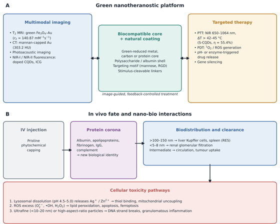
Combining diagnosis and targeted therapy in a single nanoscale vehicle is reshaping personalised oncology and the management of infectious disease (K. Y. Choi et al., 2012; Prakashan et al., 2025; Sharma, 2023). Conventional cancer therapies are often limited by systemic toxicity, poor aqueous solubility of drugs and the absence of real-time feedback on treatment response (Ahmed et al., 2012; Boehnke & Correa, 2020). Green nanotheranostic systems address these limitations with biocompatible cores, natural targeting ligands and stimulus-responsive coatings that support non-invasive multimodal imaging alongside spatially and temporally controlled therapy (Gidwani et al., 2023; Prakashan et al., 2025; Washington et al., 2025). Figure 3A illustrates this integration, and Table 3 compares representative platforms.

2.3.2. Non-Invasive Multimodal Bioimaging

Accurate clinical staging usually requires more than one imaging modality, so that the high spatial resolution of MRI or CT is complemented by the sensitivity of fluorescence or photoacoustic imaging (PAI) (Chouhan et al., 2021; Prakashan et al., 2025; Sharma, 2023). Iron oxide nanoparticles synthesised with plant polyphenols, for example from sumac, tea or Garcinia mangostana peel, are superparamagnetic, have high transverse relaxivity (r2) and can serve as T2-weighted MRI contrast agents without the nephrogenic systemic fibrosis risk associated with linear gadolinium chelates (Braim et al., 2023; Prakashan et al., 2025; Yusefi et al., 2021). Kharey et al. (2023) prepared superparamagnetic Fe3O4–Au composites with Pimenta dioica leaf extract and reported r1 = 10.67 mM−1 s−1, r2 = 140.87 mM−1 s−1 and r2/r1 = 13.20, producing strong negative contrast in ex vivo tissue.

Biogenic gold nanostructures and anisotropic metal hybrids attenuate X-rays more strongly than conventional iodinated agents and therefore provide useful CT contrast (Fazal et al., 2014; Uthaman et al., 2018). Uthaman et al. (2018) produced mannan-capped gold nanoparticles (M-GNPs) through a green polysaccharide reduction route; the mannan coating promoted uptake by antigen-presenting cells through mannose receptors and gave lymph node CT contrast of 303.2 Hounsfield units (HU) (Prakashan et al., 2025). Biologically produced magnetic particles are a further option: genetically engineered magnetosomes from magnetotactic bacteria have served as targeted MRI probes for brain tumours (Boucher et al., 2017). CQDs doped with heteroatoms (N, S, P) or co-doped with lanthanides emit in the near-infrared (NIR-I and NIR-II) windows, reducing autofluorescence and scattering and enabling visualisation of deep vasculature and tumour margins

Figure 1. Transition from conventional to bio-mediated nanomanufacturing and the resulting classes of bio-derived nanomaterials. (A) Conventional wet-chemical and physical routes depend on hazardous reductants, organic solvents and high temperatures, generating toxic effluents and surface residues. (B) Green routes replace these with renewable bio-factories (plant extracts, macroalgae, microorganisms and agro-industrial biowaste) operating in water under mild conditions. (C) Reduction and capping proceed through phytochemical, enzymatic or hydrothermal mechanisms. (D) These routes yield biogenic metal and metal oxide nanoparticles, carbon quantum dots and biopolymer matrices, although batch variability, polydispersity and heterogeneous capping remain unresolved (bottom panel).

Figure 2. Architecture and transduction mechanisms of sustainable nanobiosensors. The upper row traces the signal path from sample through the biorecognition layer (enzymes, antibodies, aptamers or nucleic acid probes) and the green nano-interface (biogenic metal nanoparticles, biowaste carbon dots, biopolymers, MXene or graphene) to the measured output. The lower left panel summarises electrochemical modes and the reasons green nanomaterials improve them, namely larger electroactive area and faster electron transfer. The lower right panel summarises optical and plasmonic modes (LSPR, SERS and FRET). Representative detection limits are taken from the platforms compiled in Table 2.

(Bouzas-Ramos et al., 2019; Sharma, 2023; Tavan et al., 2025).

2.3.3. Targeted Phototherapy and Stimulus-Responsive Therapeutics

Therapeutically, green nanoplatforms frequently act as combined photothermal therapy (PTT) and photodynamic therapy (PDT) agents (Liu et al., 2022; Prakashan et al., 2025). Under NIR irradiation (650–1064 nm), plasmon relaxation in metallic nanostructures or non-radiative decay in carbon lattices converts light into local heat (ΔT sufficient to exceed 42–45 °C), denaturing proteins and disrupting tumour cell membranes (Prakashan et al., 2025; Washington et al., 2025; R. Zhou et al., 2022). Kim et al. (2021) synthesised sulphur-doped CQDs from Camellia japonica flowers with a photothermal conversion efficiency of 55.4%, sufficient for tumour regression in vivo under NIR light (Tavan et al., 2025). Carbon-based materials more generally are emerging as versatile theranostic scaffolds, and multiwalled carbon nanotubes have shown photothermal efficacy against melanoma (Patel et al., 2019; Sobhani et al., 2017).

PDT, in turn, relies on photosensitisers that transfer energy to molecular oxygen, generating cytotoxic ROS, principally singlet oxygen (1O2), which trigger apoptosis (Prakashan et al., 2025; Z. Zhou et al., 2016). Metal oxides such as TiO2 and ZnO have been compared as photosensitisers for this purpose (Zhang et al., 2022). Natural biopolymers, including human serum albumin (HSA), hyaluronic acid and gelatin, have been engineered into self-assembled protein–nanoparticle carriers (Sheng et al., 2014; Washington et al., 2025). Sheng et al. (2014) reported HSA–indocyanine green nanoparticles (HSA-ICG NPs) that reached a tumour-to-normal tissue ratio of 36.12 ± 5.12 at 24 h in 4T1 breast tumours and eradicated tumours through combined PTT and PDT without recurrence or evident organ toxicity (Prakashan et al., 2025).

2.4. Nanotoxicology and Interfacial Nano-Bio Interactions

2.4.1. Challenging the Assumption of Intrinsic Safety

A persistent misconception in the literature is that biogenic origin or biodegradability confers biological safety automatically (Fadeel et al., 2018; Rahman, 2026; Tan et al., 2018). In vivo fate, toxicokinetics and immunogenicity are determined by nanoscale physicochemical properties, namely hydrodynamic size, zeta potential, morphology, composition and dissolution kinetics, rather than by how “green” the precursors were (Fadeel et al., 2018; Rahman, 2026; Verma et al., 2021). Figure 3B traces the main steps from injection to clearance or injury.

2.4.2. Protein Corona Formation and Clearance Kinetics

On entering the bloodstream, nanoparticles rapidly adsorb a dynamic layer of plasma proteins, including albumin, apolipoproteins, fibrinogen and immunoglobulins, forming the protein corona (Durán et al., 2015; Fadeel et al., 2018; Washington et al., 2025). This biological coating effectively replaces the original synthetic or phytochemical capping and becomes the interface that endothelial cells and immune receptors actually recognise (Fadeel et al., 2018; Kopp et al., 2017). Opsonisation by complement proteins and immunoglobulins marks particles for phagocytic clearance by the reticuloendothelial system (RES), concentrating them in hepatic Kupffer cells and splenic macrophages (Fadeel et al., 2018; Rahman, 2026). Particles larger than about 100–150 nm in hydrodynamic diameter are filtered rapidly by the liver, whereas those below about 5–8 nm can be excreted through glomerular filtration (H. S. Choi et al., 2009; K. Y. Choi et al., 2012; Rahman, 2026). Unmodified biogenic particles often aggregate in physiological buffers, changing their effective size and promoting unwanted splenic sequestration (Cardoso et al., 2025; Washington et al., 2025).

2.4.3. Mechanisms of Cytotoxicity and Genotoxicity

At the cellular level, three mechanisms dominate. First, metallic particles, particularly biogenic AgNPs and ZnO nanoparticles, oxidise in acidic lysosomes (pH ≈ 4.5–5.0) and release Ag+ and Zn2+ ions (Fadeel et al., 2018; Ferdous et al., 2024; Yu et al., 2020); these cations bind thiol groups in mitochondrial enzymes, uncouple oxidative phosphorylation and can halt respiration (Verma et al., 2019; Yu et al., 2020). Second, excess superoxide (O2•−), hydroxyl radicals (•OH) and hydrogen peroxide (H2O2) overwhelm glutathione, catalase and superoxide dismutase defences, causing lipid peroxidation, loss of membrane integrity and ferroptotic or apoptotic death (Haase et al., 2025; Yu et al., 2020). Third, ultrafine particles (<10–20 nm) can cross nuclear pores and interact with DNA directly or through radical-induced double-strand breaks, chromosomal aberrations and micronucleus formation (Fadeel et al., 2018; Singh et al., 2025; Zhang et al., 2022). High-aspect-ratio materials such as pristine carbon nanotubes may behave like asbestos fibres, sustaining granulomatous inflammation and cell transformation (Fadeel et al., 2018; Rahman, 2026). Comparable concerns have been raised for inorganic particles tested in human lung epithelial cells (S. J. Choi et al., 2009). These findings indicate that biocompatibility cannot be inferred from a green synthesis claim and must instead be demonstrated through standardised preclinical testing (Fadeel et al., 2018; FDA, 2023; Rahman, 2026).

2.5. Life-Cycle Assessment and Environmental Footprint

2.5.1. The LCA Framework

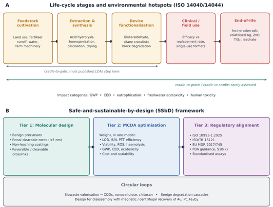
Quantifying the environmental profile of sustainable nanomaterials requires systems-level modelling through LCA conducted according to ISO 14040 and ISO 14044 (Carroccio et al., 2022; Finkbeiner et al., 2006; Rahman, 2026). An LCA tracks material and energy flows through four stages: goal and scope definition, life-cycle inventory (LCI), life-cycle impact assessment (LCIA) and interpretation (Finkbeiner et al., 2006; Rahman, 2026). Figure 4A maps these stages onto the life of a typical green nanobiosensor or theranostic agent.

2.5.2. Upstream Hotspots: Raw Material Extraction and Processing

LCAs of bio-based polymer nanocomposites, such as PLA, PHB and cellulose nanocrystals, consistently identify agricultural cultivation and primary nanofiller extraction as major hotspots (Carroccio et al., 2022; Rahman, 2026). Although plant feedstocks sequester biogenic CO2 during growth, intensive farming contributes fossil fuel combustion, stratospheric ozone depletion and eutrophication from nitrate and phosphate runoff (Carroccio et al., 2022; Rahman, 2026). The mechanical or chemical processing needed to isolate nanoscale building blocks adds further burdens (Carroccio et al., 2022; Dhali et al., 2021). Producing cellulose nanocrystals, for example, involves concentrated sulphuric acid hydrolysis, neutralisation, dialysis and high-pressure homogenisation or probe sonication, which together can raise cumulative energy demand (CED) and global warming potential (GWP) above those of synthetic polymers per kilogram (Carroccio et al., 2022; Dhali et al., 2021; Rahman, 2026). Similarly, while green synthesis of metal nanoparticles avoids hazardous reductants, thermal drying, autoclaving and calcination can still dominate the carbon footprint (Cardoso et al., 2025; Raihan et al., 2025).

2.5.3. Boundary Limitations: Cradle-to-Gate versus Cradle-to-Grave

Most nanomaterial LCAs stop at the factory gate and omit clinical use, environmental release and disposal (Carroccio et al., 2022; Dabare et al., 2025; Rahman, 2026). In clinical biosensing and theranostics, used devices and consumables are classified as biohazardous waste and are usually incinerated at high temperature (Chávez-Hernández et al., 2024; Rahman, 2026). Incineration destroys the biodegradable matrix but can transfer inorganic fillers (ZnO, TiO2, metallic Ag) into bottom ash or airborne particulates, introducing ecotoxicological hazards into soil and water (Carroccio et al., 2022; Rahman, 2026). Crosslinking strategies such as glutaraldehyde or covalent silane coupling, adopted to extend the water stability and working life of biosensors, can inhibit enzymatic biodegradation, prevent industrial composting and complicate circularity (Carroccio et al., 2022; Rahman, 2026). Sustainability therefore cannot be judged from synthesis metrics alone; it requires cradle-to-grave and cradle-to-cradle evaluation that includes realistic end-of-life recovery (Dabare et al., 2025; Rahman, 2026).

2.6. Safe-and-Sustainable-by-Design and Circular Economy Perspectives

2.6.1. The SSbD Concept

To bridge the divide between analytical engineering and environmental safety, recent international frameworks have promoted the SSbD concept together with circular-economy models (Chávez-Hernández et al., 2024; Dabare et al., 2025; Rahman, 2026). Endorsed by the European Commission and consistent with wider regulatory roadmaps, SSbD brings safety and sustainability screening into the earliest stages of molecular design rather than treating toxicology and waste management as afterthoughts (Chávez-Hernández et al., 2024; Fadeel et al., 2018). Figure 4B outlines the three-tier structure adopted in this review.

2.6.2. Circular Design Strategies

Circular design replaces the linear “take-make-dispose” model with closed loops that prioritise resource valorisation and high-value material recovery (Dabare et al., 2025; Omran & Baek, 2022; Raihan et al., 2025). Three strategies stand out. Biowaste valorisation upcycles agricultural residues, such as sugarcane bagasse, wheat straw and crustacean chitin, into carbon dots, nanocellulose and chitosan transducers (Nath et al., 2023; Omran & Baek, 2022; Sen & Sarma, 2025). Design for disassembly uses reversible supramolecular or stimulus-responsive crosslinks so that polymer matrices can be dissolved at end of life and valuable fillers (Au, Pt, magnetic Fe3O4) recovered magnetically or by centrifugation (Pavlos, 2022; Rahman, 2026). Benign degradation cascades build enzyme-cleavable bonds, for instance matrix metalloproteinase- or esterase-sensitive linkages, into theranostic carriers so that they break down into metabolisable amino acids, sugars and renal-clearable fragments smaller than 5 nm (Prakashan et al., 2025; Washington et al., 2025).

2.6.3. Multi-Criteria Decision Analysis and Regulatory Harmonisation

Because material properties involve trade-offs, so that improving analytical sensitivity or photothermal conversion may raise synthesis energy or biological reactivity, structured decision tools are needed (Rahman, 2026). MCDA provides a quantitative framework for weighing technical performance (LOD, signal-to-noise ratio, photothermal efficiency), toxicological safety (cell viability, ROS generation, haemocompatibility), environmental impact (GWP, CED, aquatic ecotoxicity) and economic scalability (Chávez-Hernández et al., 2024; Rahman, 2026). Commercial translation of green biosensors and nanotheranostics further depends on compliance with international standards, including ISO 10993-1:2025 for the biological evaluation of medical devices and ISO/TR 13121 for nanomaterial risk evaluation, and with guidance from the FDA and the European Medicines Agency under the EU Medical Device Regulation 2017/745 (Chávez-Hernández et al., 2024; FDA, 2023; International Organization for Standardization [ISO], 2025; Rodríguez-Gómez et al., 2025). Regulatory compliance demands standardised toxicological screening, precise physicochemical characterisation, verified batch consistency and validated life-cycle safety data (Rodríguez-Gómez et al., 2025). Only when these bridges are in place can next-generation nanotechnologies be judged effective, safe and sustainable across their whole life cycle.

3. Methods

3.1. Review Design and Reporting Standard

This work was designed as a structured, critical narrative review with systematic elements in searching, screening and data extraction. Reporting followed the relevant items of the PRISMA 2020 statement for literature searches, eligibility criteria, selection and data collection, adapted for a review whose evidence base mixes primary experimental studies, secondary reviews, life-cycle studies and regulatory documents. Because the included studies differ widely in materials, endpoints and test systems, no meta-analysis was attempted; instead, quantitative outcomes were extracted into harmonised tables and synthesised thematically. The approach to safety and sustainability evidence was anchored in three external frameworks: ISO 14040/14044 for life-cycle assessment (Finkbeiner et al., 2006), ISO 10993-1 for the biological evaluation of medical devices (FDA, 2023; ISO, 2025) and current nanomaterial safety-assessment practice (Fadeel et al., 2018; Rodríguez-Gómez et al., 2025).

3.2. Review Questions

Four questions defined the scope: (i) which renewable feedstocks and green synthesis routes are used to produce nanomaterials for biosensing and nanotheranostics, and how do they compare with conventional routes in process conditions and particle properties; (ii) what analytical and theranostic performance do these materials achieve; (iii) what toxicological mechanisms and biological fates have been reported for them; and (iv) what life-cycle hotspots and end-of-life constraints affect their sustainability, and which circular or SSbD strategies have been proposed to address them (Chávez-Hernández et al., 2024; Rahman, 2026).

3.3. Information Sources

PubMed/MEDLINE, Scopus and Web of Science Core Collection were searched as primary bibliographic databases, and Google Scholar was used to capture book chapters and recent articles not yet indexed elsewhere. Regulatory and standards documents were retrieved directly from the websites of the U.S. Food and Drug Administration and the International Organization for Standardization (FDA, 2023; ISO, 2025). Reference lists of key reviews on green synthesis, nanobiosensors, nanotheranostics, nanotoxicology and LCA were hand-searched (backward citation tracking), and citing articles were screened for the most relevant records (forward citation tracking) (Cardoso et al., 2025; Carroccio et al., 2022; Maparathne et al., 2026; Prakashan et al., 2025).

3.4. Search Strategy

Searches covered records published from January 2006 to March, 2026, with earlier foundational studies admitted through citation tracking. Search strings combined four concept blocks with Boolean operators and were adapted to the syntax and controlled vocabulary of each database (MeSH terms in PubMed; field codes TITLE-ABS-KEY in Scopus and TS in Web of Science). The PubMed string was:

(“green synthesis” OR “biogenic” OR “plant extract” OR “phytosynthesis” OR “biosynthesis” OR “biowaste” OR “agro-waste” OR “sustainable” OR “Green Chemistry Technology”[MeSH]) AND (“nanoparticles”[MeSH] OR nanomaterial* OR “carbon dots” OR “carbon quantum dots” OR MXene OR nanocellulose OR chitosan OR nanocomposite*) AND (“Biosensing Techniques”[MeSH] OR biosensor* OR “Theranostic Nanomedicine”[MeSH] OR theranostic* OR “photothermal therapy” OR “photodynamic therapy” OR “contrast media” OR bioimaging)

A second string, run separately, linked the material block with safety and sustainability terms:

(nanomaterial* OR nanoparticle* OR nanocomposite*) AND (“Toxicity Tests”[MeSH] OR nanotoxicolog* OR cytotoxic* OR genotoxic* OR “protein corona” OR biodistribution OR “life cycle assessment” OR “life-cycle” OR “circular economy” OR “safe-and-sustainable-by-design” OR “multi-criteria decision analysis” OR “ISO 10993”)

Filters restricted results to English-language publications. The full strings for Scopus and Web of Science, the date each search was run and the number of records returned per database are reported.

3.5. Eligibility Criteria

Records were eligible if they (a) described a nanomaterial produced from a renewable, biological or waste-derived feedstock, or by a route explicitly framed as green, low-solvent or bio-mediated, or a biopolymer nanocomposite used as a sensing or therapeutic matrix; and (b) reported at least one of the following: physicochemical characterisation linked to synthesis conditions; analytical performance of a biosensor (linear range, limit of detection, sensitivity or selectivity); imaging or therapeutic performance in vitro or in vivo; toxicological or biodistribution outcomes; or life-cycle, circularity or regulatory analysis relevant to such materials. Conventional (non-green) nanomaterials, such as CdTe/CdSe quantum dots or chemically synthesised carbon nanotubes, were included only as comparators for toxicity or life-cycle burden (Fadeel et al., 2018; Kushnir & Sandén, 2008). Peer-reviewed reviews were admitted when they supplied mechanistic, comparative or regulatory context. Records were excluded if they were conference abstracts without full data, patents, non-English publications, studies of nanomaterials used solely in non-biomedical settings without transferable mechanistic insight, or duplicate reports of the same dataset.

3.6. Study Selection

All records were exported to a reference manager and de-duplicated. Two reviewers independently screened titles and abstracts against the eligibility criteria, then assessed full texts of potentially relevant records. Disagreements were resolved by discussion, with a third reviewer consulted when consensus was not reached. Reasons for full-text exclusion were recorded.

3.7. Data Extraction

A piloted extraction form, refined after testing on five studies, was used to collect data in four domains that correspond to Tables 1–4:

  • Synthesis and characterisation (Table 1): nanomaterial class; metal salt or carbon precursor; biological source; synthesis route and conditions (temperature, time, solvent); morphology and size with the characterisation technique (e.g., XRD, SEM, TEM, DLS); surface functional groups; and stated sustainability advantages (Cardoso et al., 2025; Ferdous et al., 2024).

  • Biosensing performance (Table 2): transduction mode; nanomaterial platform; biorecognition element and immobilisation chemistry; analyte and sample matrix; linear dynamic range; limit of detection; and sensitivity or other figures of merit (Maparathne et al., 2026; Naresh & Lee, 2021).

  • Theranostic performance (Table 3): platform architecture; green or biocompatible components; imaging modality and contrast metrics (relaxivity r1, r2 and r2/r1; X-ray attenuation in HU); therapeutic mechanism and irradiation wavelength; photothermal conversion efficiency; disease model and species; and efficacy and safety outcomes (Prakashan et al., 2025; Sharma, 2023).

Table 3. Multifunctional nanotheranostic platforms: imaging modalities, therapeutic mechanisms and preclinical efficacy. The table profiles ten green-synthesised or biomacromolecular theranostic systems, including iron oxide–gold composites, albumin–dye assemblies, lignin carriers, polysaccharide-capped gold, carbon dots, magnetosomes and porphyrin nanoplatforms. For each, it lists the biocompatible or green components, the diagnostic imaging modality (MRI, CT, photoacoustic or fluorescence), the therapeutic mechanism (photothermal, photodynamic, chemotherapeutic, radiosensitising or magnetic hyperthermia) and the disease model. The final column summarises reported efficacy and safety outcomes. All entries are preclinical, and outcomes are as reported by the original authors.

Nanoplatform architecture

Biocompatible / green components

Diagnostic imaging modality

Therapeutic mechanism

Disease model and system

Preclinical outcomes and efficacy

References

Fe3O4–Au nanocomposites

Pimenta dioica leaf extract bioreductants

Transverse (T2) MRI and photoacoustic imaging (PAI)

Photothermal therapy (PTT), 808 nm NIR laser

Ex vivo poultry heart; 4T1 breast carcinoma murine model

r2 = 140.87 mM−1 s−1; temperature rise of 13.1 °C producing hyperthermic tumour ablation

Kharey et al. (2023); Tavan et al. (2025)

HSA–indocyanine green (HSA-ICG NPs)

Human serum albumin self-assembled via disulfide bonds

Dual NIR fluorescence and PAI

Synergistic photodynamic (PDT) and photothermal therapy

4T1 metastatic breast tumours in BALB/c mice

Tumour-to-normal ratio 36.12 ± 5.12 at 24 h; complete eradication without recurrence or organ toxicity

Prakashan et al. (2025); Sheng et al. (2014)

Lignin–black phosphorus (BPQDs@N-LgC)

Lignin matrix, chlorin e6, black phosphorus quantum dots

PAI and NIR-I photoluminescence

Mitochondria-targeted PTT (808 nm) and PDT (660 nm)

HeLa cervical carcinoma xenografts in nude mice

ROS-cleavable β-O-4 lignin bonds trigger degradation and excretion of fragments after therapy

Liu et al. (2022); Prakashan et al. (2025)

Mannan-capped gold (M-GNPs)

Natural mannan polysaccharide

X-ray computed tomography (CT)

Targeted photothermal ablation and immune-cell trafficking

Popliteal lymph node metastatic carcinoma in rats

Concentration-dependent attenuation (303.2 HU); receptor-mediated uptake by antigen-presenting cells

Prakashan et al. (2025); Uthaman et al. (2018)

Hollow CoPt alloy nanoparticles (HCPA-NPs)

Plant polyphenols as green template and stabiliser

Dual T2-weighted MRI and photoacoustic tomography

Hyperthermic PTT under mild NIR irradiation

HepG2 hepatocellular carcinoma xenografts

Marked negative MRI contrast; complete tumour necrosis within 8 min of irradiation

Prakashan et al. (2025); Song et al. (2016)

Sulphur-doped floral CQDs (S-CQDs)

Camellia japonica extract

NIR-I/NIR-II fluorescence imaging

High-efficiency PTT

Subcutaneous lung and breast tumour murine models

Photothermal conversion efficiency 55.4%; tumour suppression without weight loss or organ pathology

Kim et al. (2021); Tavan et al. (2025)

Gelatin–alginate silver (Gel-Alg-AgNPs)

Calendula officinalis flower extract with gelatin/alginate

Dual fluorescence imaging and optical tracking

pH-triggered chemotherapy co-delivery with photothermal ablation

Solid tumour xenograft models

Sustained stimulus-responsive release in acidic tumour microenvironment; reduced off-target toxicity

Amani et al. (2026); Publication Source (2024)

Mesoporous Pt nanoworms (Pt-NWs-PVP)

Polyvinylpyrrolidone-stabilised platinum

Dual CT and PAI

Radiosensitisation and PTT (1064 nm)

4T1 breast cancer xenografts in BALB/c mice

Rapid renal clearance; significant tumour volume reduction within 14 days; low systemic toxicity

Gidwani et al. (2023)

PLZ4–nanoporphyrin platforms (PNPs)

Self-assembled PEGylated oligomers with tumour-targeting peptide

Photodynamic diagnosis and fluorescence

Trimodal targeted chemotherapy, PTT and PDT

Orthotopic urothelial bladder carcinoma models

Tumour retention >14 days; no normal urothelial uptake, avoiding cutaneous phototoxicity

Biosensors Group (2023); T. Y. Lin et al. (2016)

Genetically engineered magnetosomes (RGD-Mags)

Biogenic magnetic Fe3O4 from magnetotactic bacteria

Ultra-high-field (17.2 T) MRI

Targeted magnetic hyperthermia and photothermia

Glioblastoma and malignant brain tumours in rodents

High magnetophoretic mobility; RGD-driven tumour retention; heating superior to synthetic iron oxides

Boucher et al. (2017); Prakashan et al. (2025)

Table 4. Toxicological profiles, life-cycle assessment (LCA) hotspots and circular design metrics of nanomaterial systems relevant to biosensing and nanotheranostics. Ten material systems are compared, including biogenic metals and metal oxides, cadmium-based quantum dots (as a conventional comparator), biopolymer nanocomposites, carbon nanotubes and thermoset sensors. For each, the table links in vitro and in vivo toxicological mechanisms and target organs to the principal LCA hotspots and the ISO 14040/14044 impact categories they affect. The penultimate column lists circular and safe-and-sustainable-by-design (SSbD) mitigation strategies proposed in the literature. Together, the rows illustrate that renewable origin alone does not determine either biological safety or environmental performance.

Nanomaterial system

In vitro / in vivo toxicological findings and pathways

Target organs and biological fate

Primary LCA hotspots and burden drivers

Impact categories affected (ISO 14040/44)

Circular design and SSbD mitigation

References

Silver nanoparticles (AgNPs / Ag-CQDs)

Lysosomal Ag+ leaching; ROS surge; glutathione depletion; mitochondrial depolarisation

Accumulation in liver Kupffer cells and spleen; dose-dependent cytotoxicity in rodents (<35 nm)

Silver ore mining; energy-intensive milling; washing effluents entering aquatic systems

Freshwater ecotoxicity; human toxicity (cancer/non-cancer); GWP

Polyphenol antioxidant capping; chitosan hybrid networks to curb leaching; recovery by centrifugal ultrafiltration

Fadeel et al. (2018); Gidwani et al. (2023); Hasan et al. (2026); Priyadarshini et al. (2024)

Zinc oxide nanoparticles (ZnONPs)

Zn2+ dissolution; G2/M cell-cycle arrest; DNA replication disorders; membrane lipid peroxidation

Hepatic and renal stress; skin irritation and possible transdermal migration under UVB

Agricultural inputs for plant-extract cultivation; calcination energy (400–600 °C)

Acidification; eutrophication; non-renewable energy use (NREU)

Albumin or PEG encapsulation; sizing particles >50 nm to avoid nuclear translocation

Hasan et al. (2026); Hou et al. (2019); Verma et al. (2021)

Copper oxide nanoparticles (CuONPs)

Promotes cytotoxic amyloid-β oligomerisation; disturbs Ca2+ homeostasis; secondary genotoxicity via lipid ROS

Neuronal tissue; central nervous system accumulation; pulmonary epithelial injury

High-energy autoclave synthesis; copper release in wastewater

Aquatic and terrestrial ecotoxicity; GWP

Reversible chelating biopolymers; closed-loop aqueous precipitation recovery

Jaragh-Alhadad & Falahati (2022); Tavan et al. (2025)

Cadmium telluride / selenide QDs (CdTe/CdSe)

Cd2+ dissolution; respiratory-chain disruption; nuclear double-strand breaks; high apoptosis

Persistent accumulation in spleen, renal cortex and liver; slow clearance

High embodied carbon in precursors; hazardous waste at end of life

Human toxicity (carcinogenic); aquatic ecotoxicity; mineral depletion

Replacement with biomass-derived CQDs or InP/ZnS cores with renal-clearable coatings

Fadeel et al. (2018); Gidwani et al. (2023); Tavan et al. (2025)

Polylactic acid / nanocellulose (PLA/CNC)

Non-cytotoxic; mild macrophage recruitment at high doses; altered platelet aggregation

Metabolised to lactic acid; renal clearance of breakdown fragments

Concentrated sulphuric acid hydrolysis; high electricity use in nanofibrillation

Cumulative energy demand (CED); photochemical ozone formation; eutrophication

Enzymatic fibre processing; solvent-free twin-screw reactive extrusion

Carroccio et al. (2022); Hasan et al. (2026)

Chitosan-based nanocomposites

Generally well tolerated (LD50 >5000 mg kg−1); positive charge may perturb coagulation

RES clearance; transient uptake in lung capillaries and liver

Deproteinisation and deacetylation of crustacean shells with strong bases and acids

Marine and freshwater eutrophication; terrestrial ecotoxicity

Chitin from fungal mycelium waste; benign ionic crosslinking (tripolyphosphate, tannic acid)

Hasan et al. (2026); Sonin et al. (2020)

Carbon nanotubes / graphene hybrids

High-aspect-ratio, asbestos-like pathogenicity; persistent granulomatous lung inflammation

Pulmonary macrophage entrapment; persistence in liver and lymph nodes

Fluidised-bed CVD (~954 MJ kg−1); mineral acid purification

Fossil fuel depletion; ozone depletion; GWP

Microwave-assisted biomass pyrolysis; hydrophilic PEG/polysaccharide functionalisation; design for disassembly

Hasan et al. (2026); Kushnir & Sandén (2008); Sobhani et al. (2017)

Polyhydroxyalkanoates / nanoclay (PHB/clay)

Biocompatible, non-immunogenic matrix; low toxicity of organo-modified montmorillonite

Enzymatic hydrolysis by microbial depolymerases into benign hydroxy acids

Bacterial fermentation requiring substantial nutrients and electricity

Land use; agricultural eutrophication; NREU

Food waste and wastewater as fermentation carbon sources; nanoclay recycling via sol–gel separation

Carroccio et al. (2022); Hasan et al. (2026); Korde & Kandasubramanian (2020)

Titanium dioxide nanoparticles (TiO2NPs)

Photo-induced ROS; S-phase arrest; genotoxicity in murine brain tissue

Translocation along olfactory pathways to brain; alveolar deposition

High-temperature calcination (>500 °C); chlorine-rich waste from sulphate process

Abiotic mineral depletion; GWP

Low-temperature sol–gel synthesis with plant polyphenols; dynamic covalent networks for magnetic reclamation

Mohamed & Hussien (2016); Raihan et al. (2025)

Thermoset nanocomposites and sensors

Leaching of uncured monomer; occupational inhalation during machining and sanding

Foreign-body encapsulation; persistence in extracellular space

Incineration volatilises metal fillers into bottom ash and emissions

Human toxicity; ecotoxicity; eutrophication from landfill leachate

Reprocessable vitrimers and covalent adaptable networks (CANs) allowing matrix dissolution and filler recovery

Hasan et al. (2026); Rezaei & Prince (2025)


  • Toxicity and life cycle (Table 4): toxicological endpoints and mechanisms; target organs and clearance route; dose or size dependence; LCA system boundary; dominant hotspots; ISO 14040/14044 impact categories affected; and proposed circular or SSbD mitigation (Carroccio et al., 2022; Fadeel et al., 2018; Rahman, 2026).

One reviewer extracted the data and a second verified every entry against the source article. When a value was reported only graphically, it was not estimated; the qualitative finding was recorded instead.

3.8. Data Harmonisation

To allow comparison, concentrations were converted to SI-consistent units (M, g L−1 or ppm, as reported) without recalculating authors’ detection limits. Relaxivities were recorded in mM−1 s−1, photothermal conversion efficiencies as percentages and CT contrast in HU. Life-cycle outcomes were mapped to standard impact categories, namely GWP, CED, non-renewable energy use, acidification, eutrophication, freshwater and terrestrial ecotoxicity and human toxicity, according to ISO 14040/14044 terminology (Finkbeiner et al., 2006). Toxicological endpoints were grouped by mechanism (ion dissolution, oxidative stress, genotoxicity, inflammation) and by the ISO 10993-1 biological evaluation categories relevant to the intended contact type and duration (FDA, 2023; ISO, 2025).

3.9. Quality and Risk-of-Bias Appraisal

Because a single validated tool does not cover the range of study designs included, appraisal was matched to study type. Experimental materials studies were judged on completeness of physicochemical characterisation (size distribution, surface chemistry, purity and stability in relevant media), the presence of appropriate controls and the number of independent replicates. Biosensor studies were additionally judged on whether performance was confirmed in real or spiked biological matrices rather than buffer alone, and on whether selectivity against common interferents was tested (Maparathne et al., 2026). In vivo studies were assessed on randomisation, blinding, sample size and reporting of dose, route and observation period. LCA studies were assessed on the transparency of the functional unit, system boundary, inventory data sources and sensitivity analysis (Carroccio et al., 2022; Dabare et al., 2025). Appraisal informed the weight given to each study in the synthesis but was not used to exclude studies.

4. Comparative Synthesis of Performance, Safety and Sustainability Evidence

4.1. Characteristics of the Evidence Base

The final body of evidence cited in this review comprises 99 sources: 90 journal articles, 7 book chapters and 2 regulatory or standards documents (FDA, 2023; ISO, 2025). The literature is notably recent. Only 10 sources were published in 2015 or earlier, 29 appeared between 2016 and 2020, 30 between 2021 and 2023, and 30 between 2024 and 2026, which suggests that the integration of green synthesis with biomedical sensing and therapy is still consolidating. Forty representative platforms or material systems, ten in each domain, were extracted into Tables 1–4. Most experimental studies concerned metallic nanoparticles (Ag, Au, Fe3O4, ZnO) or carbon dots; MXene and biopolymer composites were less common, and only a minority of the toxicological and LCA sources dealt specifically with green-synthesised materials rather than nanomaterials in general.

4.2. Synthesis Efficiencies, Precursor Sustainability and Scalability

4.2.1. Process Conditions and Particle Characteristics

The studies in Table 1 suggest that green synthesis has moved beyond single-pot curiosity, though perhaps not yet to the point of reliable industrial scale (Figure 1). Replacing sodium borohydride, hydrazine and dimethylformamide with plant extracts consistently lowered reaction temperatures and removed hazardous organic effluents from the process (Cardoso et al., 2025; Ferdous et al., 2024; Raihan et al., 2025). Extracts of Moringa oleifera, Pimenta dioica and Camellia sinensis supplied polyphenols, flavonoids and terpenoids that reduced and capped metal ions at 25–65 °C, often within one to two hours (Ashikbayeva et al., 2023; Ferdous et al., 2024; Kharey et al., 2023). Across the tabulated studies, biogenic metallic cores ranged from about 9 nm (mannan-capped gold) to 85 nm (Moringa-derived silver), and most were spherical or quasi-spherical (Table 1; Cardoso et al., 2025; Uthaman et al., 2018; Washington et al., 2025).

4.2.2. Plant versus Microbial Routes

A divergence between plant- and microbe-mediated routes is nevertheless apparent. Plant-assisted precipitation is fast and simple, yet it tends to produce broader size distributions, which most authors attribute to seasonal and geographical variation in phytochemical content (Cardoso et al., 2025; Ying et al., 2022). Bacterial and algal routes, such as bacterial cellulose pellicles from Komagataeibacter xylinus or magnetosomes from magnetotactic bacteria, give better structural control but at the cost of slower kinetics and heavier purification (Boucher et al., 2017; Lospinoso et al., 2025; Prakashan et al., 2025). Whether this trade-off favours one route over the other probably depends on the application: a diagnostic substrate may tolerate some polydispersity that an injectable contrast agent could not.

4.2.3. Biowaste Carbon Dots and Polysaccharide Templates

The valorisation of agro-industrial waste into carbon quantum dots is among the more promising trends (Table 1; Sen & Sarma, 2025; Tavan et al., 2025). Hydrothermal carbonisation of palm kernel shells, medicinal plant leaves and Camellia japonica flowers gave quantum-confined dots of roughly 3–12 nm, with reported photoluminescence quantum yields of about 20–35% (Kim et al., 2021; Meena et al., 2019; Monday et al., 2023; Tavan et al., 2025). Endogenous sulphur or nitrogen doping shifted absorption into the NIR window and, in one case, yielded a photothermal conversion efficiency of 55.4% (Kim et al., 2021). Polysaccharide templates, such as mannan or three-dimensional bacterial cellulose networks, stabilised noble-metal particles without synthetic surfactants while keeping plasmonic hot spots and bioactive motifs accessible (Lospinoso et al., 2025; Uthaman et al., 2018). Even so, very few of these studies reported yields per unit of biomass, solvent recovery or energy use, so their scalability is inferred rather than demonstrated.

4.3. Diagnostic and Analytical Performance Landscape

4.3.1. Overall Performance

The analytical data compiled in Table 2 indicate that green nanomaterials can match, and in some cases exceed, the sensing benchmarks set by conventional petroleum-derived or heavy-metal platforms (Maparathne et al., 2026; Naresh & Lee, 2021). Reported detection limits span roughly seven orders of magnitude, from 9.6 µM for a sweat glucose patch to 1.14 pM for microRNA-155, and linear ranges are generally wide enough for physiological measurement (Table 2; Hamidi et al., 2022; X. Lin et al., 2025; Pal et al., 2025). Direct comparison is difficult, however, because analytes, matrices and validation protocols vary so much (Figure 2).

4.3.2. Electrochemical Platforms

In electrochemical sensing, sustainable carbon materials and biopolymer composites accelerated interfacial electron transfer (Hamidi et al., 2022; Pal et al., 2025). Laser-induced graphene patterned on chitosan achieved a sensitivity of 457 nA mM−1 cm−2 and a detection limit of 9.6 µM across 25–1000 µM glucose in artificial sweat (Hamidi et al., 2022; Hasan et al., 2026). PANI–AgNP interfaces separated the oxidation peaks of glucose and urea well enough to detect both simultaneously in serum, at 12 µM and 45 µM, respectively (Pal et al., 2025). Combining xylan-derived CQDs with reduced graphene oxide and silver lowered the dopamine detection limit to 40 nM and limited interference from ascorbic acid (Han et al., 2021; Tavan et al., 2025). A Ti3C2Tx MXene–gold nanorod SPR sensor detected acetylcholine at 4.42 µM (Maparathne et al., 2026). It is worth noting that several of these electrodes still relied on Nafion, glassy carbon or other conventional components, so the “green” label applies to part of the device rather than the whole.

4.3.3. Optical and Plasmonic Platforms

Optical platforms showed comparable gains (Maparathne et al., 2026; Sangeetha et al., 2025). Flexible AuNPs@BC SERS swabs detected thiram at 0.24 ppm, which the authors attribute to dense hot spots anchored throughout the cellulose mesh (Lospinoso et al., 2025). Optical fibre ball resonators coated with green-tea AuNPs detected CD44 at 0.05 ng mL−1 in under a minute (Ashikbayeva et al., 2023). A FRET assay pairing palm-kernel-shell CQDs with gold quenchers measured dengue virus serotype 3 RNA at 0.35 nM (Monday et al., 2023), and NIR systems based on gold and upconversion nanoparticles pushed microRNA-155 detection to 1.14 pM while avoiding tissue autofluorescence (X. Lin et al., 2025). A carbon nitride quantum dot–polyaniline wearable sensor reached 2.5 µM for sweat glucose, and a nanoporous graphene interferometric waveguide detected C-reactive protein at 3 ng mL−1 (Chiu et al., 2025; Lisboa et al., 2025). Few of these reports, though, tested long-term storage stability or performance across many independently fabricated sensors, two properties that tend to determine whether a sensor reaches the clinic.

4.4. Theranostic Efficacy: Multimodal Imaging and Therapeutic Synergies

4.4.1. Imaging Contrast

The theranostic systems summarised in Table 3 show real progress in combining imaging and therapy within one construct (Gidwani et al., 2023; Prakashan et al., 2025; Sharma, 2023). The central design problem, achieving strong contrast without systemic chemical toxicity, appears to be at least partly solvable with green components (K. Y. Choi et al., 2012; Prakashan et al., 2025). Pimenta dioica-mediated Fe3O4–Au nanocomposites gave strong T2 contrast (r2 = 140.87 mM−1 s−1; r2/r1 = 13.20) and photoacoustic signals that guided photothermal ablation, with a temperature rise of 13.1 °C (Kharey et al., 2023). Mannan-capped gold nanoparticles exploited receptor-mediated macrophage uptake to produce lymph node CT attenuation of 303.2 HU (Uthaman et al., 2018). Polyphenol-templated hollow CoPt nanoparticles supported dual MRI and photoacoustic imaging (Song et al., 2016), and engineered magnetosomes were visualised by ultra-high-field MRI in brain tumour models (Boucher et al., 2017; Figure 3A).

4.4.2. Therapeutic Outcomes

Self-assembled protein and biopolymer carriers performed particularly well in vivo (Prakashan et al., 2025; Washington et al., 2025). HSA-ICG nanoparticles reached a tumour-to-normal tissue ratio of 36.12 ± 5.12 at 24 h in 4T1 tumours, enabling combined NIR fluorescence and photoacoustic imaging and PTT/PDT that eliminated tumours without recurrence or apparent off-target toxicity (Sheng et al., 2014). Lignin–black phosphorus platforms (BPQDs@N-LgC) used ROS-cleavable β-O-4 linkages to degrade after treatment, so that carrier fragments were cleared rather than retained (Liu et al., 2022). Sulphur-doped floral CQDs suppressed tumour growth without weight loss or organ pathology (Kim et al., 2021), and gelatin–alginate silver systems released drugs preferentially in acidic tumour environments (Amani et al., 2026; Publication Source, 2024). Peptide-targeted nanoporphyrins remained at bladder tumour sites for more than 14 days with little normal urothelial uptake (Biosensors Group, 2023; T. Y. Lin et al., 2016), and PVP-stabilised platinum nanoworms combined radiosensitisation with 1064 nm PTT and were cleared renally (Gidwani et al., 2023). These are encouraging results, but every entry in Table 3 is preclinical, most rely on subcutaneous murine tumours, and follow-up periods are short. The clinical relevance of these efficacy figures should therefore be treated with some caution.

4.5. Toxicological Reality versus the “Green” Assumption

4.5.1. Physicochemical Determinants of Toxicity

The toxicological evidence in Table 4 does not support the common assumption that green or bio-derived nanomaterials are inherently non-toxic (Fadeel et al., 2018; Hasan et al., 2026; Tan et al., 2018). Biocompatibility instead appears to depend on hydrodynamic size, surface charge, dissolution rate and protein corona dynamics more than on the biological origin of the precursor (Fadeel et al., 2018; Hasan et al., 2026; Verma et al., 2021; Figure 3B).

4.5.2. Ion Release, Oxidative Stress and Genotoxicity

Uncoated biogenic AgNPs and ZnO nanoparticles dissolve in acidic lysosomes (pH ≈ 4.5–5.0) and release Ag+ and Zn2+ (Fadeel et al., 2018; Ferdous et al., 2024; Yu et al., 2020). The resulting ion burden triggers ROS cascades, mitochondrial depolarisation and G2/M cell-cycle arrest (Hou et al., 2019; Verma et al., 2019, 2021). Size seems to matter considerably: metallic cores below about 35 nm can cross biological barriers and nuclear pores and induce DNA double-strand breaks, whereas particles above about 50 nm tend to remain in endosomes (Fadeel et al., 2018; Hasan et al., 2026; Priyadarshini et al., 2024). Other metal oxides show their own patterns. CuO nanoparticles promoted amyloid-β oligomerisation and neurotoxicity (Jaragh-Alhadad & Falahati, 2022), and TiO2 nanoparticles were genotoxic in mouse brain tissue (Mohamed & Hussien, 2016). Cadmium-based quantum dots, included as comparators, showed persistent accumulation in spleen, kidney and liver (Fadeel et al., 2018; Gidwani et al., 2023). Biopolymer systems were generally better tolerated: chitosan nanoparticles had an LD50 above 5000 mg kg−1 but could still perturb coagulation through their positive charge (Sonin et al., 2020), while carbon nanotubes retained fibre-like pathogenicity (Sobhani et al., 2017).

4.5.3. Protein Corona and Clearance

Once in circulation, biogenic particles adsorb albumin, apolipoproteins and immunoglobulins, and the resulting corona overrides the original phytochemical capping, directing particles towards Kupffer cells and splenic macrophages (Fadeel et al., 2018; Hasan et al., 2026; Washington et al., 2025). Highly charged or hydrophobic surfaces accelerate RES clearance, whereas neutral, zwitterionic or protein-passivated coatings extend circulation and reduce haemolysis (Durán et al., 2015; Hasan et al., 2026; Washington et al., 2025). Notably, few

Figure 3. Green nanotheranostic platforms and their in vivo fate. (A) A biocompatible core with a natural coating and targeting motif links multimodal imaging (MRI, CT, photoacoustic and NIR fluorescence) with targeted therapy (photothermal, photodynamic and stimulus-triggered drug release), enabling image-guided, feedback-controlled treatment; representative values are drawn from Table 3. (B) After intravenous injection, a protein corona replaces the original phytochemical capping and governs biodistribution, with large particles cleared by the reticuloendothelial system and ultrasmall particles by the kidneys. The bottom panel summarises the three main cellular toxicity pathways: ion leaching, oxidative stress and genotoxicity.

Figure 4. Life-cycle hotspots and the safe-and-sustainable-by-design (SSbD) framework proposed in this review. (A) The five life-cycle stages of a green nanobiosensor or theranostic agent, with the main environmental hotspots at each stage; arrows below indicate that most published assessments stop at the factory gate, whereas cradle-to-grave and cradle-to-cradle boundaries are rarely applied. (B) The three-tier SSbD framework links molecular design choices, multi-criteria decision analysis (MCDA) of performance, safety, environmental and cost criteria, and alignment with ISO, EU MDR and FDA requirements. Circular loops (bottom panel) close the cycle through biowaste valorisation, design for disassembly and benign degradation.

of the green-synthesis studies characterised the corona directly or measured biodistribution beyond 24–72 h, which leaves the longer-term fate of these materials largely unknown.

4.6. Life-Cycle Trade-Offs, Environmental Hotspots and Circular Realities

4.6.1. Upstream and Processing Burdens

The life-cycle evidence summarised in Table 4 points to trade-offs that synthesis-focused papers rarely acknowledge (Carroccio et al., 2022; Finkbeiner et al., 2006; Hasan et al., 2026). Biogenic synthesis reduces direct use of hazardous chemicals, but cradle-to-gate inventories show that biomass cultivation brings its own burdens in land use, fossil energy for farm equipment and fertiliser-driven eutrophication (Carroccio et al., 2022; Hasan et al., 2026). Processing biomass into nanoscale components is also energy- and resource-intensive (Carroccio et al., 2022; Dhali et al., 2021). Isolating cellulose nanocrystals by concentrated sulphuric acid hydrolysis and high-pressure homogenisation can produce CED and GWP values above those of petroleum polymers on a mass basis (Carroccio et al., 2022; Hasan et al., 2026), and chemical vapour deposition of carbon nanotubes has been estimated to require about 954 MJ kg−1 (Kushnir & Sandén, 2008; Figure 4A).

4.6.2. End-of-Life and the Circularity Gap

Cradle-to-grave and cradle-to-cradle evaluations remain scarce (Carroccio et al., 2022; Dabare et al., 2025; Rahman, 2026). Most biosensors and theranostic consumables are discarded as regulated biohazardous waste and incinerated (Chávez-Hernández et al., 2024; Hasan et al., 2026). Incineration removes the biodegradable matrix but can transfer ZnO, TiO2 or Ag into bottom ash or emissions, with consequences for landfills and watersheds (Carroccio et al., 2022; Hasan et al., 2026). Synthetic crosslinkers added for durability, such as glutaraldehyde and silane coupling agents, can block enzymatic degradation and rule out industrial composting (Carroccio et al., 2022; Hasan et al., 2026). The mitigation strategies that recur in Table 4 include covalent adaptable networks and vitrimers, reversible supramolecular crosslinks, closed-loop magnetic recovery of noble metals, enzymatic rather than acid-based cellulose processing and the use of food waste as a fermentation feedstock for PHAs (Chávez-Hernández et al., 2024; Hasan et al., 2026; Korde & Kandasubramanian, 2020; Rezaei & Prince, 2025; Figure 4B). Most of these, however, have been demonstrated for bulk nanocomposites rather than for diagnostic or theranostic devices specifically.

5. Reconciling Performance, Safety and Sustainability in Green Nanobiotechnology

5.1. Green Nanomaterials Work, but Evidence of Superior Safety and Sustainability Remains Unconvincing

Taken together, the evidence leads to a somewhat uncomfortable conclusion. Green nanomaterials clearly work: they detect biomarkers at clinically meaningful concentrations, generate useful imaging contrast and kill tumour cells in animal models (Tables 2 and 3). What the literature does not yet show, at least not convincingly, is that they are reliably safer or more sustainable than the materials they are meant to replace. The “greenness” of a feedstock removes some hazards, especially those linked to toxic reductants and solvents, but it leaves others untouched and occasionally introduces new ones (Table 4; Hasan et al., 2026; Rahman, 2026). This gap between functional promise and verified safety and sustainability is, in our reading, the central issue for the field.

5.2. Performance Is Necessary but Not Sufficient

5.2.1. Interpreting Biosensing Benchmarks

The biosensing results are impressive, and it would be unfair to downplay them. Green-tea gold, bacterial cellulose and biowaste carbon dots now support detection limits that would have seemed ambitious for any platform a decade ago (Ashikbayeva et al., 2023; Lospinoso et al., 2025; Monday et al., 2023). Yet a low detection limit in buffer is not the same as a reliable clinical test. Several studies in Table 2 validated their sensors in artificial sweat, phosphate buffer or spiked samples, and comparatively few reported batch-to-batch reproducibility, shelf life or performance in untreated patient samples (Maparathne et al., 2026; Naresh & Lee, 2021). There is also a quieter problem of scope. When a green nanomaterial is deposited on a glassy carbon electrode, coated with Nafion and read by conventional electronics, the sustainability benefit is confined to a small fraction of the device mass (Hamidi et al., 2022; Hasan et al., 2026; Figure 2). Claims of a “green biosensor” might more accurately be framed as claims about a green sensing layer, unless whole-device inventories are provided.

5.2.2. Interpreting Theranostic Efficacy

A similar caution applies to theranostics. The HSA-ICG, lignin–black phosphorus and Fe3O4–Au systems in Table 3 combine diagnosis and therapy elegantly, and some of them, notably the lignin platform with its ROS-cleavable β-O-4 bonds, address clearance at the design stage (Kharey et al., 2023; Liu et al., 2022; Sheng et al., 2014; Figure 3A). But every platform remains preclinical, and murine subcutaneous tumours are generous models: they are small, well vascularised and easy to irradiate. Translational reviews have repeatedly noted that the step from such models to humans is where most nanotheranostics fail, usually for reasons of manufacturing consistency, immunogenicity or regulatory classification rather than efficacy (Hirulkar et al., 2024; Rodríguez-Gómez et al., 2025). Combination products that merge imaging agent, drug and device further complicate this path (Chávez-Hernández et al., 2024). Integration with artificial intelligence for image analysis and treatment planning has been proposed as a way to make image-guided therapy more precise, although it is too early to judge how much it will help with green platforms in particular (Prakashan et al., 2025; Sharma, 2023).

5.3. Redefining Biocompatibility for Green Nanomaterials

5.3.1. Physicochemical Identity over Origin

The toxicological findings argue for a shift in how biocompatibility is defined. Size, surface charge, dissolution and corona formation, not botanical origin, seem to govern biological fate (Fadeel et al., 2018; Sanita et al., 2020; Verma et al., 2021). A silver nanoparticle capped with Moringa polyphenols still releases Ag+ in the lysosome (Ferdous et al., 2024; Yu et al., 2020), and a ZnO particle made with fruit-peel extract can still arrest the cell cycle (Hou et al., 2019; Nava et al., 2017). The phytochemical shell may modulate these effects, perhaps by scavenging some ROS or slowing dissolution, but once a protein corona forms in blood, the original capping is largely masked (Durán et al., 2015; Kopp et al., 2017; Figure 3B). It may be that the main toxicological advantage of green synthesis is indirect: the absence of residual borohydride, hydrazine or synthetic surfactants on the particle surface (Cardoso et al., 2025; Kharissova et al., 2019). That is a real benefit, but it is narrower than the one often implied.

5.3.2. Better Dose Metrics and Standardised Testing

Part of the difficulty lies in how toxicity is measured. Mass-based doses obscure differences in surface reactivity, and newer metrics based on reactive surface sites may better predict oxidative potential across materials (Haase et al., 2025). Advanced safety-assessment tools, including high-throughput screening, omics and computational models, are now available but are seldom applied to green nanomaterials (Fadeel et al., 2018). For devices, ISO 10993-1 provides a risk-based framework that links required testing to the nature and duration of body contact (FDA, 2023; ISO, 2025). Applying this framework from the outset, rather than retrospectively, would make toxicity data across studies far more comparable. Surface-functionalisation strategies, including PEGylation, albumin coating or zwitterionic shells, can reduce opsonisation and haemolysis, but each adds synthetic components whose own life-cycle and safety profiles then need evaluation (Table 4; Sanita et al., 2020; Washington et al., 2025).

5.4. Life-Cycle Thinking and the Burden-Shifting Problem

5.4.1. Where the Burdens Move

The LCA evidence highlights a classic burden-shifting problem. Removing toxic reductants lowers some impacts at the synthesis stage, yet the burden may simply move upstream to agriculture or downstream to energy-intensive purification (Carroccio et al., 2022; Dhali et al., 2021; Figure 4A). Nanocellulose is the clearest example: a renewable, biodegradable material whose acid hydrolysis and homogenisation can make it more carbon- and energy-intensive per kilogram than some petrochemical alternatives (Carroccio et al., 2022; Rahman, 2026). Because nanomaterials are used in very small quantities in diagnostics, the absolute impact per test may be modest, but this has rarely been quantified with a clinically meaningful functional unit, such as one diagnostic result or one treatment course (Dabare et al., 2025).

5.4.2. The Neglected End of Life

The end-of-life stage is probably the weakest link. Clinical waste rules generally require incineration, which negates the biodegradability that motivated the use of biopolymers in the first place, while potentially releasing metallic fillers into ash and air (Chávez-Hernández et al., 2024; Rahman, 2026). Thermoset and heavily crosslinked composites are particularly hard to recover (Rezaei & Prince, 2025; Table 4). For single-use point-of-care devices, which may be produced in very large numbers, this could become a substantial issue. We would argue that cradle-to-grave assessment should become the default for any study that claims sustainability benefits, and that the clinical waste pathway should be modelled explicitly (Carroccio et al., 2022; Dabare et al., 2025).

5.5. Towards a Safe-and-Sustainable-by-Design Framework

5.5.1. A Three-Tier Approach

Drawing on these findings, we propose a three-tier SSbD framework (Figure 4B). At the first tier, molecular design, developers choose benign precursors, renal-clearable cores below about 5 nm where systemic use is intended, non-leaching coatings and reversible or enzyme-cleavable linkages (H. S. Choi et al., 2009; Prakashan et al., 2025; Washington et al., 2025). At the second tier, MCDA weighs analytical or therapeutic performance against toxicological endpoints, life-cycle impacts and cost, making trade-offs explicit rather than implicit (Chávez-Hernández et al., 2024; Rahman, 2026). At the third tier, regulatory alignment ensures that characterisation, safety testing and batch-consistency data are generated in forms acceptable under ISO 10993-1, the EU Medical Device Regulation and FDA guidance (FDA, 2023; ISO, 2025; Rodríguez-Gómez et al., 2025).

5.5.2. Circular Levers

Within this framework, circular strategies act as practical levers. Biowaste valorisation can supply carbon dots, chitosan and nanocellulose from residues that would otherwise be burned or landfilled (Nath et al., 2023; Omran & Baek, 2022; Raihan et al., 2025). Magnetic retrieval, already demonstrated for plant-derived iron oxide in environmental remediation, could in principle be adapted to recover noble-metal or magnetic fillers from spent sensor chips (Pavlos, 2022). Covalent adaptable networks and vitrimers offer routes to dissolve matrices and reclaim fillers without incineration (Rezaei & Prince, 2025). None of these is yet routine for biomedical devices, and each would need to be validated under clinical waste regulations.

5.6. Research Priorities

Several priorities follow. First, minimum reporting standards are needed for green synthesis, covering biomass source and season, extract preparation, yield, polydispersity and residual impurities, so that batch variability can be quantified rather than merely acknowledged (Table 1; Cardoso et al., 2025; Ying et al., 2022). Second, biosensor studies should report validation in real clinical samples, inter-batch reproducibility and storage stability alongside detection limits (Maparathne et al., 2026). Third, theranostic studies should include long-term biodistribution, clearance and immunotoxicity in more than one species, with orthotopic or metastatic models where relevant (Hirulkar et al., 2024; Sharma, 2023). Fourth, LCAs should use clinically meaningful functional units and cradle-to-grave boundaries, and they should be performed early enough to influence design (Dabare et al., 2025; Rahman, 2026). Finally, closer collaboration between materials scientists, toxicologists, LCA practitioners and regulators would help ensure that the “green” label rests on measured evidence rather than on assumption (Chávez-Hernández et al., 2024; Contera et al., 2020).

5.7. Limitations of This study

This review has limitations. It is a structured narrative synthesis rather than a meta-analysis, so the comparisons in Tables 1–4 are descriptive and cannot establish statistical superiority of one platform over another. The evidence base is heterogeneous in materials, endpoints and test conditions, and many quantitative values come from single studies without independent replication. Several toxicological and life-cycle sources address nanomaterials generally rather than green-synthesised variants specifically, which may overstate or understate risks for the latter. Restricting the search to English-language publications may have excluded relevant work. Finally, the field is moving quickly, and some of the most recent findings may not yet have been captured.

6. Conclusion

Green nanomaterials have earned their place in biosensing and nanotheranostics on performance alone. Biogenic metal nanoparticles, biowaste carbon dots and biopolymer composites now deliver sensitive detection, useful imaging contrast and effective phototherapy in preclinical settings. Their safety and sustainability, however, cannot be taken for granted. Toxicity seems to follow particle size, surface chemistry, dissolution and protein corona formation more than feedstock origin, and life-cycle studies show that cultivation, nanocellulose processing and clinical incineration can offset the gains of benign chemistry. Most published life-cycle assessments still stop at the factory gate. We therefore suggest that green claims be tested, not assumed, through a safe-and-sustainable-by-design approach that links molecular design, multi-criteria decision analysis and regulatory alignment. Standardised reporting, real-sample validation, long-term biodistribution studies and cradle-to-grave assessment should become routine. With these in place, green nanobiotechnology could move from promising laboratory demonstrations to clinically credible and environmentally responsible tools for patients and health systems.

References


Abdelghany, T. M., Al-Rajhi, A. M. H., Al Abboud, M. A., Alawlaqi, M. M., Ganash Magdah, A., Helmy, E. A. M., & Mabrouk, A. S. (2018). Recent advances in green synthesis of silver nanoparticles and their applications: About future directions. A review. BioNanoScience, 8(1), 5–16. https://doi.org/10.1007/s12668-017-0413-3

Ahmed, N., Fessi, H., & Elaissari, A. (2012). Theranostic applications of nanoparticles in cancer. Drug Discovery Today, 17(17–18), 928–934.

Amani, A. M., Vafa, E., Mirzae, M., Abbasi, M., Vaez, A., Najdian, A., Jahanbin, A., Kasaei, S. R., Mosleh-Shirazi, S., Kamyab, H., Khademi, T., Chelliapan, S., & Rajendran, S. (2026). A comprehensive review on MXene nanostructures for biosensing, imaging, and therapeutic systems. Sensing and Bio-Sensing Research, 51, Article 100912.

Ashikbayeva, Z., Bekmurzayeva, A., Myrkhiyeva, Z., Assylbekova, N., Atabaev, T. S., & Tosi, D. (2023). Green-synthesized gold nanoparticle-based optical fiber ball resonator biosensor for cancer biomarker detection. Optics & Laser Technology, 161, Article 109136.

Atchudan, R., Chandra Kishore, S., Gangadaran, P., Jebakumar Immanuel Edison, T. N., Perumal, S., Rajendran, R. L., & Lee, Y. R. (2022). Tunable fluorescent carbon dots from biowaste as fluorescence ink and imaging human normal and cancer cells. Environmental Research, 204, Article 112365. https://doi.org/10.1016/j.envres.2021.112365

Bakirhan, N. K., & Ozkan, S. A. (2018). Quantum dots as a new generation nanomaterials and their electrochemical applications in pharmaceutical industry. In Handbook of nanomaterials for industrial applications (pp. 520–529). Elsevier.

Biosensors Group. (2023). Nanoparticle-based photosensitizers and theranostic systems in photodynamic and photothermal therapy. Biosensors, 13(1), Article 106.

Boehnke, N., & Correa, S. (2020). Theranostic layer-by-layer nanoparticles for simultaneous tumor detection and gene silencing. Angewandte Chemie International Edition, 59(7), 2776–2783.

Boucher, M., Geffroy, F., Prévéral, S., Bellanger, L., Selingue, E., Adryanczyk-Perrier, G., Péan, M., Lefèvre, C. T., Pignol, D., Ginet, N., & Lalatonne, Y. (2017). Genetically tailored magnetosomes used as MRI probe for molecular imaging of brain tumor. Biomaterials, 121, 167–178.

Bouzas-Ramos, D., Cigales Canga, J., Mayo, J. C., Sainz, R. M., Ruiz Encinar, J., & Costa-Fernandez, J. M. (2019). Carbon quantum dots codoped with nitrogen and lanthanides for multimodal imaging. Advanced Functional Materials, 29(38), Article 1903884.

Braim, F. S., Ab Razak, N. N. A. N., Aziz, A. A., Dheyab, M. A., & Ismael, L. Q. (2023). Rapid green-assisted synthesis and functionalization of superparamagnetic magnetite nanoparticles using Sumac extract and assessment of their cellular toxicity, uptake, and anti-metastasis property. Ceramics International, 49(5), 7359–7369.

Cardoso, B., Nobrega, G., Afonso, I. S., Ribeiro, J. E., & Lima, R. A. (2025). Sustainable green synthesis of metallic nanoparticle using plants and microorganisms: A review of biosynthesis methods, mechanisms, toxicity, and applications. Journal of Environmental Chemical Engineering, 13(2), Article 116921.

Carroccio, S. C., Scarfato, P., Bruno, E., Aprea, P., Dintcheva, N. T., & Filippone, G. (2022). Impact of nanoparticles on the environmental sustainability of polymer nanocomposites based on bioplastics or recycled plastics – A review of life-cycle assessment studies. Journal of Cleaner Production, 335, Article 130322.

Chaudhary, V., Vasistha, S., & Rai, M. P. (2024). Nanobiotechnology: An applicable approach for sustainable future. In Nanobiotechnology for sustainable future (pp. 1–35). Springer.

Chávez-Hernández, J. A., Velarde-Salcedo, A. J., Navarro-Tovar, G., & Gonzalez, C. (2024). Safe nanomaterials: From their use, application, and disposal to regulations. Nanoscale Advances, 6(6), 1583–1610.

Chiu, Y.-H., Rinawati, M., Chang, L.-Y., Guo, Y.-T., Chen, K.-J., Chiu, H.-C., Lin, Z.-H., Huang, W.-H., Haw, S.-C., & Yeh, M.-H. (2025). Carbon nitride quantum dots/polyaniline nanocomposites for non-invasive glucose monitoring using wearable sweat biosensor. ACS Applied Nano Materials, 8(5), 2340–2351.

Choi, H. S., Liu, W., Misra, P., Tanaka, E., Zimmer, J. P., Ipe, B. I., Bawendi, M. G., & Frangioni, J. V. (2009). Tissue-specific targeting of renal clearable quantum dots. Nature Nanotechnology, 4(1), 107–113.

Choi, K. Y., Liu, G., Lee, S., & Chen, X. (2012). Theranostic nanoplatforms for simultaneous cancer imaging and therapy: Current approaches and future perspectives. Nanoscale, 4(2), 330–342. https://doi.org/10.1039/C1NR11277E

Choi, S. J., Oh, J. M., & Choy, J. H. (2009). Toxicological effects of inorganic nanoparticles on human lung cancer A549 cells. Journal of Inorganic Biochemistry, 103(3), 463–471.

Chouhan, R. S., Horvat, M., Ahmed, J., Alhokbany, N., Alshehri, S. M., & Gandhi, S. (2021). Magnetic nanoparticles—A multifunctional potential agent for diagnosis and therapy. Cancers, 13(10), Article 2213.

Chouke, P. B., Shrirame, T., Potbhare, A. K., Mondal, A., Chaudhary, A. R., Mondal, S., Thakare, S. R., Nepovimova, E., Valis, M., Kuca, K., Sharma, R., & Chaudhary, R. G. (2022). Bioinspired metal/metal oxide nanoparticles: A road map to potential applications. Materials Today Advances, 16, Article 100314.

Contera, S., de la Bernardino Serna, J., & Tetley, T. D. (2020). Biotechnology, nanotechnology and medicine. Emerging Topics in Life Sciences, 4(6), 551–554.

Dabare, S., Rajapaksha, S., & Munaweera, I. (2025). Lifecycle analysis of nanotechnology-enhanced soft materials in the circular economy. Molecular Systems Design & Engineering, 10(6), 799–816. https://doi.org/10.1039/D5ME00067J

Dhali, K., Ghasemlou, M., Daver, F., Cass, P., & Adhikari, B. (2021). A review of nanocellulose as a new material towards environmental sustainability. Science of The Total Environment, 775, Article 145871. https://doi.org/10.1016/j.scitotenv.2021.145871

Durán, N., Silveira, C. P., Durán, M., & Martinez, D. S. T. (2015). Silver nanoparticle protein corona and toxicity: A mini-review. Journal of Nanobiotechnology, 13, Article 55.

Fadeel, B., Farcal, L., Hardy, B., Vázquez-Campos, S., Hristozov, D., Marcomini, A., Lynch, I., Valsami-Jones, E., Alenius, H., & Savolainen, K. (2018). Advanced tools for the safety assessment of nanomaterials. Nature Nanotechnology, 13(7), 537–543. https://doi.org/10.1038/s41565-018-0185-0

Fazal, S., Jayasree, A., Sasidharan, S., Koyakutty, M., Nair, S. V., & Menon, D. (2014). Green synthesis of anisotropic gold nanoparticles for photothermal therapy of cancer. ACS Applied Materials & Interfaces, 6(11), 8080–8089.

Ferdous, S., Rahman, G. M. S., Rayhan, J., Hossain, M. Z., Rahman, M. H., Khan, A., & Islam, M. S. (2024). Green synthesis of silver nanoparticles using Moringa oleifera leaf extract: Antibacterial efficacy and sustainable nanotechnology. Biosensors and Nanotheranostics, 3(1), 1–8. https://doi.org/10.25163/biosensors.319971

Finkbeiner, M., Inaba, A., Tan, R. B. H., Christiansen, K., & Klüppel, H. J. (2006). The new international standards for life cycle assessment: ISO 14040 and ISO 14044. The International Journal of Life Cycle Assessment, 11(2), 80–85. https://doi.org/10.1065/lca2006.02.002

Fong, K. E., & Yung, L.-Y. L. (2013). Localized surface plasmon resonance: A unique property of plasmonic nanoparticles for nucleic acid detection. Nanoscale, 5(24), 12043–12071.

Gidwani, B., Ansari, M. A., & Anand, S. (2023). Systematic review on nanotechnology and nanotheranostics: Basics, green synthesis, applications, limitations, and future prospects. 3 Biotech, 13(8), Article 279.

González-Ballesteros, N., Rodríguez-Argüelles, M. C., Prado-López, S., Lastra, M., Grimaldi, M., Cavazza, A., Nasi, L., Salviati, G., & Bigi, F. (2019). Macroalgae to nanoparticles: Study of Ulva lactuca L. role in biosynthesis of gold and silver nanoparticles and of their cytotoxicity on colon cancer cell lines. Materials Science and Engineering: C, 97, 498–509.

Haase, A., Coca-López, N., Portela, R., & Bañares, M. A. (2025). A refined dose metric for nanotoxicology based on surface site reactivity for oxidative potential of engineered nanomaterials. Nanoscale Advances, 7(10), 2929–2941.

Hamidi, A., et al. (2022). Laser-induced graphene enzymatic biosensors on biodegradable substrates. ACS Applied Materials & Interfaces, 14(18), 21102–21111.

Han, Y., et al. (2021). Xylan-derived carbon quantum dots nanocomposite for electrochemical dopamine sensing. Carbohydrate Polymers, 262, Article 117932.

Hasan, M. S., Rahman, M. Z., & Yasin, S. (2026). Sustainable and eco-friendly modifications of polymer nanocomposites for biomedical and smart applications. Materials Today Communications, 56, Article 116193. https://doi.org/10.1016/j.mtcomm.2026.116193

Hirulkar, R., Chaurawal, N., Alhodieb, F. S., Barkat, H., Preet, S., & Raza, K. (2024). Nanotheranostics: Clinical status, toxicity, regulatory consideration, and future prospects. In M. A. Barkat et al. (Eds.), Nanotheranostics for diagnosis and therapy (pp. 215–242). Springer.

Hou, J., Liu, H., Zhang, S., Liu, X., Hayat, T., Alsaedi, A., & Wang, X. (2019). Mechanism of toxic effects of Nano-ZnO on cell cycle of zebrafish (Danio rerio). Chemosphere, 229, 206–213.

International Organization for Standardization. (2025). Biological evaluation of medical devices — Part 1: Evaluation and testing within a risk management process (ISO Standard No. 10993-1:2025). https://www.iso.org/standard/10993-1

Jaragh-Alhadad, L. A., & Falahati, M. (2022). Copper oxide nanoparticles promote amyloid-β-triggered neurotoxicity through formation of oligomeric species as a prelude to Alzheimer’s diseases. International Journal of Biological Macromolecules, 207, 121–129.

Kharey, P., Goel, M., Husain, Z., Gupta, R., Sharma, D., Palani, I., & Gupta, S. (2023). Green synthesis of biocompatible superparamagnetic iron oxide-gold composite nanoparticles for magnetic resonance imaging, hyperthermia and photothermal therapeutic applications. Materials Chemistry and Physics, 293, Article 126859.

Kharissova, O. V., Kharisov, B. I., Oliva González, C. M., Méndez, Y. P., & López, I. (2019). Greener synthesis of chemical compounds and materials. Royal Society Open Science, 6(11), Article 191378.

Kim, D., Jo, G., Chae, Y., Subramani, S., Lee, B. Y., & Kim, E. J. (2021). High near-infrared absorbance for efficient photothermal cancer therapy utilizing sulfur-doped carbon quantum dots. Nanoscale, 13(34), 14426–14434.

Kopp, M., Kollenda, S., & Epple, M. (2017). Nanoparticle–protein interactions: Therapeutic approaches and supramolecular chemistry. Accounts of Chemical Research, 50(6), 1383–1390.

Korde, J. M., & Kandasubramanian, B. (2020). Microbiologically extracted poly(hydroxyalkanoates) and its amalgams as therapeutic nanocarriers in anti-tumor therapies. Materials Science and Engineering: C, 111, Article 110799.

Kurowiak, J., Klekiel, T., & Bedzinski, R. (2023). Biodegradable polymers in biomedical applications: A review—developments, perspectives and future challenges. International Journal of Molecular Sciences, 24(23), Article 16952. https://doi.org/10.3390/ijms242316952

Kushnir, D., & Sandén, B. A. (2008). Energy requirements of carbon nanoparticle production. Journal of Cleaner Production, 16(7), 817–826.

Lin, T. Y., et al. (2016). Multifunctional targeting nanoporphyrin platform for bladder cancer photodynamic diagnostics and therapy. Biomaterials, 83, 120–130.

Lin, X., Yang, Y., Zhu, W., He, X., & Liu, Y. (2025). Near-infrared DNA biosensors based on polysulfonate coatings for the sensitive detection of microRNAs. Nanoscale Advances, 7(2), 549–559.

Lisboa, P., et al. (2025). Nanoporous graphene integration in bimodal waveguide interferometric biosensors for C-reactive protein detection. Sensors and Actuators B: Chemical, 401, Article 135022.

Liu, X., Li, M., Li, X., Ge, M., Liu, S., Li, S., Li, J., Ding, J., Ragauskas, A. J., Sun, W., & Yang, B. (2022). A photosensitive sustainable lignin nanoplatform for multimodal image-guided mitochondria-targeted photodynamic and photothermal therapy. Materials Today Chemistry, 26, Article 101000.

Lospinoso, D., Colombelli, A., Pal, S., Cretì, P., Martucci, M. C., Giancane, G., Licciulli, A., Rella, R., & Manera, M. G. (2025). Sustainable and flexible surface-enhanced Raman scattering transducer: Gold nanoparticle-bacterial cellulose composite for pesticide monitoring in agrifood systems. Biosensors, 15(2), Article 69.

Malhotra, B. D., & Ali, M. A. (2018). Nanomaterials in biosensors: Fundamentals and applications. In Nanomaterials for biosensors (pp. 1–74). Elsevier. https://doi.org/10.1016/B978-0-323-44923-6.00001-7

Mansuriya, B. D., & Altintas, Z. (2021). Carbon dots: Classification, properties, synthesis, characterization, and applications in health care—An updated review (2018–2021). Nanomaterials, 11(10), Article 2525.

Maparathne, S., Islam, R., Ariza Gonzalez, M., Amarasekara, R., Atik, R., Chinwangso, P., & Lee, T. R. (2026). Nanomaterial-based biosensors: Sensing mechanisms, performance determinants, and applications. Journal of Nanotheranostics, 7(2), 13–41. https://doi.org/10.3390/jnt7020013

Massironi, A., Morelli, A., Grassi, L., Puppi, D., Braccini, S., Maisetta, G., Esin, S., Batoni, G., Della Pina, C., & Chiellini, F. (2019). Ulvan as novel reducing and stabilizing agent from renewable algal biomass: Application to green synthesis of silver nanoparticles. Carbohydrate Polymers, 203, 310–321.

Meena, R., Singh, R., Marappan, G., Kushwaha, G., Gupta, N., & Singh, O. (2019). Fluorescent carbon dots driven from ayurvedic medicinal plants for cancer cell imaging and phototherapy. Heliyon, 5(9), Article e02483.

Mohamed, H. R., & Hussien, N. A. (2016). Genotoxicity studies of titanium dioxide nanoparticles (TiO2NPs) in the brain of mice. Scientifica, 2016, Article 6710840.

Monday, O., et al. (2023). Carbon dots from palm kernel shell for DNA biosensing of dengue virus. Bioelectrochemistry, 151, Article 108392.

Naresh, V., & Lee, N. (2021). A review on biosensors and recent development of nanostructured materials-enabled biosensors. Sensors, 21(4), Article 1109.

Nath, P. C., Ojha, A., Debnath, S., Sharma, M., Sridhar, K., Nayak, P. K., & Inbaraj, B. S. (2023). Biogeneration of valuable nanomaterials from agro-wastes: A comprehensive review. Agronomy, 13(2), Article 561.

Nava, O. J., Soto-Robles, C. A., Gómez-Gutiérrez, C. M., Vilchis-Nestor, A. R., Castro-Beltrán, A., Olivas, A., & Luque, P. A. (2017). Fruit peel extract mediated green synthesis of zinc oxide nanoparticles. Journal of Molecular Structure, 1147, 1–6.

Omran, B. A., & Baek, K.-H. (2022). Valorization of agro-industrial biowaste to green nanomaterials for wastewater treatment: Approaching green chemistry and circular economy principles. Journal of Environmental Management, 311, Article 114806.

Pal, S., Kumar, D., Pillay, S., Shukla, S. K., & Hussain, C. M. (2025). Multiplex biosensor interface utilizing PANI-AgNP nanocomposite for glucose and urea detection. Topics in Catalysis, 69(7), 892–905.

Patel, K. D., Singh, R. K., & Kim, H. W. (2019). Carbon-based nanomaterials as an emerging platform for theranostics. Materials Horizons, 6(3), 434–469.

Patra, J. K., Das, G., Fraceto, L. F., Campos, E. V. R., del Pilar Rodriguez-Torres, M., Acosta-Torres, L. S., Diaz-Torres, L. A., Grillo, R., Swamy, M. K., Sharma, S., Habtemariam, S., & Shin, H. S. (2018). Nano based drug delivery systems: Recent developments and future prospects. Journal of Nanobiotechnology, 16, Article 71.

Pavlos, P. (2022). Magnetic retrieval and surface complexation of heavy metals using plant-derived iron oxide nanoparticles. Biosensors and Nanotheranostics, 1(2), 14–22.

Prakashan, D., PR, R., Kaushik, A., & Gandhi, S. (2025). Sustainable nanotechnology and artificial intelligence to empower image-guided therapy for precision healthcare. BME Frontiers, 6, Article 0150.

Priyadarshini, E., et al. (2024). Cytotoxic and oxidative pathways of metal-carbon dot nanoconjugates in mammalian cell models. Nano-Micro Letters, 16, Article 88.

Publication Source. (2024). Advanced nanotheranostics in cancer therapy: Integration of biopolymers, imaging, and drug delivery. In BN_39 nanobiotechnology research collection (pp. 1–32).

Rahman, M. Z. (2026). Sustainable polymer nanocomposites: A review on material selection, processing, performance, safety, life-cycle assessment, and circular end-of-life management. Materials Today Communications, 44, Article 116193. https://doi.org/10.1016/j.mtcomm.2026.116193

Raihan, J., Ali, M. S., Ferdows, M. K., Shuvo, S. M. K. R., Shadhin, M. S. F., Ahmad, R., Biswas, A., Karmaker, P., Hossain, M. S., Jakaria, M., & Islam, M. S. (2025). Green nanotechnology: Sustainable approaches for environmental remediation and waste management. Biosensors and Nanotheranostics, 4(1), 1–12. https://doi.org/10.25163/biosensors.4110284

Rezaei, Z., & Prince, E. (2025). Pathways towards a circular economy for thermoset nanocomposites. Chemical Science, 16(38), 14839–14864.

Rodríguez-Gómez, F. D., Monferrer, D., Penon, O., & Rivera-Gil, P. (2025). Regulatory pathways and guidelines for nanotechnology-enabled health products: A comparative review of EU and US frameworks. Frontiers in Medicine, 12, Article 1544393. https://doi.org/10.3389/fmed.2025.1544393

Sangeetha, P., Ayyanar, N., Prabhakar, G., & Rajaram, S. (2025). Study review of optical biosensors based on 2D materials. Plasmonics, 20(8), 7155–7169.

Sanita, G., Carrese, B., & Lamberti, A. (2020). Nanoparticle surface functionalization: How to improve biocompatibility and cellular internalization. Frontiers in Molecular Biosciences, 7, Article 587012.

Šebesta, M., Vojtková, H., Cyprichová, V., Ingle, A. P., Urík, M., & Kolencík, M. (2022). Mycosynthesis of metal-containing nanoparticles—Fungal metal resistance and mechanisms of synthesis. International Journal of Molecular Sciences, 23(22), Article 14084.

Sen, B., & Sarma, H. (2025). Recent advancements towards the green synthesis of carbon quantum dots for biosensors and optoelectronics. Communications Materials, 6, Article 1056.

Sharma, A. K. (2023). Current trends in nanotheranostics: A concise review on bioimaging and smart wearable technology. Nanotheranostics, 7(3), 258–269.

Sheng, Z., Hu, D., Zheng, M., Zhao, P., Liu, H., Gao, D., Gong, P., Gao, G., Zhang, P., Ma, Y., & Cai, L. (2014). Smart human serum albumin-indocyanine green nanoparticles generated by programmed assembly for dual-modal imaging-guided cancer synergistic phototherapy. ACS Nano, 8(12), 12310–12322.

Shobana, B., Renugadevi, K., & Prakash, P. (2025). A high sensitive microbial photoelectrochemical sensor based on cuprous oxide @ titanium dioxide/MXene for the selective detection of Staphylococcus aureus in polymicrobial environment. Journal of Electroanalytical Chemistry, 977, Article 118857.

Singh, I., Arora, R., Dhiman, H., & Pahwa, R. (2025). Carbon quantum dots: Synthesis, characterization and biomedical applications. Turkish Journal of Pharmaceutical Sciences, 22(1), 45–58.

Sobhani, Z., Behnam, M. A., Emami, F., Dehghanian, A., & Jamhiri, I. (2017). Photothermal therapy of melanoma tumor using multiwalled carbon nanotubes. International Journal of Nanomedicine, 12, 4509–4517.

Song, X. R., Yu, S. X., Jin, G. X., Wang, X., Chen, J., Li, J., Liu, G., & Yang, H. H. (2016). Plant polyphenol-assisted green synthesis of hollow CoPt alloy nanoparticles for dual-modality imaging guided photothermal therapy. Small, 12(11), 1506–1513.

Sonin, D., Pochkaeva, E., Zhuravskii, S., Postnov, V., Korolev, D., Vasina, L., Kostina, D., Mukhametdinova, D., Zelinskaya, I., & Skorik, Y. (2020). Biological safety and biodistribution of chitosan nanoparticles. Nanomaterials, 10(4), Article 810.

Tan, K. X., Barhoum, A., Pan, S., & Danquah, M. K. (2018). Risks and toxicity of nanoparticles and nanostructured materials. In Emerging applications of nanoparticles and architecture nanostructures (pp. 121–139). Elsevier.

Tavan, M., et al. (2025). Carbon dots in biosensing and theranostics: Environmental and biomedical applications. Industrial Crops & Products, 231, Article 121207.

U.S. Food and Drug Administration. (2023). Use of International Standard ISO 10993-1, “Biological evaluation of medical devices – Part 1: Evaluation and testing within a risk management process”: Guidance for industry and Food and Drug Administration staff.

Uthaman, S., Kim, H. S., Revuri, V., Min, J. J., Lee, Y., Huh, K. M., & Park, I. K. (2018). Green synthesis of bioactive polysaccharide-capped gold nanoparticles for lymph node CT imaging. Carbohydrate Polymers, 181, 27–33.

Vasudevan, M., Perumal, V., Karuppanan, S., Ovinis, M., Bothi Raja, P., Gopinath, S. C. B., & Immanuel Edison, T. N. J. (2024). A comprehensive review on biopolymer mediated nanomaterial composites and their applications in electrochemical sensors. Critical Reviews in Analytical Chemistry, 54(8), 1871–1894.

Verma, S. K., Jha, E., Panda, P. K., Thirumurugan, A., & Suar, M. (2019). Biological effects of green-synthesized metal nanoparticles: A mechanistic view of antibacterial activity and cytotoxicity. In Advanced nanostructured materials for environmental remediation (pp. 145–171). Springer.

Verma, S. K., Panda, P. K., Kumari, P., Patel, P., Arunima, A., Jha, E., Husain, S., Prakash, R., Hergenröder, R., Mishra, Y. K., & Suar, M. (2021). Determining factors for the nano-biocompatibility of cobalt oxide nanoparticles: Proximal discrepancy in intrinsic atomic interactions at differential vicinage. Green Chemistry, 23(9), 3439–3458.

Washington, A., Krishnakumar, G. S., & Kumar, R. (2025). Synergistic protein-green synthesized nanoparticles nanosystems: A sustainable and safe approach for cancer theranostics. Nanotheranostics, 9(3), 315–334.

Ying, S., Guan, Z., Ofoegbu, P. C., Clubb, P., Rico, C., He, F., & Hong, J. (2022). Green synthesis of nanoparticles: Current developments and limitations. Environmental Technology & Innovation, 26, Article 102336.

Yu, Z., Li, Q., Wang, J., Yu, Y., Wang, Y., Zhou, Q., & Li, P. (2020). Reactive oxygen species-related nanoparticle toxicity in the biomedical field. Nanoscale Research Letters, 15(1), Article 115.

Yusefi, M., Shameli, K., Su Yee, O., Teow, S.-Y., Hedayatnasab, Z., Jahangirian, H., Webster, T. J., & Kuca, K. (2021). Green synthesis of Fe3O4 nanoparticles stabilized by a Garcinia mangostana fruit peel extract for hyperthermia and anticancer activities. International Journal of Nanomedicine, 16, 2515–2532.

Zhang, H., Shan, Y., & Dong, L. (2022). A comparison of TiO2 and ZnO nanoparticles as photosensitizers in photodynamic therapy for cancer. Journal of Biomedical Nanotechnology, 18(6), 1450–1457.

Zhou, R., Zhang, M., Xi, J., Li, J., Ma, R., Ren, L., Bai, Z., Qi, K., & Li, X. (2022). Gold nanorods-based photothermal therapy: Interactions between biostructure, nanomaterial, and near-infrared irradiation. Nanoscale Research Letters, 17(1), Article 68.

Zhou, Z., Song, J., Nie, L., & Chen, X. (2016). Reactive oxygen species generating systems meeting challenges of photodynamic cancer therapy. Chemical Society Reviews, 45(23), 6597–6626.

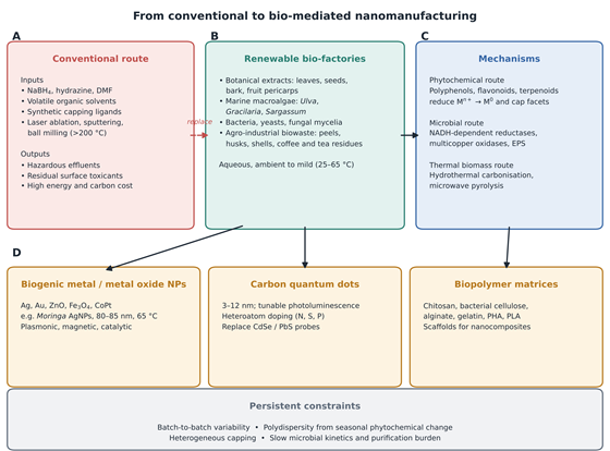
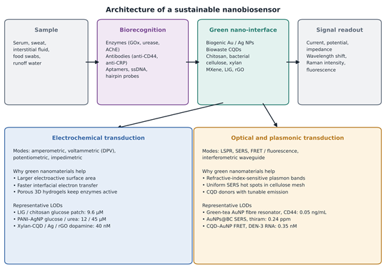
Article metrics
View details
0
Downloads
0
Citations
29
Views

View Dimensions


View Plumx


View Altmetric


0
Save
0
Citation
29
View
0
Share