Biosensors and Nanotheranostics

Bionanotechnology, Drug Delivery, Therapeutics | online ISSN 3064-7789
175
Citations
121.3k
Views
93
Articles
Your new experience awaits. Try the new design now and help us make it even better
Switch to the new experience
Figures and Tables
REVIEWS   (Open Access)

Nanomaterial Interference and Blood Compatibility Gaps in ISO 10993 Testing of Biomaterial Interfaces.

Umayyah Osman 1*, Adam Shah Abdul Majid 1, Rajesh Thangarajan 2


+ Author Affiliations

Biosensors and Nanotheranostics 5 (1) 1-8 https://doi.org/10.25163/biosensors.5111001

Submitted: 19 October 2026 Revised: 12 December 2026  Published: 22 December 2026 


Abstract

Nanostructured biomaterials now sit at the centre of many next-generation medical devices, from vascular stents and wound matrices to implant coatings and injectable carriers. Yet the very features that make them useful (an enormous surface area, tunable charge, reactive surface chemistry) also make their biological behaviour hard to predict, and the ISO 10993 series was written largely with bulk polymers and metals in mind. This review asks how well cytotoxicity testing (ISO 10993-5) and hemocompatibility testing (ISO 10993-4) hold up when applied to biomaterial–nanomaterial interfaces, reading both standards alongside EU MDR Rule 19, FDA guidance, and the nano-specific technical report ISO/TR 10993-22. Three weaknesses recur across the literature. First, colorimetric viability assays are prone to nanomaterial interference: carbon nanotubes can sequester formazan, whereas photocatalytic TiO2 reduces tetrazolium salts without cells and may inflate apparent viability by up to 14%. Second, the robust murine cell lines favoured by the standard often tolerate insults that would injure primary human cells, and stressed cultures can return metabolic signals of 100–150% that do not reflect cell number. Third, blood testing remains fragmented; protein corona formation, contact activation, platelet secretion, and complement-mediated pseudo-allergy are rarely captured together, and static incubation ignores shear, sedimentation, and air-interface artefacts. We propose a tiered framework that begins with physicochemical characterization in exposure-relevant media, pairs metabolic assays with cell-free interference controls and high-content imaging in human cells, and evaluates all five ISO 10993-4 categories under defined flow. Such an approach should make preclinical data more comparable across laboratories and, plausibly, more predictive of clinical performance.

Keywords: ISO 10993; nanomaterials; cytotoxicity; hemocompatibility; protein corona; assay interference; complement activation; microfluidic blood models; medical device regulation

1. Introduction

Few developments have reshaped medical device design as profoundly as the convergence of nanotechnology and biomaterials science. Implantable neuroprosthetics, vascular grafts, multifunctional wound matrices, and targeted nanocarriers increasingly owe their performance to features engineered at the nanoscale (D'Avenio et al., 2024; Kyriakides et al., 2021). When matter is structured in the 1–100 nm range, it acquires attributes that bulk materials simply do not possess: very high surface area-to-volume ratios, tunable surface energy, quantum confinement effects, and distinctive mechanical and topographical cues (D'Avenio et al., 2024; Kyriakides et al., 2021). Applied as coatings on conventional devices or dispersed within nanocomposites, these features can improve osteointegration, support endothelialization, discourage biofilm formation, and enable controlled therapeutic release (D'Avenio et al., 2024; Ramakrishnan & Daly, 2026).

The difficulty is that the properties responsible for these gains are also the source of unpredictable biological reactivity (D'Avenio et al., 2024; Zuckerman & Kao, 2009). A nanomaterial surface is never "seen" by the body in its pristine state. Within moments of contact, host macromolecules adsorb onto it and form a protein corona, which reshapes the material's biological identity, its cellular uptake, and its biodistribution (Aggarwal et al., 2009; Tenzer et al., 2011; Urbán et al., 2019). For this reason, the reassuring safety record of a parent polymer or bulk metal cannot be transferred automatically to its nano-engineered counterpart (D'Avenio et al., 2024; Kyriakides et al., 2021). Excessive reactive oxygen species (ROS) generation, membrane disruption, chronic inflammation, procoagulant activation, and complement-mediated pseudo-allergy remain genuine clinical hazards (Ilinskaya & Dobrovolskaia, 2013; Urbán et al., 2019). Rigorous and standardized preclinical safety screening is therefore a prerequisite, not an afterthought, if these platforms are to move from the bench into clinical practice (D'Avenio et al., 2024; Ramakrishnan & Daly, 2026).

Non-clinical biological safety of medical devices is governed internationally by the ISO 10993 series, which operates within the risk-management principles of ISO 14971 (International Organization for Standardization [ISO], 2018; Zuckerman & Kao, 2009). Its foundational part, ISO 10993-1, provides the roadmap for biological evaluation and sorts devices according to the nature and duration of patient contact, from surface-contacting and externally communicating devices to permanent implants (Food and Drug Administration [FDA], 2023; ISO, 2018). A revised edition, ISO 10993-1:2025, has since restated these requirements and general principles within the risk-management process (ISO, 2025).

Regional regulation has, meanwhile, raised the evidentiary bar for nano-enabled devices. Both the European Union Medical Device Regulation (EU MDR 2017/745) and the United States FDA now expect explicit consideration of nanoscale risks (FDA, 2023; Regulation [EU] 2017/745, 2017). Rule 19 of EU MDR Annex VIII is particularly consequential: any device that incorporates or consists of nanomaterials is placed in Class III, the most stringent risk tier, when it presents a high or medium potential for internal exposure (D'Avenio et al., 2024; Regulation [EU] 2017/745, 2017). To address the analytical problems peculiar to nanoscale systems, ISO Technical Committee 194 published ISO/TR 10993-22:2017 as dedicated guidance (ISO, 2017b; Kyriakides et al., 2021). The report expects nano-objects generated by wear, abrasion, degradation, or intentional shedding to be characterized comprehensively (hydrodynamic diameter, polydispersity, zeta potential, aggregation kinetics, and surface chemistry) both before and during biological exposure (Caputo et al., 2019; ISO, 2017b; Urbán et al., 2019).

In vitro cytotoxicity testing under ISO 10993-5 is the mandatory first tier of preclinical screening (ISO, 2009; Ramakrishnan & Daly, 2026). The standard recognizes three exposure formats, namely extract (fluid elution) testing, direct contact, and indirect diffusion by agar overlay or filter diffusion (ISO, 2009; Ruggeri, 2020). Quantitative viability is usually determined with colorimetric or metabolic reduction assays, chiefly 3-(4,5-dimethylthiazol-2-yl)-2,5-diphenyltetrazolium bromide (MTT), 2,3-bis-(2-methoxy-4-nitro-5-sulfophenyl)-2H-tetrazolium-5-carboxanilide (XTT), neutral red uptake (NRU), and colony formation assays (CFA). A material is considered non-cytotoxic when relative viability remains at or above 70% of the negative control (ISO, 2009; Ramakrishnan & Daly, 2026).

Broad international adoption has not, however, protected these assays from technical weaknesses once nanomaterials are involved (D'Avenio et al., 2024; Ramakrishnan & Daly, 2026). The first concerns extraction itself. Extraction protocols under ISO 10993-12 were conceived for soluble chemical leachables from bulk polymers, whereas surface-bound nanoparticles may shed particles that agglomerate, settle, or alter their surface energy during elution (D'Avenio et al., 2024; Wittmaack, 2011). ISO/TR 10993-22 accordingly notes that nanomaterials often need to be tested in direct contact with target cells instead of relying solely on fluid extracts (D'Avenio et al., 2024; ISO, 2017b). The second weakness is assay interference. Many nanomaterials interact with optical readouts and biochemical reagents (D'Avenio et al., 2024; Hirsch et al., 2011). Carbon nanotubes, for example, bind MTT formazan crystals and so produce false-positive cytotoxicity (Wörle-Knirsch et al., 2006), whereas photocatalytic titanium dioxide (TiO₂) can reduce tetrazolium salts non-enzymatically, inflating measured viability by up to 14% and concealing real toxicity (Lupu & Popescu, 2013). The third weakness lies in the biological model. Standard protocols rely on continuous murine lines such as L929 fibroblasts or BALB/c 3T3 cells, which are metabolically resilient and do not reproduce the vulnerability, signalling, or morphology of primary human parenchymal and endothelial cells (Goyal et al., 2025; Ramakrishnan & Daly, 2026). Multiparametric endpoints, including calcein AM/ethidium homodimer-1 live/dead staining, cytoskeletal actin organization, and annexin V apoptosis markers, are increasingly regarded as necessary companions to conventional metabolic readouts (Ramakrishnan & Daly, 2026).

For any device that contacts circulating blood, whether directly or indirectly (vascular stents, catheters, hemodialysis circuits, and intravenous drug platforms among them), hemocompatibility assessment under ISO 10993-4 is an unavoidable regulatory gate (ISO, 2017a; Weber et al., 2018). Blood is a highly reactive tissue in which cellular elements are suspended in a plasma rich in proteolytic cascades (Weber et al., 2018). ISO 10993-4 therefore organizes evaluation around five interconnected domains: thrombosis, coagulation, platelets, hematology, and immunology/complement (ISO, 2017a; Urbán et al., 2019).

In vitro hemocompatibility testing has long struggled with standardization, so much so that Ratner (1993) described a "blood compatibility catastrophe" and later revisited the problem two decades on (Ratner, 2007; see also Braune et al., 2013). Several bottlenecks persist. Hemolysis, the most standardized quantitative indicator, is measured photometrically as free plasma hemoglobin at 540 nm by the cyanmethemoglobin method (Neun & Dobrovolskaia, 2011; Weber et al., 2018), with values below 2% regarded as non-hemolytic, 2–5% as slightly hemolytic, and above 5% as hemolytic (Sossah, 2021; Totea et al., 2014; Urbán et al., 2019). Nanomaterials can nonetheless induce non-lytic erythrocyte aggregation, membrane stiffening, or optical interference that confounds these readings (Barshtein et al., 2018; Choi et al., 2011). Foreign surfaces also trigger contact activation of the intrinsic coagulation pathway through Factor XII, simultaneously mobilizing the kallikrein–kinin system and liberating bradykinin (Long et al., 2016; Weber et al., 2018). Positively charged and high-surface-area inorganic nanoparticles promote platelet adhesion, glycoprotein IIb/IIIa upregulation, and aggregation (Radomski et al., 2005; Urbán et al., 2019). Engineered nanoparticles and polymer interfaces may further activate the classical, alternative, or lectin complement pathways, generating C3a, C5a, and SC5b-9 and, in patients, complement activation-related pseudo-allergy (CARPA) (Chanan-Khan et al., 2003; Szebeni & Storm, 2015; Urbán et al., 2019). Finally, static incubation protocols do not reproduce hemodynamic shear, pre-analytical storage alters cellular reactivity, and inter-donor variability undermines reproducibility between laboratories (Blok et al., 2016; Braune et al., 2013; Weber et al., 2018).

Taken together, the expanding clinical use of nanostructured devices and the widening gap between largely static international standards and contemporary bio-interfacial science make a critical synthesis of testing methodology timely (D'Avenio et al., 2024; Ramakrishnan & Daly, 2026; Urbán et al., 2019). This review examines standardized cytotoxicity and hemocompatibility testing of biomaterial–nanomaterial interfaces within the ISO 10993 framework, with five specific objectives. The first is to analyze the regulatory evolution and applicability of ISO 10993-1, ISO 10993-5, ISO 10993-4, and ISO/TR 10993-22 under EU MDR 2017/745 and FDA requirements (D'Avenio et al., 2024; ISO, 2018; Regulation [EU] 2017/745, 2017). The second is to evaluate extraction-based, direct-contact, and indirect-contact cytotoxicity methods critically, identifying mechanisms of nanoparticle–dye interference and comparing primary human cells with standard L929 fibroblasts (D'Avenio et al., 2024; Ramakrishnan & Daly, 2026). The third is to examine hemocompatibility assessment across hemolysis, coagulation, platelet, leukocyte, and complement endpoints under both static and shear-flow conditions (Braune et al., 2013; Urbán et al., 2019; Weber et al., 2018). The fourth is to identify critical quality attributes and pre-analytical confounders, including the protein corona, surface charge, endotoxin contamination, and sterilization, that modulate biological readouts (Braune et al., 2013; Ruggeri, 2020; Urbán et al., 2019). The fifth is to propose a multi-tiered testing framework that combines validated orthogonal assays, high-content imaging, dynamic hemorheological platforms, and standardized reporting to strengthen the clinical predictability of preclinical evaluation (Braune et al., 2013; Ramakrishnan & Daly, 2026; Urbán et al., 2019).

2. Cytotoxicity and Hemocompatibility Testing of Nanostructured Biomaterials Under ISO 10993 and Global Medical Device Regulation

2.1 The Regulatory Landscape for Nanostructured Medical Devices

2.1.1 Nanoscale Engineering and Its Regulatory Consequences

Nanoscale engineering has given medical devices therapeutic, diagnostic, and regenerative capabilities that were difficult to imagine a generation ago (D'Avenio et al., 2024; Kyriakides et al., 2021). Modified surface topographies and nanocomposite formulations can enhance cellular biointegration, accelerate re-endothelialization, confer sustained antimicrobial activity, and regulate local drug kinetics (D'Avenio et al., 2024; Wu et al., 2026). The same characteristics, however (high surface area, chemical reactivity, distinctive charge density, and quantum confinement), provoke complex and often non-linear biological responses in physiological environments (Kyriakides et al., 2021; Zuckerman & Kao, 2009). Standard-setting bodies and regulators have responded by building evaluation architectures intended to capture the specific risks of nano-enabled technologies (D'Avenio et al., 2024; Ruggeri, 2020). The principal frameworks, their scope, and their limitations are compared in Table 1 and summarized schematically in Figure 1.

2.1.2 ISO 10993-1 as the Risk-Management Backbone

Internationally, biological safety assessment is anchored by ISO 10993-1, which sits within the ISO 14971 risk-management framework (FDA, 2023; ISO, 2018). The standard classifies devices by the anatomical nature and duration of contact (surface devices, externally communicating platforms, and permanent implants) and assigns an evaluation matrix accordingly (ISO, 2018, 2025). For many years, regulatory clearance leaned heavily on demonstrating equivalence to predicate materials. That logic works reasonably well for bulk materials, but chemical characterization of the bulk often fails to anticipate how a nanomaterial will behave once immersed in physiological fluids (Braune et al., 2013; Myers et al., 2017). The broader shift away from animal-based testing toward characterization-led, in vitro evaluation reinforces this point (Myers et al., 2017).

2.1.3 EU MDR Rule 19 and the US FDA Pathways

In Europe, the evidentiary challenge culminated in the replacement of the Medical Device Directive (MDD 93/42/EEC) by the EU MDR 2017/745 (D'Avenio et al., 2024; Regulation [EU] 2017/745, 2017). Rule 19 of Annex VIII classifies devices incorporating or consisting of nanomaterials according to their potential for internal exposure. A high or medium potential places a device directly in Class III, with rigorous conformity assessment and substantial clinical evidence; a low potential corresponds to Class IIb and a negligible potential to Class IIa, so that nano-specific risks remain under dedicated scrutiny at every level (D'Avenio et al., 2024; Regulation [EU] 2017/745, 2017; Ruggeri, 2020). In the United States, nano-enabled devices proceed through 510(k) clearance, De Novo classification, or Premarket Approval (PMA), each of which requires thorough characterization consistent with ISO 10993-1 (FDA, 2023). The difficulty of identifying an appropriate predicate for a nano-enabled device under 510(k) can push sponsors toward more demanding PMA data packages (Allain, 2018; FDA, 2023; Zuckerman & Kao, 2009) (Table 1).

2.1.4 ISO/TR 10993-22 and Horizontal Nanomaterial Standards

Recognizing that conventional ISO procedures were drafted for macroscopic polymers, ceramics, and metallic alloys, ISO Technical Committee 194 issued ISO/TR 10993-22:2017 (ISO, 2017b). The report guides the biological evaluation of devices that contain, generate, or consist of nanomaterials, including nanoscale wear debris, degradation products, and surface coatings (ISO, 2017b; Kyriakides et al., 2021). It cautions that extract-based paradigms may be unsuitable because extraction vehicles can promote agglomeration, surface passivation, or premature dissolution that misrepresents the physical state of the final product (D'Avenio et al., 2024; ISO, 2017b). Nano-objects should instead be characterized in their physiological exposure state, with attention to primary and secondary particle size distributions, shape, zeta potential, stability, and chemical composition before and during biological testing (Caputo et al., 2019; ISO, 2017b). Horizontal nanotechnology documents complement this guidance: ISO/TR 11360:2010 offers a methodology for classifying and categorizing nanomaterials, and ISO/TR 13121:2011 sets out a life-cycle approach to nanomaterial risk evaluation (ISO, 2010, 2011; Ramakrishnan & Daly, 2026) (Table 1; Figure 1).

2.2 Methodological Nuances and Vulnerabilities in In Vitro Cytotoxicity Testing (ISO 10993-5)

2.2.1 Exposure Paradigms

ISO 10993-5 is the mandatory initial biological gate for any

Table 1. Regulatory pathways and risk classification frameworks for nanostructured medical devices. This table compares the five principal regulatory and normative instruments that govern the biological evaluation of devices containing, consisting of, or releasing nanomaterials. For each framework it lists the scope and risk-categorization rules, the mandatory testing and characterization endpoints, the way nano-specific risks and exposure routes are addressed, and the main advantages and operational limitations. ISO 10993-1 supplies the general risk-management backbone, EU MDR Rule 19 links device class to the potential for internal exposure, ISO/TR 10993-22 adds nano-specific characterization guidance, and the FDA premarket pathways and horizontal ISO nanotechnology reports complete the landscape. Abbreviations: BET, Brunauer–Emmett–Teller; DLS, dynamic light scattering; LAL, limulus amebocyte lysate; PMA, Premarket Approval; PNCs, polymeric nanocomposites; PSD, particle size distribution; TRPS, tunable resistive pulse sensing.

Regulatory framework / standard

Scope and risk-categorization rules

Mandatory testing and characterization endpoints

Evaluation of nano-specific risks and exposure routes

Critical advantages and operational limitations

Primary references

ISO 10993-1:2018 / ISO 10993-1:2025 (Biological evaluation of medical devices within a risk management process)

Categorizes devices by contact nature: surface devices (skin, mucosal membranes, breached surfaces); externally communicating (blood path indirect, circulating blood, tissue/bone); implants (permanent, >30 days).

Chemical characterization (ISO 10993-18), cytotoxicity (Part 5), sensitization/irritation (Part 10), hemocompatibility (Part 4), systemic toxicity (Part 11), genotoxicity (Part 3), implantation (Part 6).

Recommends a stepped toxicological risk estimation before physical testing; does not explicitly detail nano-size-specific translocation phenomena.

Advantages: internationally harmonized baseline; prioritizes material characterization over animal testing. Limitations: traditional frameworks assume macroscopic bulk materials and do not address size-dependent nano-toxicokinetics.

ISO (2018, 2025); FDA (2023); Myers et al. (2017)

EU MDR (2017/745) Annex VIII, Rule 19 (European Union Medical Device Regulation)

Dedicated nano-specific classification: Class III (high or medium potential for internal exposure); Class IIb (low potential); Class IIa (negligible potential).

High-tier conformity assessment; technical documentation of nano-identity; toxicological risk assessment; clinical investigations (ISO 14155); trend reporting (Article 88).

Focuses on systemic internal exposure from free, unbound, degrading, or abraded nanoparticles (e.g., metal oxides, wear debris, nano-silver, carbon allotropes) entering the circulation or vital organs.

Advantages: stringent premarket oversight of nano-hazards; closes regulatory loopholes for invasive nano-devices. Limitations: high evidentiary burden; ambiguity in the quantitative boundary between “low” and “negligible” release.

Regulation (EU) 2017/745 (2017); D’Avenio et al. (2024); Ruggeri (2020)

ISO/TR 10993-22:2017 (Biological evaluation of medical devices: Guidance on nanomaterials)

Technical guidance for devices composed of, containing, or generating nano-objects (1–100 nm), aggregates, agglomerates, degradation nano-fragments, and wear debris.

Hydrodynamic diameter; PSD (DLS, TEM, TRPS); shape and morphology; surface charge (zeta potential); specific surface area (BET); chemical purity; dissolution kinetics.

Nano-objects shed from devices should be assessed directly rather than through conventional fluid extracts (ISO 10993-12), accounting for dynamic protein corona formation and aggregation.

Advantages: bridges standard ISO protocols and nanoscale behaviour; outlines known assay artefact mechanisms. Limitations: recommendations rather than legally binding thresholds; no specific pass/fail criteria for blood compatibility.

ISO (2017b); D’Avenio et al. (2024); Kyriakides et al. (2021)

US FDA premarket review pathways (510(k), De Novo, PMA)

Risk-tiered pathways: 510(k) (substantial equivalence to a predicate); De Novo (novel low/moderate-risk devices); PMA (Class III life-sustaining or high-risk devices).

Conformance to ISO 10993-1 (FDA guidance, 2023); ASTM F748 generic biological test methods; bacterial endotoxin (LAL); leachables and extractables; hemocompatibility; carcinogenicity.

Examines whether nanoscale modifications raise new questions of safety and effectiveness; requires evaluation of particle release, stability, and cellular uptake pathways.

Advantages: established, predictable review tracks; Breakthrough Devices Program for life-threatening conditions. Limitations: appropriate predicates for nano-enabled devices are often lacking under 510(k), triggering complex PMA data requirements.

FDA (2023); Allain (2018); Zuckerman & Kao (2009)

ISO/TR 13121:2011 and ISO/TR 11360:2010 (Nanomaterial risk evaluation and classification)

Overarching nanomanufacturing and chemical classification framework applicable across PNCs and biomedical coatings.

Identification of physicochemical properties; hazard identification; exposure assessment; life-cycle risk characterization.

Evaluates environmental, occupational, and patient exposure during manufacture, handling, surgical implantation, and ultimate material degradation.

Advantages: standardizes nanomaterial terminology, nomenclature, and tiered hazard profiling across industries. Limitations: broad scope; not tailored to clinical device clearance or human blood contact.

ISO (2010, 2011); Ramakrishnan & Daly (2026)

Table 2. In vitro cytotoxicity testing methodologies following ISO 10993-5 and advanced multiparametric modalities. This table summarizes the main assay modalities used to assess cytotoxicity of biomaterial–nanomaterial interfaces, giving for each the exposure format, biological target and reaction mechanism, standard versus primary human cell models, detection method and acceptance threshold, documented nanomaterial interferences, and methodological caveats. Tetrazolium-based assays (MTT, XTT/MTS) are susceptible to formazan adsorption and cell-free reduction, whereas neutral red uptake and clonogenic assays are affected by lysosomal disturbance and particle aggregation. Imaging-based live/dead and mechanistic panels (phalloidin, Ki67, annexin V) are presented as orthogonal confirmation. A viability of at least 70% relative to the negative control is the ISO 10993-5 criterion for a non-cytotoxic result. Abbreviations: CHO, Chinese hamster ovary; DMSO, dimethyl sulfoxide; EthD-1, ethidium homodimer-1; HDFs, human dermal fibroblasts; HUVECs, human umbilical vein endothelial cells; ROS, reactive oxygen species.

Assay modality and exposure format

Biological target and reaction mechanism

Standard vs. primary cell phenotypes

Detection method and ISO 10993-5 threshold

Nanomaterial interferences and failure modes

Advantages and methodological caveats

Primary references

MTT assay (extract elution or direct contact)

Mitochondrial dehydrogenases and NAD(P)H cleave the yellow tetrazolium ring to insoluble purple formazan.

Standard: murine L929 fibroblasts. Comparative: primary HDFs, Caco-2.

Absorbance at 540–570 nm (reference 650–690 nm) after isopropanol or DMSO dissolution. Threshold: viability ≥70% = non-cytotoxic.

Carbon nanotubes and porous scaffolds adsorb formazan (false toxicity); photocatalytic TiO₂, ascorbic acid, and metal ions reduce MTT non-enzymatically (false viability up to 14%).

Advantages: inexpensive, robust historical dataset, high throughput. Caveats: L929 shows excessive metabolic tolerance; cell-free nanoparticle controls are needed to avoid false negatives.

ISO (2009); Ramakrishnan & Daly (2026); Goyal et al. (2025); Lupu & Popescu (2013); Wörle-Knirsch et al. (2006)

XTT / MTS assay (extract elution or scaffold culture)

Extracellular reduction of a water-soluble tetrazolium salt to soluble orange formazan via electron-coupling agents (e.g., phenazine methosulfate).

Standard: L929, BALB/c 3T3. Comparative: primary human osteoblasts, endothelial cells.

Absorbance read directly at 450–490 nm without solvent. Threshold: viability ≥70% = non-cytotoxic.

Nanomaterial-induced ROS and superoxide drive cell-free XTT reduction; light sensitivity causes spontaneous background reagent degradation.

Advantages: no organic solvent step; compatible with 3D porous constructs. Caveats: highly susceptible to medium pH shifts and reducing leachables.

ISO (2009); Ramakrishnan & Daly (2026); Berillo et al. (2021)

Neutral red uptake (NRU) (fluid extract or direct monolayer)

Weakly cationic supravital dye diffuses across the plasma membrane and accumulates in lysosomes by protonation.

Standard: BALB/c 3T3 or L929. Comparative: primary human hepatocytes, epithelial cells.

Absorbance at 540 nm after acidified ethanol extraction. Threshold: viability ≥70% = non-cytotoxic.

Nanomaterials that disrupt lysosomal membranes or alter lysosomal storage bias dye accumulation independently of whole-cell viability.

Advantages: measures lysosomal integrity rather than enzymatic reduction; less interference from reducing ions. Caveats: multiple extraction steps; labour intensive.

ISO (2009); Ramakrishnan & Daly (2026); OECD (2025)

Colony formation assay (CFA, clonogenic) (dilute extract exposure)

Single-cell proliferative competence and reproductive integrity to form colonies (≥50 cells) over 6–14 days.

Standard: Chinese hamster lung V79-4, CHO-K1. Comparative: primary human somatic cells.

Counting of fixed, Giemsa- or crystal violet-stained colonies. Threshold: colony formation ≥70% of control.

Particle-induced cellular aggregation or steric hindrance lowers colony counts without killing cells; high background settling.

Advantages: reference method for long-term reproductive capacity. Caveats: technically demanding; up to 14 days; not applicable to non-dividing primary cells.

ISO (2009); Ramakrishnan & Daly (2026); Rundén-Pran et al. (2022)

Direct contact and agar overlay (semi-solid diffusion barrier)

Material placed directly on the monolayer, or separated by a 1% agar/agarose cushion, to detect a zone of lysis.

Standard: L929. Comparative: primary human dermal or gingival fibroblasts.

Microscopic grading of morphology, detachment, and lysis on a 0–4 scale. Threshold: grade ≤2 (mild) = acceptable.

Heavy or sharp aggregates crush monolayers in direct contact; hydrophobic nanoparticles fail to diffuse through agar.

Advantages: simulates intimate tissue–device contact; avoids liquid extraction artefacts. Caveats: subjective and analyst-dependent; cannot quantify sub-lethal stress.

ISO (2009); Ramakrishnan & Daly (2026); Ruggeri (2020)

Multiparametric live/dead and high-content imaging (direct and extract)

Calcein AM (esterase-cleaved, green, live) and EthD-1 (DNA-binding, red, membrane-compromised).

Primary HDFs, HUVECs, L929.

Epifluorescence or confocal microscopy with automated segmentation. Threshold: viability ≥70–80%.

Autofluorescence of polymeric matrices or quantum dots overlapping calcein (530 nm) or EthD-1 (645 nm) emission.

Advantages: distinguishes viable cells from metabolic surges; unmasks false 100–150% signals in serum-free cultures. Caveats: higher cost; threshold validation required.

Ramakrishnan & Daly (2026); Weber et al. (2018); Goyal et al. (2025)

Cytoskeletal, Ki67 and annexin V profiling (advanced mechanistic battery)

Phalloidin labels F-actin; Ki67 marks cycling cells; annexin V binds externalized phosphatidylserine.

Primary human cells (HDFs, HUVECs, macrophages) alongside standard lines.

Confocal microscopy, quantitative high-content imaging, or multicolour flow cytometry.

High particle loads may cause light scattering or non-specific binding of antibody conjugates.

Advantages: separates apoptosis, senescence, and spreading defects from necrosis; HDFs reliably display annexin V, unlike L929. Caveats: specialized; outside baseline ISO 10993-5 requirements.

Ramakrishnan & Daly (2026); Sossah (2021); Lebaudy et al. (2020)


biomaterial or device before in vivo investigation (ISO, 2009; Ramakrishnan & Daly, 2026). It recognizes three exposure formats (Figure 2A). In direct contact, cells are seeded onto the material, or the specimen is placed on an established monolayer, so that the interface itself is interrogated (ISO, 2009; Ruggeri, 2020). In indirect agar overlay or filter diffusion, a permeable barrier separates the specimen from the cells, protecting the monolayer from mechanical damage while allowing leachable constituents to diffuse (ISO, 2009; Ramakrishnan & Daly, 2026). In extract testing, governed by ISO 10993-12 sample preparation, the test item is incubated in culture medium, typically for 24–72 h at 37 °C, and the resulting eluate replaces the medium of recipient cells (Goyal et al., 2025; ISO, 2009). The strengths and failure modes of each format are compared in Table 2.

2.2.2 Colorimetric Metabolic Endpoints

Quantitative endpoints in ISO 10993-5 have traditionally relied on MTT, XTT, NRU, and CFA assays (ISO, 2009; Ruggeri, 2020). In tetrazolium chemistry, NAD(P)H-dependent mitochondrial dehydrogenases convert the yellow, membrane-permeant MTT salt into insoluble violet formazan, which must be dissolved in dimethyl sulfoxide (DMSO) or isopropanol before absorbance is read at around 540–570 nm (ISO, 2009; Ramakrishnan & Daly, 2026). Second-generation reagents such as XTT, MTS, and WST-1 use electron-coupling agents (for example, phenazine methosulfate) to generate water-soluble formazan in the supernatant, removing the dissolution step (Berillo et al., 2021; Ramakrishnan & Daly, 2026). The NRU assay instead measures lysosomal retention of a weakly cationic supravital dye and is also used in the OECD 3T3 NRU phototoxicity test (Organization for Economic Cooperation and Development [OECD], 2025). Clonogenic assays quantify the reproductive integrity of single cells over 6–14 days, and recent modifications have increased their throughput for nanomaterial testing (Rundén-Pran et al., 2022). Under ISO 10993-5, a reduction in viability of more than 30% relative to untreated controls (that is, viability below 70%) is classed as a cytotoxic response (ISO, 2009; Ramakrishnan & Daly, 2026).

2.2.3 Nanomaterial–Assay Interference

Relying solely on colorimetric metabolic assays introduces serious vulnerabilities at biomaterial–nanomaterial interfaces (D'Avenio et al., 2024; Hirsch et al., 2011). Interference can produce errors in either direction (Figure 2B). Carbon nanotubes adsorb formazan onto their hydrophobic surfaces, sequestering the chromophore and generating marked false-positive toxicity (Wörle-Knirsch et al., 2006). Redox-active and photocatalytic nanoparticles such as TiO₂ act in the opposite direction, reducing tetrazolium salts through catalytic electron transfer and raising apparent viability by up to 14% (Lupu & Popescu, 2013). Small-molecule antioxidants, polyphenols, reducing agents, and metal ions released from biodegradable composites can likewise react with tetrazolium rings in the absence of living cells, a problem documented for magnesium-based materials (Goyal et al., 2025; Ramakrishnan & Daly, 2026). Gravitational settling adds a further layer of uncertainty, because sedimenting nanostructured matter can deliver far higher doses to submerged cells than nominal concentrations imply (Wittmaack, 2011).

2.2.4 Cell Model Selection and Metabolic Artefacts

ISO 10993-5 routinely recommends immortalized rodent lines, notably mouse L929 fibroblasts, BALB/c 3T3 cells, and Chinese hamster lung V79 cells, valued for rapid doubling, stable culture, and interlaboratory reproducibility (ISO, 2009; Ramakrishnan & Daly, 2026). These lines, however, display a metabolic resilience and membrane permeability that differ from primary human tissue, and they may tolerate insults that would impair primary cells (Goyal et al., 2025; Ramakrishnan & Daly, 2026). Comparisons of L929 cells with primary human dermal fibroblasts (HDFs) show that L929 cells maintain high metabolic activity even under nutrient-deprived or serum-free extraction conditions, obscuring early stress (Ramakrishnan & Daly, 2026). There is a subtler problem as well: higher dehydrogenase activity does not necessarily mean more viable cells. Stressed cultures undergoing compensatory metabolic surges may record apparent viabilities of 100–150% in MTT and XTT assays (Ramakrishnan & Daly, 2026) (Figure 2C).

2.2.5 Orthogonal and Multiparametric Readouts

To close these blind spots, contemporary protocols increasingly combine orthogonal measurements (Ramakrishnan & Daly, 2026; Weber et al., 2018). Live/dead imaging with calcein AM and ethidium homodimer-1 discriminates intact from compromised membranes without relying on absorbance (Goyal et al., 2025; Ramakrishnan & Daly, 2026). When paired with
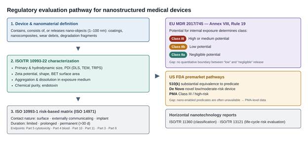
Figure 1. Regulatory evaluation pathway for nanostructured medical devices. The schematic traces how a nano-enabled device moves from definition through characterization to classification. A device that contains, consists of, or releases nano-objects (1–100 nm) is first characterized under ISO/TR 10993-22 (size, zeta potential, aggregation, dissolution, and endotoxin in the exposure medium) and then entered into the ISO 10993-1 risk matrix based on contact nature and duration. Classification diverges by jurisdiction: EU MDR Annex VIII Rule 19 assigns Class III, IIb, or IIa according to the potential for internal exposure, whereas the US FDA routes devices through 510(k), De Novo, or PMA pathways. Horizontal ISO/TR reports supply shared terminology. Noted regulatory gaps are the undefined boundary between “low” and “negligible” exposure and the frequent absence of nano-enabled predicates.
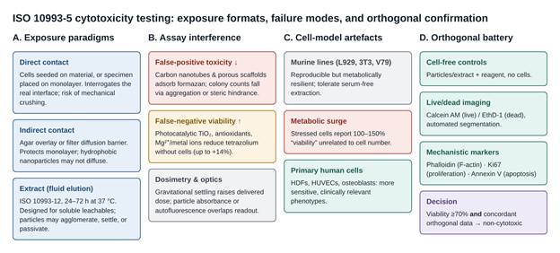
Figure 2. ISO 10993-5 cytotoxicity testing: exposure formats, failure modes, and orthogonal confirmation. Panel A shows the three standard exposure paradigms (direct contact, indirect diffusion, and fluid extract) with their intended use and principal weakness. Panel B summarizes direction-dependent assay interference, in which carbon nanotubes depress apparent viability by adsorbing formazan while photocatalytic TiO₂, antioxidants, and metal ions inflate it by reducing tetrazolium without cells; dosimetric and optical artefacts are also noted. Panel C contrasts the metabolic resilience of murine lines, including the 100–150% metabolic surge of stressed cultures, with the greater sensitivity of primary human cells. Panel D presents the orthogonal battery (cell-free controls, live/dead imaging, and mechanistic markers) that confirms a non-cytotoxic call only when a viability of at least 70% is matched by concordant imaging data.

phalloidin staining of F-actin, Ki67 proliferation indexing, and annexin V detection of externalized phosphatidylserine, it allows true biointegration to be separated from senescence, apoptosis, and necrosis (Lebaudy et al., 2020; Ramakrishnan & Daly, 2026; Sossah, 2021) (Table 2; Figure 2D).

2.3 Hemocompatibility of Biomaterial–Nanomaterial Interfaces (ISO 10993-4)

2.3.1 Blood as a Reactive Test Environment

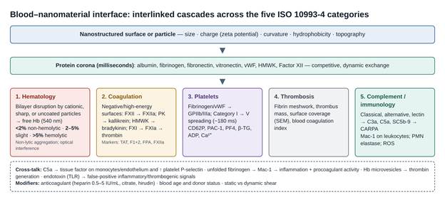
For devices intended for direct or indirect blood contact (vascular stents, heart valves, catheters, grafts, hemodialysis circuits, and intravenous nanomedicines), hemocompatibility testing under ISO 10993-4 is an essential regulatory requirement (ISO, 2017a; Weber et al., 2018). Blood comprises erythrocytes (about 44% of its volume), leukocytes and platelets (about 1%), and plasma (about 55%) containing active proteolytic enzyme systems (Weber et al., 2018). Contact with an artificial or nanostructured surface sets off immediate and interlinked interfacial cascades (Allain, 2018; Weber et al., 2018). Historically, inconsistent protocols, a lack of reference materials, and poorly predictive set-ups undermined in vitro evaluation, the situation captured by the phrase "blood compatibility catastrophe" (Braune et al., 2013; Ratner, 1993, 2007; van Oeveren, 2013). ISO 10993-4 responds by organizing evaluation into five categories, namely hematology, coagulation, thrombosis, platelets, and immunology/complement (ISO, 2017a; Urbán et al., 2019), which are depicted in Figure 3.

2.3.2 Protein Corona Dynamics and Contact Activation

Within milliseconds of blood exposure, plasma proteins adsorb onto the biomaterial–nanomaterial interface and form a dynamic protein corona (Aggarwal et al., 2009; Weber et al., 2018). Its composition, density, and conformational orientation depend on nanoscale topography, zeta potential, hydrophobicity, and curvature, with particle size emerging as a critical determinant (Tenzer et al., 2011). Abundant proteins (albumin, fibrinogen, fibronectin, vitronectin, high-molecular-weight kininogen [HMWK], and Factor XII) compete for binding sites through continual exchange (Aggarwal et al., 2009; Weber et al., 2018). Negatively charged, high-energy, or wettable nanostructured surfaces trigger the intrinsic contact pathway (Ilinskaya & Dobrovolskaia, 2013; Weber et al., 2018). This contact system consists of three serine proteases, Factor XII, Factor XI, and plasma prekallikrein (PK), together with the non-enzymatic cofactor HMWK (Long et al., 2016). On surface binding, Factor XII is converted to Factor XIIa, which cleaves PK into plasma kallikrein and liberates bradykinin from HMWK (Long et al., 2016). Factor XIIa also activates Factor XI, initiating tenase and prothrombinase assembly, a burst of thrombin generation, and polymerization of fibrinogen into fibrin (Ilinskaya & Dobrovolskaia, 2013; Weber et al., 2018). Positively charged nanoparticles act through a different route: they can unfold adsorbed fibrinogen and expose cryptic epitopes that engage the leukocyte Mac-1 receptor, promoting both inflammation and local procoagulant activity (Deng et al., 2011).

2.3.3 Platelet Adhesion, Secretion, and Aggregation

Platelet engagement is a major driver of acute device-associated thrombosis (Braune et al., 2013; Weber et al., 2018). When platelets encounter fibrinogen, fibronectin, or von Willebrand factor (vWF) adsorbed on a surface, the integrin GPIIb/IIIa (αIIbβ3) ligates these proteins and initiates outside-in signalling (Radomski et al., 2005; Weber et al., 2018). Activation can occur within roughly 180 ms of contact, after which platelets progress from resting discoid forms (Category I) through dendritic forms with early pseudopodia (Categories II and III) to fully spread cells (Category V) that build dense aggregate sheets (Braune et al., 2013; Sossah, 2021). Alpha granules release platelet factor 4 (PF4), β-thromboglobulin (β-TG), and platelet-derived growth factor, while P-selectin (CD62P) translocates to the outer membrane and serves as a key activation marker (Weber et al., 2018). Dense granules secrete ADP, serotonin, polyphosphates, and Ca²⁺, which act on P2Y1 and P2Y12 receptors of neighbouring platelets to amplify recruitment (Braune et al., 2013; Weber et al., 2018). Red cells modulate this process physically and chemically, especially under shear (Goldsmith et al., 1995). In vitro, platelet responses are quantified through PF4 and β-TG ELISA, flow cytometric CD62P and PAC-1 expression, and scanning electron or confocal microscopy of surface spreading (Sossah, 2021; Weber et al., 2018).

2.3.4 Erythrocyte Fragility and Hemolytic Profiling

Hemolysis testing is the most widely applied and best-standardized in vitro screen under ISO 10993-4 and ASTM E2524-08 (ASTM E2524-08, 2013; ISO, 2017a). Contact with foreign surfaces, high mechanical shear, or membrane-active nanoparticles such as cationic dendrimers and uncoated metal oxides can destabilize the erythrocyte bilayer and release hemoglobin (Choi et al., 2011; Zuckerman & Kao, 2009). Free hemoglobin scavenges endothelial nitric oxide, injures renal tubules, and, through procoagulant erythrocyte microvesicles, can amplify thrombin generation (Weber et al., 2018). Standard testing incubates whole blood or erythrocyte suspensions with the material or its extract for 1–3 h at 37 °C; supernatants are then centrifuged and cyanmethemoglobin absorbance is read at 540 nm against saline or phosphate-buffered saline (negative control) and Triton X-100 (1–4%) or deionized water (positive, 100% lysis) (Neun & Dobrovolskaia, 2011; Weber et al., 2018). Values below 2.0% are classified as non-hemolytic, 2.0–5.0% as slightly hemolytic, and above 5.0% as hemolytic (ASTM E2524-08, 2013; Sossah, 2021; Totea et al., 2014). Even so, non-lytic erythrocyte aggregation can alter blood rheology and tissue perfusion without registering as hemolysis (Barshtein et al., 2018).

2.3.5 Complement Activation, Leukocyte Mobilization, and CARPA

The complement system, the central humoral arm of innate immunity, comprises more than 30 soluble and membrane-bound proteins arranged in classical, alternative, and lectin cascades (Szebeni & Storm, 2015; Urbán et al., 2019). Nanostructured surfaces can initiate complement through C3 deposition or classical pathway recognition, generating the anaphylatoxins C3a and C5a and assembling the terminal complex SC5b-9 (Szebeni & Storm, 2015; Weber et al., 2018). In nanomedicine, uncontrolled activation underlies CARPA, an acute, non-IgE-mediated hypersensitivity marked by cardiopulmonary distress and hypotension, first characterized clinically with pegylated liposomal doxorubicin (Chanan-Khan et al., 2003; Szebeni & Storm, 2015). Complement also cross-talks with coagulation: C5a induces tissue factor on monocytes and endothelium, enhances platelet P-selectin, and triggers neutrophil respiratory burst with release of polymorphonuclear (PMN) elastase and ROS (Dahlgren & Karlsson, 1999; Weber et al., 2018). In practice, complement activation is monitored by ELISA for C3a, C5a, and SC5b-9 or by total hemolytic complement (CH50) assays, following practices such as ASTM F1984-99 (ASTM F1984-99, 2013; ISO, 2017a). Whether these in vitro immunotoxicity readouts predict in vivo outcomes is still debated (Dobrovolskaia & McNeil, 2013).

2.4 Pre-Analytical Variables and In Vitro Flow Dynamics

2.4.1 Donor, Handling, and Anticoagulant Effects

The clinical predictiveness of in vitro hemocompatibility data hinges on control of pre-analytical variables and on choosing a biologically appropriate incubation environment (Braune et al., 2013; van Oeveren, 2013). Donor medication, smoking history, venipuncture technique, storage time, and anticoagulant choice all shift baseline reactivity, and fresh blood is strongly preferred (Blok et al., 2016; Weber et al., 2018). Anticoagulant selection deserves particular care. Heparin mimics clinical anticoagulation, but its form and concentration (0.5–5 IU/mL) vary, and high doses can suppress contact activation and mask thrombogenicity (Weber et al., 2018). Sodium citrate (0.105–0.129 M) chelates Ca²⁺ to arrest coagulation; recalcification is then required for kinetic clotting assays and, if poorly executed, can itself activate platelets (Braune et al., 2013; Weber et al., 2018). For complement studies, the direct thrombin inhibitor hirudin (or lepirudin) preserves divalent cations and offers a more physiological environment (Weber et al., 2018). Subclinical endotoxin contamination is a further confounder, activating Toll-like receptors on monocytes and platelets and producing false-positive inflammatory and thrombogenic signals (Ruggeri, 2020; Weber et al., 2018).

2.4.2 Static Versus Dynamic Incubation Systems

The physical test environment also shapes cellular responses (Table 3). Static incubation in multiwell plates or tubes is convenient for initial screening but lacks flow, allows rapid erythrocyte sedimentation, and exposes blood to a large air interface that denatures proteins and activates platelets (Braune et al., 2013; Weber et al., 2018). Sealed rotational and agitated systems avoid an open air interface and are useful for colloidal suspensions, such as silk nanoparticles (Maitz et al., 2017; Steller et al., 2003). Flat-plate flow chambers, cone-and-plate viscometers, modified Chandler loops, and roller pump circuits generate controlled wall shear stress (Goldsmith et al., 1995; Lackner et al., 2012; Weber et al., 2018). Chandler loops, though, use an air bubble to propel blood, which can denature proteins and strip adherent cells at the gas–liquid interface (Krajewski et al., 2013; Weber et al., 2018), and peristaltic compression in roller pumps causes background trauma (Podias et al., 1995; Teligui et al., 2016). Ball-valve systems such as the Hemobile model eliminate both problems (Van Oeveren et al., 2012). Microfluidic bioMEMS platforms, increasingly preferred, need minimal blood, enable real-time fluorescence

Table 3. Comparative analysis of in vitro static and dynamic blood incubation models for biomaterial–nanomaterial interfaces. This table contrasts the main in vitro blood incubation systems used in hemocompatibility testing, classifying each by its fluid-dynamic and wall shear stress regime, operational design and blood-volume requirement, principal strengths for nano-interface testing, inherent artefactual limitations, and representative applications. Static multiwell systems are simple but lack shear and expose blood to an air interface; agitated, Chandler loop, and roller pump systems introduce defined or undefined shear at the cost of bubble- or compression-induced trauma; ball-valve and microfluidic bioMEMS platforms minimize these artefacts and support real-time imaging and endothelialized lumens. Matching the incubation model to the intended clinical flow regime is the central selection principle. Abbreviations: HUVECs, human umbilical vein endothelial cells; PDMS, polydimethylsiloxane; SIPA, shear-induced platelet aggregation; τw, wall shear stress.


Incubation system model

Fluid dynamics and wall shear stress regime

Operational design and blood volume

Primary strengths for nano-interface testing

Inherent artefactual limitations

Representative applications

Primary references

Static multiwell / tube incubation

No fluid movement (τw = 0 Pa); simulates stagnant flow or severe vascular stasis.

Multiwell polystyrene plates or test tubes; 100–500 µL per replicate.

Simple, rapid, economical; well suited to high-throughput initial screening of soluble extracts or candidate coatings.

Rapid erythrocyte sedimentation; large blood–air interface promotes protein aggregation and spontaneous platelet activation.

Basic hemolysis screening, preliminary platelet adhesion, and resting clotting time.

Weber et al. (2018); Braune et al. (2013); Sossah (2021)

Agitated and rotational models

Non-directional tumbling agitation; low, undefined wall shear stress.

Fully filled, sealed containers rotated or shaken; 1–3 mL.

Eliminates the unsealed blood–air interface; continuous agitation prevents gravitational settling of suspended nanoparticles.

Uncontrolled, non-physiological shear profiles; mechanical impacts on container walls can cause non-specific lysis.

Colloidal nanoparticle suspensions, silk nanoparticles, and particulate degradation products.

Weber et al. (2018); Steller et al. (2003); Maitz et al. (2017)

Modified Chandler loop model

Circular tubing rotation in a 37 °C bath; generates physiological arterial or venous shear.

Closed circular conduit partially filled with blood, using an internal air bubble to drive flow; ~5–10 mL.

Mimics laminar pulsatile flow; allows direct insertion of tubular devices or stents.

The circulating air–liquid interface denatures plasma proteins, strips adherent cells, and causes background hemolysis.

Stent thrombogenicity testing, silver nanoparticle circulation, and dynamic leukocyte activation.

Weber et al. (2018); Braune et al. (2013); Krajewski et al. (2013)

Roller pump closed-loop circuits

Continuous mechanical pumping; controlled directional laminar or turbulent flow.

Peristaltic roller pump compressing flexible tubing connected to a sample chamber; 15–50 mL.

Supports prolonged dynamic circulation (up to 240 min); integrates full-scale device prototypes.

Peristaltic compression induces mechanical trauma, background hemolysis, and platelet fragmentation.

Cardiopulmonary bypass circuits, membrane oxygenators, dialysis tubing, and vascular graft testing.

Weber et al. (2018); Podias et al. (1995); Teligui et al. (2016)

Hemobile (ball-valve) model

Unidirectional pulsatile flow driven by rocking motion and a one-way ball valve.

Sealed tubular loop with a non-thrombogenic ball valve; no air bubble; ~8–12 mL.

Eliminates the blood–air interface without roller pump compression, markedly reducing background trauma.

Difficult to miniaturize; requires specialized manufacturing and precise calibration of rocking angles.

Comparative thrombogenicity of vascular conduits and low-trauma platelet activation profiling.

Van Oeveren et al. (2012); Weber et al. (2018); van Oeveren (2013)

Parallel-plate and cone-plate viscometers

Highly uniform, well-defined shear stress fields (1 to >50 N·m⁻²).

Rotational plate-cone or stationary parallel-plate geometry; 2–5 mL.

Precise mathematical control of applied shear; isolates pure shear effects on platelet aggregation.

Difficult to integrate complex 3D nanostructured geometries; secondary vortices at high shear rates.

Fundamental studies of SIPA and von Willebrand factor unfolding.

Lackner et al. (2012); Goldsmith et al. (1995); Braune et al. (2013)

Microfluidic flow chambers and bioMEMS

Microscale laminar flow with precise physiological (100–1000 s⁻¹) or stenotic (>5000 s⁻¹) shear rates.

Microfabricated PDMS or glass channels; 50–200 µL.

Enables real-time confocal imaging of platelet adhesion and fibrin deposition; can integrate endothelial monolayers.

Susceptible to microchannel clogging from macroscopic clots; requires specialized microfabrication and imaging.

Real-time thrombosis monitoring, aneurysm-on-a-chip, and endothelialized nanostructure evaluation.

Allain (2018); Pham et al. (2016); Nordling et al. (2014); Westein et al. (2012)


imaging of platelet adhesion and fibrin deposition at defined shear rates, and can incorporate endothelialized lumens seeded with human umbilical vein endothelial cells (HUVECs) (Nordling et al., 2014; Pham et al., 2016; Westein et al., 2012).

2.5 Toward a Standardized, Multimodal Preclinical Framework

2.5.1 Limitations of Single-Endpoint Testing

Read together, the toxicological, regulatory, and bio-interfacial literature indicates that biomaterial–nanomaterial interfaces cannot be evaluated adequately by single-endpoint, static colorimetric assays (Braune et al., 2013; Ramakrishnan & Daly, 2026). Optical and chemical interference, over-reliance on resilient rodent lines, corona-mediated coagulation disturbances, and the absence of physiological shear are each manageable in isolation, but together they argue for a multi-tiered framework (D'Avenio et al., 2024; Urbán et al., 2019).

2.5.2 Components of a Tiered Framework

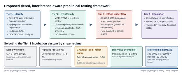
Four components appear essential (Figure 4). The first is comprehensive physicochemical characterization of primary particle size, hydrodynamic diameter, polydispersity, zeta potential, aggregation kinetics, endotoxin, and degradation profiles in physiological media before biological exposure, in line with ISO/TR 10993-22 (Caputo et al., 2019; ISO, 2017b). The second is orthogonal cytotoxicity profiling that pairs MTT, XTT, or NRU with cell-free interference controls and confirms results by high-content imaging (calcein AM/ethidium homodimer-1, phalloidin, Ki67, annexin V) in relevant primary human cells (Ramakrishnan & Daly, 2026; Rundén-Pran et al., 2022). The third is a hemocompatibility battery covering erythrocytes, contact coagulation, platelets, and complement (cyanmethemoglobin at 540 nm, Factor XIIa and thrombin–antithrombin complexes, CD62P/PAC-1 flow cytometry, and C3a/C5a/SC5b-9 ELISA) in closed-loop or microfluidic systems that reproduce arterial and venous shear (Braune et al., 2013; Weber et al., 2018). The fourth is adherence to the 3Rs through validated human cell-based models, ex ovo chorioallantoic membrane assays, and organ-on-chip systems that reduce dependence on high-variability animal models (Myers et al., 2017; Ramakrishnan & Daly, 2026).
3. Methods

3.1 Review Design and Reporting Approach

This work was designed as a structured critical review that combined a systematic literature search with a document analysis of international standards and regulatory instruments. The aim was not to pool effect sizes, since the included evidence was methodologically heterogeneous, but to map how cytotoxicity and hemocompatibility testing are specified, performed, and interpreted for biomaterial–nanomaterial interfaces, and to identify recurrent failure modes. The review question was framed in four elements: the test object (medical devices, coatings, or nanocomposites that contain, consist of, or release nanomaterials); the evaluation domain (in vitro cytotoxicity under ISO 10993-5 and blood interactions under ISO 10993-4); the comparator (standard protocol variants, cell models, anticoagulants, and incubation systems); and the outcomes (assay validity, interference, sensitivity, reproducibility, and regulatory acceptability) (D'Avenio et al., 2024; ISO, 2009, 2017a; Ramakrishnan & Daly, 2026). The search strategy, eligibility criteria, and extraction form were fixed before screening began so that the process could be repeated by independent investigators.

3.2 Information Sources

Peer-reviewed literature was sought in PubMed/MEDLINE, which was treated as the primary database, supplemented by Scopus and the Web of Science Core Collection to capture materials-science and engineering journals that are incompletely indexed in MEDLINE. Normative and regulatory sources were identified directly from the issuing bodies. These comprised the ISO 10993 series (ISO, 2009, 2017a, 2017b, 2018, 2025), the horizontal nanotechnology reports ISO/TR 11360:2010 and ISO/TR 13121:2011 (ISO, 2010, 2011), ASTM E2524-08 and ASTM F1984-99 (ASTM E2524-08, 2013; ASTM F1984-99, 2013), OECD Test Guideline 432 (OECD, 2025), the EU MDR (Regulation [EU] 2017/745, 2017), and FDA guidance on ISO 10993-1 (FDA, 2023). Doctoral dissertations addressing regulatory preclinical assessment and hemocompatibility of nanostructured carriers were included as grey literature because they report protocol-level detail rarely available in journal articles (Ruggeri, 2020; Sossah, 2021). Reference lists of key reviews (Braune et al., 2013; Urbán et al., 2019; Weber et al., 2018) were hand-searched, and forward citation tracking was applied to seminal papers on assay interference (Lupu & Popescu, 2013; Wörle-Knirsch et al., 2006) and on the blood compatibility problem (Ratner, 1993, 2007).

3.3 Search Strategy

The PubMed search combined Medical Subject Headings (MeSH) with title/abstract free-text terms in three concept blocks joined by AND, with synonyms joined by OR inside each block. Block 1 (material): "Nanostructures"[MeSH] OR "Nanoparticles"[MeSH] OR nanomaterial[tiab] OR nanoparticle[tiab] OR nanostructur[tiab] OR nanocoating[tiab] OR nanocomposite[tiab]. Block 2 (device and standard context): "Biocompatible Materials"[MeSH] OR "Equipment and Supplies"[MeSH] OR "medical device"[tiab] OR implant[tiab] OR "ISO 10993"[tiab] OR biocompatib[tiab]. Block 3 (biological endpoint): "Materials Testing"[MeSH] OR "Cell Survival"[MeSH] OR "Hemolysis"[MeSH] OR "Blood Coagulation"[MeSH] OR "Platelet Activation"[MeSH] OR "Complement Activation"[MeSH] OR "Protein Corona"[MeSH] OR cytotoxic[tiab] OR hemocompatib[tiab] OR haemocompatib[tiab] OR thrombogen[tiab] OR "assay interference"[tiab] OR "MTT"[tiab] OR "Chandler loop"[tiab] OR microfluidic*[tiab]. Equivalent strings were adapted to the syntax of Scopus (TITLE-ABS-KEY) and Web of Science (TS=). Searches covered January 1993, the year in which the blood compatibility problem was first framed explicitly (Ratner, 1993), to March 2026, with no geographical restriction. Filters limited records to English-language publications on human or mammalian in vitro, ex vivo, or in vivo systems. Full search strings, run dates, and record counts per database are provided in Supplementary Table S1.

3.4 Eligibility Criteria

Studies were eligible if they (i) evaluated a nanomaterial, nanostructured surface, or nano-enabled device, or a test method intended for such materials; (ii) reported at least one cytotoxicity endpoint aligned with ISO 10993-5 or at least one hemocompatibility endpoint within the five ISO 10993-4 categories; and (iii) described the test protocol in enough detail to identify the exposure format, cell or blood source, controls, and readout. Methodological papers that quantified assay interference or compared test systems were included even when no device was tested (Hirsch et al., 2011; Van Oeveren et al., 2012; Wittmaack, 2011). Records were excluded when they reported only in silico predictions, involved non-medical applications such as environmental or food nanotoxicology without transferable methodology, lacked controls, or were conference abstracts without full data. Regulatory and standards documents were included when they were current or had directly shaped current practice.

3.5 Study Selection

After removal of duplicates in a reference manager, two reviewers screened titles and abstracts independently against the eligibility criteria, and then assessed full texts. Disagreements were resolved by discussion and, where needed, by a third reviewer. Reasons for exclusion at the full-text stage were recorded. The numbers of records identified, screened, excluded, and included are reported in Supplementary Figure S1.

3.6 Data Extraction and Charting

A standardized extraction form was piloted on five articles and then applied to all included records. For each study, reviewers extracted the following: material class and nanoscale feature; physicochemical characterization (size by DLS, TEM, or tunable resistive pulse sensing; zeta potential; specific surface area; endotoxin; dissolution) (Caputo et al., 2019; ISO, 2017b); exposure format (extract, direct, or indirect contact; extraction vehicle, ratio, time, and temperature per ISO 10993-12); cell model (continuous line versus primary human cell, passage number, seeding density); assay and readout wavelength; cell-free interference controls; acceptance threshold; blood source and anticoagulant (type and concentration); incubation system and shear regime; hemocompatibility endpoints and their analytical methods; and principal findings with their stated limitations (Braune et al., 2013; Ramakrishnan & Daly, 2026; Weber et al., 2018). Extracted data were charted into four thematic matrices, which became Tables 1–3.

3.7 Reference Protocol Benchmarks

To allow consistent appraisal across heterogeneous studies, each included protocol was compared against benchmark conditions drawn from the standards and from widely used methodological sources.

3.7.1 Cytotoxicity Benchmarks

For extract testing, the benchmark was incubation of the test article in complete culture medium (with serum) at 37 °C for 24–72 h, at the surface-area- or mass-to-volume ratio specified in ISO 10993-12, followed by exposure of sub-confluent monolayers for 24 h (Goyal et al., 2025; ISO, 2009). Required controls were a negative control (high-density polyethylene or medium alone), a positive control (for example, latex or a cytotoxic reference material), a vehicle blank, and, for nanomaterials, a cell-free control containing the particles or extract with assay reagent but no cells, to detect direct reduction or adsorption (Lupu & Popescu, 2013; Ramakrishnan & Daly, 2026; Wörle-Knirsch et al., 2006). MTT was read at 540–570 nm with a 650–690 nm reference after DMSO or isopropanol solubilization; XTT/MTS at 450–490 nm; and NRU at 540 nm after acidified ethanol extraction (ISO, 2009; OECD, 2025). Viability at or above 70% of the negative control was taken as non-cytotoxic (ISO, 2009). Direct and agar overlay results were graded on the 0–4 reactivity scale, with grade ≤2 acceptable (ISO, 2009; Ruggeri, 2020). Orthogonal confirmation by live/dead imaging with automated segmentation (for example, FIJI/ImageJ) and by annexin V flow cytometry was recorded where present (Ramakrishnan & Daly, 2026).

3.7.2 Hemocompatibility Benchmarks

For hemolysis, the benchmark followed ASTM E2524-08: fresh human blood from consenting, medication-free donors, used within a defined time window after venipuncture with a 21-gauge needle and discard of the first tube; incubation with the test article or extract for 1–3 h at 37 °C; centrifugation; and quantification of cyanmethemoglobin at 540 nm against a hemoglobin standard curve, with saline or PBS as negative and Triton X-100 or deionized water as positive controls (ASTM E2524-08, 2013; Blok et al., 2016; Neun & Dobrovolskaia, 2011). Particle-only spectral controls were required for nanomaterials that absorb near 540 nm (Choi et al., 2011; Neun & Dobrovolskaia, 2011). Coagulation benchmarks included thrombin–antithrombin complex (TAT), prothrombin fragment 1+2, fibrinopeptide A, and Factor XIIa, with the caveat that standard aPTT reagents contain exogenous activators (Long et al., 2016; Weber et al., 2018). Platelet benchmarks included CD62P and PAC-1 flow cytometry, PF4 and β-TG ELISA, and SEM morphology scored by Categories I–V (Braune et al., 2013; Sossah, 2021). Complement benchmarks included C3a, C5a, Bb, C4d, and SC5b-9 ELISA and CH50, preferably in hirudin- or lepirudin-anticoagulated blood (ASTM F1984-99, 2013; Szebeni & Storm, 2015; Weber et al., 2018). Leukocyte activation was benchmarked against PMN elastase ELISA and DCFH-DA ROS assays, with endotoxin quantified by limulus amebocyte lysate testing (Dahlgren & Karlsson, 1999; FDA, 2023). Incubation systems were classified by wall shear regime and air-interface status, following published comparisons (Steller et al., 2003; Van Oeveren et al., 2012; Weber et al., 2018).

3.8 Critical Appraisal

Methodological quality was appraised for each primary study with a checklist built from the benchmarks above. Seven items were scored as met, partly met, or not met: adequacy of physicochemical characterization in the exposure medium; appropriateness of positive, negative, and cell-free controls; justification of the cell model; reporting of blood source, donor criteria, anticoagulant, and time to testing; appropriateness of the incubation system for the intended clinical flow regime; use of orthogonal confirmation; and statistical reporting, including the number of independent donors or biological replicates. The checklist was used to weight interpretation during synthesis rather than to exclude studies.

4. Synthesis of Regulatory, Cytotoxicity, and Hemocompatibility Evidence

4.1 Regulatory Landscape and Risk Categorization Patterns

4.1.1 From Predicate Equivalence to Size-Dependent Risk

The regulatory material points, fairly consistently, to a shift away from "predicate-only" equivalence and toward size-dependent risk management for nanostructured devices (D'Avenio et al., 2024; Myers et al., 2017). The basic architecture has not changed much. ISO 10993-1 still structures evaluation around contact type and duration (ISO, 2018), and its 2025 revision keeps that logic within the risk-management process (ISO, 2025). What has changed is the weight placed on nanoscale features, and this is where the frameworks begin to diverge (Table 1).

4.1.2 The Weight of EU MDR Rule 19

Of the instruments reviewed, Rule 19 of EU MDR Annex VIII appears to impose the highest evidentiary threshold. Devices containing or composed of nanomaterials with a high or medium potential for internal exposure are assigned automatically to Class III (D'Avenio et al., 2024; Regulation [EU] 2017/745, 2017). Low internal exposure maps to Class IIb and negligible exposure to Class IIa, so even the lightest-touch nano-device stays under notified body oversight (Ruggeri, 2020) (Figure 1). One persistent ambiguity, noted in several sources, is that no quantitative line separates "low" from "negligible" release, and this leaves classification partly to interpretation (D'Avenio et al., 2024; Ruggeri, 2020) (Table 1).

4.1.3 FDA Pathways and the Role of ISO/TR 10993-22

The US approach is structured differently. Nano-engineered platforms move through 510(k), De Novo, or PMA routes, and reviewers ask whether nanoscale dimensions introduce new toxicological or translocation behaviour (FDA, 2023). Finding a valid predicate for a nano-enabled product is not always possible, which may help explain why some sponsors end up with PMA-level data requirements (Allain, 2018; FDA, 2023; Zuckerman & Kao, 2009). Bridging both systems, ISO/TR 10993-22 offers guidance on nano-objects, aggregates, degradation fragments, and wear debris (ISO, 2017b). The broad consensus, if one can call it that, is that conventional ISO 10993-12 extracts do not capture the reactivity of particulate debris; nano-objects should be characterized directly in physiologically relevant media for hydrodynamic diameter, zeta potential, aggregation, and dissolution (Caputo et al., 2019; D'Avenio et al., 2024; Kyriakides et al., 2021). Horizontal documents on nanomaterial classification and life-cycle risk evaluation help with terminology, although they were not written with blood-contacting devices in mind (ISO, 2010, 2011) (Table 1).

4.2 In Vitro Cytotoxicity: Discrepancies and Assay Interference

4.2.1 A Clear Threshold, Less Clear Mechanisms

The ISO 10993-5 literature shows a tension that is hard to ignore. The acceptance criterion is simple, with viability at or above 70% of untreated controls counting as non-cytotoxic (ISO, 2009; Ramakrishnan & Daly, 2026), yet the biochemistry underneath that number can produce both false-positive and false-negative outcomes when nanomaterials are present (D'Avenio et al., 2024; Hirsch et al., 2011). The main assay modalities, their mechanisms, and the ways they fail are set out in Table 2 and Figure 2.

4.2.2 Direction-Dependent Interference

Interference does not run in one direction. Carbon nanotubes, for instance, adsorb insoluble formazan onto their hydrophobic surfaces, resisting solvent extraction and artificially depressing viability (Wörle-Knirsch et al., 2006). Photocatalytic and redox-active oxides such as TiO₂ seem to do the reverse, reducing tetrazolium salts by direct electron transfer and inflating apparent viability by as much as 14% (Lupu & Popescu, 2013). Antioxidant-functionalized biopolymers and biodegradable metals, magnesium-based implants in particular, release reducing species into extracts that react with tetrazolium in cell-free conditions (Goyal et al., 2025; Ramakrishnan & Daly, 2026). XTT and MTS add a further complication: their sensitivity to light, pH shifts, and nanomaterial-generated superoxide can raise background signal (Berillo et al., 2021). NRU is less exposed to reducing ions, but nanomaterials that disturb lysosomes can still bias dye accumulation (OECD, 2025), and clonogenic counts may fall through particle-induced aggregation or steric hindrance rather than cell death (Rundén-Pran et al., 2022) (Table 2; Figure 2B).

4.2.3 Cell Model Resilience and Metabolic Surges

Cell model choice turns out to matter more than the standard's wording might suggest. L929 and BALB/c 3T3 cells consistently appear more metabolically resilient than primary human cells (Goyal et al., 2025; Ramakrishnan & Daly, 2026). L929 cells keep high metabolic turnover under serum-depleted extraction, which may hide sub-lethal stress (Ramakrishnan & Daly, 2026). Perhaps more troubling, stressed cells can mount compensatory mitochondrial surges that register as 100–150% viability while bearing no clear relation to cell number (Ramakrishnan & Daly, 2026) (Figure 2C).

4.2.4 Uptake of Orthogonal Methods

Partly in response to these problems, more recent evaluations integrate fluorescence-based, multiparametric readouts. Calcein AM/ethidium homodimer-1 imaging combined with phalloidin, Ki67, and annexin V appears to separate viable adherence from metabolic stress and early apoptosis more reliably than any single colorimetric assay (Lebaudy et al., 2020; Ramakrishnan & Daly, 2026; Ruggeri, 2020). These methods have their own caveats, however, including autofluorescence from polymeric matrices or quantum dots and higher instrument costs (Goyal et al., 2025; Weber et al., 2018) (Table 2; Figure 2D).

4.3 Hemocompatibility Profiles Across the Blood–Material Interface

4.3.1 Overview of the Five ISO 10993-4 Domains

The evidence across the five ISO 10993-4 categories describes an interconnected set of responses rather than five independent tests (Braune et al., 2013; ISO, 2017a; Weber et al., 2018). Specific nanoscale attributes, especially charge, curvature, and surface area, map with some regularity onto particular hematological outcomes (Ilinskaya & Dobrovolskaia, 2013; Urbán et al., 2019) (Figure 3).

4.3.2 Erythrocyte Damage and Hemolysis

Cyanmethemoglobin absorbance at 540 nm remains the main standardized quantitative metric (ASTM E2524-08, 2013; Neun & Dobrovolskaia, 2011). Cationic nanoparticles, sharp-edged nanomaterials, and unpassivated metal surfaces quite often breach the erythrocyte bilayer and exceed the 5.0% threshold (Choi et al., 2011; Sossah, 2021; Urbán et al., 2019). Silver nanoparticles illustrate the measurement problem well, because their optical absorbance near 540 nm can generate false readings unless particle-only controls are included (Choi et al., 2011). Alloys such as Zr-binary systems, by contrast, have been reported to fall within acceptable limits in whole human blood (Totea et al., 2014) (Figure 3).

4.3.3 Contact Activation and Coagulation

High-energy, negatively charged nanostructured surfaces bind Factor XII, PK, and HMWK rapidly, triggering intrinsic pathway activation and raising thrombin–antithrombin complexes and fibrinopeptide A (Long et al., 2016; Weber et al., 2018). The extent of activation seems to track the composition of the protein corona, which particle size in particular modulates (Aggarwal et al., 2009; Tenzer et al., 2011). A methodological caveat recurs throughout: clinical aPTT reagents contain exogenous activators that may mask nanomaterial-driven contact activation (Weber et al., 2018) (Figure 3).

4.3.4 Platelet Adhesion and Secretion

Adsorbed fibrinogen and vWF, especially in denatured form, bind GPIIb/IIIa and drive transitions from resting Category I to spread Category V platelets, sometimes within about 180 ms (Braune et al., 2013; Sossah, 2021). Activated platelets display CD62P and release PF4, β-TG, and ADP, which accelerates local thrombosis (Braune et al., 2013; Weber et al., 2018). Particulate nanomaterials have also been shown to induce platelet aggregation and vascular thrombosis directly (Radomski et al., 2005) (Figure 3).

4.3.5 Complement Activation and CARPA

Nanoparticulate and polymeric nanocomposite platforms can activate complement, producing C3a, C5a, and SC5b-9 (Szebeni & Storm, 2015; Urbán et al., 2019). Clinically, this has been linked with CARPA and acute cardiopulmonary reactions (Chanan-Khan et al., 2003). Heparin may suppress parts of the cascade in vitro, so hirudin or lepirudin is generally recommended for this domain (ASTM F1984-99, 2013; Weber et al., 2018) (Figure 3).

4.3.6 Leukocyte Activation and Inflammatory Cross-Talk

Unfolded surface proteins expose cryptic epitopes that engage leukocyte Mac-1, stimulating PMN elastase release and respiratory burst ROS that in turn amplify procoagulant activity (Dahlgren & Karlsson, 1999; Deng et al., 2011; Weber et al., 2018). Endotoxin contamination, if left unmeasured, could produce much the same picture for very different reasons (Braune et al., 2013; Weber et al., 2018) (Figure 3).

4.4 Impact of Hydrodynamic Regimes on Blood Interactions

4.4.1 Static Systems

One of the clearer findings concerns the limits of static blood incubation (Table 3). Static multiwell assays are simple and need little blood, but they lack shear, allow erythrocytes to sediment, and expose blood to an open air interface that denatures plasma proteins and causes background platelet activation (Braune et al., 2013; Sossah, 2021; Weber et al., 2018).

4.4.2 Agitated, Loop, and Pump-Driven Systems

Dynamic models are more representative physiologically, but they are not free of artefacts (Weber et al., 2018). Sealed agitated systems prevent sedimentation of suspended nanoparticles, although their shear fields are poorly defined (Maitz et al., 2017; Steller et al., 2003). Chandler loops depend on an internal air bubble that strips adherent cells and denatures proteins, a problem documented, for example, in silver nanoparticle testing (Krajewski et al., 2013; Weber et al., 2018). Roller pump circuits support long circulation times but compress tubing in ways that damage erythrocytes and platelets (Podias et al., 1995; Teligui et al., 2016). Ball-valve (Hemobile) models avoid both a gas interface and peristaltic crushing and show noticeably lower background hemolysis (Van Oeveren et al., 2012; van Oeveren, 2013) (Table 3).

4.4.3 Shear-Defined and Microfluidic Systems

Parallel-plate and cone-plate devices offer precise control over shear and are well suited to studies of shear-induced platelet activation, although they are difficult to adapt to complex three-dimensional nanostructures (Goldsmith et al., 1995; Lackner et al., 2012). Microfluidic bioMEMS chambers allow control over both physiological (100–1000 s⁻¹) and stenotic (>5000 s⁻¹) shear rates (Allain, 2018; Pham et al., 2016). They use only 50–200 µL of blood, permit real-time confocal tracking of platelet deposition and thrombus growth, and can incorporate functional HUVEC monolayers (Nordling et al., 2014; Westein et al., 2012). Of the systems reviewed, these may come closest to bridging benchtop assays and vascular performance, although clogging and fabrication demands remain practical obstacles (Pham et al., 2016; Westein et al., 2012) (Table 3; Figure 4).

5. Reconciling Static Standards with Dynamic Nano–Bio Interfaces

5.1 Principal Findings in Context

What this synthesis shows, more than anything, is a mismatch of timescales and assumptions. The ISO 10993 series was built for materials whose biological identity is more or less fixed at the moment of manufacture, whereas a nanomaterial's identity is renegotiated continuously, first by the medium it is extracted into, then by the proteins that coat it, and finally by the flow conditions it meets (Aggarwal et al., 2009; D'Avenio et al., 2024; Tenzer et al., 2011). The regulatory frameworks summarized in Table 1 acknowledge this, at least in principle. ISO/TR 10993-22 says plainly that nano-objects should be characterized in their exposure state (ISO, 2017b), and EU MDR Rule 19 ties classification to internal exposure (Regulation [EU] 2017/745, 2017). The test methods themselves, however, have not fully caught up. It would be unfair to call the standards obsolete, since they still provide a common language and a defensible baseline (FDA, 2023; ISO, 2018). They do, though, seem to leave the most nano-specific questions to the discretion of individual laboratories.

5.2 Regulatory Implications: Classification Without Quantification

5.2.1 Exposure Thresholds and Predicates

Rule 19 is arguably the most decisive regulatory development reviewed here, yet its practical force depends on something it does not define, namely a quantitative boundary between low and negligible internal exposure (D'Avenio et al., 2024; Ruggeri, 2020) (Table 1). Without such a boundary, two manufacturers with similar coatings could reasonably arrive at different classes. The FDA's reliance on predicates poses a parallel problem (FDA, 2023). A predicate is meaningful only if the nanoscale features that drive biological behaviour are comparable, and that is precisely what bulk characterization fails to establish (Myers et al., 2017; Zuckerman & Kao, 2009).

5.2.2 Characterization as the Regulatory Anchor

The more promising direction, we think, is the one already implied by ISO/TR 10993-22: treating physicochemical characterization in biologically relevant media as the anchor of the dossier rather than an appendix to it (Caputo et al., 2019; ISO, 2017b). Orthogonal sizing methods developed jointly by European and US nanomedicine characterization laboratories offer a workable template (Caputo et al., 2019). Horizontal nanomaterial documents could support shared terminology (ISO, 2010, 2011), and the 2025 revision of ISO 10993-1 may provide an opportunity to reference such methods more explicitly (ISO, 2025) (Figure 1).

5.3 Rethinking the 70% Viability Threshold

5.3.1 What the Threshold Assumes

The 70% viability criterion of ISO 10993-5 (ISO, 2009) is attractive because it is simple. Its weakness is that it presumes the signal reflects living cells, and for nanomaterials that presumption often does not hold. The interference mechanisms in Table 2 are not exotic edge cases. Formazan adsorption by carbon nanotubes (Wörle-Knirsch et al., 2006), cell-free tetrazolium reduction by TiO₂ (Lupu & Popescu, 2013), and reducing ions from degrading magnesium (Goyal et al., 2025) cover a large share of the materials now in development. Hirsch et al. (2011) asked whether nanomaterial–cell interaction research was "on the right track"; more than a decade later, the question still seems apt.

5.3.2 Cell Models and Metabolic Signals

The cell model problem is harder to settle. L929 cells were chosen for their reproducibility, and that remains a real virtue (ISO, 2009). Yet their tolerance of serum-free extraction and their tendency toward compensatory metabolic surges of 100–150%

Figure 3. Interlinked biological cascades at the blood–nanomaterial interface across the five ISO 10993-4 categories. The figure depicts how surface properties (size, charge, curvature, hydrophobicity, and topography) first shape a protein corona that forms within milliseconds, and how this corona seeds the five evaluation domains: hematology (hemolysis thresholds), coagulation (contact activation through Factor XII, prekallikrein, and HMWK), platelets (GPIIb/IIIa-mediated adhesion and Category I–V spreading with granule secretion), thrombosis (fibrin and thrombus formation), and complement/immunology (C3a, C5a, SC5b-9, and CARPA). The lower band lists the cross-talk linking these domains, for example C5a induction of tissue factor and fibrinogen-driven Mac-1 engagement, together with the pre-analytical modifiers (anticoagulant, blood age, and shear) that influence every readout.

Figure 4. Proposed tiered, interference-aware preclinical testing framework and shear-based selection of the blood incubation system. The upper row sets out four sequential tiers: Tier 1 establishes physicochemical identity in the exposure medium; Tier 2 applies interference-controlled cytotoxicity in both reference and primary human cells with orthogonal imaging; Tier 3 evaluates all five ISO 10993-4 blood domains under clinically matched flow; and Tier 4 escalates to endothelialized microfluidics, ex ovo assays, or targeted in vivo work only when earlier tiers raise concerns, consistent with the 3Rs. The lower row arranges candidate Tier 3 incubation systems along a gradient from lower to higher physiological fidelity, from static wells through agitated, Chandler loop, roller pump, and ball-valve systems to microfluidic bioMEMS, guiding selection by the intended clinical shear regime.

(Ramakrishnan & Daly, 2026) suggest that a pass on L929 may not translate into safety for human endothelium or parenchyma. Replacing murine lines outright would sacrifice comparability with decades of historical data. A more pragmatic route, perhaps, is to run the reference line in parallel with at least one primary human cell type relevant to the intended contact, as some magnesium and extracellular-matrix studies have done (Goyal et al., 2025; Ramakrishnan & Daly, 2026).

5.3.3 Making Orthogonal Readouts Routine

Orthogonal imaging-based endpoints are well positioned to resolve ambiguous metabolic signals (Lebaudy et al., 2020; Ramakrishnan & Daly, 2026) (Figure 2D). Their limitations (autofluorescence, image thresholding, and cost) are real but tractable, and arguably less consequential than a false pass. Including cell-free interference controls costs very little, and there seems to be little justification for omitting them in any nanomaterial study (Lupu & Popescu, 2013; Wörle-Knirsch et al., 2006). Higher-throughput clonogenic formats may also help bridge screening and mechanistic work (Rundén-Pran et al., 2022).

5.4 Hemocompatibility as an Integrated System

5.4.1 Interdependent Domains

Perhaps the most important lesson from Figure 3 is that the five ISO 10993-4 categories describe one system viewed from five angles. Corona formation sets the stage (Aggarwal et al., 2009; Tenzer et al., 2011). Contact activation then generates thrombin and bradykinin (Long et al., 2016), unfolded fibrinogen recruits leukocytes through Mac-1 (Deng et al., 2011), and C5a links complement back to tissue factor and platelet P-selectin (Weber et al., 2018) (Figure 3). A material that passes hemolysis can still be strongly procoagulant or complement-activating. Evaluating domains in isolation may therefore miss emergent risks, and this could partly explain the inconsistent correlation between in vitro immunotoxicity tests and in vivo outcomes (Dobrovolskaia & McNeil, 2013; Ilinskaya & Dobrovolskaia, 2013).

5.4.2 Hemolysis Beyond the Thresholds

Hemolysis deserves a specific comment. Its numerical cut-offs (<2%, 2–5%, >5%) are among the clearest in the whole framework (ASTM E2524-08, 2013), but clarity is not the same as completeness. Optical interference from particles such as silver (Choi et al., 2011) and non-lytic erythrocyte aggregation with rheological consequences (Barshtein et al., 2018) both fall outside what a supernatant absorbance reading can detect.

5.4.3 Complement and CARPA

The clinical experience with CARPA (Chanan-Khan et al., 2003; Szebeni & Storm, 2015) is a reminder that complement testing is not a formality. For lipid-based, PEGylated, or high-curvature systems, it should probably be considered mandatory rather than optional, ideally in hirudin-anticoagulated blood (ASTM F1984-99, 2013; Weber et al., 2018).

5.5 Pre-Analytical Control and Hydrodynamic Realism

5.5.1 Pre-Analytical Reporting

Much of the irreproducibility in blood testing may come down to variables that are rarely reported. Blood age (Blok et al., 2016), heparin dose, and anticoagulant type (Weber et al., 2018) can each shift results by amounts comparable to the material effects being measured. Endotoxin can mimic material-driven inflammation (Ruggeri, 2020). Van Oeveren (2013) catalogued many of these obstacles, and Braune et al. (2013) asked, pointedly, whether existing standards are sufficient. Our reading is that they are not yet sufficient, mainly because pre-analytical reporting is not mandated in sufficient detail.

5.5.2 Matching the Incubation System to Clinical Flow

The choice of incubation system is, in effect, a choice about which artefacts one is willing to accept (Table 3). Static assays accept air-interface activation and sedimentation; Chandler loops accept bubble-induced denaturation (Krajewski et al., 2013); roller pumps accept mechanical trauma (Podias et al., 1995; Teligui et al., 2016). Ball-valve and microfluidic systems reduce these compromises (Van Oeveren et al., 2012; Westein et al., 2012), and endothelialized channels add a biological wall that no static well can provide (Nordling et al., 2014). The difficulty is that each system defines shear differently, so results are not easily transferable between them (Goldsmith et al., 1995; Lackner et al., 2012; Pham et al., 2016). Matching the system to the intended clinical flow regime (venous, arterial, or stenotic) seems more defensible than any single universal choice (Allain, 2018; Weber et al., 2018).

5.6 Toward a Tiered, Interference-Aware Testing Framework

5.6.1 Structure of the Framework

Drawing these threads together, we propose a four-tier framework (Figure 4). Tier 1 establishes physicochemical identity in the exposure medium, including endotoxin and dissolution (Caputo et al., 2019; ISO, 2017b). Tier 2 applies interference-controlled cytotoxicity in both the reference and human primary cells, with orthogonal imaging confirmation (Ramakrishnan & Daly, 2026). Tier 3 evaluates all five ISO 10993-4 domains in fresh, appropriately anticoagulated blood under flow that matches clinical use (Braune et al., 2013; Weber et al., 2018). Tier 4 escalates to complex models, such as endothelialized microfluidics, ex ovo assays, or targeted in vivo studies, only when earlier tiers raise questions, in keeping with the 3Rs (Myers et al., 2017; Nordling et al., 2014). None of these elements is new on its own. The contribution lies in sequencing them so that each tier informs the interpretation of the next.

5.6.2 Application to Emerging Device Classes

Infection-responsive biodegradable coatings (Wu et al., 2026) and anti-inflammatory material designs (Lebaudy et al., 2020) illustrate why such a framework matters: they are designed to change over time, so a single static snapshot is unlikely to capture their safety profile.

5.7 Limitations of This study

Several limitations should be acknowledged. The review is narrative and thematic rather than meta-analytic, and heterogeneity in exposure metrics prevented quantitative pooling. Some sources are doctoral dissertations or book chapters (Allain, 2018; Berillo et al., 2021; Ruggeri, 2020; Sossah, 2021), which, though rich in protocol detail, have not always been peer reviewed in the conventional sense. Standards evolve, and ISO 10993-1:2025 (ISO, 2025) may already shift some of the emphases discussed here. Finally, the review concentrates on in vitro methods. Long-term implantation, genotoxicity, and systemic toxicity, while essential, lay outside its scope.

5.8 Future Directions

Three priorities stand out. The first is the development of certified nano-reference materials with known interference profiles, so that laboratories can benchmark their assays against a common standard (Caputo et al., 2019; Hirsch et al., 2011). The second is the adoption of minimum reporting standards for blood source, anticoagulant, time to test, and shear regime (Blok et al., 2016; Braune et al., 2013). The third is interlaboratory validation of microfluidic hemocompatibility platforms, which could eventually allow their incorporation into a future revision of ISO 10993-4 (Pham et al., 2016; Westein et al., 2012). Progress on any one of these would, we suspect, do more for clinical predictability than adding further endpoints to an already crowded test matrix.

6. Conclusion

Nanostructured biomaterials offer real clinical promise, but their safety cannot be inferred from bulk materials or captured by single, static assays. ISO 10993-5 and ISO 10993-4 remain indispensable baselines, yet they are vulnerable to nanomaterial-specific artefacts. Formazan adsorption, cell-free tetrazolium reduction, resilient murine models, and misleading metabolic surges can distort cytotoxicity data. Hemocompatibility, in turn, is shaped by an interlinked network of corona formation, contact activation, platelet secretion, complement, and leukocyte responses that static incubation can neither isolate nor reproduce. Regulatory frameworks, notably EU MDR Rule 19 and ISO/TR 10993-22, already recognize these risks, although quantitative criteria are still lacking. A tiered strategy that starts with exposure-state characterization, adds interference controls and human primary cells, evaluates all blood domains under clinically matched flow, and escalates only when necessary offers a practical route toward more reproducible, predictive, and ethically efficient preclinical evaluation.

References


Aggarwal, P., Hall, J. B., McLeland, C. B., Dobrovolskaia, M. A., & McNeil, S. E. (2009). Nanoparticle interaction with plasma proteins as it relates to particle biodistribution, biocompatibility and therapeutic efficacy. Advanced Drug Delivery Reviews, 61(6), 428–437. https://doi.org/10.1016/j.addr.2009.03.009

Allain, J. P. (2018). Multiscale interactions between blood and metallic biomaterials. In Metallic biomaterials: Processing and interface science (pp. 317–352). Elsevier.

ASTM E2524-08. (2013). Standard test method for analysis of hemolytic properties of nanoparticles. ASTM International.

ASTM F1984-99. (2013). Standard practice for testing for whole complement activation in serum by solid materials. ASTM International.

Barshtein, G., Pajic-Lijakovic, I., & Gural, A. (2018). Role of erythrocyte aggregation in blood rheology and tissue perfusion. Current Opinion in Chemical Engineering, 22, 148–154.

Berillo, D., Al-Jwaid, A., & Caplin, J. (2021). Cytotoxicity and biocompatibility of macroporous cryogels. In Nanomaterials: Biomedical applications and toxicity (pp. 15–48). Springer.

Blok, S. L. J., Engels, G. E., & van Oeveren, W. (2016). In vitro hemocompatibility testing: The importance of fresh blood. Biointerphases, 11(2), Article 029802.

Braune, S., Grunze, M., Straub, A., & Jung, F. (2013). Are there sufficient standards for the in vitro hemocompatibility testing of biomaterials? Biointerphases, 8(1), Article 33. https://doi.org/10.1186/1559-4106-8-33

Caputo, F., Clogston, J., Calzolai, L., Rösslein, M., & Prina-Mello, A. (2019). Measuring particle size distribution of nanoparticle enabled medicinal products, the joint view of EUNCL and NCI-NCL: A step by step approach combining orthogonal measurements with increasing complexity. Journal of Controlled Release, 299, 31–43.

Chanan-Khan, A., Szebeni, J., Saville, S., Le, N., Baxevanis, C., Gordon, M., & Muggia, F. (2003). Complement activation by pegylated liposomal doxorubicin (Doxil®) in cancer patients: Association with hypersensitivity reactions. Annals of Oncology, 14(9), 1430–1437.

Choi, J., Reipa, V., Hitchins, V. M., Goering, P. L., & Malinauskas, R. A. (2011). Physicochemical characterization and in vitro hemolysis evaluation of silver nanoparticles. Toxicological Sciences, 123(1), 133–143. https://doi.org/10.1093/toxsci/kfr149

D’Avenio, G., Daniele, C., & Grigioni, M. (2024). Nanostructured medical devices: Regulatory perspective and current applications. Materials, 17(8), Article 1787. https://doi.org/10.3390/ma17081787

Dahlgren, C., & Karlsson, A. (1999). Respiratory burst in human neutrophils. Journal of Immunological Methods, 232(1–2), 3–14.

Deng, Z. J., Liang, M., Monteiro, M., Toth, I., & Minchin, R. F. (2011). Nanoparticle-induced unfolding of fibrinogen promotes Mac-1 receptor activation and inflammation. Nature Nanotechnology, 6(1), 39–44. https://doi.org/10.1038/nnano.2010.250

Dobrovolskaia, M. A., & McNeil, S. E. (2013). Understanding the correlation between in vitro and in vivo immunotoxicity tests for nanomedicines. Journal of Controlled Release, 172(2), 456–466.

Food and Drug Administration. (2023). Use of International Standard ISO 10993-1, “Biological evaluation of medical devices — Part 1: Evaluation and testing within a risk management process”: Guidance for industry and Food and Drug Administration staff. U.S. Department of Health and Human Services.

Goldsmith, H. L., Bell, D. N., & Braovac, S. (1995). Physical and chemical effects of red cells in the shear-induced aggregation of human platelets. Biophysical Journal, 69(4), 1584–1595.

Goyal, A., Pandey, A., Marwaha, J., & Matai, J. (2025). Standardized in vitro cytotoxicity and degradation assessment of magnesium-based biomaterials in compliance with ISO 10993-5. Journal of Materials Science: Materials in Medicine, 36(1), Article 92.

Hirsch, C., Roesslein, M., Krug, H. F., & Wick, P. (2011). Nanomaterial cell interactions: Are we on the right track? Beilstein Journal of Nanotechnology, 2, 894–902.

Ilinskaya, A. N., & Dobrovolskaia, M. A. (2013). Nanoparticles and the blood coagulation system. Part II: Safety concerns. Nanomedicine, 8(5), 773–784.

International Organization for Standardization. (2009). Biological evaluation of medical devices — Part 5: Tests for in vitro cytotoxicity (ISO Standard No. 10993-5:2009).

International Organization for Standardization. (2010). Nanotechnologies — Methodology for the classification and categorization of nanomaterials (ISO Technical Report No. ISO/TR 11360:2010).

International Organization for Standardization. (2011). Nanotechnologies — Nanomaterial risk evaluation (ISO Technical Report No. ISO/TR 13121:2011).

International Organization for Standardization. (2017a). Biological evaluation of medical devices — Part 4: Selection of tests for interactions with blood (ISO Standard No. 10993-4:2017).

International Organization for Standardization. (2017b). Biological evaluation of medical devices — Part 22: Guidance on nanomaterials (ISO Technical Report No. ISO/TR 10993-22:2017).

International Organization for Standardization. (2018). Biological evaluation of medical devices — Part 1: Evaluation and testing within a risk management process (ISO Standard No. 10993-1:2018).

International Organization for Standardization. (2025). Biological evaluation of medical devices — Part 1: Requirements and general principles for the evaluation of biological safety within a risk management process (ISO Standard No. 10993-1:2025).

Krajewski, S., Prucek, R., Panacek, A., Avci-Adali, M., Nolte, A., Straub, A., & Wendel, H. P. (2013). Hemocompatibility evaluation of different silver nanoparticle concentrations employing a modified Chandler-loop in vitro assay on human blood. Acta Biomaterialia, 9(7), 7460–7468.

Kyriakides, T. R., Raj, A., Tseng, T. H., Xiao, H., Nguyen, P., & Hirschi, K. K. (2021). Biocompatibility of nanomaterials and their interface with host tissues. Journal of Materials Chemistry B, 9(33), 6432–6449.

Lackner, J. M., Waldhauser, W., Hartmann, P., Bruckert, F., Weidenhaupt, M., Major, R., & Sanak, M. (2012). Hemocompatibility of inorganic physical vapor deposition (PVD) coatings on thermoplastic polyurethane polymers. Journal of Functional Biomaterials, 3(2), 283–297.

Lebaudy, E., Fournel, S., Lavalle, P., Vrana, N. E., & Gribova, V. (2020). Recent advances in antiinflammatory material design. Advanced Healthcare Materials, 10(1), Article 2001373.

Long, A. T., Kenne, E., Jung, R., Fuchs, T. A., & Renné, T. (2016). Contact system revisited: An interface between inflammation, coagulation, and innate immunity. Journal of Thrombosis and Haemostasis, 14(3), 427–437.

Lupu, A. R., & Popescu, T. (2013). The noncellular reduction of MTT tetrazolium salt by TiO2 nanoparticles and its implications for cytotoxicity assay. Toxicology in Vitro, 27(5), 1445–1450.

Maitz, M. F., Sperling, C., Wongpinyochit, T., Herklotz, M., Werner, C., & Seib, F. P. (2017). Biocompatibility assessment of silk nanoparticles: Hemocompatibility and internalization by human blood cells. Nanomedicine: Nanotechnology, Biology and Medicine, 13(8), 2633–2642.

Myers, D. D., Wolf, M. F., & Anderson, J. M. (2017). From in vivo to in vitro: The medical device testing paradigm shift. ALTEX — Alternatives to Animal Experimentation, 34(4), 479–500.

Neun, B. W., & Dobrovolskaia, M. A. (2011). Method for analysis of nanoparticle hemolytic properties in vitro. In M. A. Dobrovolskaia & S. E. McNeil (Eds.), Characterization of nanoparticles intended for drug delivery (pp. 215–224). Humana Press.

Nordling, S., Nilsson, B., & Magnusson, P. U. (2014). A novel in vitro model for studying the interactions between human whole blood and endothelium. Journal of Visualized Experiments, (96), Article e52112.

Organization for Economic Cooperation and Development. (2025). Test No. 432: In vitro 3T3 NRU phototoxicity test. OECD Publishing.

Pham, T. T., Wiedemeier, S., Maenz, S., Gastrock, G., Settmacher, U., Jandt, K. D., Zanow, J., Lüdecke, C., & Bossert, J. (2016). Hemodynamic aspects of reduced platelet adhesion on bioinspired microstructured surfaces. Colloids and Surfaces B: Biointerfaces, 145, 502–509.

Podias, A., Groth, T., & Missirlis, Y. (1995). The effect of shear rate on the adhesion/activation of human platelets in flow through a closed-loop polymeric tubular system. Journal of Biomaterials Science, Polymer Edition, 6(5), 399–410.

Radomski, A., Jurasz, P., Alonso-Escolano, D., Drews, M., Morandi, M., Malinski, T., & Radomski, M. W. (2005). Nanoparticle-induced platelet aggregation and vascular thrombosis. British Journal of Pharmacology, 146(6), 882–893.

Ramakrishnan, R., & Daly, A. C. (2026). Revisiting ISO 10993-5 in vitro cytotoxicity standard tests for evaluation of extracellular matrix-based biomaterials. International Journal of Biomaterials, 2026, Article 7395612. https://doi.org/10.1155/ijbm/7395612

Ratner, B. D. (1993). The blood compatibility catastrophe. Journal of Biomedical Materials Research, 27(3), 283–287.

Ratner, B. D. (2007). The catastrophe revisited: Blood compatibility in the 21st century. Biomaterials, 28(34), 5144–5147.

Regulation (EU) 2017/745 of the European Parliament and of the Council of 5 April 2017 on medical devices. (2017). Official Journal of the European Union, L 117, 1–175.

Ruggeri, M. (2020). Nanotechnology-based medical devices: From critical quality attributes to regulatory preclinical assessment [Doctoral dissertation, University of Pavia].

Rundén-Pran, E., Mariussen, E., El Yamani, N., Elje, E., Longhin, E. M., & Dusinska, M. (2022). The colony forming efficiency assay for toxicity testing of nanomaterials — Modifications for higher-throughput. Frontiers in Toxicology, 4, Article 983316.

Sossah, F. (2021). Preclinical evaluation and hemocompatibility testing of micro- and nanostructured hemoglobin carrier systems [Doctoral dissertation, Charité — Universitätsmedizin Berlin].

Steller, U., Sperling, C., Hübner, J., Hanke, R., & Werner, C. (2003). Design and evaluation of novel blood incubation systems for in vitro hemocompatibility assessment of planar solid surfaces. Journal of Biomedical Materials Research Part B: Applied Biomaterials, 66B(1), 379–390.

Szebeni, J., & Storm, G. (2015). Complement activation-related pseudoallergy: A new class of adverse reactions to nanomedicines. Journal of Controlled Release, 219, 437–444.

Teligui, L., Dalmayrac, E., Corbeau, J. J., Bouquet, E., Godon, A., Denommé, A. S., ... & Baufreton, C. (2016). Ex vivo simulation of cardiopulmonary bypass with human blood for hemocompatibility testing. Perfusion, 31(5), 376-383.

Tenzer, S., Docter, D., Rosfa, S., Wlodarski, A., Kuharev, J., Rekik, A., Knauer, S. K., Bantz, C., Nawroth, T., Bier, C., Sirirungruang, J., Mann, W. R., Treuel, L., Zellner, R., & Stauber, R. H. (2011). Nanoparticle size is a critical physicochemical determinant of the human blood plasma corona: A comprehensive quantitative proteomic analysis. ACS Nano, 5(9), 7155–7167.

Totea, G., Ionita, D., Demetrescu, I., & Mitache, M. (2014). In vitro hemocompatibility and corrosion behavior of new Zr-binary alloys in whole human blood. Open Chemistry, 12(7), 796–803.

Urbán, P., Liptrott, N. J., & Bremer, S. (2019). Overview of the blood compatibility of nanomedicines: A trend analysis of in vitro and in vivo studies. WIREs Nanomedicine and Nanobiotechnology, 11(2), Article e1546.

van Oeveren, W. (2013). Obstacles in haemocompatibility testing. Scientifica, 2013, Article 392584.

Van Oeveren, W., Tielliu, I. F., & De Hart, J. (2012). Comparison of modified Chandler, roller pump, and ball valve circulation models for in vitro testing in high blood flow conditions: Application in thrombogenicity testing of different materials for vascular applications. International Journal of Biomaterials, 2012, Article 673163.

Weber, M., Steinle, H., Golombek, S., Hann, L., Schlensak, C., Wendel, H. P., & Avci-Adali, M. (2018). Blood-contacting biomaterials: In vitro evaluation of the hemocompatibility. Frontiers in Bioengineering and Biotechnology, 6, Article 99.

Westein, E., de Witt, S., Lamers, M., Cosemans, J. M., & Heemskerk, J. W. (2012). Monitoring in vitro thrombus formation with novel microfluidic devices. Platelets, 23(7), 501–509.

Wittmaack, K. (2011). Excessive delivery of nanostructured matter to submersed cells caused by rapid gravitational settling. ACS Nano, 5(5), 3766–3778.

Wörle-Knirsch, J. M., Pulskamp, K., & Krug, H. F. (2006). Oops they did it again! Carbon nanotubes hoax scientists in viability assays. Nano Letters, 6(6), 1261–1268.

Wu, Y., Zhang, L., Wang, Q., Li, H., & Liu, Z. (2026). Infection-responsive biodegradable magnesium implant coatings. Bioactive Materials, 58, 632–649.

Zuckerman, S. T., & Kao, W. J. (2009). Nanomaterials and biocompatibility: BioMEMS and dendrimers. In M. M. de Villiers, P. Aramwit, & G. S. Kwon (Eds.), Nanotechnology in drug delivery (pp. 193–216). Springer.


Article metrics
View details
0
Downloads
0
Citations
29
Views

View Dimensions


View Plumx


View Altmetric


0
Save
0
Citation
29
View
0
Share