Biosensors and Nanotheranostics

Bionanotechnology, Drug Delivery, Therapeutics | online ISSN 3064-7789
175
Citations
118.9k
Views
88
Articles
Your new experience awaits. Try the new design now and help us make it even better
Switch to the new experience
Figures and Tables
REVIEWS   (Open Access)

Nanoparticle-Mediated Ferroptosis Induction Combined with MRI Monitoring in Therapy-Resistant Cancers

Long Chiau Ming 1* Ong Khang Wei 2, Nurul Dayana Binti Mahizir 2


+ Author Affiliations

Biosensors and Nanotheranostics 5 (1) 1-8 https://doi.org/10.25163/biosensors.5110997

Submitted: 06 September 2026 Revised: 01 November 2026  Published: 09 November 2026 


Abstract

Therapy-resistant solid tumours remain among the most intractable problems in clinical oncology, in large part because the mainstays of treatment converge on a single death programme—apoptosis—that resistant cells and cancer stem cells learn to evade. Ferroptosis, an iron-dependent form of regulated cell death driven by unchecked lipid peroxidation, sidesteps these classical escape routes and has therefore drawn intense interest. Yet the small-molecule inducers that first revealed its promise are limited by poor solubility, rapid clearance, and off-target toxicity. This review examines how rationally engineered nanomaterials—superparamagnetic iron oxide nanoparticles, metal–organic frameworks, catalytic nanozymes, and biomimetic membrane-camouflaged carriers—address these liabilities while simultaneously acting as magnetic resonance imaging (MRI) contrast agents, uniting therapy and diagnosis within one platform. We trace the molecular logic of ferroptosis through the System X??–glutathione–GPX4 axis, the labile iron pool, and Fenton chemistry, and describe how nanoplatforms exploit tumour iron addiction to deplete antioxidant defences, reverse multidrug efflux, and eradicate stem-like populations. We then consider the biophysics of T1, T2, and switchable dual-mode contrast that permits real-time tracking of biodistribution, accumulation, and early therapeutic response. Across preclinical models, nanoferroptosis combined with photothermal, sonodynamic, and radiotherapeutic actuation yields supra-additive responses and, through damage-associated molecular pattern release and cGAS–STING activation, converts immunologically cold tumours into inflamed niches responsive to checkpoint blockade. Finally, we weigh the translational obstacles—protein corona formation, long-term iron clearance, manufacturing reproducibility, and predictive modelling—that still separate compelling benchtop data from clinical reality. On balance, the evidence invites cautious optimism rather than certainty, since dependable clinical translation is not yet assured.

Keywords: ferroptosis; iron oxide nanoparticles; MRI theranostics; multidrug resistance; cancer stem cells; cGAS–STING; tumour microenvironment; immunogenic cell death.

1. Introduction

Malignant neoplasms remain a leading cause of death worldwide, and much of that mortality is attributable to two linked phenomena: the capacity of tumours to acquire resistance to therapy and their propensity for metastatic spread (Han et al., 2024; Kesharwani & Gao, 2024; Wang et al., 2025). The conventional pillars of cancer treatment—systemic chemotherapy, fractionated radiotherapy, and molecularly targeted agents—share a common mechanistic dependence: they kill cells chiefly by engaging the apoptotic cascade (Zhang, 2026). This shared dependence is also a shared vulnerability. Under sustained therapeutic pressure, tumours evolve a repertoire of anti-apoptotic adaptations, including upregulation of anti-apoptotic Bcl-2 superfamily members, loss or mutational inactivation of the pro-apoptotic effectors Bax and Bak, and overexpression of adenosine triphosphate (ATP)-binding cassette efflux transporters such as P-glycoprotein (P-gp/MDR1) (Kesharwani & Gao, 2024; Wang et al., 2025).

Resistance is compounded at the cellular and microenvironmental levels. Cancer stem cells (CSCs)—a subpopulation endowed with self-renewal, multilineage plasticity, and quiescence—tolerate standard cytotoxic agents and reseed tumours after apparently successful treatment (Wang et al., 2025). Surrounding these cells, the immunosuppressive tumour microenvironment (TME) of therapy-resistant lesions presents dense desmoplastic matrix, hypoxia, acidosis, pro-tumoral M2-polarised tumour-associated macrophages (TAMs), and regulatory T cells, which together exclude cytotoxic lymphocytes and hold checkpoint-blockade response rates below roughly 30% (C. Yang et al., 2025; Zhang, 2026). Confronting apoptosis-competent machinery with agents that require that same machinery is, in this light, a losing proposition.

A compelling way around the apoptotic impasse is to engage an alternative, non-apoptotic mode of regulated cell death (Ensoy et al., 2024; Kesharwani & Gao, 2024; Zhang, 2026). Ferroptosis is the most thoroughly developed of these. Morphologically it is distinct—shrunken mitochondria with condensed membranes, diminished cristae, and outer-membrane rupture—and biochemically it is governed by the intersection of iron metabolism, amino acid/glutathione (GSH) dynamics, and lipid peroxidation (Ensoy et al., 2024; Han et al., 2024). The canonical brake on ferroptosis is the cystine/glutamate antiporter System Xᴄ⁻ (SLC7A11/SLC3A2), which imports cystine for GSH biosynthesis, together with glutathione peroxidase 4 (GPX4), the selenoprotein that uses GSH to reduce toxic polyunsaturated-fatty-acid phospholipid hydroperoxides into benign lipid alcohols (Bilbao-Asensio et al., 2022; Ensoy et al., 2024; Han et al., 2024).

When this surveillance axis is disabled, redox balance collapses and the labile iron pool (LIP) fuels Fenton chemistry: ferrous iron reacts with hydrogen peroxide to generate hydroxyl radicals that propagate chain-reaction lipid peroxidation across membranes (Bilbao-Asensio et al., 2022; Roshanghias et al., 2026; Z. Wang et al., 2025). Because malignant and stem-like cells carry an elevated iron requirement—often described as iron addiction—and heightened basal oxidative stress, they are selectively vulnerable to ferroptosis, which opens an exploitable therapeutic window (Bilbao-Asensio et al., 2022; Wang et al., 2025). Translating that window into a treatment, however, has proven difficult: canonical inducers such as erastin, RSL3, sulfasalazine, and sorafenib suffer from negligible aqueous solubility, rapid clearance, poor tumour selectivity, and dose-limiting toxicity (Ensoy et al., 2024; Kesharwani & Gao, 2024).

Nanotechnology offers a route through these barriers, defining the field of nanoferroptosis (Ensoy et al., 2024; Kesharwani & Gao, 2024; Wang et al., 2025). Engineered nanomaterials—superparamagnetic iron oxide nanoparticles (SPIONs), metal–organic frameworks (MOFs), biomimetic membrane-camouflaged carriers, and catalytic nanozymes—combine high payload capacity, prolonged circulation, and preferential intratumoral accumulation through passive enhanced permeability and retention (EPR) or ligand-mediated targeting (Anani et al., 2021; Ensoy et al., 2024; Kesharwani & Gao, 2024; C. Yang et al., 2025). Crucially, iron-based carriers disassemble in the acidic, reducing endolysosomal milieu to release catalytic Fe²⁺/Fe³⁺, while incorporated components deplete GSH, downregulate GPX4, silence iron exporters, and disable efflux pumps—reversing multidrug resistance and triggering lethal lipid peroxidation (Ensoy et al., 2024; Kesharwani & Gao, 2024; Wang et al., 2025).

What distinguishes magnetic nanoferroptosis from other delivery strategies is that the therapeutic core is also an imaging agent. Integrating diagnosis and therapy in one construct—theranostics—is central to precision oncology (Anani et al., 2021; Roshanghias et al., 2026; Zhang, 2026). Magnetic resonance imaging (MRI) is an almost ideal partner: it offers outstanding soft-tissue contrast, sub-millimetre resolution, unlimited penetration depth, and no ionising radiation (Anani et al., 2021; Roshanghias et al., 2026; C. Yang et al., 2025). Iron oxide cores shorten transverse relaxation to produce negative (dark) T₂ contrast, whereas ultrasmall cores and surface-coordinated clusters raise longitudinal relaxivity for positive T₁ or dual-mode imaging (Anani et al., 2021; Roshanghias et al., 2026). This lets investigators follow biodistribution, confirm passage across physiological barriers, quantify accumulation, and detect early microenvironmental change before gross tumour shrinkage is visible (Roshanghias et al., 2026; C. Yang et al., 2025; Zhang, 2026). Beyond direct ablation, ferroptosis is immunogenic, and the resulting cGAS–STING activation and M2-to-M1 macrophage repolarisation can turn cold tumours hot (Kesharwani & Gao, 2024; C. Yang et al., 2025; Zhang, 2026). A mechanistic synthesis of how these elements combine—and of the chemical, biological, and regulatory hurdles that remain—is the purpose of this review.

2. Nanoferroptosis Theranostics Target Cancer Stem Cell Niches and Reprogram the Tumour Immune Microenvironment

2.1 Conceptual foundations: why ferroptosis, and why nanoscale delivery

The rationale for nanoferroptosis rests on a simple mismatch. Apoptosis-based therapy asks resistant cells to execute a programme they have specifically learned to suppress, whereas ferroptosis attacks a liability those same cells cannot easily discard—their dependence on iron and on continuous antioxidant repair of peroxidised membranes (Ensoy et al., 2024; Han et al., 2024; Wang et al., 2025). CSC niches, defined by self-renewal, plasticity, and quiescence, are precisely the compartments in which this vulnerability is most pronounced (Wang et al., 2025). The obstacle has never been whether ferroptosis can kill resistant cells, but whether an inducer can reach them selectively and be observed doing so; nanoscale carriers address delivery and, when magnetic, observation as well (Anani et al., 2021; Ensoy et al., 2024; Roshanghias et al., 2026).

2.2  Mechanistic architecture of ferroptotic cell death

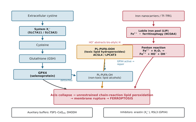
Ferroptosis is governed at the intersection of three metabolic pillars—iron handling, antioxidant defence, and lipid metabolism (Figure 1) (Ensoy et al., 2024; Han et al., 2024). The principal brake is the System Xᴄ⁻–GSH–GPX4 axis. System Xᴄ⁻, an antiporter of catalytic SLC7A11 and regulatory SLC3A2 subunits, imports cystine that is reduced to cysteine, the rate-limiting substrate for GSH (Ensoy et al., 2024; Han et al., 2024). GPX4 then uses GSH to convert cytotoxic PL-PUFA-OOH into harmless PL-PUFA-OH; inhibiting the axis with erastin (System Xᴄ⁻) or RSL3 (GPX4) collapses this defence (Bilbao-Asensio et al., 2022; Ensoy et al., 2024; Han et al., 2024). Auxiliary systems—the FSP1–CoQ₁₀ pathway and dihydroorotate dehydrogenase—provide partial, compartment-specific buffering (C. Li et al., 2025).

Execution requires redox-active ferrous iron (Figure 1). Tumour cells elevate transferrin receptor 1 expression and accelerate NCOA4-mediated ferritinophagy, enlarging the labile iron pool that sustains proliferation (Bilbao-Asensio et al., 2022; Han et al., 2024; Kesharwani & Gao, 2024). That same iron, under pathological overload, drives Fenton chemistry: Fe²⁺ + H₂O₂ → Fe³⁺ + HO• + OH⁻. The hydroxyl radicals abstract bis-allylic hydrogens from membrane polyunsaturated fatty acids esterified by ACSL4 and LPCAT3, initiating the radical chain reactions that rupture membranes and lyse the cell (Ensoy et al., 2024; Han et al., 2024). The selective lethality of this chemistry in iron-addicted cells is the biochemical foundation on which every platform discussed below is built.

2.3  Multifunctional nanoplatforms for targeted ferroptosis activation

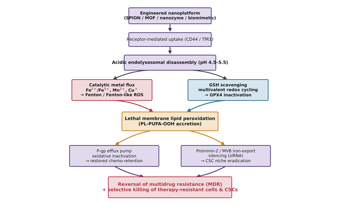
Four broad nanomaterial classes have been engineered to deliver and amplify ferroptosis, each with distinctive chemistry and representative constructs summarised in Table 1 (Anani et al., 2021; Ensoy et al., 2024; Kesharwani & Gao, 2024). The cascade they share—receptor-mediated uptake, acidic disassembly, catalytic metal release with concurrent GSH depletion, and the resulting collapse of multidrug efflux and stem-cell iron export—is depicted in Figure 2.

Iron oxide nanoparticles and SPIONs act as classic nano-inducers: internalised by receptor-mediated endocytosis, they dissolve in the acidic endolysosome (pH 4.5–5.5) to release ferrous and ferric ions, and their surfaces display intrinsic peroxidase- and catalase-mimetic activity that drives chemodynamic therapy in situ (Bilbao-Asensio et al., 2022; Roshanghias et al., 2026; Zhang, 2026). Surface ligands tune selectivity: hyaluronic acid homes to CD44-overexpressing CSCs, and Angiopep-2 enables transcytosis across the blood–brain barrier via LRP1 (M. Xie et al., 2022; Zhang, 2026). Metal–organic frameworks add very high surface area and tunable coordination for multi-agent co-delivery; high-valence Fe³⁺, Mn³⁺/Mn⁴⁺, and Cu²⁺ nodes undergo stoichiometric reduction by GSH, depleting antioxidant stores while generating catalytic reduced ions that accelerate secondary Fenton-like reactions (Y. Chen et al., 2024; C. Li et al., 2025; Wang et al., 2025). Catalytic nanozymes extend this logic with sustained, enzyme-

Figure 1. The molecular machinery of ferroptosis. The schematic integrates the three metabolic pillars that govern ferroptotic cell death. The antioxidant arm (left) traces cystine import through System Xᴄ⁻ (SLC7A11/SLC3A2), its reduction to cysteine, glutathione (GSH) synthesis, and GPX4-mediated detoxification of toxic PL-PUFA-OOH into harmless PL-PUFA-OH. The iron arm (right) shows labile-iron-pool expansion and the Fenton reaction generating hydroxyl radicals that abstract bis-allylic hydrogens from membrane lipids. Collapse of the System Xᴄ⁻–GSH–GPX4 axis, together with unrestrained Fenton chemistry, drives chain-reaction lipid peroxidation, membrane rupture, and ferroptosis; auxiliary buffers (FSP1–CoQ₁₀, DHODH) and canonical inhibitors (erastin, RSL3) are indicated.

Figure 2. Nanoplatform-triggered ferroptosis and reversal of therapeutic resistance. The flow depicts how an engineered nanoplatform (SPION, MOF, nanozyme, or biomimetic carrier) is internalised by receptor-mediated uptake (CD44/TfR1) and disassembles in the acidic endolysosome. Two converging engines follow—catalytic metal flux driving Fenton/Fenton-like ROS, and multivalent redox cycling scavenging GSH to inactivate GPX4—which together produce lethal membrane lipid peroxidation. Downstream, oxidative inactivation of P-glycoprotein restores chemo-retention while siRNA-mediated silencing of Prominin-2/MVB iron export eradicates the stem-cell niche, yielding reversal of multidrug resistance and selective killing of therapy-resistant cells and cancer stem cells.

mimetic H₂O₂ conversion and GSH consumption (Qin et al., 2025; Roshanghias et al., 2026).

Biomimetic camouflage solves the delivery problem from a different angle. Coating carriers in red-blood-cell, platelet, macrophage, or cancer-cell membranes extends circulation, reduces non-specific hepatic uptake, and confers homologous tumour homing (Jiang et al., 2020; Wang et al., 2025). Targeting CSCs further demands defeating active iron export: Wu et al. (2023) developed spindle-like FeOOH nanoparticles carrying siRNA against Prominin-2, which in stem-like breast cancer cells normally packages ferritin into multivesicular bodies and expels iron. Silencing Prominin-2 while releasing catalytic iron and consuming GSH traps iron inside the cell and selectively eliminates the resistant niche (Wu et al., 2023; Wang et al., 2025).

2.4 Biophysical principles and applications of MRI theranostics

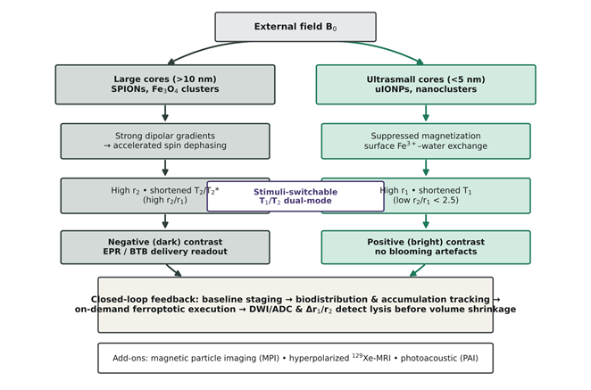
The same magnetic cores that drive ferroptosis report on it, and the governing variable is core size (Figure 3; Table 2) (Anani et al., 2021; Roshanghias et al., 2026; Zhang, 2026). Larger SPIONs (>10 nm) create strong microscopic field gradients that accelerate transverse spin dephasing, shorten T₂/T₂* relaxation, and yield hypointense contrast useful for confirming delivery across the blood–brain tumour barrier and tracking EPR-mediated retention (Anani et al., 2021; Roshanghias et al., 2026; Song et al., 2025). Because T₂ voids can be confounded by clots, calcification, and air interfaces, ultrasmall cores (<5 nm) have been engineered as positive T₁ agents: surface canting suppresses bulk magnetisation, lowers the r₂/r₁ ratio, and allows paramagnetic surface Fe³⁺ to accelerate longitudinal relaxation (Suh et al., 2024; L. Wang et al., 2017; Q. Xie et al., 2024).

The most elegant designs switch modes in response to the microenvironment. Xie et al. (2024) assembled ultrasmall Fe₃O₄ nanoclusters stabilised with DMSA and conjugated to RGD, which give strong T₂ signal in circulation but disassemble into dispersed sub-units under acidic, reducing intracellular conditions to switch toward bright T₁ enhancement (L. Wang et al., 2017; Q. Xie et al., 2024). Core–shell CuMnO@Fe₃O₄ and manganese-doped platforms achieve ratiometric dual-mode imaging by shedding Mn²⁺ (a T₁ agent) while retaining the Fe₃O₄ T₂ core (Qin et al., 2025; Roshanghias et al., 2026). Beyond anatomy, relaxometry, diffusion-weighted imaging and ADC mapping, and emerging modalities such as magnetic particle imaging and hyperpolarised ¹²⁹Xe-MRI capture ferroptotic destruction days before volume changes appear, enabling closed-loop therapy (Gupta et al., 2024; Nejadnik et al., 2017; Zhang, 2026).

2.5  Multimodal synergies and external physical actuation

Because small-molecule ferroptosis is often local and transient, nanoplatforms are increasingly combined with external energy to force supra-additive responses, as catalogued in Table 3 (Kesharwani & Gao, 2024; Z. Wang et al., 2025; Zhang, 2026). Photothermal therapy uses near-infrared absorption to generate mild hyperthermia (42–45 °C), which accelerates Fenton kinetics by Arrhenius behaviour, downregulates heat-shock defences, and speeds GSH depletion (Ensoy et al., 2024; Kesharwani & Gao, 2024). Sonodynamic therapy activates embedded sonosensitisers with deep-penetrating ultrasound, relieving hypoxia and triggering autophagy-dependent ferroptosis (Ensoy et al., 2024; Zhao et al., 2023). High-Z elements in core–shell constructs such as Fe₃O₄@Bi₂S₃ concentrate radiation and raise baseline H₂O₂ to fuel secondary Fenton loops, overcoming radioresistance (Song et al., 2025). Piezoelectric and chemodynamic combinations extend the same principle to platinum- and heavy-metal-resistant disease (Ge et al., 2022; Xu et al., 2021).

2.6  Immunological reprogramming of the tumour microenvironment

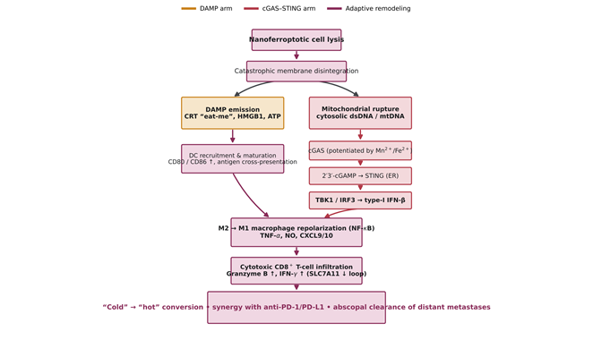
Perhaps the most consequential property of nanoferroptosis is that its membrane catastrophe is immunogenic, and the representative immunomodulatory platforms are compiled in Table 4 (Figure 4) (C. Li et al., 2025; C. Yang et al., 2025; Zhang, 2026). Unlike tolerogenic apoptosis, ferroptotic lysis exposes calreticulin as an “eat-me” signal and releases HMGB1, ATP, and fragmented mitochondrial DNA (C. Li et al., 2025; Z. Wang et al., 2025; C. Yang et al., 2025). Cytosolic mtDNA engages cyclic GMP-AMP synthase (cGAS), producing 2′3′-cGAMP that activates STING on the endoplasmic reticulum; STING recruits TBK1 to phosphorylate IRF3 and drive type-I interferon transcription (C. Li et al., 2025; C. Yang et al., 2025). Released Mn²⁺ from multimetallic carriers sharpens cGAS DNA-binding and STING sensitivity, acting as an innate-immune adjuvant, while paracrine interferon downregulates SLC7A11 to form a self-amplifying ferroptotic loop

Figure 3. MRI contrast mechanisms and closed-loop theranostic monitoring. The diagram shows how magnetic core size, under an external field (B₀), dictates MRI behaviour. Large cores (>10 nm) create strong dipolar gradients that accelerate spin dephasing, giving high r₂, shortened T₂/T₂*, and negative (dark) contrast for EPR/blood-brain tumour-barrier readout; ultrasmall cores (<5 nm) suppress magnetisation and exploit surface Fe³⁺–water exchange, giving high r₁, low r₂/r₁, and positive (bright) contrast without blooming. Stimuli-switchable designs bridge the two regimes. The lower panel summarises the closed-loop workflow—baseline staging, biodistribution and accumulation tracking, on-demand ferroptotic execution, and DWI/ADC and Δr₁/r₂ detection of lysis before volume shrinkage—augmented by MPI, ¹²⁹Xe-MRI, and photoacoustic imaging.

Figure 4. Ferroptosis-driven immunogenic cell death and tumour-microenvironment reprogramming. The cascade begins with nanoferroptotic cell lysis and catastrophic membrane disintegration, which split into two arms. The DAMP arm (left) releases calreticulin, HMGB1, and ATP, recruiting and maturing dendritic cells (CD80/CD86↑). The cGAS–STING arm (right) shows cytosolic dsDNA/mtDNA engaging cGAS—potentiated by Mn²⁺/Fe²⁺—producing 2′3′-cGAMP, activating STING, and driving TBK1/IRF3-dependent type-I IFN-β. The arms converge on M2-to-M1 macrophage repolarisation and cytotoxic CD8⁺ T-cell infiltration (with an SLC7A11-downregulating feedback loop), converting cold niches into hot lesions that synergise with anti-PD-1/PD-L1 blockade and support abscopal clearance of distant metastases.

(H. Chen et al., 2025; C. Li et al., 2025).

In parallel, iron and catalytic metal ions activate macrophage NF-κB signalling, repolarising immunosuppressive M2 TAMs into tumoricidal M1 cells that secrete TNF-α and nitric oxide and release CXCL9/CXCL10 to recruit cytotoxic CD8⁺ T lymphocytes (Bilbao-Asensio et al., 2022; C. Yang et al., 2025; Zhang, 2026). The net effect—dismantling local immunosuppression and expanding tumour-infiltrating lymphocytes—resensitises previously unresponsive tumours to anti-PD-1/anti-PD-L1 blockade and, in the strongest preclinical examples, drives durable systemic responses that prevent metastatic recurrence (C. Li et al., 2025; H. Wang et al., 2023; Z. Wang et al., 2025; Zhang, 2026).

2.7  Translational bottlenecks addressed by the literature

The same reviews that document these advances are candid about their limits (Ensoy et al., 2024; Z. Wang et al., 2025; Zhang, 2026). Colloidal instability, protein-corona formation, reticuloendothelial clearance, uncontrolled systemic iron release, batch-to-batch variability under scalable manufacturing, and the absence of settled regulatory frameworks for drug–device combination nanoformulations are recurring themes (Wang et al., 2025; Zhang, 2026). These concerns frame the critical appraisal that follows in the synthesis and discussion.

3. Methods

This article is a narrative, mechanism-oriented review assembled with a structured, reproducible search-and-synthesis protocol adapted from Preferred Reporting Items for Systematic Reviews and Meta-Analyses (PRISMA) guidance for narrative syntheses. The protocol is reported below in sufficient detail to allow independent replication, and all extracted evidence is tabulated in Tables 1–4.

3.1  Information sources and search dates

Three bibliographic databases—PubMed/MEDLINE, Scopus, and Web of Science Core Collection—were searched from database inception through the review preparation window, with a final update search executed immediately before manuscript submission. The reference lists of the principal anchor reviews on nanoferroptosis and magnetic nanotheranostics were hand-searched to capture primary studies not retrieved by the electronic queries (Ensoy et al., 2024; Han et al., 2024; Kesharwani & Gao, 2024; Roshanghias et al., 2026; Wang et al., 2025; Zhang, 2026). No language restriction was applied at the screening stage, although only English-language full texts were ultimately included.

3.2  Search strategy and controlled vocabulary

Search strings combined Medical Subject Headings (MeSH) with free-text terms using Boolean operators. The core PubMed string was: (“ferroptosis”[MeSH] OR “lipid peroxidation” OR “GPX4” OR “SLC7A11” OR “Fenton reaction”) AND (“nanoparticles”[MeSH] OR “iron oxide” OR “SPION” OR “metal-organic framework” OR “nanozyme” OR “biomimetic nanoparticle”) AND (“magnetic resonance imaging”[MeSH] OR “MRI” OR “relaxivity” OR “theranostic*”) AND (“drug resistance, neoplasm”[MeSH] OR “multidrug resistance” OR “cancer stem cell*” OR “tumor microenvironment”). The string was translated into the native syntax of Scopus and Web of Science. Additional targeted queries paired ferroptosis with specific actuation or immune terms (“photothermal,” “sonodynamic,” “radiosensitization,” “cGAS-STING,” “immunogenic cell death,” “macrophage repolarization”) to ensure adequate coverage of the multimodal and immunological literature (C. Li et al., 2025; C. Yang et al., 2025).

3.3  Eligibility criteria

Records were eligible if they (i) investigated an engineered nanomaterial that induces or amplifies ferroptosis in cancer; (ii) addressed at least one dimension relevant to this review—therapeutic resistance reversal, MRI or multimodal imaging guidance, external physical actuation, or immune reprogramming; and (iii) reported original mechanistic, in vitro, or in vivo data, or provided an authoritative mechanistic synthesis. Reports were excluded if they concerned ferroptosis without a nanomaterial component, described nanomaterials without a ferroptotic mechanism, were conference abstracts without full data, or duplicated a dataset already captured. Priority was given to primary studies for quantitative claims and to recent peer-reviewed reviews for conceptual framing (Ensoy et al., 2024; Kesharwani & Gao, 2024; Wang et al., 2025; Zhang, 2026).

3.4  Study selection and screening

Retrieved records were de-duplicated and screened in two stages—title/abstract, then full text—against the criteria above. Screening was performed independently by the reviewing authors, with disagreements resolved by discussion and consensus. The flow of records through identification, screening, eligibility, and inclusion was tracked in a PRISMA-style record so that the count of included primary studies could be reconstructed.

3.5  Data extraction and charting

A standardised extraction form captured, for each included study: nanoplatform composition and surface chemistry; target malignancy and resistance model; the ferroptotic mechanism and the specific resistance pathway reversed; imaging modality and relaxivity parameters where reported; external actuation modality; immunological readouts; and in vitro and in vivo outcomes. Extracted items were charted into four thematic matrices corresponding to this review’s organisation: engineered ferroptosis-inducing platforms (Table 1), MRI theranostic systems and relaxivity dynamics (Table 2), multimodal synergistic regimens (Table 3), and immunological reprogramming via DAMP and cGAS–STING signalling (Table 4). Quantitative values—relaxivities, tumour growth-inhibition rates, and fold changes in ferroptosis markers—were recorded verbatim as reported by the source studies to avoid secondary transformation error.

3.6  Synthesis and figure construction

Because the included studies differ widely in cancer type, model system, and outcome metric, a quantitative meta-analysis was neither feasible nor appropriate; evidence was therefore synthesised narratively around mechanism and model, with convergent and divergent findings noted explicitly. The four schematic figures (Figures 1–4) were constructed de novo as mechanistic syntheses of the extracted literature—respectively depicting the molecular machinery of ferroptosis, nanoplatform-triggered ferroptosis and resistance reversal, MRI contrast and closed-loop monitoring, and the immunogenic cGAS–STING cascade—rather than reproducing any single published figure. Referencing throughout follows APA (7th edition) conventions, and the complete bibliography lists only sources cited in the body text, tables, and figures.

4. In Vivo Efficacy, Relaxivity Dynamics, and Immune Remodelling Across Preclinical Nanoferroptosis Platforms

4.1  What the aggregate picture actually shows

Read as a whole, the preclinical record is more encouraging than it is tidy. Across the studies charted here, nanoparticle-mediated ferroptosis does seem to shift the treatment-response landscape of resistant solid tumours, and it does so fairly consistently—yet the models are so heterogeneous that one hesitates to put a single number on the effect (Ensoy et al., 2024; Zhang, 2026). The recurring contrast is familiar: free inducers such as erastin, RSL3, and sorafenib remain hamstrung by low bioavailability, rapid clearance, and off-target toxicity, whereas the engineered platforms in [Table 1] coordinate iron and multivalent metal delivery, concentrate in tumours by active or passive targeting, and dismantle antioxidant defences in a reasonably reproducible way [Figure 2] (Kesharwani & Gao, 2024; Wang et al., 2025). When those carriers also carry a magnetic core, accumulation can be measured rather than assumed [Table 2] (Roshanghias et al., 2026; Zhang, 2026). Where head-to-head comparison is possible—triple-negative breast cancer, orthotopic glioblastoma, multidrug-resistant hepatocellular carcinoma, desmoplastic pancreatic cancer—primary tumour growth inhibition commonly lands somewhere between about 70% and 90%, with longer survival and fewer metastatic foci (C. Yang et al., 2025; Zhang, 2026). That range is wide on purpose; averaging across such different systems would flatter the data more than it should.

4.2  Reversing multidrug resistance and clearing stem-like cells

The clearest single outcome, reported again and again, is reversal of acquired multidrug resistance together with selective loss of the stem-cell compartment [Table 1] (Wang et al., 2025). The logic is straightforward even when the chemistry is not: resistant cells and CSCs lean on P-glycoprotein efflux and on the System Xᴄ⁻–GSH–GPX4 axis, and the nanoplatforms attack both at once [Figure 2] (Kesharwani & Gao, 2024; Wang et al., 2025). Wu et al. (2023) offer perhaps the cleanest illustration in breast cancer stem cells: HA-functionalised FeOOH nanospindles carrying siProminin-2 home to CD44, and by silencing Prominin-2 they arrest the multivesicular-body route that normally exports iron, trapping catalytic Fe²⁺ inside the cell. Combined with GSH depletion and acid-triggered Fe³⁺ release, GPX4 falls, lipid hydroperoxides pile up, and the stem-like subpopulation is eliminated in vivo (Wu et al., 2023; Wang et al., 2025).

Framework chemistries reach the same endpoint by other means. A ferrous MIL-101 carrier co-loaded with

Table 1. Engineered nanomaterials for ferroptosis induction in therapy-resistant cancers and cancer stem cells (CSCs). The table charts representative ferroptosis-inducing nanoplatforms by core composition and surface chemistry, the resistant malignancy or CSC model targeted, the molecular mechanism of ferroptotic execution and the specific resistance pathway reversed, and the reported in vitro and in vivo outcomes. Collectively, the entries illustrate how iron oxide carriers, metal–organic frameworks, peptide assemblies, dendrimers, and biomimetic shells combine catalytic iron delivery with glutathione depletion to overcome P-glycoprotein efflux and eradicate stem-like niches across breast, hepatocellular, and triple-negative models.

Nanoplatform formulation

Core composition & surface modifications

Targeted malignancy & resistance model

Mechanism of ferroptotic execution & resistance reversal

In vitro & in vivo outcomes

Reference(s)

FeOOH/siPROM2@HA

Spindle-like iron oxyhydroxide (FeOOH) core loaded with siProminin-2, functionalised with hyaluronic acid (HA)

Breast cancer stem cells (BCSCs; MCF-7-derived CSCs)

HA targets CD44 on CSCs; acidic TME triggers Fe³⁺ release and GSH consumption; siProminin-2 halts multivesicular-body (MVB) iron export, locking Fe²⁺ intracellularly

Suppresses iron export; downregulates GPX4; drives massive lipid-hydroperoxide (LPO) accumulation; eradicates the BCSC subpopulation in vivo

Wu et al. (2023); Wang et al. (2025)

FMN (MIL-101(Fe)@siSLC7A11/DOX)

Ferrous metal–organic framework (MIL-101(Fe)); surface-adsorbed siSLC7A11; encapsulated doxorubicin (DOX)

Multidrug-resistant breast adenocarcinoma (MCF-7/ADR)

Framework releases catalytic iron; siSLC7A11 silences cystine uptake, depleting GSH; oxidative membrane damage suppresses P-glycoprotein (P-gp) efflux

Restores doxorubicin sensitivity; decreases DOX efflux; achieves marked regression in MCF-7/ADR xenograft models

R. Yang et al. (2021); Wang et al. (2025)

Gi-F-CAA

Self-assembling GPX4-inhibitory peptide (GACNWLPLYPCPV) conjugated to iron porphyrin (FeTCPP) via cis-aconitic anhydride (CAA)

Multidrug-resistant breast cancer (MCF-7/MDR)

CAA enables deep penetration and self-assembly into dense nanofibrils; iron porphyrin drives Fenton catalysis while the peptide competitively inhibits GPX4

Enhances retention via assembly-enhanced binding (AEB); amplifies ROS cascades; reverses multidrug chemoresistance

Hou et al. (2024); Wang et al. (2025)

PMNP-DOX@RBC

Iron(III)-based MOF core loaded with doxorubicin (DOX) and pseudolaric acid B (PAB), camouflaged with red-blood-cell (RBC) membranes

Doxorubicin-resistant breast carcinoma (MCF-7/ADR) & malignant glioma

RBC membrane prevents MPS clearance; PAB suppresses System Xᴄ⁻ via p53 modulation; Fe³⁺ oxidises GSH to GSSG, releasing Fe²⁺ for Fenton reactions

Decreases membrane fluidity; downregulates P-gp; combines ferroptotic membrane lysis with nuclear apoptotic chemotherapy

Peng et al. (2023); Wang et al. (2025)

RSL3@CPT-SS-Fc-RGD

Ferrocene (Fc) conjugated to camptothecin (CPT) via disulfide bonds, co-loaded with RSL3, stabilised by DSPE-PEG-cRGD

Triple-negative breast cancer (TNBC; 4T1 resistant models)

αᵥβ₃ integrin homing; intracellular GSH cleaves the disulfide linker, releasing CPT and Fc; Fc catalyses HO• generation while RSL3 covalently inhibits GPX4

Synergistic apoptosis and ferroptosis; eliminates primary tumours and suppresses spontaneous pulmonary metastases

Y. Chen et al. (2023); Wang et al. (2025)

DDTF

Generation-5 PAMAM dendrimer loaded with DOX, surface-crosslinked with tannic acid (TA) and Fe³⁺ ions

Multidrug-resistant breast carcinoma (MCF-7/ADR)

“Proton-sponge” effect promotes lysosomal escape; TA reduces Fe³⁺ to Fe²⁺, driving intracellular Fenton loops; oxidative stress disables ATP-driven drug transporters

Overcomes P-gp-mediated DOX resistance; enhances intracellular drug accumulation; triggers concurrent apoptosis and ferroptosis

H. Chen et al. (2021); Wang et al. (2025)

TA-Fe/ART@ZIF

Zeolitic imidazolate framework-8 (ZIF-8) loaded with artemisinin (ART), cloaked in tannic acid–iron (TA-Fe) networks

Triple-negative breast adenocarcinoma (MDA-MB-231)

Acidic endolysosomes trigger framework collapse; ferrous iron cleaves artemisinin’s endoperoxide bridge, generating destructive carbon-centred radicals

Decreases viability by ~60% in vitro; ~33% greater tumour shrinkage in vivo vs monotherapies, without weight loss

Ensoy et al. (2024); Z. Li et al. (2021)

Au/Fe-GA/SFB@PEG

Gold nanoparticle core decorated with iron–gallic acid (Fe-GA) networks and sorafenib (SFB), coated with DSPE-PEG

Sorafenib-resistant hepatocellular carcinoma (SMMC-7721, Huh-7)

Sorafenib downregulates SLC7A11/SLC3A2; Fe-GA supplies continuous catalytic iron for Fenton reactions; Au cores enable photothermal amplification

Downregulates P-gp, GPX4, and SLC3A2; restores sorafenib chemosensitivity; significantly impedes tumour proliferation in vivo

Han et al. (2024); X. Wang et al. (2023)

Table 2. Magnetic nanoparticles and advanced MRI theranostics for imaging guidance and therapeutic monitoring. Each row pairs a magnetic nanoparticle formulation with its core and surface engineering, measured relaxivity parameters, the MRI modality and signal dynamics it supports, and its diagnostic–therapeutic–monitoring capabilities. The compilation shows how core size dictates contrast behaviour—large cores and clusters giving negative T₂ contrast, ultrasmall cores giving positive T₁ contrast, and switchable core–shell designs giving ratiometric dual-mode imaging—and how relaxometry, diffusion imaging, and emerging modalities report ferroptotic response and macrophage turnover in vivo.

Nanoparticle formulation

Magnetic core & surface engineering

Relaxivity parameters (r₁, r₂, or r₂/r₁)

MRI modality & signal dynamics

Diagnostic, therapeutic & monitoring capabilities

Reference(s)

Fe₃O₄-DMSA-RGD nanoclusters

Ultrasmall Fe₃O₄ nanocores (~3–4 nm) self-assembled via 2,3-dimercaptosuccinic acid (DMSA); RGD peptide-conjugated

r₁ = 0.296 mM⁻¹ s⁻¹; r₂ = 2.9 mM⁻¹ s⁻¹ (r₂/r₁ ≈ 9.8)

Dual-mode T₁/T₂-weighted switchable MRI

Provides T₁ bright and T₂ dark ratiometric imaging; maps real-time accumulation and monitors DOX-mediated CDT in vivo

Roshanghias et al. (2026); Q. Xie et al. (2024)

FBM (Fe₃O₄@Bi₂S₃-GL261m)

Core–shell Fe₃O₄@Bi₂S₃ functionalised with zinc hydroxide and cloaked with GL261 glioma cell membranes

High transverse relaxivity (r₂ > 120 mM⁻¹ s⁻¹)

T₂-weighted negative-contrast MRI

Homologous glioma targeting; crosses the blood–brain barrier; monitors magnet-guided accumulation and ferroptotic radiosensitisation (~75% tumour inhibition)

Roshanghias et al. (2026); Song et al. (2025)

Ferumoxytol (Feraheme)

Non-stoichiometric magnetite/maghemite core (~4–6 nm; hydrodynamic ~20–30 nm) with carboxymethyl dextran

r₂ ≈ 89 mM⁻¹ s⁻¹ at 1.5 T (high r₂/r₁)

T₂/T₂*-weighted negative contrast

FDA-approved; maps TAM infiltration and iron turnover; tracks macrophage repolarisation from pro-tumoral M2 to tumoricidal M1

Anani et al. (2021); Nejadnik et al. (2017); C. Yang et al. (2025); Zhang (2026)

CLIO-ICT

Cross-linked iron oxide nanoparticle (core ~5 nm; hydrodynamic ~22 nm) conjugated to an MMP-14-cleavable peptide-inhibitor

r₂ = 276.07 mM⁻¹ s⁻¹ (7 T scanner)

High-resolution T₂-weighted MRI

MMP-14-activatable homing in glioblastoma; high-contrast depiction of infiltrating margins and therapeutic tracking

Anani et al. (2021); Zhang (2026)

uIONPs (ultrasmall SPIONs)

Monodisperse iron oxide cores (<5 nm), encapsulated in hydrophilic oligosaccharide or micellar shells

r₁ ≈ 4.8 mM⁻¹ s⁻¹; suppressed r₂/r₁ (<2.5)

Positive T₁-weighted contrast MRI

Eliminates blooming and bleeding artefacts; delineates tumour microvasculature across intact BBB; tracks vascular normalisation and leakage

Suh et al. (2024); M. Xie et al. (2022); Zhang (2026)

PCMF (CuMnO@Fe₃O₄)

Dual-responsive core–shell nanozyme coated with PEG–platelet-derived growth factor-B (PEGPDGFB)

Dynamically activatable (T₁/T₂ divergence upon shell degradation)

Dual-mode switchable T₁ and T₂ MRI

Acid/GSH triggers shell dissolution; releases Mn²⁺ (T₁ increase) and disperses Fe₃O₄ (T₂ enhancement); guides CDT/PTT and GPX4 suppression

Qin et al. (2025); Roshanghias et al. (2026)

Fe₃O₄@Cu₁.₇₇Se

Protease-activatable core–shell magnetic semiconductor nanozyme

High transverse relaxivity (r₂ enhanced upon enzymatic cleavage)

MRI coupled with NIR-II photoacoustic imaging (PAI)

Overexpressed metalloproteinases degrade the shell; releases ferrous iron to trigger Fenton cascades; guides photothermal ferroptotic ablation and M1 repolarisation

Qin et al. (2025); Roshanghias et al. (2026)

FGTL nanoparticles

Glucose oxidase (GOD), tuftsin, and lipid-functionalised magnetic Fe₃O₄ nanoplatform

Multi-nuclear relaxivity responsive to ROS and lipid peroxidation

Hyperpolarised ¹²⁹Xe MRI & T₂* MRI

High-H₂O₂ TME triggers payload release; Xe-MRI non-invasively evaluates efficacy and immune recruitment in metastatic lung cancer without ionising radiation

Gupta et al. (2024); C. Yang et al. (2025)

doxorubicin and siSLC7A11 suppressed cystine uptake, drained GSH, and—almost as a side effect—crippled P-gp efflux, which raised intracellular drug levels and produced marked regression in MCF-7/ADR xenografts (R. Yang et al., 2021; Wang et al., 2025). Bimetallic designs push harder still: a disulfide-bridged Fe/Cu MOF studded with in situ-grown gold consumed GSH stoichiometrically while Fe³⁺ and Cu²⁺ drove secondary Fenton and Fenton-like reactions, overcoming both radio- and chemoresistance in 4T1 models (Kesharwani & Gao, 2024; S. Liang et al., 2021). The through-line is that resistance is rarely defeated by a single hit; it tends to give way only when iron catalysis and antioxidant depletion are delivered together.

4.3  Relaxivity dynamics and quantitative tracking in vivo

On the imaging side, the magnetic core doubles as a reporter, and the numbers behave largely as the physics predicts [Table 2; Figure 3] (Roshanghias et al., 2026; Zhang, 2026). For negative contrast, MMP-14-activatable cross-linked iron oxide (CLIO-ICT) reached a transverse relaxivity of about 276 mM⁻¹ s⁻¹ at 7 T; after intravenous dosing in orthotopic glioblastoma, quantitative multi-echo acquisitions showed a statistically significant drop in tumour T₂ (p < 0.005) and clear hypointensity (Anani et al., 2021; Zhang, 2026). Membrane-camouflaged Fe₃O₄@Bi₂S₃ (FBM), with r₂ above roughly 120 mM⁻¹ s⁻¹, let investigators steer the carrier across the blood–brain barrier under external magnetism and confirm localisation in glioma (Roshanghias et al., 2026; Song et al., 2025).

Positive and switchable contrast tell a complementary story. Ultrasmall SPIONs, by suppressing bulk magnetisation, lower the r₂/r₁ ratio and sidestep the blooming and bleeding artefacts that plague T₂ imaging (Suh et al., 2024; Zhang, 2026). The self-assembled Fe₃O₄-DMSA-RGD nanoclusters of Xie et al. (2024) are a nice case in point: phantom studies gave r₁ ≈ 0.296 and r₂ ≈ 2.9 mM⁻¹ s⁻¹, and in vivo the construct provided coordinated bright and dark contrast while verifying simultaneous chemodynamic and chemotherapeutic delivery (Q. Xie et al., 2024). Core–shell CuMnO@Fe₃O₄ went further, using acidic, GSH-rich conditions to dissolve its shell, release Mn²⁺ for a T₁ jump, and disperse the Fe₃O₄ core for continued T₂ mapping (Qin et al., 2025; Roshanghias et al., 2026). What matters clinically, though, may be the functional readout rather than the anatomical one: diffusion-weighted imaging and ADC mapping—and, in clinically approved form, ferumoxytol-based probes—detected the cellular consequences of ferroptotic lysis days before tumour volume changed (Nejadnik et al., 2017; Zhang, 2026).

4.4  Outcomes under external actuation

Layering an external trigger onto nanoferroptosis generally produces more than the sum of its parts, though the magnitude varies with modality [Table 3] (Kesharwani & Gao, 2024). Under sonodynamic activation, Zhou et al. (2021) co-encapsulated protoporphyrin IX with clinically approved ferumoxytol; low-intensity focused ultrasound generated ROS bursts while ferumoxytol supplied catalytic iron, and western blots showed a 5.4-fold rise in ACSL4 alongside a 2.8-fold fall in GPX4 in 4T1 tumours—enough to clear primary lesions without recurrence (Zhou et al., 2021). In pancreatic cancer, an NRP2-targeted ferrous MOF opened the mitochondrial permeability transition pore under ultrasound and upregulated NCOA4, driving ferritinophagy and extensive breakdown of 3D spheroids (Z. Zhao et al., 2023). Photothermal designs exploit Arrhenius kinetics: a ferritin-modified polydopamine–iron oxide carrier used 808-nm light to raise local temperature to 42–45 °C, accelerating Fenton turnover and sustaining regression in doxorubicin-resistant breast xenografts (Y. Chen et al., 2024). And where high-Z radiosensitisers were added—bismuth in the FBM system—radiation deposition and concurrent Fe²⁺ release raised in vivo tumour growth inhibition to roughly 75% under magnetic guidance (Song et al., 2025). The pattern is consistent; the exact gain is not, and it plainly depends on how well the trigger reaches the tumour.

4.5  Reprogramming the microenvironment and awakening systemic immunity

The immunological results are, to my reading, the most striking part of the record—and the hardest to be certain about [Table 4; Figure 4] (C. Li et al., 2025). Ferroptotic lysis behaves as immunogenic cell death: carriers delivering catalytic iron and chemotherapeutic cargo, such as a self-assembled nanoelicitor, drove calreticulin exposure and HMGB1/ATP release, recruited dendritic cells, and raised CD80/CD86 with cross-presentation in draining nodes (C. Chen et al., 2022; C. Yang et al., 2025). The cGAS–STING axis is the mechanistic hinge: mtDNA liberated during ferroptosis binds cGAS, cGAMP activates STING, and TBK1/IRF3 drive type-I interferon and CXCL9/CXCL10 (C. Li et al., 2025). Manganese-doped frameworks—Mn(III)-SS nanoenzymes and FeMn@cGAMP carriers among them—amplify this by

Table 3. Multimodal synergistic therapies combining nanoferroptosis with external physical and chemical actuation. The table maps nanoplatforms to the external stimulus and combined modality they employ, the catalytic and sensitisation mechanism invoked, the resistance pathway reversed, and the resulting in vivo anti-tumour efficacy and synergy metrics. Entries span sonodynamic, photothermal, photodynamic, radiosensitising, and piezoelectric strategies, and together demonstrate that pairing ferroptosis with external energy yields supra-additive responses by accelerating Fenton kinetics, relieving hypoxia, amplifying ROS, and dismantling drug-efflux machinery across breast, pancreatic, hepatic, ovarian, and osteosarcoma models.

Nanoplatform / formulation

External stimulus & combined modality

Catalytic & sensitization mechanisms

Reversal of tumour resistance

In vivo anti-tumour efficacy & synergy

Reference(s)

Lipo-PpIX@Ferumoxytol

Low-intensity focused ultrasound (LIFU); sonodynamic therapy (SDT)

Ferumoxytol delivers Fe²⁺/Fe³⁺; protoporphyrin IX generates acoustic ROS bursts, initiating autophagy-dependent ferroptosis

Co-induces apoptosis and ferroptosis; upregulates ACSL4 5.4-fold and decreases GPX4 2.8-fold, overcoming apoptotic blocks

Complete ablation and eradication of primary breast lesions without local recurrence

Kesharwani & Gao (2024); Zhou et al. (2021)

GDMCN2 (GEM-DVDMS@NH₂-MIL-101(Fe)-NRP2)

Deep-tissue ultrasound (US); sonodynamic + chemotherapy

NRP2 mAb targets pancreatic cancer; US triggers DVDMS to produce ROS, opening the MPTP and upregulating NCOA4

Initiates ferritinophagy-mediated ferroptosis alongside gemcitabine; overcomes dense desmoplastic gemcitabine resistance

Extensive necrosis and architecture breakdown in 3D pancreatic spheroids and orthotopic models

Z. Zhao et al. (2023); Wang et al. (2025)

Au-FCP-MOF-NP

Fractionated ionising radiation (X-ray); radiosensitisation

Bimetallic Fe/Cu MOF with in situ-grown gold; Au mimics glucose oxidase to oxidise glucose and yield H₂O₂

Au nanoseeds act as high-Z radio-enhancers; Fe/Cu ions catalyse continuous Fenton loops while disulfide cleavage consumes GSH

Prevents radio-adaptive GPX4 upregulation; halts colony formation under 6–8 Gy; pronounced regression

Kesharwani & Gao (2024); S. Liang et al. (2021)

I@P-SS-FRT

Near-infrared (NIR; 808 nm) laser; photothermal therapy (PTT)

Polydopamine-coated iron oxide core conjugated to ferritin via disulfide bonds; hyperthermia accelerates Fenton kinetics (Arrhenius behaviour)

DOX-resistant cells over-internalise ferritin; NIR triggers heat-mediated disassembly, releasing vast Fe²⁺ and consuming GSH

Overcomes uneven PTT heating; induces extensive lipid peroxidation; suppresses MCF-7/ADR xenografts

Y. Chen et al. (2024); Wang et al. (2025)

C-R-AuNC

NIR laser (808 nm); photothermal + targeted therapy

Porous gold nanocage loaded with GPX4 inhibitor RSL3, camouflaged with osteosarcoma cell membranes

Membrane coating gives homologous targeting; NIR hyperthermia increases vascular permeability and perfusion, accelerating RSL3 release

Synergistic photothermal destruction and GPX4 blockade; eliminates multidrug resistance in refractory 143B-R osteosarcoma

Y. Chen et al. (2023); Wang et al. (2025)

CSO-SS-Cy7-Hex/SPION/Srfn

660 nm / 808 nm light; photodynamic therapy (PDT)

Glycol chitosan-SS-cyanine-7 polymer carries SPIONs and sorafenib, anchoring to mitochondrial membranes

Light generates singlet oxygen and ROS while SPION releases Fe²⁺, driving lethal mitochondrial lipid-peroxide bursts

Halts epithelial–mesenchymal transition; suppresses invasive and metastatic behaviour in therapy-resistant breast carcinoma

Ensoy et al. (2024); Sang et al. (2019)

HA@MoCF

Low-intensity ultrasound (US); piezoelectric catalysis + SDT

Piezoelectric MoS₂ nanosheets loaded with CF₃SO₂Na and cisplatin, coated with hyaluronic acid

US induces piezocatalytic electron transfer; MoS₂ oxidises GSH to GSSG; radicals dismantle the ATP7B cisplatin-efflux transporter

Restores cisplatin sensitivity; reverses heavy-metal drug resistance in refractory ovarian adenocarcinoma

Ge et al. (2022); Wang et al. (2025)

Mn-MOF

Ultrasound (US); sonodynamic + chemodynamic therapy

Manganese porphyrin-based MOF acting as an acoustic nano-sensitiser

Deep-penetrating US produces singlet oxygen and hydroxyl radicals; manganese framework consumes GSH and suppresses GPX4

Alleviates hypoxia; stimulates dendritic-cell infiltration; reverses the immunosuppressive microenvironment

Ensoy et al. (2024); Xu et al. (2021)

Table 4. Immunological reprogramming of the tumour microenvironment via nanoferroptosis and DAMP/cGAS–STING activation. Each entry links an immunomodulatory nanocarrier to its cargo and composition, the primary immune cascades and damage-associated molecular pattern (DAMP) signatures it elicits, the resulting tumour-microenvironment reprogramming, and its synergy with immune checkpoint blockade. The compilation shows a consistent mechanistic logic—ferroptotic DAMP release and manganese-potentiated cGAS–STING signalling drive dendritic-cell maturation, M2-to-M1 macrophage repolarisation, and CD8⁺ T-cell infiltration—that converts immunologically cold tumours into inflamed lesions responsive to anti-PD-1/PD-L1 therapy across breast, lung, and bladder models.

Nanocarrier formulation

Immunomodulatory cargo & composition

Primary immune cascades & DAMP signatures

TME reprogramming

Synergy with checkpoint blockade (ICB)

Reference(s)

Mn(III)-SS NEs

Manganese-based nanozyme with disulfide-thiol exchange networks

Disulfide redox cycling depletes GSH; Mn²⁺ drives Fenton-like ROS; membrane rupture liberates HMGB1, CRT, and mtDNA to activate cGAS-STING

Promotes DC maturation (CD80/CD86↑); drives CD8⁺ T-cell infiltration; infiltrating T cells secrete IFN-γ, downregulating SLC7A11

Establishes a self-amplifying “closed loop” that eliminates primary tumours and halts distant metastases in 4T1 models

He et al. (2023); C. Li et al. (2025)

DHA@MIL-101

Iron-based MOF (MIL-101(Fe)) nanoreactor encapsulating dihydroartemisinin (DHA)

Preferentially phagocytosed by TAMs; releases iron and DHA (GPX4 inhibitor) to trigger ferroptotic DNA damage, activating STING/IRF3/NF-κB

Reprograms M2 macrophages into tumoricidal M1; upregulates MHC-II, CD80, CD86, IL-1β, and IL-12

Transforms immunosuppressive TME into an inflamed state, halting primary Lewis lung carcinoma progression

L. G. Li et al. (2023); C. Li et al. (2025)

Fe-THBQ/SR

Iron-tetrahydroxy-1,4-benzoquinone MOF loaded with STING agonist SR-717; NIR-II responsive

NIR-II irradiation depletes GSH and downregulates HSPs; ROS induces double-stranded DNA damage; GSH-triggered SR-717 release amplifies STING

Promotes vascular normalisation; alleviates hypoxia; stimulates infiltration of mature DCs and CD8⁺ cytotoxic T lymphocytes

Strong synergy with anti-PD-1; establishes durable systemic immunological memory

H. Zhao et al. (2025); C. Li et al. (2025)

CMS nanostimulator

Bimetallic Ca²⁺ and Mn²⁺ hybrid nanoplatform

Acidic dissolution releases Ca²⁺ (mitochondrial stress) and Mn²⁺; ferroptotic debris and mtDNA trigger the STING pathway in DCs

Repolarises M2-like TAMs into M1 via Mn²⁺; stimulates DC antigen presentation; expands systemic CD8⁺ T-cell pools

Overcomes immune exclusion in TNBC; converts cold tumours into immune-hot lesions

Deng et al. (2024); C. Li et al. (2025)

HBMn-FA

Hemin, buthionine sulfoximine (BSO), and Mn²⁺ co-encapsulated in PLGA-PEG-folate

BSO halts GSH synthesis; hemin supplies iron; Mn²⁺ binds cGAS; ferroptosis releases nuclear dsDNA and mtDNA to amplify STING

Stimulates DC maturation; elevates type-I interferon (IFN-β) transcription; activates NK cells and CD4⁺/CD8⁺ effector T cells

Potentiates anti-PD-L1 efficacy; near-complete eradication of primary lesions and suppression of lung metastases

J. L. Liang et al. (2023); C. Li et al. (2025)

MnFe₅O₈@(M1M-DOX)

Manganese ferrite nanocore loaded with DOX, cloaked with pro-inflammatory M1 macrophage membranes

Fe²⁺/Fe³⁺ cycling triggers ferroptosis; DOX causes nuclear DNA breakage; Mn²⁺ activates cGAS-STING; releases CRT and HMGB1

Homologous inflammatory homing; co-activates NF-κB and STING in resident macrophages; repolarises TAMs to M1

Inhibits primary growth, prevents post-surgical local recurrence, and prolongs overall survival in 4T1 models

H. Chen et al. (2025); C. Li et al. (2025)

FeMn@cGAMP@M

Core–shell Fe₃O₄@MnO loaded with 2′3′-cGAMP, coated with cancer cell membranes

Intracellular redox cycling depletes GSH; Fenton reaction produces HO•; acid-triggered release of Mn²⁺ and cGAMP activates STING

Direct cytosolic cGAMP delivery bypasses enzymatic degradation; triggers robust TBK1/IRF3 phosphorylation; induces high TNF-α and IFN-β

Achieves ~76% primary growth inhibition and drives durable systemic regression of distant metastases

Chai et al. (2025); C. Li et al. (2025)

BQR@MLipo

Liposomal nanocarrier co-delivering the DHODH inhibitor brequinar (BQR) and manganese ions

BQR inhibits mitochondrial DHODH, elevating mitochondrial lipid peroxides; outer-membrane rupture releases mtDNA; Mn²⁺ increases cGAS DNA-binding affinity

Engages cytosolic cGAS; induces high-level IFN-β secretion and DC maturation

Sensitises refractory bladder and breast cancers to anti-PD-1; prevents metastatic outgrowth

Ding et al. (2023); C. Li et al. (2025)

sharpening cGAS DNA-binding, and paracrine interferon then lowers SLC7A11 to feed ferroptosis back on itself (Chai et al., 2025; He et al., 2023; C. Li et al., 2025).

Downstream, iron uptake flips macrophage polarity. Fe²⁺/Fe³⁺ activates NF-κB, shifting M2 TAMs toward an M1 state marked by MHC-II, CD80/CD86, TNF-α, and nitric oxide (C. Yang et al., 2025; Zhang, 2026). Coupled with STING activation, this converts cold tumours into hot ones: across syngeneic 4T1 and B16-F10 models, nanoferroptosis regimens reportedly yielded roughly a 6.5-fold increase in tumour-infiltrating CD8⁺ T cells, higher intratumoral Granzyme B, and complete regression in over half of treated animals (C. Li et al., 2025; Zhang, 2026). Added to anti-PD-1 or anti-PD-L1 antibodies, several platforms established durable memory and cleared distant, non-irradiated lung metastases through an abscopal effect (C. Li et al., 2025; H. Wang et al., 2023; Z. Wang et al., 2025). It is an impressive convergence—though, as the next section argues, impressive in mice is not yet the same as dependable in patients.

5. Weighing the Promise and the Perils of MRI-Guided Nanoferroptosis

5.1  How the mechanistic threads connect

Stepping back from the individual platforms, what impresses is less any one construct than the way the mechanisms reinforce one another. A single carrier can, in principle, deliver catalytic iron, drain glutathione, silence an iron exporter, disable a drug pump, and then—through the debris it creates—prime an immune response, all while being watched by MRI [Table 1; Table 4; Figure 2] (Kesharwani & Gao, 2024; C. Li et al., 2025; Zhang, 2026). That convergence is genuinely attractive, because resistance is itself multi-layered; attacking it at one point has rarely sufficed. Still, I would be cautious about reading the preclinical consistency as inevitability. The same property that makes these platforms powerful—their multifunctional complexity—is also what makes them fragile in translation, and the remainder of this discussion dwells on that tension (Ensoy et al., 2024; Z. Wang et al., 2025).

5.2  Colloidal stability, the protein corona, and pharmacokinetics

The first difficulty begins the moment a nanoparticle enters blood. Plasma proteins adsorb almost immediately, forming a dynamic corona that rewrites hydrodynamic size, surface charge, and—most frustratingly—targeting specificity (Zhang, 2026). Heavy opsonisation tends to route carriers to Kupffer cells and splenic macrophages before they ever reach the tumour, which can quietly erase a targeting strategy that looked convincing in a cuvette (Zhang, 2026). PEGylation and zwitterionic coatings help, but they are not free: repeated dosing of PEGylated carriers can raise anti-PEG antibodies and accelerate clearance, the so-called ABC phenomenon (Zhang, 2026). Durable, low-immunogenicity stealth chemistry is therefore not a finishing touch but a prerequisite (Wang et al., 2025; Zhang, 2026).

5.3  Long-term biocompatibility, iron clearance, and toxicology

A second concern is harder to design around, because it is intrinsic to the therapeutic mechanism. The very Fenton chemistry that kills tumour cells can, if metal is released in healthy tissue, damage it (Z. Wang et al., 2025; Zhang, 2026). Chronic iron deposition in liver, spleen, and myocardium raises the spectre of hemosiderosis, arrhythmia, and fibrosis via non-specific radical generation (Z. Wang et al., 2025; Zhang, 2026). Clinically approved agents such as ferumoxytol are reassuring here—they clear through physiological ferritin recycling—but bespoke, slowly degrading constructs do not automatically inherit that safety profile [Table 2] (Z. Wang et al., 2025; Zhang, 2026). Engineering either complete renal clearance through ultrasmall (<5.5 nm) hydrodynamic design or frameworks that break down into benign metabolites looks, on current evidence, less optional than essential (Suh et al., 2024; Q. Xie et al., 2024; Zhang, 2026).

5.4  Manufacturing reproducibility and CMC standardisation

There is also a less glamorous obstacle that may, in practice, prove decisive: making the same particle twice. The multifunctionality celebrated above—inorganic core, organic linkers, targeting peptides, payload, imaging probe, all in one construct—translates into real difficulty controlling core diameter, loading efficiency, ligand density, and relaxivity from batch to batch (Zhang, 2026). Without standardised Chemistry, Manufacturing, and Controls guidance, continuous-flow microfluidic synthesis, and stringent quality control, reproducibility at scale remains an open question, and regulators are unlikely to overlook it (Zhang, 2026).

5.5  Preclinical modelling and patient stratification

Finally, much of the enthusiasm rests on models that may flatter the technology. Subcutaneous human xenografts in immunocompromised mice simply do not reproduce the desmoplasia of pancreatic cancer, the restrictive barrier of glioblastoma, or—crucially for the immune claims above—an intact immune system [Table 4] (Zhang, 2026). More faithful systems (patient-derived xenografts and organoids, immunocompetent syngeneic models, tumour-on-a-chip platforms) would sharpen the predictions considerably (Zhang, 2026). Alongside them, companion diagnostics that read baseline tumour iron, GPX4, and System Xᴄ⁻ density could identify the patients most likely to respond, turning a broad strategy into a stratified one (C. Li et al., 2025; Zhang, 2026).

5.6  Where this leaves the field

On balance, the honest verdict is cautious optimism. The biology is sound, the engineering is inventive, and the imaging integration is, frankly, a genuine advantage that few other modalities offer (Anani et al., 2021; Roshanghias et al., 2026). But the distance between a compelling mouse experiment and a dependable clinical product is paved with corona effects, clearance kinetics, manufacturing variance, and model mismatch—none fatal, all non-trivial. The platforms that ultimately reach patients will probably be the simpler, more reproducible ones, even if they sacrifice a little of the elegance on display in the current literature (Z. Wang et al., 2025; Zhang, 2026).

6. Conclusion

Nanoparticle-mediated ferroptosis paired with MRI monitoring is a genuinely versatile answer to therapy resistance. By exploiting tumour iron addiction, draining antioxidant reserves, and delivering catalytic metals straight to resistant cells and stem-like populations, engineered carriers bypass the apoptotic blocks that defeat conventional drugs, while the magnetic core reports biodistribution, accumulation, and early response in real time. The immunogenic character of ferroptotic lysis—amplified through cGAS–STING signalling and M2-to-M1 repolarisation—adds a systemic dimension, reheating cold tumours toward checkpoint-blockade sensitivity. Yet the gap between elegant mouse data and dependable clinical benefit remains real, hinging on colloidal stability, controlled iron clearance, scalable and reproducible manufacturing, and models that mirror human tumours. None of these is insurmountable, and simpler, more reproducible designs will probably reach patients first, likely in neuro-oncology. As biosafety evaluation, standardised production, and predictive preclinical systems mature, MRI-guided nanoferroptosis appears well positioned to move, cautiously but credibly, from proof-of-concept toward clinical oncology.

References


Anani, T., Rahmati, S., Sultana, N., & David, A. E. (2021). MRI-traceable theranostic nanoparticles for targeted cancer treatment. Theranostics, 11(2), 579-601. https://doi.org/10.7150/thno.48811

Bilbao-Asensio, M., Zabaleta, A., Sosnowska, D., Arnold, J. N., & Mareque-Rivas, J. C. (2022). Peroxidase-mimetic magnetic iron oxide nanoparticles loaded with platinum(IV) prodrugs trigger ferroptosis and chemodynamic therapy in cancer. Advanced Therapeutics, 5(10), Article 2200088.

Chai, H., Xia, R., Ouyang, N., Yao, J., Hu, J., Sun, M., & Miao, P. (2025). Membrane encapsulated Fe3O4@MnO@cGAMP nanocatalyst for cancer therapy: cGAS-STING immune pathway activation. Advanced Healthcare Materials, 14(15), Article e2501587. https://doi.org/10.1002/adhm.202501587

Chen, C., Du, W., Jing, W., Sun, P., Shi, C., & Zhang, Y. (2022). Leveraging tumor cell ferroptosis for colorectal cancer treatment via nanoelicitor-activated tumoricidal immunity. Chemical Engineering Journal, 430, Article 132983. https://doi.org/10.1016/j.cej.2021.132983

Chen, H., Wang, D., Liu, J., Chen, J., Hu, Y., & Ni, Y. (2025). Augmenting antitumor immune effects through the coactivation of cGAS-STING and NF-κB crosstalk in dendritic cells and macrophages by engineered manganese ferrite nanohybrids. ACS Applied Materials & Interfaces, 17(9), 13375-13390. https://doi.org/10.1021/acsami.4c18570

Chen, H., Yan, Z., Wu, S., & Li, F. (2021). A glutathione-responsive polyphenol-constructed nanodevice for double roles in apoptosis and ferroptosis. Colloids and Surfaces B: Biointerfaces, 205, Article 111902. https://doi.org/10.1016/j.colsurfb.2021.111902

Chen, Y., Li, X., Luo, K., Wang, T., Liu, T., Lu, E., & Shen, Q. (2024). Hyperthermia/glutathione-triggered ferritin nanoparticles amplify the ferroptosis for synergistic tumor therapy. Materials Today Bio, 26, Article 101085. https://doi.org/10.1016/j.mtbio.2024.101085

Chen, Y., Yao, Z., Liu, P., Hu, Q., Huang, Y., Ping, L., & Shen, Q. (2023). A self-assembly nano-prodrug for triple-negative breast cancer combined treatment by ferroptosis therapy and chemotherapy. Acta Biomaterialia, 159, 275-288. https://doi.org/10.1016/j.actbio.2023.01.050

Deng, X., Liu, T., Zhu, Y., Chen, J., Song, Z., Shi, Z., & Chen, H. (2024). Ca & Mn dual-ion hybrid nanostimulator boosting anti-tumor immunity via ferroptosis and innate immunity awakening. Bioactive Materials, 33, 483-496. https://doi.org/10.1016/j.bioactmat.2023.11.017

Ding, Q., Tang, W., Li, X., Ding, Y., Chen, X., Cao, W., Wang, X., Mo, W., Su, Z., Zhang, Q., & Shen, Q. (2023). Mitochondrial-targeted brequinar liposome boosted mitochondrial-related ferroptosis for promoting checkpoint blockade immunotherapy in bladder cancer. Journal of Controlled Release, 363, 221-234. https://doi.org/10.1016/j.jconrel.2023.09.024

Ensoy, M., Bumin, Z. S., Jama, H. A., & Cansaran-Duman, D. (2024). Inducing ferroptosis via nanomaterials: A novel and effective route in cancer therapy. Journal of Physics: Materials, 7(3), Article 032003. https://doi.org/10.1088/2515-7639/ad4d1e

Ge, H., Du, J., Zheng, J., Xu, N., Yao, Q., Long, S., & Xiao, Y. (2022). Viscosity-dependent photocatalysis triggers ferroptosis and type-I photodynamic therapy to kill drug-resistant tumors. Chemical Engineering Journal, 449, Article 136565. https://doi.org/10.1016/j.cej.2022.136565

Gupta, A., Niveria, K., Chandpa, H. H., Singh, M., Kumar, V., Panda, A. K., & Meena, S. (2024). Monitoring ROS responsive Fe3O4-based nanoparticle mediated ferroptosis and immunotherapy via ¹²?Xe MRI. Angewandte Chemie International Edition, 63(26), Article e202403771.

Han, S., Zou, J., Xiao, F., Xian, J., Liu, Z., Li, M., Luo, W., Feng, C., & Kong, N. (2024). Nanobiotechnology boosts ferroptosis: Opportunities and challenges. Journal of Nanobiotechnology, 22(1), Article 606. https://doi.org/10.1186/s12951-024-02842-5

He, H., Du, L., Xue, H., An, Y., Zeng, K., Huang, H., He, Y., Zhang, C., Wu, J., & Shuai, X. (2023). Triple tumor microenvironment-responsive ferroptosis pathways induced by manganese-based imageable nanoenzymes for enhanced breast cancer theranostics. Small Methods, 7(9), Article e2300230. https://doi.org/10.1002/smtd.202300230

Hou, D. Y., Cheng, D. B., Zhang, N. Y., Wang, Z. J., Hu, X. J., Li, X., & Liu, Z. (2024). In vivo assembly enhanced binding effect augments tumor specific ferroptosis therapy. Nature Communications, 15(1), Article 454. https://doi.org/10.1038/s41467-023-44665-2

Jiang, Q., Wang, K., Zhang, X., Ouyang, B., Liu, H., Pang, Z., & Yang, W. (2020). Platelet membrane-camouflaged magnetic nanoparticles for ferroptosis-enhanced cancer immunotherapy. Small, 16(22), Article 2001704. https://doi.org/10.1002/smll.202001704

Kesharwani, P., & Gao, F. (2024). Nanoparticle-mediated ferroptosis: Overcoming barriers in cancer therapy. Nano-Micro Letters, 16(1), Article 199. https://doi.org/10.1007/s40820-024-01399-0

Li, C., Zhao, W., Geng, D., Jin, Y., & Guan, W. (2025). Targeting the interplay of cGAS-STING and ferroptosis by nanomedicine in the treatment of cancer. Journal of Hematology & Oncology, 18(1), Article 45. https://doi.org/10.1186/s13046-025-03520-6

Li, L. G., Yang, X. X., Xu, H. Z., Yu, T. T., Li, Q. R., Hu, J., Peng, X. C., Han, N., Xu, X., Chen, N. N., & Wang, H. (2023). A dihydroartemisinin-loaded nanoreactor motivates anti-cancer immunotherapy by synergy-induced ferroptosis to activate cGAS/STING for reprogramming of macrophage. Advanced Healthcare Materials, 12(31), Article e2301561. https://doi.org/10.1002/adhm.202301561

Li, Z., Wu, X., Wang, W., Gai, C., Zhang, W., Li, W., & Ding, D. (2021). Fe(II) and tannic acid-cloaked MOF as carrier of artemisinin for supply of ferrous ions to enhance treatment of triple-negative breast cancer. Nanoscale Research Letters, 16(1), Article 37. https://doi.org/10.1186/s11671-021-03497-z

Liang, J. L., Jin, X. K., Zhang, S. M., Huang, Q. X., Ji, P., Deng, X. C., Cheng, S. X., Chen, W. H., & Zhang, X. Z. (2023). Specific activation of cGAS-STING pathway by nanotherapeutics-mediated ferroptosis evoked endogenous signaling for boosting systemic tumor immunotherapy. Science Bulletin, 68(6), 622-636. https://doi.org/10.1016/j.scib.2023.02.027

Liang, S., Yao, J., Liu, D., Rao, L., Chen, X., & Wang, Z. (2021). Harnessing nanomaterials for cancer sonodynamic immunotherapy. Advanced Materials, 33(35), Article e2101224. https://doi.org/10.1002/adma.202211130

Nejadnik, H., Pandit, P., Loening, A., Gambhir, S. S., & Daldrup-Link, H. E. (2017). Ferumoxytol-derived dual-modality imaging probe for monitoring stem cell therapies and tumor margins. Nature Biomedical Engineering, 1(8), Article 0092.

Peng, H., Zhang, X., Yang, P., Zhao, J., Zhang, W., Feng, N., & Shen, Q. (2023). Defect self-assembly of metal-organic framework triggers ferroptosis to overcome resistance. Bioactive Materials, 19, 1-11. https://doi.org/10.1016/j.bioactmat.2021.12.018

Qin, Y., Liu, X., Zhang, M., Chen, T., & Wang, L. (2025). Dual-responsive CuMnO\@Fe3O4 core-shell nanozyme for MRI-guided tumor theranostics and synergistic ferroptosis-photothermal ablation. Chemical Communications, 61(12), 1740-1755.

Roshanghias, H., Ganjavinia, N., Zarei-Shokat, S., & Maleki, A. (2026). An overview on the multifunctional nanozyme activities of iron oxide magnetic nanoparticles for nanomedicine. Materials Advances, 7(13), 6971-6992. https://doi.org/10.1039/D5MA01508A

Sang, M., Luo, R., Bai, Y., Dou, J., Zhang, Z., Liu, F., Feng, F., Xu, J., & Liu, W. (2019). Mitochondrial membrane anchored photosensitive nano-device for lipid hydroperoxides burst and inducing ferroptosis to surmount therapy-resistant cancer. Theranostics, 9(21), 6209-6223. https://doi.org/10.7150/thno.36283

Song, X., Xiao, L., Xu, L., Jiang, Y., Fu, W., Chen, B., Zheng, W., Qian, H., & Yu, Y. (2025). Fe3O4@Bi2S3 nanoparticles mediated MRI-guided precision radiosensitization for orthotopic glioblastoma via external magnetism. ACS Applied Materials & Interfaces, 17(14), 21478-21490. https://doi.org/10.1021/acsami.5c01498

Suh, M., Park, J. Y., Ko, G. B., Kim, J. Y., Hwang, D. W., Rees, L., Conway, G. E., Doak, S. H., Kang, H., & Lee, N. (2024). Optimization of micelle-encapsulated extremely small sized iron oxide nanoparticles as a T1 contrast imaging agent: Biodistribution and safety profile. Journal of Nanobiotechnology, 22(1), Article 419. https://doi.org/10.1186/s12951-024-02699-8

Wang, H., Guan, Y., Li, C., Chen, J., Yue, S., Qian, J., Dai, B., Jiang, C., Wen, C., & Wen, L. (2023). PEGylated manganese-zinc ferrite nanocrystals combined with intratumoral implantation of micromagnets enabled synergetic prostate cancer therapy via ferroptotic and immunogenic cell death. Small, 19(38), Article 2207077. https://doi.org/10.1002/smll.202207077

Wang, L., Huang, J., Chen, H., Wu, H., Xu, Y., Li, Y., Yi, H., Wang, Y. A., Yang, L., & Mao, H. (2017). Exerting enhanced permeability and retention effect driven delivery by ultrafine iron oxide nanoparticles with T1-T2 switchable magnetic resonance imaging contrast. ACS Nano, 11(5), 4582-4592. https://doi.org/10.1021/acsnano.7b00038

Wang, X., Luo, J., Zeng, X., & Yan, S. (2025). Ferroptosis by nanomaterials: targeting cancer stem cells and overcoming drug resistance. Acta Materia Medica, 4(3), 358-389.

Wang, X., Zhao, L., Wang, C., Wang, L., Wu, H., Song, X., & Shen, Q. (2023). Potent nanoreactor-mediated ferroptosis-based strategy for the reversal of cancer chemoresistance to sorafenib. Acta Biomaterialia, 159, 237-246. https://doi.org/10.1016/j.actbio.2023.01.053

Wang, Z., Xiao, P., Guo, Y., Zheng, Z., Wang, X., & Yang, C. (2025). Inducing ferroptosis in breast cancer with nanoparticles: A promising and challenging therapeutic strategy. Journal of Materials Chemistry B, 13(42), 13567-13588. https://doi.org/10.1039/D5TB00768B

Wu, K., Zhang, W., Chen, H., Wu, J., Wang, X., Yang, X., & Liu, Z. (2023). An iron oxyhydroxide-based nanosystem sensitizes ferroptosis by a "three-pronged" strategy in breast cancer stem cells. Acta Biomaterialia, 160, 281-296. https://doi.org/10.1016/j.actbio.2023.02.015

Xie, M., Li, Y., Xu, Y., Wang, L., Huang, J., & Mao, H. (2022). Brain tumor imaging and delivery of sub-5 nm magnetic iron oxide nanoparticles in an orthotopic murine model of glioblastoma. ACS Applied Nano Materials, 5(7), 9706-9718. https://doi.org/10.1021/acsanm.2c01930

Xie, Q., Wang, X., Zhang, G., Zhou, D., Zhao, Y., Liu, H., Duan, J., Yu, D., & Sang, Y. (2024). Ultrasmall Fe3O4 nanoparticles self-assembly induced dual-mode T1/T2-weighted magnetic resonance imaging and enhanced tumor synergetic theranostics. Scientific Reports, 14(1), Article 10646. https://doi.org/10.1038/s41598-024-59525-2

Xu, Q., Zhan, G., Zhang, Z., Yong, T., Yang, X., & Gan, L. (2021). Manganese porphyrin-based metal-organic framework for synergistic sonodynamic therapy and ferroptosis in hypoxic tumors. Theranostics, 11(4), 1937-1952. https://doi.org/10.7150/thno.45511

Yang, C., Li, S., & Wang, L. (2025). Engineered iron oxide nanoplatforms: Reprogramming immunosuppressive niches for precision cancer theranostics. Molecular Cancer, 24(1), Article 225. https://doi.org/10.1186/s12943-025-02443-2

Yang, R., Li, Y., Wang, X., Yan, J., Pan, D., Xu, Y., Wang, L., & Yang, M. (2021). Doxorubicin loaded ferritin nanoparticles for ferroptosis enhanced targeted killing of cancer cells. RSC Advances, 9(49), 28548-28553. https://doi.org/10.1039/C9RA04478G

Zeng, X., & Yan, S. (2025). Targeting tumor ferroptosis with nanomaterials: Eradicating cancer stem cells and overcoming drug resistance. Antioxidants & Redox Medicine, 2(2), Article AMM20240083.

Zhang, F. (2026). Magnetic nanoparticles as a theranostic platform for brain tumors: Mechanistic framework, translational barriers, and clinical perspectives. International Journal of Nanomedicine, 21, 1-28. https://doi.org/10.2147/IJN.S611920

Zhao, H., Jin, S., Liu, Y., Wang, Q., Tan, B., Wang, S., Han, W. K., Niu, X., & Zhao, Y. (2025). A second near-infrared window-responsive metal-organic-framework-based photosensitizer for tumor immunotherapy via synergistic ferroptosis and STING activation. Journal of the American Chemical Society, 147(6), 4871-4885. https://doi.org/10.1021/jacs.4c13241

Zhao, Z., Wu, Y., Liang, X., Liu, J., Luo, Y., Zhang, Y., & Shen, Q. (2023). Sonodynamic therapy of NRP2 monoclonal antibody-guided MOFs@COF targeted disruption of mitochondrial and endoplasmic reticulum homeostasis to induce autophagy-dependent ferroptosis. Advanced Science, 10(31), Article e2303872. https://doi.org/10.1002/advs.202303872

Zhou, L., Dong, C., Ding, L., Feng, W., Yu, L., Cui, X., & Chen, Y. (2021). Targeting ferroptosis synergistically sensitizes apoptotic sonodynamic anti-tumor nanotherapy. Nano Today, 39, 101212.



Article metrics
View details
0
Downloads
0
Citations
18
Views

View Dimensions


View Plumx


View Altmetric


0
Save
0
Citation
18
View
0
Share