Integrative Biomedical Research

Integrative Biomedical Research (Journal of Angiotherapy) | Online ISSN  3068-6326
463
Citations
1.9m
Views
792
Articles
Your new experience awaits. Try the new design now and help us make it even better
Switch to the new experience
Figures and Tables
REVIEWS   (Open Access)

AI-Guided Nanoparticle Design for Pancreatic Cancer Drug Delivery in Precision Oncology

Most Farhana Akter1*, Md. Robiul Islam1, Md Abu Bakar Siddique2, Muhammad Asif 3,  Hafiza Sidra Yaseen 4

+ Author Affiliations

Integrative Biomedical Research 10 (2) 1-8 https://doi.org/10.25163/biomedical.10210951

Submitted: 15 August 2026 Revised: 04 October 2026  Published: 13 October 2026 


Abstract

Pancreatic ductal adenocarcinoma remains, even now, one of oncology's most stubborn problems: five-year survival still hovers near 10-13%, a figure that has barely moved despite genuinely impressive gains elsewhere in cancer care. Much of that stubbornness traces back to biology rather than to any single missing drug - a dense, fibrotic stroma, collapsed microvasculature, and an immunologically "cold" microenvironment conspire to keep even well-designed therapeutics from ever reaching their target. This review reveals, how far artificial intelligence has actually moved the needle on nanoparticle-based delivery for this disease - not just what is theoretically possible, but what has been demonstrated. We trace the field across four interlocking layers: the physiological and genomic barriers that necessitate smart delivery in the first place; the stimuli-responsive and cell-based platforms engineered to overcome them; the machine learning and deep learning architectures now used to predict nanoparticle behavior before a single batch is synthesized; and the translational bottlenecks - fragmented datasets, black-box interpretability, immunogenicity, manufacturing variability - that still stand between a promising algorithm and a patient. Drawing on the peer-reviewed and preprint literature synthesized here, we find that AI-guided design has matured well beyond proof-of-concept for narrow prediction tasks (particle size, encapsulation efficiency, biodistribution) but remains far short of an integrated, clinically validated pipeline. We close with a pragmatic roadmap - FAIR data infrastructures, explainable AI, organoid-based validation, and quality-by-design manufacturing - that we believe represents the most realistic path toward translating computationally designed nanotherapeutics into precision pancreatic oncology.

Keywords: pancreatic ductal adenocarcinoma; nanomedicine; artificial intelligence; machine learning; stimuli-responsive nanoparticles; precision oncology

1. Introduction

Pancreatic ductal adenocarcinoma, or PDAC, is not an easy disease to write about optimistically, and we will not pretend otherwise here. It remains one of the most lethal solid malignancies in oncology, with five-year overall survival rates that have crept only slowly upward, still sitting below 10% to 13% even in recent registry data (Li et al., 2026; Siegel et al., 2026; Wang et al., 2025). Part of what makes PDAC so difficult, frankly, is timing - most patients present late, after the disease has already progressed silently for months or years, and the tumor itself seems almost purpose-built to resist the therapies we throw at it, whether cytotoxic chemotherapy, surgical resection, or radiotherapy (Amin et al., 2017; Li et al., 2026; Wang et al., 2025).

Underneath that clinical picture sits a biological one that is, if anything, even less forgiving. The PDAC tumor microenvironment is defined by a dense, fibroinflammatory desmoplasia - an overgrowth of extracellular matrix so extensive that it can physically compress the tumor's own blood supply (Hosein et al., 2020; Li et al., 2025; Nia et al., 2019). That compression is not a minor architectural detail; it restricts fluid convection and macromolecular transport, drives regional hypoxia, pushes tissue pH down to somewhere around 6.5-7.2, and helps sustain an immunosuppressive, "cold" immune landscape that keeps both drugs and immune cells at arm's length from the tumor core (Cai et al., 2021; Li et al., 2026). The practical consequence, seen again and again in the clinic, is that standard cytotoxic agents simply never reach therapeutic concentrations where they are needed most - and disease recurs, often quickly (Harwansh et al., 2025; Wang et al., 2025).

Nanotechnology was supposed to be, and in some ways has been, an answer to exactly this kind of delivery problem. Sub-micron carriers - liposomes, polymeric nanoparticles, inorganic hybrids, stimuli-responsive hydrogels, lipid nanoparticles - offer a genuinely useful set of advantages: better drug solubility, longer circulation, less off-target toxicity, and passive tumor accumulation through the enhanced permeability and retention (EPR) effect (Allen & Cullis, 2004; Escalera-Anzola et al., 2025; Li et al., 2025; Mitchell et al., 2021; Sun et al., 2023). Layered on top of that passive accumulation, active targeting strategies - ligands directed against overexpressed markers like CD44, EGFR, MUC1, or FAP, combined with stimuli-responsive triggers keyed to acidic pH, elevated glutathione, hypoxia, or overexpressed matrix metalloproteinases - have shown real preclinical promise (Carney et al., 2023; Kara et al., 2025; Spada & Gerber-Lemaire, 2025). And this is not purely hypothetical: nab-paclitaxel and liposomal irinotecan (Onivyde) are already approved, already used, already extending survival in metastatic PDAC, which is proof enough that nanomedicine can clear the bar of clinical relevance (Kunzmann et al., 2021; Milano et al., 2022).

And yet - here is where the story gets less tidy - most nanoparticle formulation work is still done the old way: synthesize, test, adjust, repeat, largely by empirical trial and error (Esmaeilpour et al., 2026; Grumezescu et al., 2025). That approach is slow almost by design, and it struggles badly with the sheer combinatorial complexity of the problem. Core composition, particle size, surface charge, polydispersity, ligand density, and their downstream biological consequences form a parameter space that is, for practical purposes, too large to explore by hand (Beam & Kohane, 2018; Mendes et al., 2024).

This is the gap artificial intelligence is being asked to fill. Machine learning and deep learning methods are increasingly used to mine heterogeneous formulation and outcome datasets for the nonlinear relationships that link design parameters - size, charge, PDI, ligand density - to functional endpoints such as encapsulation efficiency, colloidal stability, cellular uptake, and endosomal escape (Adir et al., 2020; Chou et al., 2023; Grumezescu et al., 2025; Hoseini et al., 2023; Li et al., 2026). Algorithms ranging from Support Vector Machines and Random Forests through XGBoost, Artificial Neural Networks, Convolutional Neural Networks, and Graph Neural Networks now let researchers optimize formulations in silico, at least in principle, before committing bench time and reagents to synthesis (Esmaeilpour et al., 2026; Long et al., 2024). Pairing these models with physiologically based pharmacokinetic (PBPK) simulation and quantitative structure-activity relationship (n-QSAR) frameworks extends prediction further still, toward organ-level biodistribution, protein corona formation, and tumor delivery efficiency across simulated patient populations (Chou et al., 2023; Lin et al., 2022; Ouassil et al., 2022). Add multi-omics data and dynamic tumor microenvironment radiomics, and the ambition becomes patient stratification itself - identifying who is likely to respond via the EPR effect, and tailoring formulations accordingly (Li et al., 2026; Patel et al., 2020; Setia et al., 2025).

It would be easy to stop the story there, on an upbeat note. We think that would be a mistake. Despite these genuinely impressive computational advances, clinical translation of AI-guided nanomedicine for pancreatic cancer remains, honestly, limited (Esmaeilpour et al., 2026; Li et al., 2026). The most immediate problem is data: high-quality, well-annotated nanoparticle datasets are scarce, fragmented, and unevenly distributed, with public repositories skewed heavily toward inorganic materials while soft-matter lipid and polymeric carriers - arguably the more clinically relevant category for PDAC - remain underrepresented and poorly standardized against FAIR (Findable, Accessible, Interoperable, Reusable) reporting principles (Ching et al., 2018; Grumezescu et al., 2025; Mendes et al., 2024). Many of the deep learning architectures doing the heavy lifting are, in a meaningful sense, black boxes - powerful, but not readily interpretable, which is a real problem when regulators and clinicians alike are being asked to trust their outputs (Barua et al., 2026; Blanco-González et al., 2023). Preclinical models add another layer of uncertainty: standard 2D cultures and murine xenografts do not really capture the spatial complexity, dense stroma, or immune architecture of human PDAC, so predictions made in silico or in mice do not always survive contact with human trials (Li et al., 2026; Mallya et al., 2021). And even formulations that do translate can run into unexpected biology - anti-PEG antibody responses, reticuloendothelial clearance - or into the unglamorous but very real problem of batch-to-batch variability under Good Manufacturing Practice constraints (Hasan et al., 2026; Zhang et al., 2016).

Given all of this - the promise and the friction together - this review sets out to do something fairly specific: to evaluate, as even-handedly as we can manage, the current capabilities, emerging innovations, and genuine limits of AI-guided nanoparticle design for pancreatic cancer drug delivery. More concretely, our objectives are to: (1) evaluate the biological and physiological barriers in PDAC that make intelligent nanocarrier engineering necessary in the first place; (2) synthesize the computational paradigms - supervised, unsupervised, deep learning, and generative - used to predict nanoparticle physicochemical properties, release kinetics, cellular internalization, and pharmacokinetic biodistribution; (3) examine state-of-the-art AI-driven applications, including stimuli-responsive nanocarriers, tumor microenvironment remodeling, multi-omics-guided personalization, and toxicogenomic safety screening; (4) critically delineate the data-centric, mechanistic, and translational limits that persist, including dataset fragmentation, interpretability, immunogenicity, and manufacturing scalability; and (5) propose a strategic roadmap - built around FAIR data infrastructure, organoid-based validation, and regulatory alignment—that might plausibly accelerate clinical translation.

2. Smart Nanomedicine and Artificial Intelligence for Overcoming Stromal Barriers in Pancreatic Cancer

2.1. The Intractable Reality of Pancreatic Ductal Adenocarcinoma

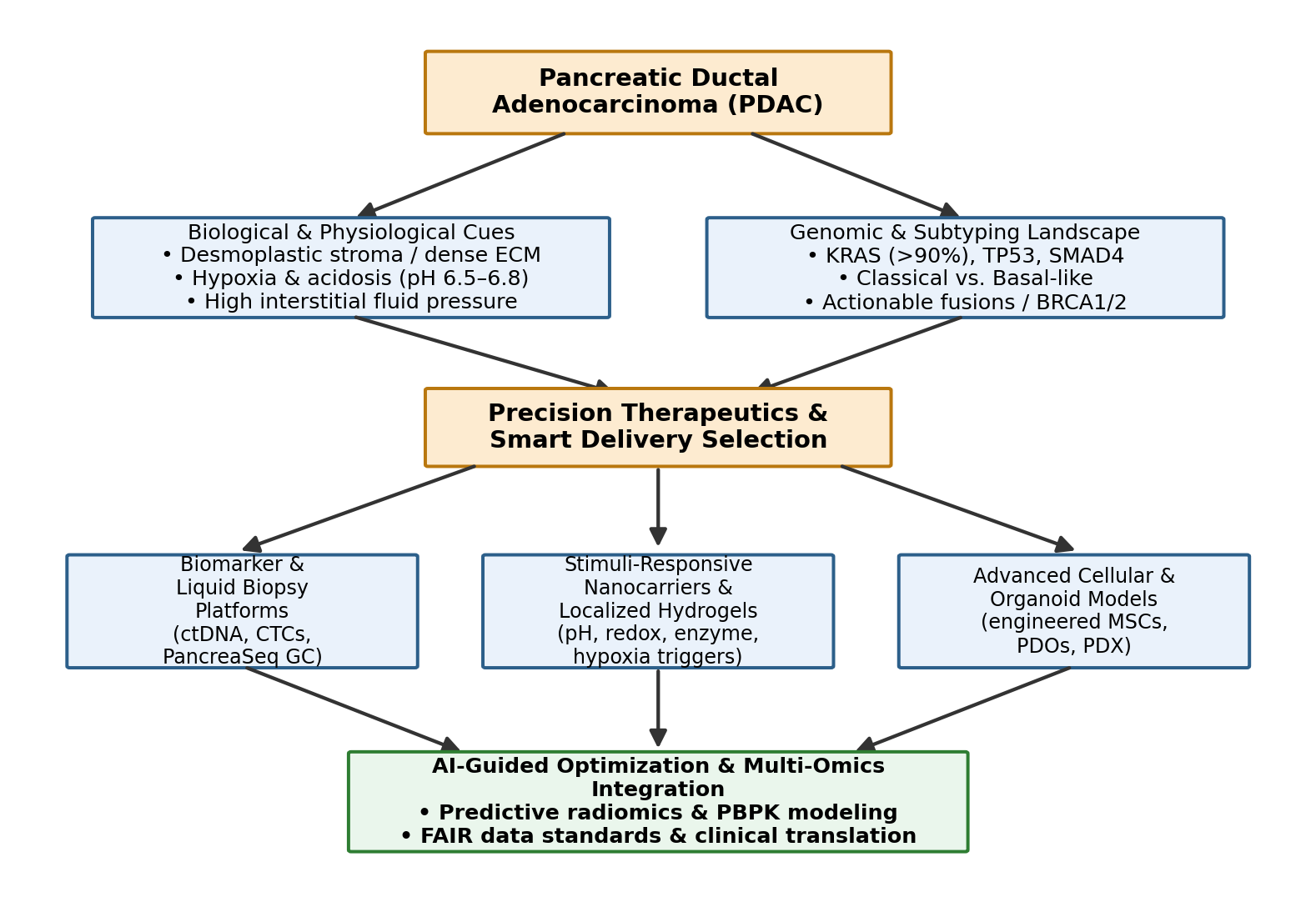
It is worth pausing, before getting into delivery systems and algorithms, on just how unforgiving PDAC's underlying biology actually is. Five-year survival has stagnated between roughly 10% and 13% for years now, notwithstanding real, incremental progress elsewhere in the field (Murthy et al., 2026; Siegel et al., 2026). Several factors converge to produce this grim number: an insidious, largely asymptomatic onset; rapid systemic progression once symptoms appear; and a level of intrinsic resistance to standard regimens like FOLFIRINOX or gemcitabine/nab-paclitaxel that other solid tumors simply do not exhibit to the same degree (Li et al., 2026; Wang et al., 2025). At the anatomical and cellular level, activated pancreatic stellate cells and cancer-associated fibroblasts secrete an extracellular matrix so dense - rich in fibrillar collagens, fibronectin, and hyaluronan - that it can account for 80% to 90% of total tumor mass (Kara et al., 2025; Li et al., 2026) (see Figure 1).

That fibrotic stroma is not passive scaffolding; it generates real mechanical solid stress, compressing the tumor's own microvasculature and pushing interstitial fluid pressure up to somewhere between 10 and 40 mmHg (Kara et al., 2025; Meskher et al., 2026). The downstream effects cascade from there - macromolecular therapeutics get trapped in perivascular spaces rather than penetrating the tumor core, while the tissue itself becomes hypoxic, acidic (pH roughly 6.5-6.8), and immunologically excluded (Kara et al., 2025; Li et al., 2026). None of this is compatible with a "one-size-fits-all" systemic therapy model, which is really the underlying reason the field has been pushed, somewhat out of necessity, toward precision oncology - toward therapies tailored to the molecular, cellular, and microenvironmental signature of each patient's tumor (Murthy et al., 2026; Wang et al., 2025).

2.2. Genomic Drivers, Molecular Taxonomy, and the Liquid Biopsy Revolution

The molecular picture underneath PDAC is, in one sense, remarkably consistent across patients, even as the clinical course varies enormously. Activating KRAS mutations occur in over 90-95% of cases, functioning as the near-universal initiating event that drives unchecked proliferation through persistent MAPK and PI3K/AKT signaling (Li et al., 2026; Wang et al., 2025). Layered on top of that are inactivating mutations in key tumor suppressors - TP53 in roughly 75% of tumors, CDKN2A in 40-60%, SMAD4 in about 50% - each pushing the disease further toward cell cycle dysregulation, apoptotic evasion,

Figure 1. Conceptual pipeline linking PDAC's biological and genomic landscape to precision therapeutic selection. The diagram traces the path from core disease biology (desmoplastic stroma, hypoxia, high interstitial fluid pressure) and genomic subtyping (KRAS, TP53, SMAD4, Classical versus Basal-like subtypes) through precision therapeutic and smart-delivery selection, culminating in biomarker/liquid-biopsy platforms, stimuli-responsive nanocarriers, and organoid-based validation that are ultimately integrated through AI-guided optimization and multi-omics data fusion.

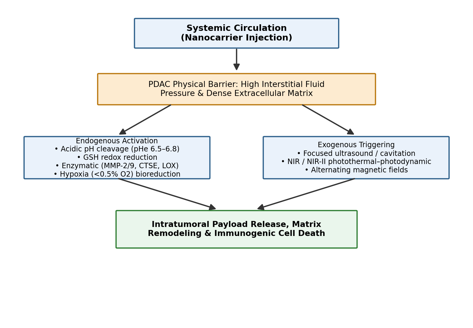
Figure 2. Endogenous and exogenous activation pathways governing stimuli-responsive nanocarrier drug release within the PDAC tumor microenvironment. After systemic injection, nanocarriers must first cross the physical barrier posed by high interstitial fluid pressure and dense extracellular matrix before being activated by either endogenous cues (acidic pH, glutathione-driven redox reduction, enzymatic cleavage, or hypoxia) or exogenous physical triggers (focused ultrasound, NIR-II light, or alternating magnetic fields), converging on intratumoral payload release, matrix remodeling, and immunogenic cell death.


and metastatic spread (Murthy et al., 2026; Wang et al., 2025). Beyond this mutational core, transcriptomic profiling efforts (Collisson, Moffitt, Bailey, and TCGA among them) have converged on two broad epithelial subtypes: Classical, or Pancreatic Progenitor, tumors, which express differentiation markers like GATA6 and tend to be more chemosensitive; and Basal-like, or Squamous, tumors, enriched for mesenchymal and EMT programs, which are considerably more chemoresistant and carry a worse prognosis (Murthy et al., 2026) (Figure 1).

Translating this molecular landscape into actual treatment decisions requires accurate, timely patient stratification (Murthy et al., 2026). Direct inhibitors against KRAS G12C (sotorasib, adagrasib) and newer switch-II pocket inhibitors against KRAS G12D (MRTX1133) are beginning to change what "undruggable" means in this disease, while KRAS wild-type tumors - a smaller but clinically important subset - can sometimes be matched to NTRK fusion inhibitors (larotrectinib), BRAF V600E-directed therapy, or PARP inhibitors like olaparib for BRCA1/2-mutant disease (Murthy et al., 2026).

Because CA19-9, the traditional serum biomarker, simply is not sensitive or specific enough for early detection or real-time monitoring, liquid biopsy has emerged - somewhat out of necessity - as a critical complementary tool (Murthy et al., 2026; Raufi et al., 2023). Serial profiling of circulating tumor DNA, circulating tumor cells, extracellular vesicles, and microRNA panels allows clinicians to track minimal residual disease, catch recurrence months before it would show up on imaging, and flag emerging resistance mechanisms early (Chen et al., 2026; Murthy et al., 2026; Yin et al., 2025). International consortia - GUIDE.MRD and FRENCH.MRD.PDAC validating ctDNA-guided adjuvant decisions, PANCAID developing an AI-assisted multi-omics blood test, PANLIPSY prospectively integrating multi-analyte panels in at-risk cohorts - are gradually standardizing these platforms for clinical use (Bardol et al., 2024; Murthy et al., 2026). In parallel, genomic classifiers such as PancreaSeq GC, applied to pancreatic cyst fluid, have meaningfully improved diagnostic accuracy for mucinous precursor lesions like IPMNs before they progress to invasive disease (Paniccia et al., 2023; Singhi et al., 2026) (Table 1).

2.3. Overcoming the Stromal Fortress: Smart and Adaptive Nanomedicine Platforms

Given a tumor that so actively resists passive drug entry, delivery systems have had to evolve from simply accumulating in tumor tissue toward actively navigating - and in some cases actively dismantling - the barriers described above (Kara et al., 2025; Li et al., 2026). The clinically approved formulations mentioned earlier, nab-paclitaxel and liposomal irinotecan, rely substantially on passive EPR-mediated accumulation, extended circulation, and reduced off-target toxicity (Kunzmann et al., 2021; Milano et al., 2022; Wang-Gillam et al., 2016). But because PDAC's hypovascular, heterogeneous stroma blunts EPR far more than it does in many other tumor types, the next generation of smart polymeric nanoparticles leans instead on active, receptor-mediated transcytosis and on stimuli-responsive activation triggered by the tumor microenvironment itself (Kara et al., 2025; Li et al., 2026).

These smart polymeric nanoparticles are functionalized with targeting ligands - antibodies, aptamers, peptides - directed at biomarkers overexpressed on tumor cells (EGFR, CD44, MSLN), cancer stem cells, or stromal components such as fibroblast activation protein on cancer-associated fibroblasts (Kara et al., 2025; Li et al., 2026). Several distinct triggering strategies have emerged, each exploiting a different feature of the hostile PDAC microenvironment, and it is worth walking through them individually because they are not interchangeable (Figure 2):

pH-responsive systems exploit the extracellular acidosis characteristic of the tumor (pHe roughly 6.5-6.8) through acid-labile linkers - acetal, amide, hydrazone bonds - or protonatable polymer backbones, converting an otherwise neutral, stealthy carrier into a positively charged particle once it reaches acidic tumor tissue, which in turn promotes cellular uptake and deeper matrix penetration (Ahmed et al., 2026; Kara et al., 2025; Li et al., 2026).

Redox-responsive systems take advantage of a rather elegant biochemical gradient: extracellular glutathione concentrations are low, while intracellular concentrations inside cancer cells run into the millimolar range. Disulfide bonds built into the carrier remain stable in circulation but cleave rapidly once inside the cell, releasing payload directly into the cytosol (Bhairam et al., 2026; Kara et al., 2025; Li et al., 2026).

Enzyme-responsive systems rely on TME enzymes that are overexpressed in PDAC specifically - matrix

Table 1. Molecular targets, diagnostic biomarkers, and precision therapeutic modalities in pancreatic ductal adenocarcinoma (PDAC). The table summarizes six major biomarker classes identified in the reviewed literature, spanning KRAS and DNA-damage-repair mutations to liquid-biopsy analytes and cyst-fluid genomic classifiers. For each class, the prevalence, associated diagnostic modality, matched targeted therapeutic, and current clinical status are reported to illustrate how molecular subtyping increasingly informs biomarker-matched treatment selection in PDAC.

Molecular Target / Biomarker Class

Prevalence & Biological Role

Diagnostic Modality

Targeted Therapeutics

Clinical Status

Oncogenic KRAS (G12D, G12C, G12R, G12V)

>90-95% of cases; drives MAPK/PI3K-AKT signaling and desmoplasia (Drizyte-Miller et al., 2025; Murthy et al., 2026)

NGS of tissue/EUS-FNB; ultra-deep ctDNA sequencing (Murthy et al., 2026; Yousef et al., 2025)

Sotorasib, adagrasib (G12C); MRTX1133 (G12D); AMPLIFY-201 mKRAS vaccine (Murthy et al., 2026; Pant et al., 2024)

Shifts KRAS from undruggable to targetable; combination strategies under evaluation (Murthy et al., 2026)

DDR deficiencies (BRCA1/2, PALB2, ATM)

~5-10% of patients; homologous recombination repair defects (Kindler et al., 2022; Perkhofer et al., 2017)

Germline testing; NGS panels (FoundationOne, MSK-IMPACT) (Murthy et al., 2026; Reiss et al., 2021)

PARP inhibitors (olaparib, rucaparib); platinum-based chemotherapy (Kindler et al., 2022)

Olaparib extended PFS in BRCA-mutant PDAC in the POLO trial (Kindler et al., 2022)

dMMR / MSI-H & SWI/SNF alterations

<1% of PDAC; altered epigenetic and immune states (Botta et al., 2021; Murthy et al., 2026)

IHC for MMR proteins; NGS-based MSI scoring (Souza da Silva et al., 2024)

Pembrolizumab and combination checkpoint inhibitors (Hilmi et al., 2023)

Regulatory approval established for dMMR/MSI-H solid tumors (Coston et al., 2023)

Rare actionable alterations (NTRK, BRAF V600E, MTAP deletion)

<2-5% of KRAS wild-type tumors (Murthy et al., 2026; Nikas et al., 2022)

RNA-seq fusion panels; targeted NGS; FISH (Murthy et al., 2026)

Larotrectinib, entrectinib; dabrafenib+trametinib; PRMT5/MAT2A inhibitors (Hu et al., 2021)

High objective response in biomarker-matched patients (O'Reilly & Hechtman, 2019)

Liquid biopsy analytes (ctDNA, CTCs, EVs, miRNA)

Real-time systemic sampling of tumor evolution and MRD (Chen et al., 2026; Raufi et al., 2023)

Methylation profiling (PDACatch); cfDNA fragmentomics; ddPCR (Wu et al., 2022; Yin et al., 2025)

Informs adjuvant therapy timing and drug switching (Chen et al., 2026)

Detects recurrence months before imaging (Murthy et al., 2026)

Pancreatic cyst fluid classifier (PancreaSeq GC)

Sequential alterations in IPMN/MCN precursor lesions (Paniccia et al., 2023)

EUS-FNA cyst fluid NGS (Singhi et al., 2026)

Guides risk-stratified resection vs. surveillance (Paniccia et al., 2023)

Improved sensitivity/specificity over CEA/imaging alone (Singhi et al., 2026)

Table 2. Endogenous and exogenous stimuli-responsive nanocarrier triggering mechanisms engineered to overcome the desmoplastic PDAC microenvironment. Rows are organized by trigger class (pH, redox, enzyme, hypoxia, exogenous physical, and localized depot systems), with representative named platforms, their underlying biological mechanism, and the delivery or therapeutic outcome reported for each system in the synthesized literature. The table is intended to show, side by side, how distinct microenvironmental cues are being converted into precise, site-specific drug release strategies.

Trigger Class

Mechanism

Representative Platform

Biological Effect

Reported Outcome

pH-responsive (endogenous)

Acid-labile linkers / protonatable backbones activate at pHe 6.5-6.8 (Kara et al., 2025)

DMMA-PLGA-PEI micelles; iCluster dendrimer (Kara et al., 2025; Li et al., 2025)

Charge reversal and size shrinkage (100 nm to 5 nm) for deep ECM penetration (Li et al., 2026)

Enhanced cytosolic delivery of platinum prodrugs (Li et al., 2026)

Redox / GSH-responsive (endogenous)

Disulfide/diselenide cleavage at intracellular GSH (2-10 mM) (Kara et al., 2025)

PAMAM dendrimer nanogels; PDSA cores (Kara et al., 2025; Li et al., 2025)

GSH depletion and elevated ROS (Li et al., 2026)

Targeted cytosolic siRNA delivery (Li et al., 2026)

Enzyme-responsive (endogenous)

Cleavage by MMP-2/9, cathepsin E, or lysyl oxidase (Kara et al., 2025)

AuHQ gold nanoparticles; LOXAb-NPs (Li et al., 2026)

Nanoparticle aggregation for PTT/imaging; reduced collagen crosslinking (Li et al., 2025)

Improved perfusion and stromal softening (Li et al., 2026)

Hypoxia-responsive (endogenous)

Bioreductive cleavage of nitroaromatic/azobenzene linkers under <0.5% O2 (Kara et al., 2025)

s(DGL)n@Apt aptamer-coated nanoparticle (Li et al., 2026)

Aptamer shedding; STAT3 inhibitor release (Li et al., 2026)

Molecular softening of fibrotic ECM (Li et al., 2026)

Exogenous physical (US / NIR-II / AMF)

Acoustic cavitation, NIR photothermal/photodynamic activation, magnetic actuation (Li et al., 2026)

Col-H-TiO2 nanoremodelers; M1@PAP; TPC CNPs (Li et al., 2026)

Endothelial gap opening, ICD induction, ROS/hyperthermia (Li et al., 2026)

Tumor regression under image guidance in preclinical models (Li et al., 2026)

Localized depots (non-systemic)

Sustained regional release from injectable matrices (Wang et al., 2025)

PPP thermoresponsive hydrogel; post-resection immunostimulatory hydrogel (Wang et al., 2025)

Weeks-long gemcitabine/cytokine release at the tumor site (Wang et al., 2025)

Reduced local recurrence and pancreatic fistula incidence (Wang et al., 2025)

metalloproteinases MMP-2/9, hyaluronidase, cathepsin E, lysyl oxidase - to degrade protective shells or cleave peptide tethers, achieving genuinely site-specific matrix remodeling alongside drug release (Kara et al., 2025; Li et al., 2026).

Hypoxia-responsive systems, meanwhile, incorporate nitroaromatic or azobenzene linkers that undergo enzymatic reduction only under the extreme hypoxia (below roughly 0.5% oxygen) found in avascular tumor cores, triggering disassembly precisely where oxygen tension is lowest (Kara et al., 2025; Li et al., 2026).

Alongside these endogenous triggers, exogenous physical modalities - focused ultrasound, near-infrared (NIR/NIR-II) light, alternating magnetic fields - offer a complementary and arguably more controllable route to spatiotemporal precision, driving local hyperthermia, photodynamic reactive oxygen species generation, and acoustic cavitation that physically loosens dense stroma (Li et al., 2026).

Nanoparticles are not the only tool in this toolbox, either. Three-dimensional hydrogels have emerged as versatile, localized depots, administered directly into tumor tissue or applied to resection margins via endoscopic ultrasound or intraoperative placement (Wang et al., 2025). These depots sustain high local concentrations of chemotherapeutics, STING agonists, or RNA-loaded lipid nanoparticles over weeks while limiting systemic exposure - and, notably, helping prevent local recurrence or fistula formation after pancreatectomy (Hasan et al., 2026; Wang et al., 2025). Even further afield from conventional nanoparticles, engineered stem-cell platforms - mesenchymal stem cells, neural stem cells, iPSCs - exploit their natural tumor-homing tropism via chemokine signaling (CXCR4-CXCL12 among others) to actively infiltrate PDAC stroma, and can be genetically engineered to locally secrete pro-apoptotic ligands, immunostimulatory cytokines, or matrix-degrading enzymes, effectively converting a "cold" tumor into something more treatment-responsive (Khan et al., 2026).

2.4. Computational Intelligence: AI-Guided Design, Radiomics, and Functional Models

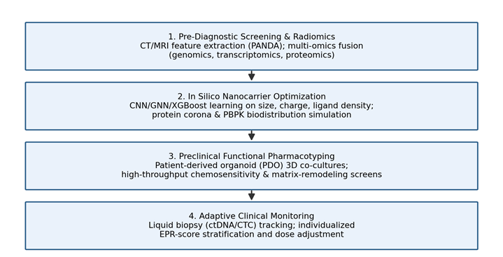
If the preceding section described what smart nanocarriers are built to do, this one turns to how artificial intelligence is reshaping the process of designing them in the first place. The core shift is away from empirical, trial-and-error formulation and toward predictive design, driven by machine learning and deep learning methods applied across essentially the entire discovery pipeline (Barua et al., 2026; Grumezescu et al., 2025; Li et al., 2026). Support Vector Machines, Random Forests, XGBoost, Artificial Neural Networks, Convolutional Neural Networks, and Graph Neural Networks are all in active use, each suited to somewhat different tasks (Barua et al., 2026; Grumezescu et al., 2025; Long et al., 2024) (Figure 3; Table 3).

In nanocarrier synthesis specifically, machine learning models take high-dimensional physicochemical descriptors - core composition, particle diameter, surface charge, polydispersity index, ligand density - and use them to predict functional outcomes such as encapsulation efficiency, colloidal stability, cellular endocytosis, and endosomal escape, generally with reasonably good accuracy for narrow, well-defined prediction tasks (Grumezescu et al., 2025; Hoseini et al., 2023; Li et al., 2026).

Coupling these models with physiologically based pharmacokinetic simulation extends the prediction further outward, toward whole-organism behavior: systemic organ biodistribution, reticuloendothelial clearance rates, and protein corona formation across simulated patient populations (Chou et al., 2023; Lin et al., 2022; Ouassil et al., 2022). Separately, deep learning radiomics models process non-contrast CT, MRI, and multimodal electronic health record data to surface subtle pre-diagnostic imaging biomarkers, estimate individual EPR effect magnitude, and stratify patients by likely treatment response (Barua et al., 2026; Cao et al., 2023; Setia et al., 2025). The PANDA (Pancreatic Cancer Detection with Artificial Intelligence) framework is a good illustration here - a deep learning system trained on non-contrast CT that has demonstrated notably high sensitivity and specificity for occult early-stage lesions, outperforming conventional radiological assessment in reported evaluations (Cao et al., 2023; Murthy et al., 2026).

None of this computational work means much clinically, though, unless it can be validated against genuinely human-relevant biology - which is where functional precision oncology platforms come in, attempting to bridge the historically wide gap between preclinical prediction and clinical trial outcome (Murthy et al., 2026). Patient-derived organoids and patient-derived xenografts preserve three-dimensional architecture, the primary tumor's mutational signature (KRAS, TP53, SMAD4), transcriptomic subtype,

Figure 3. Four-stage artificial intelligence and computational pipeline supporting precision nanomedicine design in PDAC. The stages proceed sequentially from pre-diagnostic radiomic and multi-omic screening, through in silico nanocarrier formulation optimization using CNN/GNN/XGBoost architectures, to preclinical functional pharmacotyping using patient-derived organoids, and finally to adaptive clinical monitoring via liquid biopsy and individualized EPR-score-guided dosing.

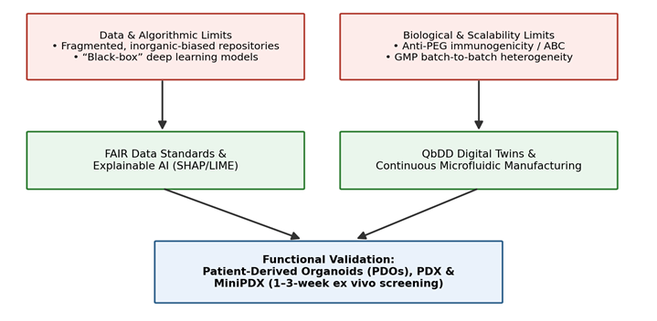
Figure 4. Roadmap for resolving data-centric, algorithmic, biological, and manufacturing bottlenecks limiting the clinical translation of AI-guided PDAC nanotherapeutics. Data and algorithmic limitations (fragmented, inorganic-biased repositories; black-box deep learning) and biological/scalability limitations (anti-PEG immunogenicity; GMP batch heterogeneity) are each paired with a corresponding mitigation strategy (FAIR data standards and Explainable AI; Quality-by-Digital-Design manufacturing), both converging on patient-derived organoid and xenograft platforms as a shared functional validation layer.

and stromal heterogeneity far more faithfully than standard 2D culture (Fawzy et al., 2026; Khan et al., 2026). High-throughput organoid pharmacotyping can return actionable chemosensitivity data within one to three weeks - fast enough, potentially, to inform treatment decisions for chemo-refractory patients and to shape adaptive clinical trial design before systemic therapy even begins (Khan et al., 2026; Murthy et al., 2026).

2.5. Translational Bottlenecks and Strategic Horizons

For all of this progress - and it is real progress - several stubborn bottlenecks still stand between AI-designed, microenvironment-responsive nanotherapeutics and routine clinical use in PDAC (Esmaeilpour et al., 2026; Li et al., 2026) (Figure 4; Table 4). The most fundamental is, again, data: public nanomedicine repositories remain fragmented and heavily skewed toward inorganic materials, while the soft-matter lipid and polymeric carriers most relevant to PDAC delivery lack standardized, FAIR-compliant reporting (Grumezescu et al., 2025; Mendes et al., 2024). Layered on top of that is the interpretability problem - many of the deep learning models doing the predictive heavy lifting function as black boxes, which is a genuine obstacle to both clinical trust and regulatory approval, not merely an academic inconvenience (Barua et al., 2026; Blanco-González et al., 2023).

Biologically and industrially, the picture is similarly mixed. Simplified 2D cultures and subcutaneous murine xenografts do not really reproduce the mechanical solid stress and dense desmoplasia that define human PDAC, so computational predictions calibrated against these models can drift away from what actually happens in patients (Li et al., 2026; Meskher et al., 2026). Unintended immunogenicity - anti-PEG IgM antibody induction leading to accelerated blood clearance, for instance - adds a further layer of risk that is not always caught early (Hasan et al., 2026; Zhang et al., 2016). And scaling multi-component, stimuli-responsive nanoassemblies up to GMP-compliant, batch-consistent manufacturing remains, frankly, a substantial industrial challenge in its own right (Jameel et al., 2026; Li et al., 2026).

None of these problems looks unsolvable in isolation, but addressing them will require a genuinely cross-disciplinary roadmap: multi-institutional, FAIR-compliant biobanks; microfluidic, automated synthesis suited to continuous GMP manufacturing; vascularized organ-on-a-chip and humanized PDX models that better approximate human biology; and Explainable AI frameworks built into the design pipeline from the outset rather than bolted on afterward (Barua et al., 2026; Li et al., 2026). It is this kind of integration - computational design, material innovation, and functional tumor biology working together rather than in parallel - that we believe offers the most realistic route from experimental nanotherapeutics to genuine bedside impact.

3. Methods

3.1. Review Design and Reporting Standards

This work is a narrative-synthesis literature review rather than a formal systematic review or meta-analysis, and we want to be upfront about that distinction rather than overstate our methodology. That said, we tried, wherever it was feasible, to borrow the transparency and reproducibility conventions of PRISMA-style systematic reviews (search strategy documentation, explicit inclusion and exclusion criteria, structured data extraction) so that another researcher could, in principle, reconstruct our search and arrive at a substantially similar evidence base (Page et al., 2021, as applied methodologically). We report the process below in enough detail that it should be reproducible by an independent reviewer using the same databases and search terms.

3.2. Information Sources and Search Strategy

We searched PubMed/MEDLINE, Scopus, Web of Science, and, for the most recent preprint literature not yet indexed in these databases, medRxiv and bioRxiv, covering records published between January 2015 and the most recent date practicable prior to manuscript submission, with particular emphasis on literature from 2020 onward given the rapid pace of change in both AI methodology and nanomedicine formulation science. Search strings combined controlled vocabulary (MeSH terms where applicable) with free-text keywords across three conceptual domains, connected with Boolean operators: (1) disease terms - "pancreatic ductal adenocarcinoma," "pancreatic cancer," "PDAC"; (2) delivery-system terms - "nanoparticle," "nanomedicine," "drug delivery," "liposome," "polymeric nanoparticle," "stimuli-responsive," "hydrogel," "lipid nanoparticle"; and (3) computational terms - "artificial intelligence," "machine learning," "deep learning," "neural network," "predictive modeling," "in silico." A representative PubMed query took the form: ("pancreatic ductal adenocarcinoma" OR "pancreatic cancer") AND ("nanoparticle" OR "nanomedicine" OR "drug delivery system") AND ("artificial intelligence" OR "machine learning" OR "deep learning"), with equivalent syntax adapted for each database's field-tag conventions. Reference lists of key retrieved articles and recent reviews were additionally hand-searched (backward citation chasing) to identify further eligible studies not captured by the primary search strings.

3.3. Eligibility Criteria

We included peer-reviewed primary research articles, systematic reviews, and high-quality narrative reviews published in English that addressed at least one of the following: (a) the biological, physiological, or genomic barriers to systemic drug delivery in PDAC; (b) the design, synthesis, or preclinical/clinical evaluation of nanoparticle- or hydrogel-based delivery systems for pancreatic cancer; or (c) the application of AI, machine learning, or deep learning methods to nanoparticle design, formulation optimization, biodistribution prediction, or patient stratification in oncology, with priority given to pancreatic cancer-specific applications where available. We excluded conference abstracts without full-text availability, non-English publications, case reports lacking generalizable methodological detail, and articles focused exclusively on cancers other than PDAC unless they described a computational or nanomedicine methodology of direct, explicit relevance to pancreatic cancer applications discussed elsewhere in the retrieved literature.

3.4. Study Selection and Data Extraction

Titles and abstracts identified through the search strategy above were screened for topical relevance against the eligibility criteria; full texts of potentially eligible articles were then retrieved and assessed in full. For each included study, we extracted, where reported: study design and model system (in silico, in vitro, murine, organoid/PDX, or clinical); nanoparticle or delivery-platform composition and targeting strategy; the specific AI/ML architecture employed and its input feature descriptors; reported performance metrics (e.g., R², accuracy, mean absolute error, mean squared error, sensitivity/specificity); and, where applicable, clinical trial identifiers and regulatory status. Extracted data were organized thematically into the four synthesis domains that structure this review - biological/genomic barriers, stimuli-responsive delivery platforms, computational modeling architectures, and translational bottlenecks - and are summarized in Tables 1 through 4.

3.5. Synthesis Approach and Reproducibility

Given the substantial methodological heterogeneity across the included literature - spanning wet-lab formulation studies, computational modeling papers, and clinical trial reports - a quantitative meta-analysis was not appropriate, and we did not attempt one. Instead, we performed a structured narrative synthesis, organizing findings by conceptual domain and cross-referencing quantitative performance metrics (model accuracy, R² values, clinical trial identifiers) directly in the text and summary tables so that specific claims can be traced back to their source and, in principle, independently verified against the cited primary literature. All sources synthesized in this review are cited in APA 7th edition format in-text and compiled in the reference list; no data external to the cited literature were generated, and no original wet-laboratory or animal experiments were performed as part of this review.

4. Synthesis of AI-Guided Nanoparticle Design Capabilities and Limits Across the PDAC Delivery Pipeline

4.1. Molecular Subtyping, Actionable Targets, and Diagnostic Biomarker Innovations

Taken together, the literature synthesized here paints a picture of a field that has made real, measurable progress at the molecular level even as overall survival numbers have moved only modestly. Genomic sequencing has firmly established that PDAC tumorigenesis follows a near-universal mutational cascade dominated by oncogenic KRAS alterations, present in more than 90-95% of cases, alongside inactivation of TP53 (75%), CDKN2A (40-60%), and SMAD4 (50%) (Li et al., 2026; Murthy et al., 2026; Wang et al., 2025). Allele-specific analysis further resolves KRAS variants into distinct missense subtypes - G12D at roughly 40%, G12V at 30%, G12R at 15%, and Q61H at 5% - a level of granularity that matters clinically, since allele-specific inhibitors (sotorasib and adagrasib for G12C, MRTX1133 for G12D) are only effective against the variant they target (Murthy et al., 2026) (Table 1).

Beyond single-gene drivers, integrated transcriptomic classification - drawing on TCGA and several international consortia - consistently separates PDAC into Classical (Pancreatic Progenitor) and Basal-like (Squamous) subtypes, distinguished respectively by GATA6/FOXA2 expression and relative chemosensitivity versus basal keratin expression, EMT programming, and pronounced chemoresistance (Murthy et al., 2026). This subtyping is not merely academic; it increasingly informs regulatory-approved, biomarker-matched therapy. Maintenance olaparib meaningfully extended progression-free survival in germline BRCA1/2-mutant PDAC in the Phase III POLO trial (Kindler et al., 2022; Murthy et al., 2026), while mismatch repair-deficient or microsatellite instability-high tumors - admittedly under 1% of PDAC cases - show durable responses to pembrolizumab (Coston et al., 2023; Hilmi et al., 2023; Murthy et al., 2026). In KRAS wild-type disease, targeting NTRK fusions, BRAF V600E, or MTAP co-deletion (via synthetic lethality with PRMT5/MAT2A inhibition) has produced clinically meaningful responses, reinforcing the case for routine, broad-panel genomic profiling rather than single-gene testing (Drizyte-Miller et al., 2025a; Hu et al., 2021; Murthy et al., 2026; O'Reilly & Hechtman, 2019).

On the diagnostic side, liquid biopsy platforms have moved from research curiosity to genuine clinical tool, at least in the settings where they have been most extensively validated. Multi-parametric profiling of ctDNA, CTCs, and extracellular vesicles enables minimal residual disease assessment and recurrence detection that can precede radiographic visualization by months (Chen et al., 2026; Murthy et al., 2026). Assays such as the PDACatch cfDNA methylation panel and fragmentomics-based classifiers report high specificity for distinguishing early PDAC from non-malignant controls (Wu et al., 2022; Yin et al., 2025), while the PancreaSeq Genomic Classifier, applied to EUS-FNA cyst fluid, has improved risk stratification for mucinous precursor lesions such as IPMNs (Paniccia et al., 2023; Singhi et al., 2026). International initiatives - GUIDE.MRD, FRENCH.MRD.PDAC, PANCAID, PANLIPSY - are now working to standardize these blood-based, multi-omics signatures across institutions (Bardol et al., 2024; Murthy et al., 2026) (Table 1).

4.2. Microenvironment-Responsive Nanomedicine Platforms and Localized Depots

Systemic delivery in PDAC is, as established above, fundamentally constrained by anatomical and physical barriers rather than by any deficiency in the drugs themselves (Kara et al., 2025; Li et al., 2026; Wang et al., 2025). Given that the desmoplastic stroma can occupy 80-90% of total tumor volume and drives interstitial fluid pressures of 10-40 mmHg - enough to substantially blunt passive EPR-mediated accumulation - the delivery platforms reviewed here have had to become considerably more sophisticated than "accumulate and hope" (Kara et al., 2025; Li et al., 2026; Meskher et al., 2026) (Figure 2; Table 2).

Several specific, well-characterized platform examples illustrate what this looks like in practice. DMMA-modified PLGA-PEI micelles switch from negative to positive surface charge specifically at tumor pH, strengthening membrane interaction and deep matrix penetration (Kara et al., 2025; Li et al., 2026); the "iCluster" platform undergoes pH-triggered size shrinkage from roughly 100 nm down to 5 nm dendrimer fragments to promote deep penetration and cytosolic delivery of platinum prodrugs (Kara et al., 2025; Li et al., 2026). On the redox-responsive side, polyamidoamine dendrimer nanogels and polydithioamide cores deplete intracellular glutathione, elevate reactive oxygen species, and achieve targeted cytosolic siRNA delivery (Kara et al., 2025; Li et al., 2026). Enzyme-responsive systems are similarly specific: the AuHQ platform relies on cathepsin E cleavage of a defined peptide sequence to trigger gold nanoparticle aggregation for near-infrared photothermal therapy and simultaneous imaging, while LOX-targeted antibody-functionalized nanoparticles inhibit collagen crosslinking to reduce stromal stiffness and improve perfusion (Li et al., 2026). Under hypoxia, the s(DGL)n@Apt system sheds its aptamer coating to release gemcitabine monophosphate conjugates alongside STAT3 inhibitors that molecularly soften fibrotic networks (Kara et al., 2025; Li et al., 2026).

Exogenous physical triggers add a further, largely complementary layer of control. Ultrasound-activated cavitation - using platforms such as Col-H-TiO2 nanoparticles or semiconductor polymer nanoremodelers - transiently opens endothelial junctions, disrupts stroma, and induces immunogenic cell death (Kara et al., 2025; Li et al., 2026). NIR-II-responsive systems, including M1 macrophage membrane-coated nanoparticles (M1@PAP) and conjugated small-molecule nanoparticles (TPC CNPs), generate Type I/II reactive oxygen species or localized hyperthermia under near-infrared light, with reported tumor regression under image guidance in preclinical models (Li et al., 2026).

Local, non-systemic delivery has also matured considerably. Injectable thermoresponsive hydrogels - PLGA-PEG-PLGA (PPP) matrices, for instance - administered via endoscopic ultrasound-guided fine-needle injection form sustained-release gemcitabine reservoirs directly within pancreatic tissue (Wang et al., 2025), while postoperative dual-crosslinked immunostimulatory hydrogels applied to resection margins release platelet-conjugated antibodies and cytokines to suppress local recurrence while reducing pancreatic fistula rates (Wang et al., 2025) (Table 2).

4.3. Artificial Intelligence Architectures and Predictive Modeling in Nanomedicine

Turning specifically to computational performance, the reported results across AI/ML modeling frameworks are genuinely encouraging in narrow, well-defined tasks, even if they do not yet add up to an integrated, end-to-end design pipeline (Table 3; Figure 3). Supervised tree-based methods - LightGBM, XGBoost, Random Forest - perform well at modeling formulation stability, particle size, and drug encapsulation efficiency; comparative benchmarking indicates LightGBM achieves particularly low mean absolute error (approximately 119-120) for nanocrystal sizing across independent evaluation datasets (Grumezescu et al., 2025). Kernel-based methods, particularly Support Vector Regression, have been used to predict protein corona adsorption with reported accuracy around 78% and low mean squared error (approximately 0.0016) for encapsulation efficiency modeling (Grumezescu et al., 2025).

Deep neural network architectures capture the nonlinear relationships that simpler models tend to miss. Trained across a substantial multi-organ mouse biodistribution dataset - spanning more than 500 tumor and 1,900 organ-level endpoints - DNN models identified an optimal hydrodynamic size window of roughly 50-75 nm, striking a balance between avoiding rapid renal filtration below about 6 nm and reticuloendothelial sequestration above roughly 100 nm (Meng et al., 2026). Convolutional neural network-based radiomics, exemplified by the PANDA framework, achieved reportedly high sensitivity and specificity for detecting occult pancreatic lesions from non-contrast CT, outperforming conventional radiological assessment in the evaluations described (Cao et al., 2023; Murthy et al., 2026). Graph neural networks, meanwhile, extend prediction into molecular structure space - the siRNADiscovery platform, for instance, uses message-passing GNNs to predict siRNA silencing efficacy directly from sequence features, screening over 1.6 million candidate lipid structures in silico to identify organ-selective lipid nanoparticle candidates (Long et al., 2024; Meskher et al., 2026).

Finally, hybrid AI-PBPK and n-QSAR frameworks integrate physicochemical descriptors with tissue perfusion, capillary permeability, and vascular stress parameters to simulate systemic biodistribution and reticuloendothelial clearance across species, reportedly achieving superior R² values relative to conventional PBPK modeling alone and, notably, reducing reliance on empirical animal testing at the formulation-screening stage (Chou et al., 2023; Grumezescu et al., 2025; Lin et al., 2022) (Table 3).

4.4. Translational Bottlenecks and Functional Preclinical Horizon

Set against these computational successes, several structural bottlenecks remain, and it would be misleading to present the field's progress without equal attention to its limits (Table 4; Figure 4). On the data side, existing nanomedicine repositories suffer from fragmentation, reporting heterogeneity, and a persistent bias toward inorganic materials, leaving soft-matter lipid and polymeric carriers - again, arguably the more clinically relevant category for PDAC - comparatively underrepresented (Grumezescu et al., 2025; Mendes et al., 2024). Implementing ontology-based FAIR standards and frameworks such as MIRIBEL reporting is, in our reading of the literature, a necessary rather than optional step toward building the kind of robust, multi-institutional training pipelines this field will eventually need (Grumezescu et al., 2025).

The interpretability problem compounds this. Multi-layer deep learning architectures frequently function as black boxes, which limits mechanistic interpretability and, in turn, complicates both clinical trust and regulatory dossier review (Barua et al., 2026; Blanco-González et al., 2023). Explainable AI methods - SHAP and LIME feature attribution being the most commonly reported - offer a partial remedy by providing human-interpretable rationale for algorithmic predictions, though their routine adoption in nanomedicine-specific pipelines remains, at present, more the exception than the rule (Grumezescu et al., 2025).

Biologically, repeated systemic administration of PEGylated nanocarriers can induce anti-PEG IgM antibodies, triggering accelerated blood clearance and, in some cases, hypersensitivity reactions - a real and under-discussed safety consideration for repeat-dosing regimens (Hasan et al., 2026; Zhang et al., 2016). And industrially, transitioning multi-component, stimuli-responsive nanostructures from bench-scale synthesis to continuous,

Table 3. Landscape of artificial intelligence and machine learning architectures applied to nanoparticle design and precision oncology. Six modeling families are compared according to their representative algorithms, the physicochemical or biological input descriptors they rely upon, the specific prediction task addressed, and the quantitative performance metrics reported in the source literature (R², accuracy, MAE, MSE). The table is meant to give readers a single reference point for comparing where each computational approach has demonstrated the strongest predictive capability.

AI/ML Framework

Representative Algorithms

Input Descriptors

Prediction Task

Reported Performance

Supervised tree-based ML

Random Forest, XGBoost, LightGBM

Particle size, zeta potential, PDI, core/ligand composition (Esmaeilpour et al., 2026)

Formulation stability; encapsulation efficiency

R² > 0.90 for green ZnO NP size; lowest MAE (~119-120) for nanocrystal sizing (Grumezescu et al., 2025)

Kernel-based ML

SVM, Support Vector Regression

Surface charge density, coating thickness, proteomics data (Grumezescu et al., 2025)

Protein corona composition; colloidal stability

78% accuracy; MSE 0.0016 for encapsulation efficiency (Grumezescu et al., 2025)

Deep neural networks

ANN, DNN, multilayer perceptrons

Formulation composition, sonication time, polymer MW (Grumezescu et al., 2025)

Hydrodynamic diameter; organ accumulation

R² = 0.966 (polymeric NP size); optimal 50-75 nm EPR window (Meng et al., 2026)

CNN / radiomics

Convolutional Neural Networks, GANs

Non-contrast CT scans; TEM/SEM images (Cao et al., 2023)

Automated early PDAC lesion detection

High sensitivity/specificity, outperforming radiologists (Cao et al., 2023)

Graph neural networks

GNN, message-passing neural networks

RNA sequence graphs; lipid/polysaccharide coating structures (Long et al., 2024)

siRNA silencing efficacy; lipid nanoparticle screening

Screened >1.6 million lipid structures in silico (Long et al., 2024; Meskher et al., 2026)

Hybrid AI-PBPK / n-QSAR

ML-augmented PBPK models

Size, charge, logP, tissue perfusion, vascular stress (Chou et al., 2023)

Systemic biodistribution; RES clearance

Superior R² across multi-organ predictions; reduced animal testing need (Chou et al., 2023; Lin et al., 2022)

Table 4. Key translational bottlenecks, associated platforms or trial initiatives, their underlying mechanism or clinical impact, and proposed mitigation strategies for advancing AI-guided nanomedicine toward clinical use in PDAC. Categories span functional preclinical models, international liquid-biopsy consortia, physical/immunological tumor barriers, manufacturing scalability, and computational/regulatory constraints, reflecting the five domains most frequently raised as obstacles in the reviewed literature.

Translational Category

Key Platforms / Bottlenecks

Mechanism / Impact

Mitigation Strategy

Functional precision models

Patient-derived organoids (PDOs), PDX, MiniPDX (Tiriac et al., 2018)

1-3 week ex vivo chemosensitivity testing guiding personalized therapy (Murthy et al., 2026)

Active validation trials (NCT04777604, NCT04736043) (Murthy et al., 2026)

International liquid biopsy initiatives

GUIDE.MRD, FRENCH.MRD.PDAC, PANCAID, PANLIPSY (Bardol et al., 2024)

Validates ctDNA-based MRD detection for adjuvant therapy guidance (Chen et al., 2026)

Prospective multi-parametric, multi-institutional standardization (Murthy et al., 2026)

Physical & immunological TME barriers

Dense desmoplasia; IFP 10-40 mmHg (Hosein et al., 2020)

Restricts macromolecular convection; drives hypoxia, acidosis, immune exclusion (Kara et al., 2025)

Stimuli-responsive nanoparticles; enzymatic matrix degradation; CAF reprogramming (Li et al., 2026)

Manufacturing & scalability barriers

Microfluidic nanoprecipitation; Quality by Digital Design (Jameel et al., 2026)

Batch-to-batch variability in size, charge, and encapsulation under GMP (Jameel et al., 2026)

Continuous-flow microfluidics; Process Analytical Technology; QbDD digital twins (Li et al., 2026)

Computational & regulatory bottlenecks

“Black-box” AI; FAIR data gaps; anti-PEG immunogenicity (Zhang et al., 2016)

Impairs clinician trust; delays regulatory dossier approval; accelerated blood clearance (Esmaeilpour et al., 2026)

Explainable AI (SHAP/LIME); MIRIBEL reporting standards; non-PEG stealth coatings (Grumezescu et al., 2025)

GMP-compliant manufacturing requires Quality by Digital Design and Process Analytical Technology frameworks capable of maintaining batch-to-batch consistency at scale - a challenge that, candidly, remains only partially solved (Jameel et al., 2026; Li et al., 2026).

Functional precision oncology platforms offer perhaps the most promising bridge across these gaps. Patient-derived organoids, patient-derived xenografts, and rapid MiniPDX platforms preserve primary tumor histology, transcriptomic subtype, and genomic mutational profile (KRAS, TP53, SMAD4) far better than 2D culture or subcutaneous xenografts (Fawzy et al., 2026; Khan et al., 2026; Tiriac et al., 2018). High-throughput organoid pharmacotyping can return actionable chemosensitivity profiles within one to three weeks, fast enough, at least in principle, to inform both personalized drug selection and prospective, adaptive clinical trial design (Khan et al., 2026; Murthy et al., 2026) (Table 4).

5. Reconciling Computational Promise with Clinical Reality in AI-Guided PDAC Nanomedicine

Stepping back from the individual findings summarized above, a fairly consistent pattern emerges across this literature, and we think it is worth stating plainly. Artificial intelligence has demonstrably improved the efficiency and precision of narrow, well-bounded prediction tasks within nanoparticle design - particle size, encapsulation efficiency, protein corona composition, biodistribution windows - each validated, at least to some degree, against experimental or clinical benchmarks (Grumezescu et al., 2025; Meng et al., 2026) (Table 3). What AI has not yet done, at least not convincingly across the literature we reviewed, is deliver an integrated, end-to-end design-to-clinic pipeline that spans molecular target selection, nanocarrier engineering, and patient stratification within a single validated framework. The field, in other words, has assembled a genuinely impressive set of components without yet having fully assembled the machine.

This gap matters clinically because PDAC's biology is precisely the kind of problem where an integrated approach would help most. The disease's molecular heterogeneity - KRAS-driven, Classical versus Basal-like, occasionally BRCA- or NTRK-actionable (Murthy et al., 2026) - argues for nanocarriers whose targeting strategy is matched not just to generic tumor markers but to a specific patient's subtype and mutational profile. Some of the platforms reviewed in Section 4.2 - stimuli-responsive systems keyed to pH, redox state, or specific enzymes - already move in this direction, but their deployment has so far been largely disconnected from the genomic and liquid-biopsy stratification tools described in Section 4.1. Bringing these two threads together - using ctDNA-based subtyping, for instance, to select among a menu of stimuli-responsive nanocarriers - seems, to us, like one of the more tractable near-term opportunities in this field, and one that the current literature has not yet fully explored.

The interpretability and data-fragmentation problems documented in Section 4.4 deserve to be taken seriously rather than treated as footnotes, because they are not merely inconveniences - they are, in a real sense, gatekeeping issues. A model that cannot explain why it recommends a particular formulation is a model that a regulator is unlikely to approve and a clinician is unlikely to trust, however strong its reported R² (Barua et al., 2026; Blanco-González et al., 2023). We would go further and suggest that Explainable AI should not be treated as an optional add-on layered onto existing black-box architectures after the fact, but built into model development from the outset - a design philosophy rather than a post-hoc audit. Similarly, the dataset fragmentation problem (Grumezescu et al., 2025; Mendes et al., 2024) is not simply an inconvenience for researchers; it is a genuine structural constraint on what these models can learn, particularly for soft-matter lipid and polymeric systems that remain underrepresented relative to inorganic nanoparticles in most public repositories. Until FAIR-compliant, multi-institutional data infrastructure exists at meaningful scale, predictive models trained on today's fragmented data will likely continue to generalize imperfectly beyond the narrow conditions under which they were trained - a limitation that no amount of algorithmic sophistication can fully compensate for.

On the biological side, the persistent gap between preclinical prediction and human clinical outcome (Li et al., 2026; Mallya et al., 2021) points, we think, toward patient-derived organoid and xenograft platforms as an unusually promising near-term validation layer - not a replacement for computational prediction, but a faster, more human-relevant filter positioned between in silico design and first-in-human testing (Fawzy et al., 2026; Khan et al., 2026; Tiriac et al., 2018). A one-to-three-week organoid pharmacotyping turnaround is genuinely fast enough to be clinically actionable, and integrating this kind of functional readout as a routine validation step before nanoparticle formulations advance further would, in our view, meaningfully de-risk translation without materially slowing it down.

Manufacturing and immunogenicity concerns (Hasan et al., 2026; Jameel et al., 2026; Zhang et al., 2016) round out the picture, and here we admit some genuine uncertainty about how quickly the field can move. Quality by Digital Design and continuous microfluidic manufacturing are conceptually sound solutions to the batch-consistency problem, but their adoption at industrial scale for the complex, multi-component stimuli-responsive systems described in Section 4.2 remains, as far as we can tell from the available literature, largely aspirational rather than demonstrated at scale. Anti-PEG immunogenicity is a related concern that the field has, we would argue, somewhat under-addressed relative to its clinical significance, particularly for therapies intended for repeat dosing over extended treatment courses.

This review is not without limitations, and it seems only fair to name them directly. As a narrative synthesis rather than a formal systematic review, our literature identification, while structured and documented in Section 3, was not independently duplicated by multiple reviewers in the manner PRISMA guidelines would formally require, which introduces some risk of selection bias that a fully systematic approach would better control. Much of the AI performance literature synthesized here (Table 3) also reports internal validation metrics rather than independent external validation, meaning that reported accuracy figures should be interpreted as an upper bound on real-world performance rather than a guarantee of it. And because the field is moving quickly, some of the most recent preprint literature may not yet be fully represented, or may have since been revised prior to peer review.

Taken together, what we take from this literature is a picture of a field with real, demonstrated computational capability that has not yet been matched by equivalent clinical infrastructure - data standards, interpretability tooling, manufacturing scale, and validation pathways. Closing that gap looks, on the evidence reviewed here, less like a single breakthrough and more like sustained, coordinated investment across several fronts simultaneously.

6. Conclusion

AI-guided nanoparticle design has genuinely reshaped how researchers approach pancreatic cancer drug delivery, moving portions of the field from slow empirical iteration toward faster, computationally informed formulation. The evidence synthesized here shows real predictive strength for narrow tasks - particle sizing, biodistribution, protein corona behavior - alongside stimuli-responsive nanocarriers that meaningfully address PDAC's uniquely hostile microenvironment. Yet the translational path from algorithm to bedside remains incomplete. Fragmented datasets, black-box interpretability, anti-PEG immunogenicity, and GMP-scale manufacturing variability continue to limit clinical adoption, even where individual technical components perform well in isolation. Closing this gap will likely require FAIR-compliant data infrastructure, Explainable AI built in from the outset, organoid-based functional validation, and quality-by-design manufacturing working in concert rather than in isolation. We remain cautiously optimistic: the underlying science is sound, and the remaining barriers, while real, appear addressable with sustained cross-disciplinary effort.

References


Ahmed, S., Mujahid, M. H., Kour, M., Fatima, S., Ahamad, S., Mishra, S. K., Bhat, A. R., Abu-Rayyan, A., & Tebeje, B. A. (2026). Molecular design and therapeutic applications of polymeric nanoparticles for drug delivery: A comprehensive review. Results in Chemistry, 28, 103459. https://doi.org/10.1016/j.rechem.2026.103459        

Allen, T. M., & Cullis, P. R. (2004). Drug delivery systems: entering the mainstream. Science, 303(5665), 1818-1822. https://doi.org/10.1126/science.1095833              

Amin, M. B., Greene, F. L., Edge, S. B., Compton, C. C., Gershenwald, J. E., Brookland, R. K., Meyer, L., Gress, D. M., Byrd, D. R., & Winchester, D. P. (2017). The eighth edition AJCC cancer staging manual: continuing to build a bridge from a population-based to a more "personalized" approach to cancer staging. CA: A Cancer Journal for Clinicians, 67(2), 93-99. https://doi.org/10.3322/caac.21388       

Bardol, T., Dujon, A. M., Taly, V., Dunyach-Remy, C., Lavigne, J. P., Costa-Silva, B., Kurma, K., Eslami, S. Z., Cayrefourcq, L., Canivet, C., Muscari, F., Bournet, B., & Alix-Panabières, C. (2024). Early detection of pancreatic cancer by liquid biopsy "PANLIPSY": A french nation-wide study project. BMC Cancer, 24(1), 709. https://doi.org/10.1186/s12885-024-12463-8            

Barua, S., Balaji, B., & Balaji, S. (2026). Bridging diagnosis and therapeutics: The role of AI in cancer detection and drug development. Health Sciences Review, 19, 100270. https://doi.org/10.1016/j.hsr.2026.10027

Beam, A. L., & Kohane, I. S. (2018). Big data and machine learning in health care. JAMA, 319(13), 1317-1318. https://doi.org/10.1001/jama.2017.18391

Bhairam, M., Shukla, S. S., Pandey, R. K., & Gidwani, B. (2026). Stimuli-responsive nanomaterials in targeted drug delivery. OpenNano, 29, 100299. https://doi.org/10.1016/j.onano.2026.100299

Blanco-González, A., Cabezón, A., Seco-González, A., Conde-Torres, D., Antelo-Riveiro, P., Piñeiro, Á., & Garcia-Fandino, R. (2023). The role of AI in drug discovery: Challenges, opportunities, and strategies. Pharmaceuticals, 16(6), 891. https://doi.org/10.3390/ph16060891

Botta, G. P., Kato, S., Patel, H., Fanta, P., Banks, K. C., & Kurzrock, R. (2021). SWI/SNF complex gene alterations in pancreatic adenocarcinoma and response to immune checkpoint inhibition. JCO Precision Oncology, 5, 840-849.

Cai, H., Wang, R., Guo, X., Song, M., Yan, F., Ji, B., & Liu, Y. (2021). Combining gemcitabine-loaded macrophage-like nanoparticles and erlotinib for pancreatic cancer therapy. Molecular Pharmaceutics, 18(7), 2495-2506. https://doi.org/10.1021/acs.molpharmaceut.0c01225

Cao, K., Xia, Y., Yao, J., Han, X., Lambert, L., Zhang, T., Tang, W., Jin, G., Jiang, H., Fang, X., Nogues, I., Li, X., Guo, W., Wang, Y., Fang, W., Qiu, M., Hou, Y., Kovarnik, T., Vocka, M., … Lu, L. (2023). Large-scale pancreatic cancer detection via non-contrast CT and deep learning. Nature Medicine, 29(12), 3033-3043. https://doi.org/10.1038/s41591-023-02640-w

Carney, C. P., Pandey, N., Kapur, A., Saadi, H., Ong, H. L., Chen, C., Winkles, J. A., Woodworth, G. F., & Kim, A. J. (2023). Impact of targeting moiety type and protein corona formation on the uptake of Fn14-targeted nanoparticles by cancer cells. ACS Nano, 17(20), 19667-19684. https://doi.org/10.1021/acsnano.3c02575

Chen, J., Zou, L., Bai, X., Tong, F., Ni, J., Tang, H., Liu, Y., Kong, X., Yin, J., Wang, F., Sha, H., Meng, F., & Du, J. (2026). Personalized tumor-informed circulating tumor DNA monitoring for early detection of recurrence in postoperative pancreatic cancer. Frontiers in Oncology, 16, 1745466. https://doi.org/10.5772/intechopen.1008585

Ching, T., Himmelstein, D. S., Beaulieu-Jones, B. K., Kalinin, A. A., Do, B. T., Way, G. P., & Greene, C. S. (2018). Opportunities and obstacles for deep learning in biology and medicine. Journal of the Royal Society Interface, 15(141), 20170387. https://doi.org/10.1098/rsif.2017.0387

Chou, W. C., Chen, Q., Yuan, L., Cheng, Y. H., He, C., Monteiro-Riviere, N. A., Riviere, J. E., & Lin, Z. (2023). An artificial intelligence-assisted physiologically-based pharmacokinetic model to predict nanoparticle delivery to tumors in mice. Journal of Controlled Release, 361, 53-63. https://doi.org/10.1016/j.jconrel.2023.07.040

Coston, T., Desai, A., Babiker, H., Sonbol, M. B., Chakrabarti, S., Mahipal, A., McWilliams, R., Ma, W. W., Bekaii-Saab, T. S., Stauffer, J., & Starr, J. S. (2023). Efficacy of immune checkpoint inhibition and cytotoxic chemotherapy in mismatch repair-deficient and microsatellite instability-high pancreatic cancer: Mayo Clinic experience. JCO Precision Oncology, 7, e2200706. https://doi.org/10.1200/PO.22.00706

Drizyte-Miller, K., Engstrom, L. D., Klomp, J. A., Stalnecker, C. A., Stamey, A. G., Taylor, K. E., Roach, M. K., Robb, R., Waters, L. M., Calinisan, A., Degan, S., Chang, W. H., Helu, X. M., Nguyen, D., Baldelli, E., Pierobon, M., Petricoin, E. F., Briere, D. M., Hallin, J., … Der, C. J. (2025a). Combination of the MTA-cooperative PRMT5 inhibitor BMS-986504 and KRAS inhibitors is an effective treatment strategy for MTAP-deleted KRAS-mutant pancreatic cancer. Cancer Research, 85(18), 3540-3557. https://doi.org/10.1158/0008-5472.CAN-25-1507

Drizyte-Miller, K., Talabi, T., Somasundaram, A., Cox, A. D., & Der, C. J. (2025). KRAS: The Achilles' heel of pancreas cancer biology. Journal of Clinical Investigation, 135(16), e182900. https://doi.org/10.1172/JCI191939

Escalera-Anzola, S., Rosado, M., Yang, Y., Parra-Sanchez, D., Pedro-Liberal, C. S., & Acedo, P. (2025). Breakthroughs in nanoparticle-based strategies for pancreatic cancer therapy. Biochemical Pharmacology, 232, 116685. https://doi.org/10.1016/j.bcp.2024.116685

Esmaeilpour, D., Sivakumar, P. M., Khosravi, A., Zarepour, A., Taj, M. B., Zare, E. N., Sillanpää, M., & Karimi-Maleh, H. (2026). AI-driven design and optimization of polysaccharide-functionalized metallic nanoparticles for siRNA delivery in cancer nanomedicine. Materials Today Bio, 37, 102981. https://doi.org/10.1016/j.mtbio.2026.102981

Fawzy, M. P., Nafie, M. S., Nabih, N. W., Diab, M. K., Babker, A., Mohamed, H., Hassan, H. A. F. M., & Fahmy, S. A. (2026). Reprogramming cancer therapeutics through artificial intelligence-guided drug repurposing: Computational innovation, experimental validation, and translational challenges. Artificial Intelligence in the Life Sciences, 9, 100168. https://doi.org/10.1016/j.ailsci.2026.100168

Grumezescu, M., Niculescu, A. G., Han, Y., Kim, D. H., & Pack, S. P. (2025). Nanomaterials in drug delivery: Leveraging artificial intelligence and big data for predictive design. Cell Biomaterials, 1, 100279.

Harwansh, R. K., Hamid, J. A., Wal, P., Deshmukh, R., Patra, P. K., & Gasmi, A. (2025). Recent advancement in drug targeting therapies in the treatment of pancreatic cancer. Current Pharmaceutical Design, 31(31), 2504-2524. https://doi.org/10.2174/0113816128334659241223113743

Hasan, N., Aftab, M., Ikram, S., Mustofa, A. Z., Sriwidodo, S., Darusman, H. S., Amir, M. N., Tockary, T. A., & Uchida, S. (2026). mRNA-LNP cancer vaccines: Delivery systems, immunological mechanisms, and translational opportunities. International Journal of Pharmaceutics: X, 11, 100484. https://doi.org/10.1016/j.ijpx.2026.100484

Hilmi, M., Delaye, M., Muzzolini, M., Nicolle, R., Cros, J., Hammel, P., Cardot-Ruffino, V., & Neuzillet, C. (2023). The immunological landscape in pancreatic ductal adenocarcinoma and overcoming resistance to immunotherapy. The Lancet Gastroenterology & Hepatology, 8(12), 1129-1142. https://doi.org/10.1016/S2468-1253(23)00207-8

Hosein, A. N., Brekken, R. A., & Maitra, A. (2020). Pancreatic cancer stroma: An update on therapeutic targeting strategies. Nature Reviews Gastroenterology & Hepatology, 17(8), 487-505. https://doi.org/10.1038/s41575-020-0300-1

Hoseini, B., Jaafari, M. R., Golabpour, A., Momtazi-Borojeni, A. A., Karimi, M., & Eslami, S. (2023). Application of ensemble machine learning approach to assess the factors affecting size and polydispersity index of liposomal nanoparticles. Scientific Reports, 13(1), 18012. https://doi.org/10.1038/s41598-023-43689-4

Hu, Q., Qin, Y., Ji, S., Shi, X., Dai, W., Fan, G., Li, S., Xu, W., Liu, W., Liu, M., Zhang, Z., Ye, Z., Zhou, Z., Yang, J., Zhuo, Q., Yu, X., Li, M., & Xu, X. (2021). MTAP deficiency-induced metabolic reprogramming creates a vulnerability to cotargeting de novo purine synthesis and glycolysis in pancreatic cancer. Cancer Research, 81(19), 4964-4980. https://doi.org/10.1158/0008-5472.CAN-20-0414

Jameel, M. S., Wahab, H. A., Ahmad, W., Mehrdel, B., Alanazi, M. A., Abdullah, W., & Dheyab, M. A. (2026). Quality by design and regulatory pathways for nanomedicine manufacturing. Materials Today Chemistry, 55, 103781. https://doi.org/10.1016/j.mtchem.2026.103781

Kara, N. P., Lin, J., HassankhaniRad, A., Li, W., Aboushanab, A. R., Li, Y., & Sun, J. (2025). Smart polymeric nanoparticles for targeted delivery and microenvironment-responsive therapy in pancreatic cancer. Smart Materials in Medicine, 6, 368-386. https://doi.org/10.1016/j.smaim.2025.09.003

Khan, T. B., Choudhary, N., Sharma, S., Kumar, D., Kaushik, U., Gupta, V., Faiyazuddin, M., & Webster, T. J. (2026). Smart stem cell platforms: Rebuilding the pancreatic tumor microenvironment for better therapy. Advances in Cancer Biology - Metastasis, 17, 100188. https://doi.org/10.1016/j.adcanc.2026.100188

Kindler, H. L., Hammel, P., Reni, M., Van Cutsem, E., Macarulla, T., Hall, M. J., Park, J. O., Hochhauser, D., Arnold, D., Oh, D. Y., Reinacher-Schick, A., Tortora, G., Algül, H., O'Reilly, E. M., McGuinness, D., Cui, K., Locker, G. Y., & Golan, T. (2022). Overall survival results from the POLO trial: A phase III study of active maintenance olaparib versus placebo for germline BRCA-mutated metastatic pancreatic cancer. Journal of Clinical Oncology, 40(34), 3929-3939. https://doi.org/10.1200/JCO.21.01604

Kunzmann, V., Siveke, J. T., Algül, H., Goekkurt, E., Siegler, G., Martens, U. M., & Heinemann, V. (2021). Nab-paclitaxel plus gemcitabine versus gemcitabine alone in patients with metastatic pancreatic cancer. New England Journal of Medicine, 384, 1200-1210.

Li, F., Tang, Z., Zheng, Y., Jiang, T., Dai, W., Zhao, Y., Zheng, N., Lu, S., Chen, Y., Liu, G., Zhang, Y., & Xiong, Y. (2025). Responsive nanomedicine strategies achieve precise cancer theranostics. Bioactive Materials, 55, 334-375. https://doi.org/10.1016/j.bioactmat.2025.08.012

Li, Y., Luo, R., Qin, S., He, Z., Zhi, L., Duan, X., Liu, S., & Li, Y. (2026). Translational barriers and clinical perspectives of precision nanomedicine. Biomedicine & Pharmacotherapy, 203, 119888. https://doi.org/10.1016/j.biopha.2026.119888

Lin, Z., Chou, W. C., Cheng, Y. H., He, C., Monteiro-Riviere, N. A., & Riviere, J. E. (2022). Predicting nanoparticle delivery to tumors using machine learning and artificial intelligence approaches. International Journal of Nanomedicine, 17, 1365-1379. https://doi.org/10.2147/IJN.S344208

Long, R., Guo, Z., Han, D., Liu, B., Yuan, X., Chen, G., & Zhang, L. (2024). siRNADiscovery: A graph neural network for siRNA efficacy prediction via deep RNA sequence analysis. Briefings in Bioinformatics, 25(6), bbae563. https://doi.org/10.1093/bib/bbae563

Mallya, K., Gautam, S. K., Aithal, A., Batra, S. K., & Jain, M. (2021). Modeling pancreatic cancer in mice for experimental therapeutics. Biochimica et Biophysica Acta - Reviews on Cancer, 1876(2), 188554. https://doi.org/10.1016/j.bbcan.2021.188554

Mendes, B. B., Zhang, Z., Conniot, J., Sousa, D. P., Ravasco, J. M. J. M., Onweller, L. A., Lorenc, A., Rodrigues, T., Reker, D., & Conde, J. (2024). A large-scale machine learning analysis of inorganic nanoparticles in preclinical cancer research. Nature Nanotechnology, 19(6), 867-878. https://doi.org/10.1038/s41565-024-01673-7

Meng, F., Li, Z., & Wang, Y. (2026). Deep learning modeling of systemic biodistribution and EPR-mediated tumor delivery windows for nanotherapeutics. Biomedicine & Pharmacotherapy, 202, 119739. https://doi.org/10.1016/j.biopha.2026.119739

Meskher, H., Bassim, S., Al Hinai, A., & Al-Maliki, R. M. (2026). Green nanoparticles in drug delivery: Translational challenges and AI solutions. Next Materials, 13, 103081. https://doi.org/10.1016/j.nxmate.2026.103081

Milano, G., Innocenti, F., & Minami, H. (2022). Liposomal irinotecan (Onivyde): Exemplifying the benefits of nanotherapeutic drugs. Cancer Science, 113(7), 2224-2231. https://doi.org/10.1111/cas.15377

Mitchell, M. J., Billingsley, M. M., Haley, R. M., Wechsler, M. E., Peppas, N. A., & Langer, R. (2021). Engineering precision nanoparticles for drug delivery. Nature Reviews Drug Discovery, 20(2), 101-124. https://doi.org/10.1038/s41573-020-0090-8

Murthy, D., Seyhan, A. A., den Hollander, P., Lu, S., Kuburich, N. A., Gould, C., Karam, A. A., Sun, K., Schupp, P. G., Safran, H., Kurzrock, R., El-Deiry, W., & Mani, S. A. (2026). Advancing precision medicine in pancreatic cancer using novel biomarkers and clinical targets. Molecular Aspects of Medicine, 109, 101476. https://doi.org/10.1016/j.mam.2026.101476

Nia, H. T., Munn, L. L., & Jain, R. K. (2019). Mapping physical tumor microenvironment and drug delivery. Clinical Cancer Research, 25(7), 2024-2026. https://doi.org/10.1158/1078-0432.CCR-18-3724

Nikas, I. P., Mountzios, G., Sydney, G. I., Ioakim, K. J., Won, J. K., & Papageorgis, P. (2022). Evaluating pancreatic and biliary neoplasms with small biopsy-based next generation sequencing (NGS): Doing more with less. Cancers, 14(2), 381. https://doi.org/10.3390/cancers14020397

O'Reilly, E. M., & Hechtman, J. F. (2019). Tumour response to TRK inhibition in a patient with pancreatic adenocarcinoma harbouring an NTRK gene fusion. Annals of Oncology, 30(Suppl. 8), viii36-viii40. https://doi.org/10.1093/annonc/mdz385

O'Reilly, E. M., Lee, J. W., Zalupski, M., Capanu, M., Park, J., Golan, T., Tahover, E., Lowery, M. A., Chou, J. F., Sahai, V., Brenner, R., Kindler, H. L., Yu, K. H., Zervoudakis, A., Vemuri, S., Stadler, Z. K., Do, R. K. G., Dhani, N., Chen, A. P., … Kelsen, D. P. (2020). Randomized, multicenter, phase II trial of gemcitabine and cisplatin with or without veliparib in patients with pancreas adenocarcinoma and a germline BRCA/PALB2 mutation. Journal of Clinical Oncology, 38(13), 1378-1388. https://doi.org/10.1200/JCO.19.02931

Ouassil, N., Pinals, R. L., Del Bonis-O'Donnell, J. T., Wang, J. W., & Landry, M. P. (2022). Supervised learning model predicts protein adsorption to carbon nanotubes. Science Advances, 8(22), eabm0898. https://doi.org/10.1126/sciadv.abm0898

Paniccia, A., Polanco, P. M., Boone, B. A., Wald, A. I., McGrath, K., Brand, R. E., Khalid, A., Kubiliun, N., O'Broin-Lennon, A. M., Park, W. G., Klapman, J., Tharian, B., Inamdar, S., Fasanella, K., Nasr, J., Chennat, J., Das, R., DeWitt, J., Easler, J. J., … Singhi, A. D. (2023). Prospective, multi-institutional, real-time next-generation sequencing of pancreatic cyst fluid reveals diverse genomic alterations that improve the clinical management of pancreatic cysts. Gastroenterology, 164(1), 117-133. https://doi.org/10.1053/j.gastro.2022.09.028

Pant, S., Wainberg, Z. A., Weekes, C. D., Furqan, M., Kasi, P. M., Devoe, C. E., Leal, A. D., Chung, V., Basturk, O., VanWyk, H., Tavares, A. M., Seenappa, L. M., Perry, J. R., Kheoh, T., McNeil, L. K., Welkowsky, E., DeMuth, P. C., Haqq, C. M., & O'Reilly, E. M. (2024). Lymph-node-targeted, mKRAS-specific amphiphile vaccine in pancreatic and colorectal cancer: The phase 1 AMPLIFY-201 trial. Nature Medicine, 30(2), 531-542. https://doi.org/10.1038/s41591-023-02760-3

Patel, S. K., George, B., & Rai, V. (2020). Artificial intelligence to decode cancer mechanism: beyond patient stratification for precision oncology. Frontiers in Pharmacology, 11, 1177. https://doi.org/10.3389/fphar.2020.01177

Perkhofer, L., Schmitt, A., Romero Carrasco, M. C., Ihle, M., Hampp, S., Ruess, D. A., Hessmann, E., Russell, R., Lechel, A., Azoitei, N., Lin, Q., Liebau, S., Hohwieler, M., Bohnenberger, H., Lesina, M., Algül, H., Gieldon, L., Schrock, E., Gaedcke, J., … Seufferlein, T. (2017). ATM deficiency generating genomic instability sensitizes pancreatic ductal adenocarcinoma cells to therapy-induced DNA damage. Cancer Research, 77(20), 5576-5590. https://doi.org/10.1158/0008-5472.CAN-17-0634

Raufi, A. G., May, M. S., Hadfield, M. J., Seyhan, A. A., & El-Deiry, W. S. (2023). Advances in liquid biopsy technology and implications for pancreatic cancer. International Journal of Molecular Sciences, 24(4), 3808. https://doi.org/10.3390/ijms24044238

Reiss, K. A., Mick, R., O'Hara, M. H., Teitelbaum, U., Karasic, T. B., Schneider, C., Cowden, S., Southwell, T., Romeo, J., Izgur, N., Hannan, Z. M., Tondon, R., Nathanson, K., Vonderheide, R. H., Wattenberg, M. M., Beatty, G., & Domchek, S. M. (2021). Phase II study of maintenance rucaparib in patients with platinum-sensitive advanced pancreatic cancer and a pathogenic germline or somatic variant in BRCA1, BRCA2, or PALB2. Journal of Clinical Oncology, 39(22), 2497-2505. https://doi.org/10.1200/JCO.21.00003

Setia, A., Challa, R. R., Vallamkonda, B., & Satti, P. (2025). AI-assisted EPR effect prediction and patient stratification in cancer nanomedicine. Green Nanoparticles in Drug Delivery, 12, 100588.

Siegel, R. L., Giaquinto, A. N., & Jemal, A. (2026). Cancer statistics, 2026. CA: A Cancer Journal for Clinicians, 76(1), 7-48. https://doi.org/10.3322/caac.70043

Singhi, A. D., Wald, A. I., Smith, K., Wolkenstein, L., Bubar, R. D., Diaz-Aragon, R., Grupillo, M., Guckin, D., Lai, Y. T., Brand, R. E., McGrath, K., Park, W. G., Pfau, P. R., Polanco, P. M., Kubiliun, N., DeWitt, J., Easler, J. J., Dam, A., Mok, S. R., … Nikiforova, M. N. (2026). PancreaSeq genomic classifier (PancreaSeq GC) improves pancreatic cyst classification and detection of advanced neoplasia: A multi-institutional validation study. Annals of Surgical Oncology, 33(4), 2865-2875. https://doi.org/10.1245/s10434-025-18848-8

Souza da Silva, C., Bunduc, S., Iwaya, H., & Takano, S. (2024). Optimizing next-generation sequencing pipelines for low-cellularity pancreatic endoscopic biopsy specimens. Diagnostics, 14(3), 188.

Spada, A., & Gerber-Lemaire, S. (2025). Surface functionalization of nanocarriers with anti-EGFR ligands for cancer active targeting. Nanomaterials, 15(3), 158. https://doi.org/10.3390/nano15030158

Sun, L., Liu, H., Ye, Y., Lei, Y., Islam, R., Tan, S., Tong, R., Miao, Y. B., & Cai, L. (2023). Smart nanoparticles for cancer therapy. Signal Transduction and Targeted Therapy, 8(1), 418. https://doi.org/10.1038/s41392-023-01642-x

Tiriac, H., Belleau, P., Engle, D. D., Plenker, D., Deschenes, A., Somerville, T. D. D., Froeling, F. E. M., Burkhart, R. A., Denroche, R. E., Jang, G. H., Miyabayashi, K., Young, C. M., Patel, H., Ma, M., LaComb, J. F., Palmaira, R. L. D., Javed, A. A., Huynh, J. C., Johnson, M., … Tuveson, D. A. (2018). Organoid profiling identifies common responders to chemotherapy in pancreatic cancer. Cancer Discovery, 8(9), 1112-1129. https://doi.org/10.1158/2159-8290.CD-18-0349

Wang, H., Yu, H., Bai, X., Wang, X., Shao, H., Liao, J., & Yang, X. (2025). Recent advances in hydrogel-based delivery systems for the treatment of pancreatic cancer. Materials Today Bio, 35, 102487. https://doi.org/10.1016/j.mtbio.2025.102487

Wang, K., Wang, X., Pan, Q., & Zhao, B. (2023). Liquid biopsy techniques and pancreatic cancer: Diagnosis, monitoring, and evaluation. Molecular Cancer, 22(1), 167. https://doi.org/10.1186/s12943-023-01870-3

Wang-Gillam, A., Li, C. P., Bodoky, Hungarian, G., Dean, A., Shan, Y. S., Jameson, G., Macarulla, T., Lee, K. H., Cunningham, D., Blanc, J. F., Hubner, R. A., Chiu, C. F., Schwartsmann, G., Siveke, J. T., Braiteh, F., Moyo, V., Belanger, B., Dhindsa, N., Bayever, E., … Chen, L. T. (2016). Nanoliposomal irinotecan with fluorouracil and folinic acid in metastatic pancreatic cancer after previous gemcitabine-based therapy (NAPOLI-1): A global, randomised, open-label, phase 3 trial. The Lancet, 387(10018), 545-557. https://doi.org/10.1016/S0140-6736(15)00986-1

Wu, H., Guo, S., Liu, X., Li, Y., Su, Z., He, Q., Liu, X., Zhang, Z., Yu, L., Shi, X., Gao, S., Wang, H., Pan, Y., Ma, C., Liu, R., Dai, M., Jin, G., & Liang, Z. (2022). Noninvasive detection of pancreatic ductal adenocarcinoma using the methylation signature of circulating tumour DNA. BMC Medicine, 20(1), 458. https://doi.org/10.1186/s12916-022-02647-z

Yin, L., Cao, C., Lin, J., Wang, Z., Peng, Y., Zhang, K., Xu, C., Yang, R., Zhu, D., Wang, F., Chang, S., Bao, H., Yang, S., Li, N., Wu, X., Shao, Y., Wu, Z., Wu, S., Pu, N., … Jiang, K. (2025). Development and validation of a cell-free DNA fragmentomics-based model for early detection of pancreatic cancer. Journal of Clinical Oncology, 43(26), 2863-2874. https://doi.org/10.1200/JCO.24.00287

Yousef, M., Yousef, A., Hurd, M. W., Pillai, A., Chowdhury, S., Snyder, R., Knafl, M., Lewis, R. L., Roy, P. M., Fanaeian, M., Albarouki, S., Castelnovo, L. F., Peterson, J., Smaglo, B. G., Wolff, R. A., Pant, S., Willis, J., Huey, R., Overman, M., … Zhao, D. (2025). KRAS mutation detection by liquid biopsy for pancreatic ductal adenocarcinoma. Journal of Hematology & Oncology, 18(1), 44. https://doi.org/10.1186/s13045-025-01696-0

Zhang, P., Sun, F., Liu, S. J., & Jiang, S. (2016). Anti-PEG antibodies in the clinic: Current issues and beyond PEGylation. Journal of Controlled Release, 244, 184-193. https://doi.org/10.1016/j.jconrel.2016.06.040