The sections that follow work through this landscape in roughly the order a structural biologist would encounter it: first the disease biology itself, then the computational tools now used to visualize it, then the therapeutic modalities those tools have made possible, and finally the delivery and translational obstacles that still stand between a promising structure and an actual medicine.
2.1. Pathophysiological Foundations: From Intrinsically Disordered Tau to Polymorphic Fibril Assemblies
Alzheimer’s disease is, at its structural core, a disease of two aggregating proteins — extracellular amyloid-beta plaques and intracellular neurofibrillary tangles — and although both have received enormous attention, the clinical evidence increasingly favors tau as the more proximate driver of neurodegeneration (Dominguez-Gortaire et al., 2025; Jiang et al., 2025). Decades of therapeutic effort were poured into the amyloid cascade hypothesis, not unreasonably given the genetics of early-onset familial disease, yet longitudinal and neuropathological studies keep returning the same finding: it is the spread of tau pathology, not amyloid burden, that correlates most strongly with synaptic loss, cortical atrophy, and the clinical slope of cognitive decline (Al Khzem & Gomaa, 2026; Arar et al., 2023).
Tau itself is encoded by the MAPT gene on chromosome 17 and exists, under normal conditions, as a soluble intrinsically disordered protein — a somewhat unusual state of affairs for a protein whose job depends on precise molecular interactions (Cehlar et al., 2024). Rather than folding into one fixed shape, healthy tau samples a broad conformational ensemble that stabilizes axonal microtubules and supports motor-protein-mediated transport along the axon (Cehlar et al., 2024; Job et al., 2023). Alternative splicing of exons 2, 3, and 10 generates six distinct isoforms in the adult human brain, distinguished chiefly by whether they carry three (3R) or four (4R) microtubule-binding repeats — a detail that turns out to matter a great deal, since different tauopathies show marked isoform-specific inclusion patterns (Cehlar et al., 2024).
Disease intervenes at the level of phosphorylation. Dysregulated kinase activity — glycogen synthase kinase-3β (GSK-3β), cyclin-dependent kinase 5 (CDK5), protein kinase A, and protein kinase C chief among the culprits — drives hyperphosphorylation across roughly eighty candidate serine, threonine, and tyrosine residues (Job et al., 2023; Setiawan et al., 2026). The consequence is fairly direct: hyperphosphorylation weakens tau’s electrostatic grip on microtubules, the protein detaches, and it relocates — somewhat inappropriately — into the somatodendritic compartment of the neuron (Arar et al., 2023; Job et al., 2023). From there, monomeric tau misfolds and self-assembles, passing through soluble oligomers and protofibrils on its way to mature paired helical filaments (PHFs) and straight filaments (SFs) (Cehlar et al., 2024; Taylor & Radford, 2026). Two hexapeptide motifs within the microtubule-binding repeat region do most of the structural work here: ₂₇₅VQIINK₂₈₀, in the R2 segment, and ₃₀₆VQIVYK₃₁₁ — often called PHF6 — in the R3 segment (Al Khzem & Gomaa, 2026; Job et al., 2023). Both motifs nucleate inter- and intra-protofilament steric zippers and cross-β sheet spines that ultimately stabilize the hydrophobic core running through the length of a mature tau filament (Cehlar et al., 2024; Taylor & Radford, 2026). Figure 1 traces this cascade schematically, from the physiological monomer through hyperphosphorylation-driven detachment, oligomer formation, and the recently characterized intermediate species described below, to the polymorphic, disease-specific filament folds that close out the pathway.
It is cryo-EM, more than any single technique, that has rewritten this part of the story. Reconstructions of filaments extracted directly from patient brain tissue reveal that tau exhibits genuine structural polymorphism — the same primary sequence folding into distinct, highly ordered amyloid architectures depending on the neurodegenerative context (Arar et al., 2023; Taylor & Radford, 2026). Alzheimer’s disease is characterized by C-shaped protofilament folds that assemble into PHFs and SFs, whereas chronic traumatic encephalopathy, Pick’s disease, corticobasal degeneration, and progressive supranuclear palsy each display their own distinct filament topology (Cehlar et al., 2024; Taylor & Radford, 2026). Time-resolved cryo-EM work by Lövestam et al. (2024) has since added a kinetic dimension to this structural picture, showing that both Alzheimer’s and chronic traumatic encephalopathy tau filaments nucleate from a shared structural ancestor — the ‘first intermediate amyloid,’ or FIA (Cehlar et al., 2024; Taylor & Radford, 2026). FIA is, in essence, an antiparallel dimer stabilized by a tight cross-β interface centered on the ₃₀₆VQIVYK₃₁₁ segment; over time, it undergoes secondary nucleation and solvent-guided structural refinement, gradually maturing into the disease-specific protofilament folds
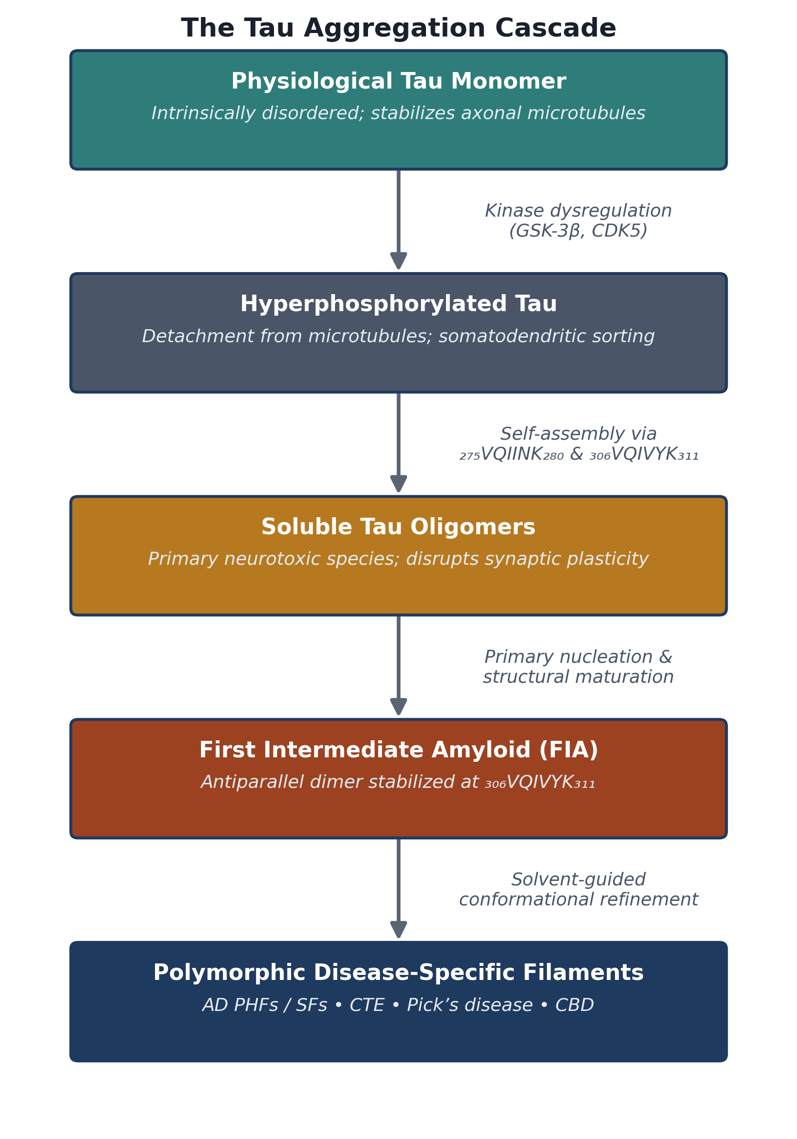
Figure 1. The Tau Aggregation Cascade: From Physiological Monomer to Polymorphic Disease-Specific Fibrils. This Figure trace the pathological transformation of tau from a soluble, intrinsically disordered microtubule-associated monomer into mature, disease-specific amyloid fibrils. Kinase-driven hyperphosphorylation (GSK-3β, CDK5) first causes tau to detach from microtubules and relocate to the somatodendritic compartment, after which self-assembly through the ₂₇₅VQIINK₂₈₀ and ₃₀₆VQIVYK₃₁₁ hexapeptide hotspots generates neurotoxic soluble oligomers. These oligomers nucleate into the recently characterized first intermediate amyloid (FIA), an antiparallel dimeric species that subsequently undergoes solvent-guided structural maturation into the polymorphic, tauopathy-specific protofilament folds — including paired helical and straight filaments in Alzheimer’s disease.
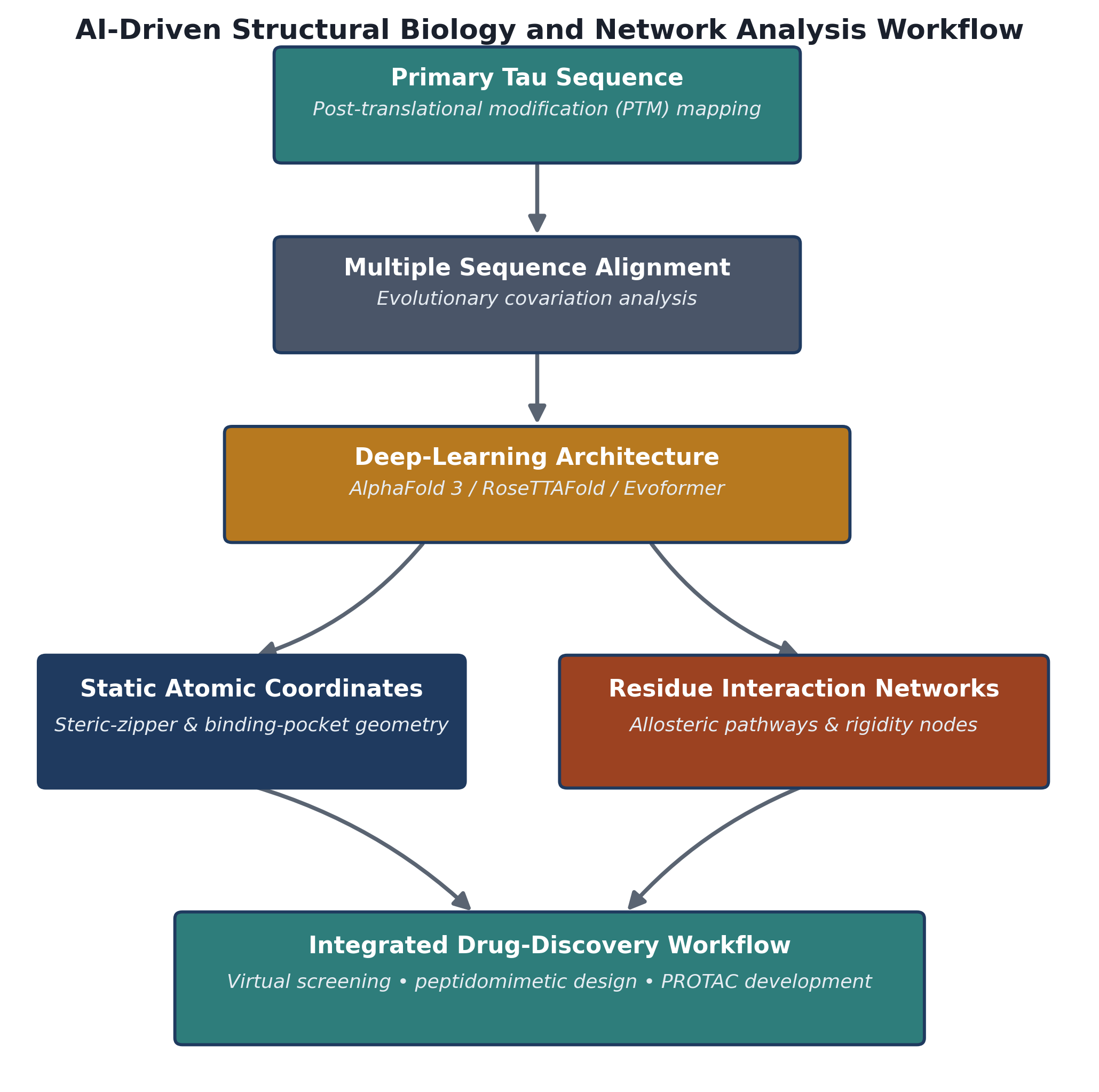
Figure 2. AI-Driven Structural Biology and Network Analysis Workflow for Tau Drug Discovery. This figure summarizes the computational pipeline by which primary tau sequence and post-translational modification data are converted into actionable drug-design inputs. Multiple sequence alignment and evolutionary covariation data feed deep-learning architectures such as AlphaFold 3 and RoseTTAFold, which generate both static atomic coordinates (steric-zipper and binding-pocket geometry) and, through downstream Residue Interaction Network analysis, allosteric and rigidity maps of the protein. These two complementary structural outputs converge on an integrated drug-discovery workflow encompassing virtual screening, peptidomimetic design, and PROTAC development.
recovered from postmortem tissue (Cehlar et al., 2024; Taylor & Radford, 2026). Whether this common-intermediate model generalizes across the full spectrum of tauopathies remains, admittedly, an open question — but it already offers a genuinely new handle for intervention, since a therapeutic capable of intercepting tau before it commits to a disease-specific fold might, in principle, prove effective across several tauopathies at once.
2.2. The Deep Learning Revolution: Structure Prediction and Network Structural Biology
Structure-based drug design against tau was, for a long time, held back less by a lack of ambition than by a lack of tools. Traditional biophysical methods — X-ray crystallography in particular, and to a lesser extent solution NMR — simply were not built to resolve the dynamic, heterogeneous conformational ensembles that intrinsically disordered proteins like tau adopt (Arar et al., 2023; Jeyaraj et al., 2025). What eventually broke this bottleneck was not a single breakthrough so much as a wave of deep-learning architectures: AlphaFold, AlphaFold 2, AlphaFold 3, and RoseTTAFold, each building on the last (Esmaeilpour et al., 2026; Jeyaraj et al., 2025; Jumper et al., 2021). By extracting evolutionary covariation signals from multiple sequence alignments and integrating them with spatial geometric modules — the Evoformer architecture being the best known — these platforms can now predict three-dimensional protein folds directly from primary sequence with a level of accuracy that would have seemed implausible a decade ago (Jeyaraj et al., 2025; Jiang et al., 2025). AlphaFold 3 extended this further still, moving toward unified multimodal prediction capable of modeling proteins alongside nucleic acids, small-molecule ligands, ions, and post-translationally modified residues within a single framework (Fang et al., 2025; Jeyaraj et al., 2025).
Complementary tools have opened up adjacent questions. Deep-learning platforms such as D-I-TASSER now support structural annotation of specific proteoforms — the individual protein variants produced by mutation, alternative splicing, or post-translational modification — which, in tau research, allows computational chemists to ask how a particular phosphorylation, acetylation, or truncation event reshapes local backbone flexibility and promotes β-sheet-prone, aggregation-competent conformations (Fang et al., 2025; Setiawan et al., 2026). To capture the higher-order topology that emerges from these local changes, structural biologists have increasingly turned to Residue Interaction Networks (RINs), which represent individual amino acids as nodes and their non-covalent contacts — van der Waals interactions, hydrogen bonds, salt bridges, π–π stacking — as edges (Miotto et al., 2025; Tosatto et al., 2025). Network metrics derived from this representation, including degree centrality, betweenness centrality, and average shortest path length, offer a reasonably direct way to quantify structural rigidity, trace allosteric communication pathways, and flag mutation-sensitive hotspots across a protein assembly (Miotto et al., 2025; Tosatto et al., 2025). Figure 2 sketches how these pieces typically fit together in practice: primary sequence and post-translational modification data feed a deep-learning prediction step, which in turn generates both static atomic coordinates and residue interaction graphs that jointly inform downstream drug discovery workflows.
None of this, it must be said plainly, has fully solved the disordered-protein problem — and arguably it was never going to. Because AlphaFold-family models are trained predominantly on the well-ordered, crystallographically resolved structures deposited in the Protein Data Bank, they continue to struggle with intrinsically disordered regions, with liquid–liquid phase separation condensates, and with fibril polymorphs whose folds shift with solution conditions (Jeyaraj et al., 2025; Taylor & Radford, 2026). For disordered regions specifically, per-residue confidence scores — the predicted Local Distance Difference Test, or pLDDT — routinely fall below 50, which reflects genuine conformational mobility rather than a modeling failure as such, though it is easy to conflate the two if one is not careful (Jeyaraj et al., 2025). More troubling, at least for peptide-based therapeutics, is that AlphaFold 3 shows chiral violation rates near 51% when modeling D-amino acid peptides, a limitation of real consequence given that D-peptides represent a leading therapeutic class for amyloid capping strategies (Al Khzem & Gomaa, 2026). The practical upshot is that AI-generated structures are best treated as testable hypotheses rather than finished answers, requiring cross-validation through molecular dynamics simulation, hydrogen–deuterium exchange mass spectrometry, and cryo-EM before any real confidence can be placed in them (Fang et al., 2025; Jeyaraj et al., 2025).
2.3. Structure-Guided Inhibitor Modalities: Small Molecules, Peptidomimetics, and Multitarget-Directed Ligands
With atomic-resolution tau structures finally in hand, medicinal chemists have pursued a genuinely broad range of chemical strategies, spanning small organic molecules, peptidomimetics, engineered antibody fragments, and multitarget-directed ligands (Al Khzem & Gomaa, 2026; Job et al., 2023; Masri, 2026). On the small-molecule side, Job et al. (2023) designed and evaluated a series of seven piperidine-substituted 3-nitrobenzaldehyde derivatives, labeled L1 through L7, using extensive in silico docking and molecular dynamics. Docking against the ₂₇₅VQIINK₂₈₀ segment (PDB ID: 5V5B) identified compound L3 as the strongest candidate, binding with notable affinity to the charged residues Lys280 and Lys281 — an interaction that appeared to interfere with primary nucleation and destabilize pre-formed oligomeric seeds, while the compound also retained reasonably favorable blood–brain barrier permeability characteristics (Job et al., 2023). A rather different chemotype, natural coumarin derivatives, works by a complementary mechanism: these rigid, planar scaffolds intercalate directly into the cross-β fibril groove through π–π stacking and hydrophobic contacts, and several synthetic pyrazole and pyrazolopyridine hybrids extend this idea further by simultaneously inhibiting GSK-3β kinase activity while interrupting tau self-assembly (Arslan et al., 2026; Masri, 2026).
Single-target drugs have, frankly, had a rough run in late-stage Alzheimer’s trials — isolated BACE1 inhibitors and γ-secretase inhibitors both failed, as did several early monoclonal antibodies — and this string of disappointments has pushed the field toward multitarget-directed ligands, or MTDLs (Habtemariam, 2026; Masri, 2026; Mella et al., 2026). The underlying logic is fairly intuitive once stated: because Alzheimer’s pathology involves an interconnected web of Aβ toxicity, tau hyperphosphorylation, cholinergic depletion, metal dyshomeostasis, and oxidative stress, a single molecule engineered to engage several of these nodes at once may simply have better odds than one aimed at only one (Job et al., 2023; Mella et al., 2026; Setiawan et al., 2026). Advanced computational frameworks — quantitative structure–activity relationship models, Graph Transformer representation learning, and multi-objective reinforcement learning approaches such as EvoPlay-MuZero — now support the rational design of MTDLs that balance cholinesterase inhibition against tau anti-aggregation activity, iron chelation, and radical-scavenging capacity within a single scaffold (Huang et al., 2026; Mella et al., 2026; Zhou & Chen, 2026).
Peptidomimetic design occupies a particularly interesting niche here, since it targets protein–protein interfaces that remain, for the most part, undruggable by conventional small molecules (Al Khzem & Gomaa, 2026). Rational sequence engineering has produced D-amino acid retro-inverso peptides such as RI-AG03, built specifically to dual-cap the ₂₇₅VQIINK₂₈₀ and ₃₀₆VQIVYK₃₁₁ hotspots simultaneously (Al Khzem & Gomaa, 2026). In Drosophila tauopathy models, RI-AG03 suppressed tau oligomerization quite potently, extended survival, and — perhaps its most clinically relevant property — resisted proteolytic degradation once in systemic circulation (Al Khzem & Gomaa, 2026). Cyclic peptides such as cTau-binder and slightly larger mini-protein inhibitors, in the 35–48 residue range, take a related but distinct approach, capping the growth tips of tau protofilaments and sterically blocking lateral association and secondary nucleation (Al Khzem & Gomaa, 2026). Structural biology has informed antibody engineering in much the same way: Abskharon et al. (2023) designed bispecific nanobodies that selectively recognize and cap the ends of patient-derived tau fibrils, and Kayed et al. (2024) subsequently developed toxic-conformation-specific monoclonal antibodies — TTCM2 among them — that, when delivered intranasally via polymeric micelles, penetrate neurons and clear intracellular seed-competent tau aggregates by engaging the cytosolic antibody receptor and E3 ligase TRIM21. Figure 3 draws these modalities together schematically, showing how structurally distinct chemical strategies — small molecules, peptidomimetics, and, as described next, targeted degraders — converge on a common functional outcome: suppression of seeding and enhanced clearance of pathological tau.
2.4. Targeted Protein Degradation and Advanced Central Nervous System Delivery
A genuinely different therapeutic logic altogether comes from targeted protein degradation, executed most commonly through proteolysis-targeting chimeras, or PROTACs (Jiang et al., 2025; Zhou et al., 2026). Where a classical occupancy-based inhibitor must maintain sustained binding to an active or enzymatic pocket to exert its effect, a PROTAC instead operates catalytically (Jiang et al., 2025; Zhou et al., 2026). Structurally, a heterobifunctional PROTAC molecule links a tau-binding warhead through an optimized spacer to a moiety that recruits an E3 ubiquitin ligase — commonly von Hippel–Lindau (VHL) or Cereblon (CRBN) (Zhou et al.,
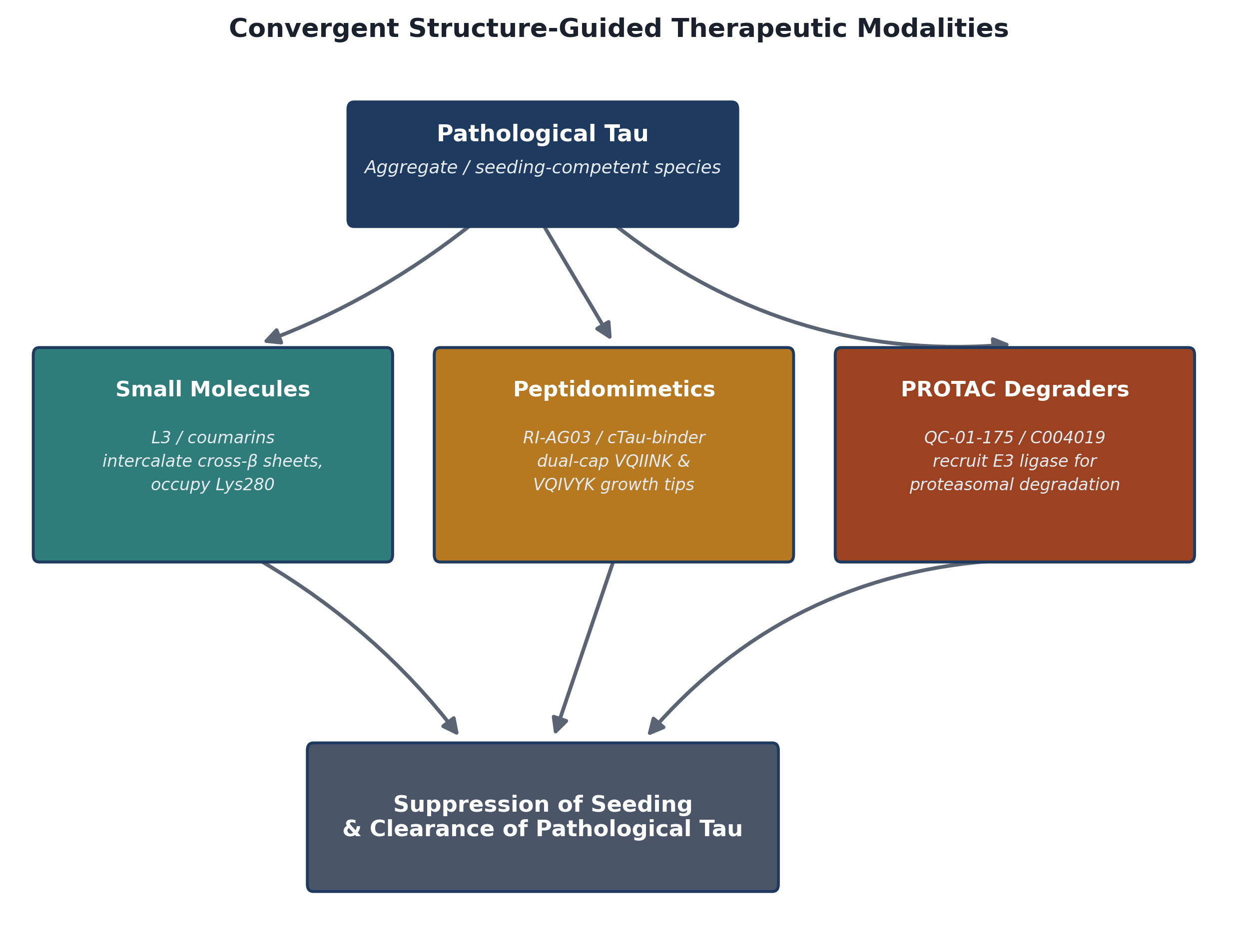
Figure 3. Convergent Structure-Guided Therapeutic Modalities Targeting Tau Aggregation and Clearance. This figure illustrates how three mechanistically distinct classes of structure-based tau therapeutics — small-molecule and coumarin intercalators, dual-capping peptidomimetics, and catalytic PROTAC degraders — each engage a pathological tau aggregate or seed through a different molecular mechanism, yet converge on the same functional outcome. Small molecules intercalate cross-β sheets and occupy the Lys280 nucleation interface; peptidomimetics sterically cap the ₂₇₅VQIINK₂₈₀ and ₃₀₆VQIVYK₃₁₁ growth tips; and PROTACs recruit E3 ubiquitin ligases for proteasomal clearance. The three converging arrows summarize the shared endpoint.
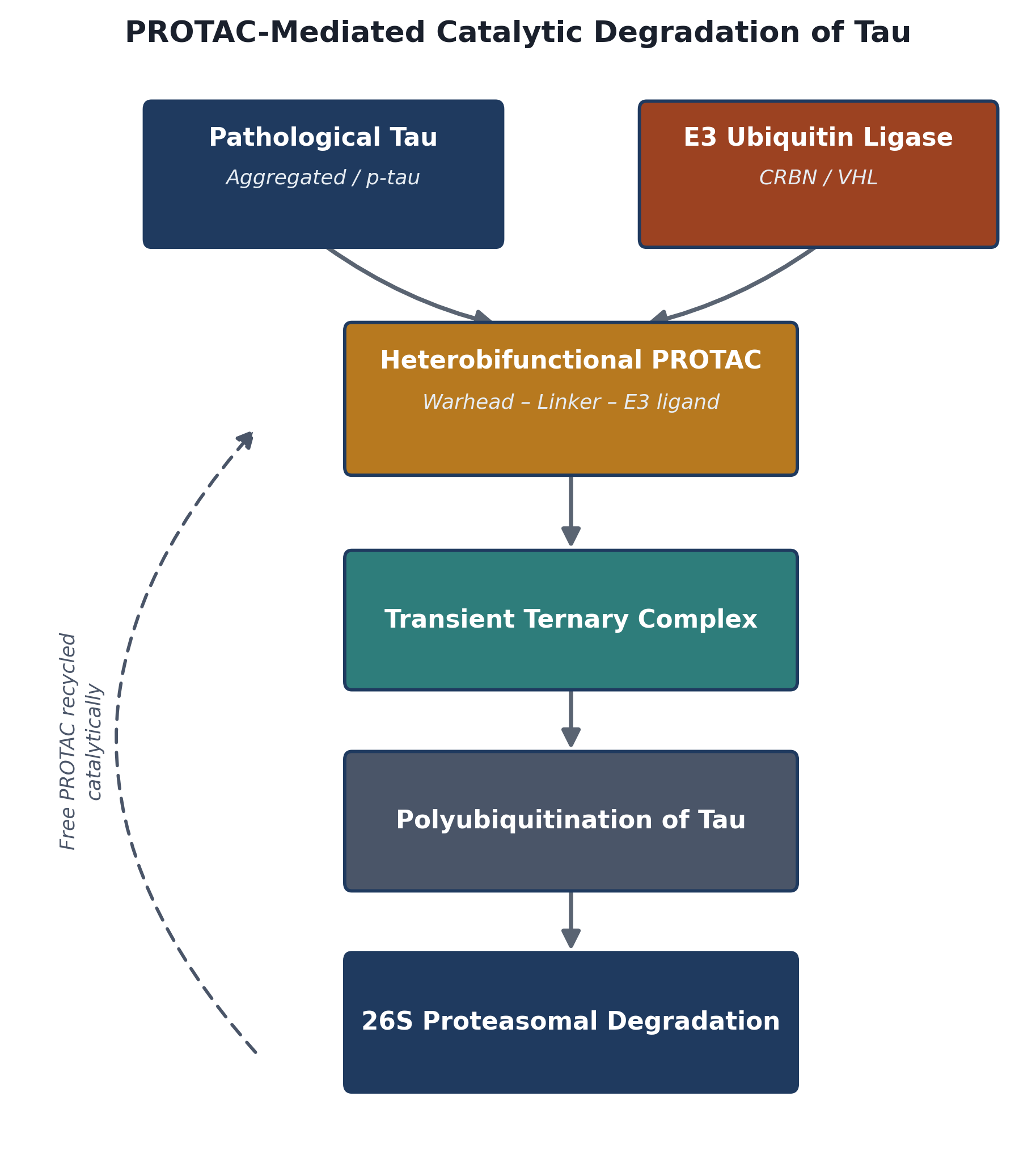
Figure 4. PROTAC-Mediated Catalytic Degradation of Pathological Tau via the Ubiquitin–Proteasome System. This figure details the catalytic mechanism by which heterobifunctional PROTAC molecules clear pathological tau. A tau-binding warhead and an E3 ubiquitin ligase-recruiting ligand (targeting CRBN or VHL), joined by an optimized linker, bring aggregated or hyperphosphorylated tau into direct spatial proximity with the E3 ligase, forming a transient ternary complex. This complex drives polyubiquitination of the bound tau species, marking it for recognition and cleavage by the 26S proteasome, after which the PROTAC molecule is released intact to catalytically repeat the cycle — the mechanistic basis for the tau-clearing activity of candidates such as QC-01-175 and C004019
2026). By bringing pathological tau into close spatial proximity with the E3 ligase, the PROTAC induces formation of a transient ternary complex, which triggers polyubiquitination of tau and directs it toward destruction by the 26S proteasome (Jiang et al., 2025; Zhou et al., 2026). Figure 4 outlines this cycle step by step, from initial ternary complex formation through polyubiquitination, proteasomal cleavage, and the eventual release of the PROTAC molecule to continue the cycle catalytically. Preclinical tau-directed PROTACs such as QC-01-175 and C004019 have already demonstrated fairly robust clearance of hyperphosphorylated and oligomeric tau in neuronal models derived from frontotemporal dementia patients and in transgenic mice, restoring synaptic plasticity and, in at least some paradigms, measurable cognitive function (Zhou et al., 2026). To accelerate the discovery of further candidates, machine-learning platforms such as PrePROTAC now use pre-trained protein language models together with random-forest classifiers to predict degradation-susceptible proteoforms directly from sequence, without waiting on structural data (Zhou et al., 2026).
Highly selective as these degraders and biologics are, getting them into the brain at all remains a formidable obstacle — arguably the central bottleneck of the entire field (Al Khzem & Gomaa, 2026; Rajkumar et al., 2026). The blood–brain barrier, formed by non-fenestrated endothelial cells joined by tight junctions, excludes upward of 98% of small-molecule drugs and essentially all large hydrophilic biologics from the brain parenchyma (Al Khzem & Gomaa, 2026; Rajkumar et al., 2026). PROTACs, unhelpfully, tend to violate Lipinski’s rule of five outright, owing to molecular weights that routinely exceed 800 Da and correspondingly high topological polar surface area, which together preclude passive transcellular diffusion (Zhou et al., 2026). Meeting this challenge has driven a wave of drug-delivery engineering. Smart nanocarriers — poly(lactic-co-glycolic acid) nanoparticles, solid lipid nanoparticles, liposomes, and neurotransmitter-derived lipidoids among them — extend circulation half-life, improve systemic stability, and can be functionalized to exploit receptor-mediated transcytosis across brain endothelium (Al Khzem & Gomaa, 2026; Rajkumar et al., 2026). Surface functionalization with brain-targeting peptides, such as the rabies-virus-glycoprotein-derived RVG29 peptide or ligands for the transferrin receptor (TfR1), substantially increases brain uptake through receptor-mediated endocytosis (Al Khzem & Gomaa, 2026). That said, TfR1-targeted shuttles are not without their own liabilities — peripheral tissue accumulation and, more seriously, reticulocyte depletion have been recurring safety concerns (Lofgren, 2024). High-throughput in vivo screening platforms, such as Manifold Bio’s mCode technology, have since identified alternative brain-endothelial receptor ‘portals,’ including one designated PX1, that appear to achieve comparable or superior parenchymal enrichment without triggering the same hematological toxicity (Lofgren, 2024). Direct nose-to-brain delivery via mucoadhesive nasal sprays or polymeric micelles offers yet another route, bypassing the blood–brain barrier altogether by exploiting the olfactory and trigeminal nerve pathways to deliver therapeutic peptides and PROTACs into the olfactory bulb and hippocampal circuitry directly (Jiang et al., 2025; Kayed et al., 2024).
2.5. Methodological Bottlenecks and Integrated Computational–Experimental Pipelines
Underlying much of the preceding discussion is a translational gap that has come to be known, only half-jokingly, as the ‘valley of death’ — the persistent disconnect between encouraging computational predictions, promising in vitro binding data, and eventual failure in human clinical trials (Esmaeilpour et al., 2026; Setiawan et al., 2026). Part of the problem is structural in a different sense: static AI models tend to over-optimize candidate molecules for enthalpy-driven binding against a single, fixed structural target, which is a reasonable simplification computationally but a poor approximation of physiological reality (Esmaeilpour et al., 2026). In an actual living system, drugs must contend with dense macromolecular crowding, competitive binding to human serum albumin, active P-glycoprotein efflux, ongoing conformational transitions in the target itself, and a surprising degree of post-translational proteoform diversity (Esmaeilpour et al., 2026; Fang et al., 2025). There is, additionally, a subtler risk worth naming: over-reliance on insufficiently validated generative models can quietly introduce dataset bias, with models repeatedly proposing familiar, narrow chemotypes — an over-representation of aromatic residues, for instance — rather than generalizing meaningfully into novel chemical space (Esmaeilpour et al., 2026; Huang et al., 2026).
The more promising response to this gap, and the one increasingly adopted across the field, is what might be called an integrated design platform — a closed feedback loop connecting computation and wet-lab experimentation rather than treating them as sequential, one-way stages (Al Khzem & Gomaa, 2026; Esmaeilpour et al., 2026). In such workflows, structural models generated by AlphaFold 3 or RoseTTAFold function as initial hypotheses rather than finished deliverables (Esmaeilpour et al., 2026; Fang et al., 2025). These predicted poses are then subjected to microsecond-scale, explicit-solvent molecular dynamics simulations, MM/PBSA free-energy calculations, and Residue Interaction Network topology analysis to probe dynamic binding stability and allosteric conformational behavior (Job et al., 2023; Miotto et al., 2025). In parallel, automated fast-flow peptide synthesis platforms with inline UV-vis monitoring can rapidly produce physical lead candidates of high structural purity, which then undergo biophysical profiling by cryo-EM, surface plasmon resonance, and hydrogen–deuterium exchange mass spectrometry — with the resulting experimental data fed back into the machine-learning models to iteratively refine scoring functions and weed out false positives (Al Khzem & Gomaa, 2026; Esmaeilpour et al., 2026; Jeyaraj et al., 2025). It is this closed-loop character, more than any single algorithmic advance, that seems likely to determine whether the current wave of AI-guided tau therapeutics ultimately reaches patients — a theme we return to, in more practical detail, in the Discussion.
