Idiopathic pulmonary fibrosis (IPF) is a devastating, progressive interstitial lung disease of older adults that leads to irreversible destruction of alveolar architecture and fatal respiratory failure. While historically viewed as a chronic inflammatory disorder, contemporary research has redefined IPF as a cellular-systems problem driven by repetitive epithelial microinjury, aberrant mesenchymal activation, and compromised cellular quality-control networks. At the epicenter of this pathogenesis is cellular senescence, a state of permanent cell-cycle arrest that actively orchestrates remodeling of the lung parenchyma. This literature review adopts a multiscale conceptual mind map framework to dissect the pathogenic networks of cellular senescence in IPF. We trace how microinjuries and genetic susceptibilities trigger aberrant, senescent progenitor states---such as the KRT8⁺ alveolar differentiation intermediate (ADI) and KRT5⁻/KRT17⁺ aberrant basaloid cells---which disrupt the epithelial stem cell niche and lock cells in regenerative arrest. We explore the bidirectional mechanical and metabolic crosstalk between senescent epithelial cells and a heterogeneous fibroblast landscape, showing how metabolic reprogramming (aerobic glycolysis) and mechanical memory drive persistent myofibroblast activation. Furthermore, we examine the reciprocal regulation of the senescence-autophagy axis, detailing how disabled macroautophagy and mitophagy fuel reactive oxygen species (ROS) accumulation and stabilize senescent phenotypes. Finally, we review emergent senotherapeutic and regenerative reversal strategies---encompassing selective senolytic clearance, senomorphic modulation of the pro-fibrotic secretome, and autologous basal cell transplantation---as pioneering roadmaps to move the clinical management of IPF beyond palliative deceleration toward structural and functional restoration. This organization is summarized conceptually in Figure 1, which maps the four thematic branches synthesized below and their convergence on a shared, self-reinforcing pathogenic network.
2.1 Branch 1: Reprogrammed Epithelial Trajectories and Alveolar Regeneration Failure
The classical paradigm of idiopathic pulmonary fibrosis (IPF) as a linear, passive tissue response to chronic inflammation has been substantially reshaped by single-cell RNA sequencing (scRNA-seq) and spatial transcriptomics (Deng et al., 2026). The contemporary consensus establishes IPF as an epithelial-driven, aberrant wound-healing disorder initiated by recurrent, subclinical microinjuries to the alveolar epithelium (Baurzhan et al., 2026; Torres-Machorro et al., 2025). In genetically susceptible individuals, these microinjuries---compounded by environmental toxicants such as silica dust, particulate matter, and tobacco smoke---drive alveolar type II (AEC2) progenitor cells into states of persistent regenerative arrest and cellular senescence (Gao et al., 2025; Jones et al., 2025).
Rather than undergoing simple apoptotic depletion, injured alveolar progenitors enter a highly active, reprogrammed transitional state. A primary node in this aberrant epithelial trajectory is the accumulation of the KRT8⁺ alveolar differentiation intermediate (ADI) stem cell state (Deng et al., 2026).
This trajectory — summarized schematically in Figure 2 — begins during healthy alveolar repair, where the ADI state represents a transient, developmental bridge through which AEC2 progenitors differentiate into flat, gas-exchanging alveolar type I (AEC1) cells (Jones et al., 2025). In the chronic fibrotic milieu of IPF, however, these cells fail to exit this transitional phase. They become locked in a persistent regenerative bottleneck characterized by the continuous activation of p53, NF-κB, and DNA damage response (DDR) pathways (Deng et al., 2026; Longhin et al., 2026). This trajectory arrest is accompanied by the up-regulation of the cellular senescence markers p21 (CDKN1A) and p16 (CDKN2A), converting a physiological repair intermediate into a pathological signaling engine (Baurzhan et al., 2026; Deng et al., 2026).
Spatially segregated at the active borders of fibroblastic foci and within remodeled honeycomb cysts lie ectopic epithelial cells termed aberrant basaloid cells (Deng et al., 2026; Svobodová et al., 2025). Characterized transcriptomically as KRT5⁻/KRT17⁺, these cells co-express markers of senescence (p21, p16, GDF15), epithelial-to-mesenchymal transition (EMT), and matricellular remodeling, including collagen VII (Svobodová et al., 2025). Spatial transcriptomic profiling demonstrates that these aberrant basaloid cells line the highly remodeled epithelial-mesenchymal interface immediately overlying pathological fibroblasts, where they serve as crucial paracrine hubs promoting fibrogenesis (Deng et al., 2026; Jones et al., 2025). Bulk and single-cell transcriptomic analyses indicate that these senescent epithelial populations undergo substantial epigenetic and transcriptional modifications, acquiring an altered, secretory phenotype that further drives tissue stiffness and suppresses healthy stem cell renewal (Melo-Narváez et al., 2024).This epithelial dysfunction extends into the conducting and small airways. Accumulating evidence reveals that small airway ciliary impairment and compromised mucociliary clearance (MCC) are early, active drivers of IPF pathogenesis rather than secondary consequences of parenchymal collapse (Wang et al., 2025). Mature multiciliated cells in the conducting airway epithelium are responsible for clearing inhaled particulates, pathogens, and cellular debris (Wang et al., 2025). In patients with IPF, small airway epithelial cells (SAECs) demonstrate a significant, intrinsic decline in ciliogenesis potency, characterized by a marked reduction in the expression of Forkhead box protein J1 (FOXJ1)---the master transcriptional orchestrator of ciliogenesis---and fewer β-IV-tubulin-positive ciliated cells (Wang et al., 2025). When SAECs are subjected to external injuries (such as bleomycin challenge) or serial cell passage, ciliary differentiation is selectively impaired, while mucin production (MUC5AC and MUC5B) remains unaffected
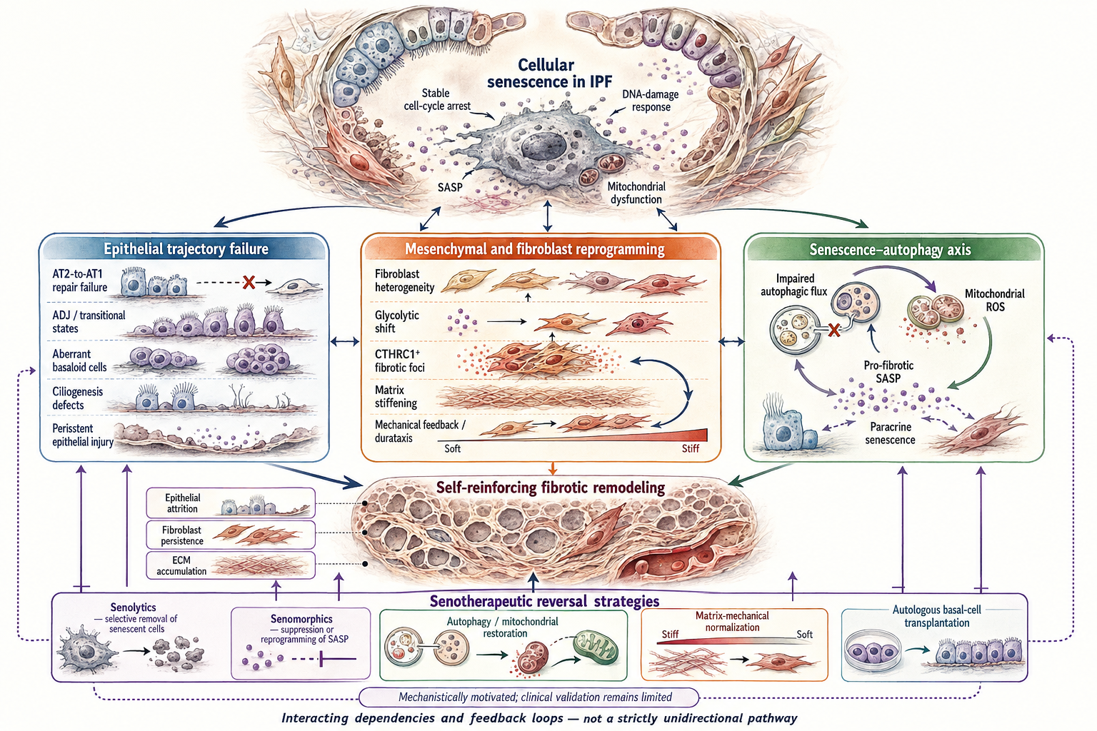
Figure 1 | Conceptual organization of cellular-senescence pathways in idiopathic pulmonary fibrosis. Cellular senescence forms a pathogenic hub characterized by stable cell-cycle arrest, persistent DNA-damage signalling, mitochondrial dysfunction and secretion of the senescence-associated secretory phenotype (SASP). This state interacts with three overlapping mechanistic branches. Epithelial trajectory failure involves unsuccessful alveolar type 2-to-type 1 cell differentiation, accumulation of transitional and aberrant basaloid populations, ciliogenesis defects and persistent epithelial injury. Mesenchymal reprogramming encompasses fibroblast heterogeneity, glycolytic shifts, CTHRC1-positive fibrotic foci and mechanically reinforced migration and activation within stiffened extracellular matrix. The senescence–autophagy axis links impaired autophagic flux with mitochondrial reactive oxygen species, profibrotic SASP signalling and paracrine propagation of senescence. These processes converge on epithelial attrition, fibroblast persistence and extracellular-matrix accumulation, producing self-reinforcing fibrotic remodeling. Potential reversal strategies include senolytics, senomorphics, restoration of autophagy and mitochondrial function, normalization of matrix mechanics and autologous basal-cell transplantation. Arrows represent interacting mechanistic dependencies and feedback relationships rather than strictly unidirectional causation; clinical validation of these therapeutic approaches remains limited.
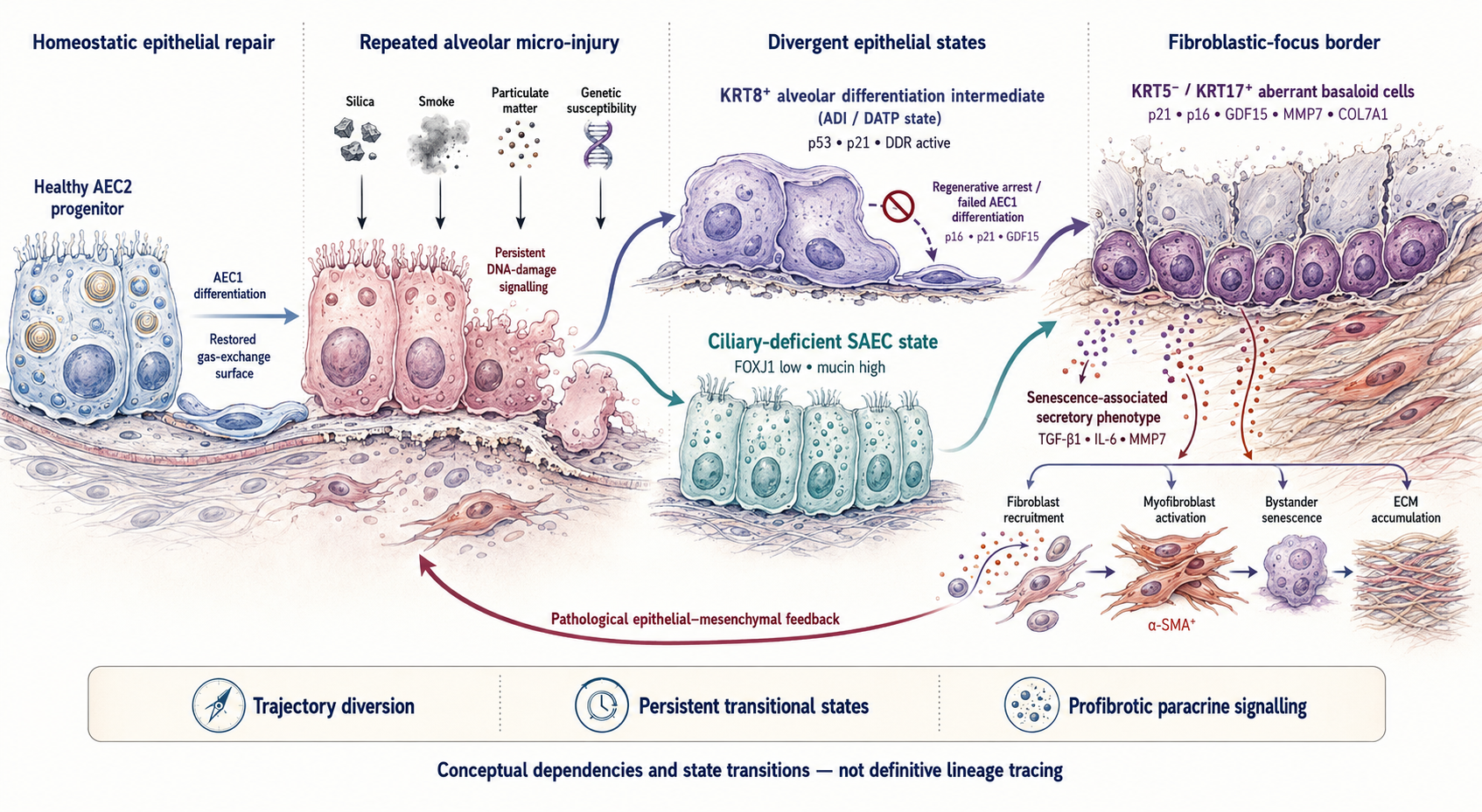
Figure 2 | Reprogrammed epithelial trajectories following alveolar micro-injury. Under homeostatic conditions, alveolar epithelial type 2 (AEC2) progenitors differentiate into flattened alveolar type 1 (AEC1) cells to restore the gas-exchange surface. Repeated injury associated with inhaled particulates, smoke, silica or genetic susceptibility disrupts this regenerative program and sustains DNA-damage signalling. Injured progenitors are consequently diverted into persistent KRT8-positive alveolar differentiation intermediate or damage-associated transient progenitor states and ciliary-deficient small-airway epithelial states characterized by low FOXJ1 and increased mucin expression. Regenerative arrest prevents completion of AEC1 differentiation and promotes the accumulation of KRT5-negative/KRT17-positive aberrant basaloid cells at the epithelial–fibroblastic-focus interface. These persistent epithelial populations express senescence-associated markers and release SASP mediators, including TGF-β1, IL-6 and MMP7, which recruit fibroblasts, promote myofibroblast differentiation, propagate bystander senescence and increase extracellular-matrix deposition. The resulting epithelial–mesenchymal feedback reinforces tissue injury and fibrotic remodeling. Arrows denote conceptual state transitions and mechanistic dependencies rather than definitive lineage tracing.
(Wang et al., 2025).
Importantly, conditioned medium harvested from SAECs with defective ciliogenesis or silenced FOXJ1 potently stimulates healthy lung fibroblasts to differentiate into α-smooth muscle actin (α-SMA)-positive and fibronectin- positive myofibroblasts, demonstrating that ciliary impairment mediates pro-fibrotic epithelial-mesenchymal crosstalk (Wang et al., 2025). This deficiency is further compounded by the gain-of-function MUC5B promoter variant (rs35705950), the strongest genetic risk factor for IPF, which drives massive mucin accumulation in the distal bronchioles and alveoli, triggering chronic endoplasmic reticulum (ER) stress and further stabilizing the senescent, basaloid state (Deng et al., 2026).
2.2 Branch 2: Fibroblast Heterogeneity, Glycolytic Reprogramming, and Biomechanical Feedback
The mesenchymal compartment of the IPF lung exhibits profound cellular heterogeneity, containing distinct subpopulations of fibroblasts that occupy different pathological niches (Deng et al., 2026), summarized across their markers, metabolic programs, and apoptotic sensitivity in Table 2. Single-cell transcriptomics has identified that the active core of the fibroblastic focus---the primary site of active matrix deposition---is dominated by a highly specialized, collagen-producing fibroblast subpopulation marked by the expression of Collagen Triple Helix Repeat Containing 1 (CTHRC1) (Deng et al., 2026; Melo-Narváez et al., 2024). CTHRC1⁺ fibroblasts exhibit extreme invasiveness, apoptotic resistance, and robust collagen synthesis, representing the primary engine of structural scarring (Deng et al., 2026; Melo-Narváez et al., 2024). Spatiomics and lineage-tracing studies describe a dynamic cellular trajectory wherein quiescent lung fibroblasts and lipid-storing lipofibroblasts undergo a phenotypic switch toward these pathogenic, CTHRC1⁺ myofibroblasts under the influence of local biochemical and biophysical cues (Deng et al., 2026; Shanmugasundaram et al., 2024).
The persistence and aggressiveness of these activated mesenchymal populations are tightly regulated by metabolic reprogramming and biomechanical feedback loops (Alvarado-Vasquez et al., 2024; Zhang et al., 2022). In the fibrotic lung, myofibroblasts shift their primary energy production from oxidative phosphorylation to aerobic glycolysis, a metabolic phenomenon analogous to the Warburg effect in cancer cells (Alvarado-Vasquez et al., 2024). This glycolytic reprogramming is driven by the up-regulation of rate-limiting enzymes, including 6-phosphofructo-2-kinase/fructose-2,6-bisphosphatase 3 (PFKFB3) and hexokinase 2 (HK2), which are induced downstream of transforming growth factor-beta 1 (TGF-β1) signaling (Alvarado-Vasquez et al., 2024). Elevated glycolysis results in the excessive accumulation and extracellular secretion of lactic acid, which establishes a local acidic microenvironment (Alvarado-Vasquez et al., 2024). This extracellular acidity further activates latent, matrix-bound TGF-β1, creating a self-sustaining, pro-fibrotic feed-forward circuit (Alvarado-Vasquez et al., 2024).
Concurrently, this metabolic shift is reinforced by a highly altered biomechanical microenvironment (Zhang et al., 2022). As myofibroblasts deposit disorganized fibrillar collagens and matricellular proteins, the mechanical stiffness of the extracellular matrix (ECM) increases exponentially (Svobodová et al., 2025; Zhang et al., 2022). This local stiffening is sensed by cell-surface mechanoreceptors, which transduce physical forces into intracellular biochemical signals (such as the MRTF-actin and YAP/TAZ axes), a process known as mechanotransduction (Zhang et al., 2022).
Stiff substrates persistently stimulate lung fibroblasts to remain in an activated, contractible, and synthetic state (Zhang et al., 2022). Over time, fibroblasts exposed to stiff environments acquire a \"mechanical memory\" through chromatin remodeling and stable epigenetic modifications (such as altered histone acetylation and DNA methylation), allowing them to maintain their pro-fibrotic, senescent state even if they are removed from the stiff microenvironment (Zhang et al., 2022). Consequently, the integration of mechanical forces with glycolytic reprogramming locks the mesenchymal compartment into an irreversible, self-perpetuating fibrotic state that resists physiological resolution (Alvarado-Vasquez et al., 2024; Zhang et al., 2022).
2.3 Branch 3: The Senescence-Autophagy Axis, Mitochondrial ROS, and the SASP Secretome
At the molecular scale, cellular senescence and macroautophagy are best conceptualized as intertwined components of a single, disrupted cellular quality-control network (Min et al., 2026); the principal signaling axes underlying this network, including their mechanobiological and autophagy-regulatory effects, are catalogued in Table 3. Autophagy is a critical homeostatic mechanism
Table 1. Epithelial cell states, metaplasia, and regenerative arrest in idiopathic pulmonary fibrosis. This table catalogues the major senescent and transitional epithelial cell phenotypes identified across single-cell and spatial transcriptomic studies of the IPF lung, including aberrant basaloid cells, alveolar differentiation intermediates (ADI/DATP), metaplastic airway basal cells, and ciliary-deficient small airway epithelial cells. For each phenotype, the table lists defining molecular markers, spatial localization within the alveolar or airway niche, presumed cellular origin and lineage trajectory, the senescence pathway(s) activated, the dominant SASP secretory profile, and the resulting functional impact on the surrounding alveolar microenvironment, with supporting citations.
|
Epithelial Cell Phenotype / Subtype
|
Key Molecular Markers
|
Primary Spatial Localization
|
Cellular Origins & Lineage Trajectories
|
Senescence Pathways Activated
|
SASP Secretory Profile & Cytokines
|
Functional Impact on Alveolar Microenvironment
|
Grounding References (APA Style)
|
|
Aberrant Basaloid Cells (AbBa / KRT5⁻/KRT17⁺)
|
KRT17, TP63, LAMB3,
LAMC2, CDH2, VIM, FN1, COL1A1,
GDF15, MMP7
|
Located at the direct borders/edges of active fibroblastic foci, adjacent to diseased stroma, and lining honeycomb cysts.
|
Failed or ectopic differentiation path originating from mature alveolar type II (AT2) cells and SCGB3A2⁺ airway secretory lineages.
|
Co-expression of stable cell-cycle arrest pathways governed by CDKN1A (p21) and CDKN2A (p16).
|
Robust secretion of stroma-remodeling matrix metalloproteinases (MMP7), GDF15, TGF-β1, IL-6, and IL-1β.
|
Drives fibroblast-to-myofibroblast transition (FMT), induces paracrine "bystander" senescence, and deposits pathological fibrillar ECM.
|
Adams et al. (2020); Habermann et al. (2020); Mayr et al. (2024); Svobodová et al. (2025)
|
|
Alveolar Differentiation Intermediate (ADI) Cells
|
KRT8, CLDN4, YAP1, TAZ
|
Preserved alveolar regions with fibrotic septa and at the margins of collapsing alveoli.
|
Arise from injured mature AT2 cells or distal small airway Scgb1a1⁺ club cell progenitors during aberrant repair.
|
Persistent, non-mitotic arrest characterized by chronic p53 activation, DNA damage responses (DDR), and telomere attrition.
|
Secretes high levels of TGF-β1, IL-6, and various pro-inflammatory SASP chemokines.
|
Locks alveolar regeneration, failing to transition into gas-exchanging AT1 cells, and promotes chronic tractional alveolar collapse.
|
Jones et al. (2026); Min et al. (2026); Svobodová et al. (2025)
|
|
Metaplasic Airway Basal Cells (KRT17^high / PTEN^low)
|
KRT17, p63, GPR87
|
Dilated distal bronchioles and lining the pseudostratified epithelium of pathological honeycomb cysts.
|
Proximal cartilaginous airway basal stem cells undergoing aberrant distal migration and expansion ("bronchiolization").
|
Replicative senescence marked by severe down-regulation of PTEN and loss of normal ciliary-fate program program.
|
High release of IL-6, IL-8, and macrophage-activating factors such as osteopontin (SPP1).
|
Directly stimulates mesenchymal proliferation, recruits neutrophils and monocytes, and impairs local mucociliary clearance.
|
Deng et al. (2026); Jones et al. (2026); Svobodová et al. (2025)
|
|
Activated Airway Club Cells (Club_MMP1_hi Subpopulation)
|
KRT17, CC10 (SCGB1A1), MMP1
|
First-line epithelial barrier of remodeled bronchioles and terminal conducting small airways.
|
Homeostatic ciliated club cells undergoing injury-induced activation and basaloid metaplasia.
|
Stress-induced premature senescence (SIPS) driven by environmental inhaled toxicants and oxidative injury.
|
Off-on switch-like upregulation of MMP1 and lysosomal proteases like CTSB.
|
Drives extensive basement membrane degradation, facilitating active fibroblast invasion into the alveolar space.
|
Fu et al. (2026)
|
|
Ciliary-Deficient Small Airway Epithelial Cells (SAECs)
|
FOXJ1 (low), β-IV-tubulin (low), MUC5B, MUC5AC
|
Conducting small bronchioles and distal terminal bronchioles in advanced stages of IPF.
|
Mature ciliated small airway epithelial cells undergoing phenotypic degradation and loss of multiciliogenesis.
|
DNA damage response (DDR) activation and stable cycle withdrawal via the TP73-MYB-FOXJ1 axis.
|
Enhanced secretion of pro-fibrotic cytokines, particularly TGF-β1, IL-6, and MMP1.
|
Triggers severe impairment in mucociliary clearance (MCC), promoting alveolar mucus plugging, and drives fibroblast activation.
|
Wang et al. (2025)
|
|
Proximal Basal Stem Cells (Small Bronchi-Derived / Healthy State)
|
p63⁺, Krt5⁺, ITGA6⁺, NGFR⁺, Ki67⁺
|
Proximal cartilaginous small bronchi spared from severe distal parenchymal distortion.
|
Healthy, resident tissue-specific adult stem cell pool maintained in a protected, homeostatic niche.
|
Distinctly non-senescent phenotype, preserving robust proliferative and clonogenic properties across multiple passages.
|
Lacks inflammatory SASP; secretes homeostatic and regenerative growth factors.
|
Retains complete capacity to differentiate into functional ciliated and club cells, serving as a clean therapeutic transplant reservoir.
|
Snyder et al. (2025)
|
|
Injured Alveolar Type II (AT2) Cells with Proteostatic Stress
|
SFTPC (mutant), BIP, ATF4, CHOP, p53
|
Alveolar niches adjacent to active fibrotic areas and collapsing septa.
|
Alveolar progenitor AT2 cells exposed to genomic instability, microinjuries, or genetic mutations (e.g., SFTPC/TERT).
|
UPR-induced endoplasmic reticulum stress (ERS) triggering stable senescence arrest via the PERK/eIF2α axis.
|
High-level production of TGF-β1, PAI-1, IL-6, and CXCL1 chemokines.
|
Compromises lung surfactant production, induces apoptosis resistance, and recruits macrophages to establish a pro-inflammatory microenvironment.
|
Gong et al. (2026); Jones et al. (2026); Torres-Machorro et al. (2025)
|
|
SIRT3-Deficient Alveolar Progenitor Cells
|
SIRT3 (low), SFTPC, p21
|
Distal gas-exchange alveoli and active transitional zones.
|
Alveolar type II cells experiencing age-related metabolic and mitochondrial decay.
|
Mitochondrial-driven senescence due to increased mitochondrial oxidative stress and bioenergetic crash.
|
Elevated levels of PAI-1, TNF-α, endothelin-1, and PDGF.
|
Enhances adjacent myofibroblast proliferation, disrupts stem cell renewal, and accelerates spatial propagation of fibrotic niches.
|
Baurzhan et al. (2026); Min et al. (2026)
|
|
MUC5B-Overexpressing Metaplasic Alveolar Cells
|
MUC5B (rs35705950 promoter variant), KRT5
|
Pathological honeycomb cysts and distal airspace lesions.
|
AT2 cells or club cells undergoing metaplastic lineage conversion under chronic endoplasmic reticulum stress.
|
Stress-induced senescence driven by proteostatic collapse and accumulated mucin-protein folding stress.
|
High levels of mucin secretion, IL-6, and ADAM17.
|
Promotes severe mucus retention, plugs alveolar gas exchange surfaces, and induces a self-perpetuating fibrotic niche via mechanochemical stress.
|
Deng et al. (2026); Svobodová et al. (2025); Wang et al. (2025)
|
|
Damage-Associated Transitional Progenitors (DATPs / KRT8⁺)
|
KRT8, CLDN4, p21
|
Active transition zones linking normal alveoli to dense fibrotic scars.
|
Alveolar epithelial progenitor cells arrested midway during differentiation into gas-exchanging AT1 cells.
|
Characterized by persistent DNA damage response (DDR) and p21-mediated cycle withdrawal.
|
Secretes TGF-β1, IL-1β, and IL-6.
|
Perpetuates alveolar collapse and tissue traction forces, establishing a positive feedback loop of mechanobiological injury.
|
Deng et al. (2026); Jones et al. (2026); Svobodová et al. (2025)
|
Table 2. Mesenchymal lineages, heterogeneity, and activation dynamics in the fibrotic lung. This table summarizes the major fibroblast and mesenchymal-derived subpopulations described in IPF, including CTHRC1+ invasive myofibroblasts, hyaluronan-producing fibroblasts, lipofibroblasts, and several senescent fibroblast phenotypes (e.g., FABP5-overexpressing, EPDR1-upregulated, GSR-depleted). Columns detail key molecular markers, spatial niche, transcriptional/epigenetic regulators, metabolic reprogramming and bioenergetics, secreted biomarkers and ECM components, and apoptosis sensitivity/proteostasis status, with supporting citations.
|
Mesenchymal Lineage / Phenotype
|
Key Molecular Markers
|
Spatial Niche / Tissue Localization
|
Primary Transcriptional & Epigenetic Regulators
|
Metabolic Reprogramming & Bioenergetics
|
Secreted Biomarkers & ECM Components
|
Apoptosis Sensitivity & Proteostasis
|
Grounding References (APA Style)
|
|
Invasive Myofibroblasts (CTHRC1⁺ / α-SMA⁺)
|
CTHRC1, ACTA2 (α-SMA), COL1A1, FN1
|
Highly concentrated in the active core of fibroblastic foci, directly underlying the aberrant epithelial cap.
|
TGF-β/SMAD3 axis, mechanical YAP/TAZ nuclear translocation, and MRTF-actin signaling.
|
Massive shift toward aerobic glycolysis (Warburg effect) with lactate accumulation driven by PFKFB3/HK2.
|
Disorganized fibrillar collagens (types I, III), fibronectin, and tenascin-C.
|
Highly resistant to apoptosis due to upregulation of Bcl-2/Bcl-xL and downregulation of caspases.
|
Deng et al. (2026); Jones et al. (2026); Torres-Machorro et al. (2025); Zhang et al. (2026)
|
|
Hyaluronan-Producing Fibroblasts (HAS1^high / HAS2^high)
|
HAS1, HAS2, CD44
|
Subpleural region, adjacent to aberrant basaloid epithelial cells at the fibrotic front.
|
Highly responsive to IL-1β and IL-4/IL-13 pro-inflammatory signaling.
|
Shifted toward glycan and glycosaminoglycan synthesis to sustain rapid matrix hydration.
|
Highly fragmented, low-molecular-weight hyaluronic acid (HA) (Jones et al., 2026).
|
Moderate resistance; exhibits high migratory and tissue-invasive dynamics via CD44 activation.
|
Jones et al. (2026)
|
|
Lipofibroblasts (LIFs / Resolution-Capable State)
|
PLIN2, PDGFRα, FABP4
|
Healthy/preserved alveolar niches; can transdifferentiate reversibly in young lungs.
|
Active Wnt/β-catenin and PPAR-γ pathways maintaining adipogenic homeostasis.
|
Dominated by fatty acid storage, lipid droplet accumulation, and oxidative phosphorylation.
|
Matrix-remodeling enzymes and alveolar-trophic factors.
|
Retains physiological apoptosis sensitivity; lacks pro-survival senescence markers.
|
Alvarado-Vasquez et al. (2024); Deng et al. (2026)
|
|
FABP5-Overexpressing Senescent Myofibroblasts
|
FABP5, FASN, UCP2, p21, p16
|
Fibrotic foci, particularly in aged mouse models and occupational silicosis.
|
Induced by chronic silica-derived or bleomycin-derived oxidative stress and GSK3β activation.
|
Relentless fatty acid synthesis coupled to UCP2-mediated mitochondrial uncoupling and ROS overproduction.
|
Secretes pro-fibrotic lipid-binding chaperones as an unconventional SASP factor.
|
Profoundly apoptosis-resistant; persistent accumulation drives paracrine β-catenin signaling in neighboring stroma.
|
Sun et al. (2026)
|
|
EPDR1-Upregulated Pathological Fibroblasts
|
EPDR1, LAMP1 (normal levels), LC3B, p62, p21
|
Active fibrotic lesions in lung tissue, also detectable in BALF and serum.
|
Epigenetic remodeling and TGF-β transcriptional activation.
|
Suppressed lysosomal acidification and compromised autophagic degradation.
|
Ependymin-related protein 1 (EPDR1), a novel biomarker correlating with high mortality.
|
Highly resistant; EPDR1 knockdown restores lysosomal function and reverses cellular senescence.
|
Southern & Gadre (2025)
|
|
Glutathione Reductase (GSR)-Depleted Senescent Fibroblasts
|
GSR (low), FN1, COL1A1, α-SMA, p53, p21
|
Distal lung parenchyma undergoing chronic oxidative stress.
|
Driven by age-related glutathione depletion and cellular redox imbalance.
|
Compromised glutathione-redox state (low GSH/GSSG ratio), shifting cells to hyper-oxidative senescence.
|
High levels of type I collagen and fibronectin deposition.
|
Apoptosis-resistant; exhibits marked increases in β-galactosidase activity and migratory capabilities.
|
Zhang et al. (2025)
|
|
Senescent Primary Human Lung Fibroblasts (phLFs - Stimulus-Induced)
|
CDKN1A (p21), CDKN2A (p16), yH2Ax, SA-β-gal
|
Remodeled alveolar interstitial spaces and fibrotic zones.
|
Triggered by DNA damage (bleomycin), oxidative stress (H₂O₂), or canonical TGF-β1 (5 ng/ml).
|
Metabolic rewiring from oxidative phosphorylation to glycolysis, driven by STAT3 and glucose transporters.
|
Prominent release of GDF-15, IL-6, and MMP-3 into the extracellular space.
|
Stable cycle arrest; TGF-β1 primarily drives a pro-fibrotic rather than pure senescent phenotype at physiological doses.
|
Min et al. (2026); Torres-Machorro et al. (2025)
|
|
Endothelial-Derived Fibroblast-like Cells (EndMT State)
|
CD31 (low), α-SMA (high), LGALS3 (galectin-3)
|
Perivascular niches and severely remodeled pulmonary microvasculature.
|
Governed by endothelial senescence, TGF-β1, and AKT/β-catenin/GSK3β signaling.
|
Glycolytic shift driven by cellular aging and chronic tissue hypoxia.
|
Expresses mesenchymal contractile proteins, fibronectin, and galectin-3.
|
High resistance to apoptosis, actively contributing to perivascular collagen deposition.
|
Alvarado-Vasquez et al. (2024); Deng et al. (2026)
|
|
Pericyte-Derived Myofibroblasts
|
ABCG2 (loss), PDGFRβ, PDGFRα
|
Alveolar capillaries and interstitial boundary layers.
|
Stimulated by chronic microvascular leakage, PDGF-BB, and mechanical shear stress.
|
Metabolic transition supporting high-contractility structural stabilization.
|
Deposition of specialized basement membrane collagens (e.g., type IV, type VI).
|
Epigenetic transition drives robust survival advantages and cellular invasiveness.
|
Alvarado-Vasquez et al. (2024)
|
|
Inflammatory Fibroblasts (Subpopulations 1 & 3)
|
SERPINE1 (PAI-1), IL-6, GDF15, p21
|
Interstitial septa displaying high immune-cell infiltration.
|
Activated by extracellular granzyme K from senescent CD8+ T cells and alveolar macrophage crosstalk.
|
Dominated by pro-inflammatory cytokine translation and high protein synthesis/folding stress.
|
Massive levels of IL-6, PAI-1, and MMP-3.
|
High proteostatic stress; survives via persistent anti-apoptotic cellular defenses.
|
Min et al. (2026); Okereke et al. (2026); Snyder et al. (2026)
|
responsible for the lysosomal degradation and recycling of misfolded proteins and damaged organelles, thereby preserving intracellular proteostasis (Baurzhan et al., 2026; Min et al., 2026). Under physiological conditions, healthy autophagic flux acts as a protective barrier that suppresses premature senescence (Min et al., 2026). In IPF, however, this protective barrier is completely disabled in both the alveolar epithelium and the mesenchymal compartments (Min et al., 2026; Torres-Machorro et al., 2025).
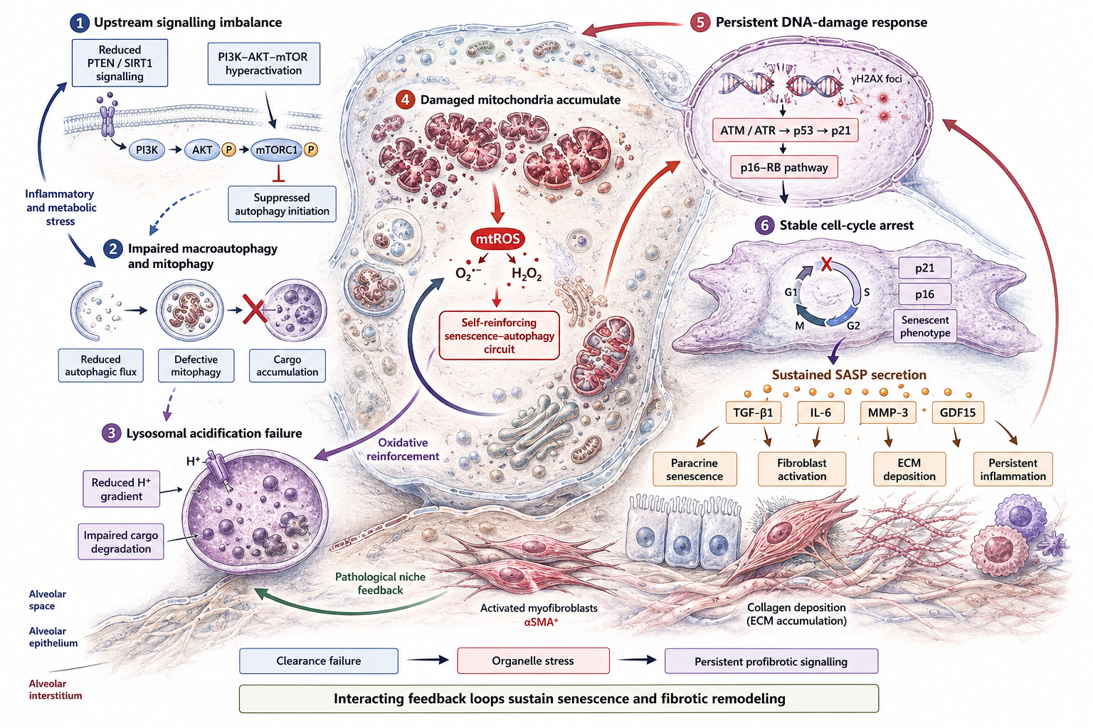
In the lungs of patients with IPF, impaired autophagic flux is biochemically characterized by a marked reduction in the lipidated form of microtubule-associated protein 1 light chain 3B (LC3B-II) and the pathological accumulation of p62/sequestosome-1 (Min et al., 2026; Torres-Machorro et al., 2025). This autophagic impairment is driven by hyperactive nutrient-sensing signaling via the PI3K-Akt-mTOR pathway, which directly suppresses the initiation of autophagy and the formation of autophagosomes (Min et al., 2026; Torres-Machorro et al., 2025). Furthermore, novel effectors such as ependymin-related 1 (EPDR1)---a lysosome-associated transmembrane glycoprotein that is significantly up-regulated in IPF fibroblasts and biological fluids---have been shown to negatively regulate lysosomal acidification (Southern & Gadre, 2025). The resulting loss of lysosomal acidity halts autophagic degradation, causing the intracellular accumulation of damaged macromolecular aggregates, persistent cellular stress, and the activation of p21- and p16-dependent senescent pathways (Southern & Gadre, 2025).
Figure 3 summarizes this cascade schematically, tracing the sequence from upstream PTEN/Sirt1 loss and mTOR hyperactivation through disabled macroautophagy, lysosomal acidification failure, mitochondrial ROS accumulation, chronic DNA damage signaling, and, ultimately, stable p21/p16-driven senescence with SASP output.
This proteostatic collapse is directly linked to the failure of mitophagy---the selective autophagic clearance of damaged mitochondria (Min et al., 2026). Impaired mitophagy leads to the progressive accumulation of large, dysmorphic, and dysfunctional mitochondria within senescent cells (Min et al., 2026; Zhang et al., 2024). These abnormal mitochondria exhibit decreased membrane potential, impaired fatty acid oxidation, and uncoupled respiration, resulting in the massive overproduction of mitochondrial reactive oxygen species (ROS) (Min et al., 2026; Zhang et al., 2024). This persistent oxidative stress acts as a potent DNA-damaging agent, sustaining the activation of ATM and ATR kinases, stabilizing p53, and locking the cells into an irreversible senescent state (Baurzhan et al., 2026; Li et al., 2023). Under chronic stress, senescent fibroblasts also up-regulate fatty acid binding protein 5 (FABP5), which competitively inhibits the ubiquitination and degradation of fatty acid synthase (FASN), disrupting lipid homeostasis and engaging a downstream uncoupling protein 2 (UCP2)-dependent mitochondrial axis that amplifies ROS generation and reinforces the senescent phenotype (Sun et al., 2026).
Senescent cells remain highly active metabolically and exert their pathological influence on the lung microenvironment through the robust secretion of the senescence-associated secretory phenotype (SASP) (Baurzhan et al., 2026; Torres-Machorro et al., 2025). The SASP is a complex, context-dependent cocktail of pro-inflammatory cytokines (IL-6, IL-8, IL-1β), chemokines, growth factors (TGF-β1, connective tissue growth factor \[CTGF\]), and extracellular matrix-remodeling metalloproteinases (such as MMP-3 and MMP-12) (Baurzhan et al., 2026; Melo-Narváez et al., 2024; Torres-Machorro et al., 2025). Additionally, metabolic chaperones like FABP5 are actively secreted as unconventional SASP factors by senescent myofibroblasts, directly activating paracrine β-catenin signaling in neighboring quiescent cells (Sun et al., 2026).
The chronic secretion of these SASP factors into the interstitial space induces paracrine \"bystander\" senescence in adjacent healthy epithelial cells and fibroblasts, creating a self-reinforcing, pathogenic circuit that drives spatial fibrotic expansion (Baurzhan et al., 2026; Min et al., 2026). Furthermore, the SASP establishes a highly immunosuppressive and pro-fibrotic niche by recruiting and metabolically reprogramming SPP1⁺/MERTK⁺ monocyte-derived alveolar macrophages, which actively cooperate with activated fibroblasts to write the physical scar at the fibroblast-focus edge (Deng et al., 2026).
2.4 Branch 4: Emergent Senotherapeutic Targets and Functional Reversal Strategies
Table 4 summarizes the translational landscape described below, spanning approved antifibrotics through early-phase senotherapeutics. The recognition of cellular
Table 3. Intercellular signaling pathways, crosstalk axes, and mechanical feedback loops sustaining fibrotic remodeling. This table details the principal ligand-receptor signaling axes that connect epithelial, mesenchymal, macrophage, and T-cell compartments in the IPF lung, including the canonical TGF-β1/Smad axis, PI3K/Akt/mTOR signaling, the Sirt1/HSF1/HSPs quality-control pathway, and the mitochondrial cGAS-STING mtDNA leakage axis. Each row specifies the ligands/receptors involved, the sender-receiver cell dialogue, downstream intracellular cascades, mechanobiological feedback mechanics, effects on cellular senescence programs, and autophagy/mitophagy regulatory consequences, with supporting citations.
|
Intercellular Crosstalk / Pathogenic Axis
|
Principal Ligands & Receptors Involved
|
Cell Type Dialogue (Sender → Receiver)
|
Intracellular Downstream Cascades
|
Mechanobiological Feedback Mechanics
|
Impact on Cellular Senescence Programs
|
Autophagy & Mitophagy Regulatory Effects
|
Grounding References (APA Style)
|
|
The Canonical TGF-β1/Smad Signaling Axis
|
TGF-β1 → TβRII and TβRI (ALK5)
|
Epithelial cells/Macrophages → Fibroblasts & Myofibroblasts.
|
Smad2/Smad3 phosphorylation and nuclear translocation with Smad4 co-regulator.
|
Promotes high-tension stress fiber formation and α-SMA upregulation, stiffness-driven integrin αv monomer feedback.
|
Directly induces epithelial senescence via p53 and p21 pathways.
|
Strong inhibitor of autophagic flux; suppresses metabolic clearance to reinforce the pro-fibrotic state.
|
Ding et al. (2021); Deng et al. (2026); Shakeel et al. (2023); Torres-Machorro et al. (2025)
|
|
The Non-Canonical PI3K/Akt/mTOR Pathway
|
Growth factors (PDGF, FGF, VEGF) → Tyrosine Kinase Receptors (RTKs)
|
Metaplastic Basal Cells → Resident Fibroblasts.
|
Phosphorylation of PI3K, activation of Akt, and hyperactivation of mTORC1 and mTORC2 complexes.
|
Upregulates the translation of structural matrix proteins and triggers EMT transcription factors SNAIL1/ZEB1.
|
Drives replicative senescence and persistent SASP translation.
|
Powerful inhibitor of macroautophagy and lysosomal biogenesis; accumulates damaged organelles and p62.
|
Deng et al. (2026); Gao et al. (2025); Min et al. (2026); Shakeel et al. (2023)
|
|
The Sirt1/HSF1/HSPs Quality-Control Pathway
|
GLP-1 receptor activation (e.g., via semaglutide)
|
Autocrine/Paracrine loops in Alveolar Epithelial cells.
|
Sirt1-mediated deacetylation of HSF1 (Heat Shock Factor 1).
|
Mitigates cellular tension and halts stiffness-induced DNA double-strand breaks.
|
Potently suppresses hydrogen peroxide-induced and age-associated cellular senescence.
|
Rescues mitochondrial bioenergetics and protects cells from oxidative damage via chaperone expression.
|
Qian et al. (2026)
|
|
The PERK/eIF2α/ATF4 Integrated Stress Response (ISR)
|
Endoplasmic reticulum (ER) stress sensors (p-PERK)
|
Injury-stressed AEC2 cells → Interstitial Myofibroblasts.
|
eIF2α phosphorylation leading to the selective translation of transcription factor ATF4.
|
Promotes alternative splicing of matrix components under biomechanical strain.
|
Drives cellular senescence via activation of ATM/ATR and upregulation of p53, p21, and p16.
|
Linked to defective mitophagy; ISR inhibition with ISRIB reverses cellular senescence and restores proteostasis.
|
Baurzhan et al. (2026); Li et al. (2023)
|
|
The Osteopontin (SPP1) / CD44 Macrophage-Fibroblast Axis
|
SPP1 (Osteopontin) → CD44 and integrin receptors
|
Proliferating SPP1⁺/MERTK⁺ Macrophages → CTHRC1⁺ Myofibroblasts.
|
Intracellular AKT and β-catenin nuclear signaling pathways.
|
Drives cell motility and directed migration along pathological extracellular matrix stiffness gradients.
|
SASP-derived SPP1 reinforces paracrine "bystander" senescence in adjacent healthy epithelium.
|
Suppresses basic autophagic clearance while reinforcing survival advantages and apoptosis resistance.
|
Deng et al. (2026)
|
|
The Interleukin-11 (IL-11) "TIME" Signaling Loop
|
IL-11 → gp130 and IL-11RA receptor complexes
|
KRT5⁻/KRT17⁺ Metaplastic Basaloid cells → AECs & Fibroblasts.
|
Activation of MEK/ERK ("TIME" pathway) and gp130-mediated STAT3 phosphorylation.
|
Upregulates matrix stiffness and myofibroblast contractility.
|
Amplifies the pro-inflammatory SASP profile and triggers stress-induced premature senescence.
|
Interferes with autophagic degradation pathways, locking AT2 cells in arrested transitional states.
|
Yu et al. (2025)
|
|
The FABP5/FASN Extracellular Paracrine Loop
|
Secreted extracellular FABP5 → adjacent fibroblast membrane receptors
|
Senescent Myofibroblasts → Quiescent resident Fibroblasts.
|
Intracellular β-catenin signaling activation.
|
Amplifies tissue-wide tension and establishes a mechanical memory loop via matrix remodeling.
|
Induces stable cell-cycle arrest in neighboring normal fibroblasts via paracrine propagation.
|
Impairs mitophagy, accumulating dysfunctional and dysmorphic mitochondria.
|
Sun et al. (2026)
|
|
The CD8⁺ T Cell-Derived Granzyme K Pro-Fibrotic Axis
|
Extracellular granzyme K (GZMK)
|
Senescent exhausted CD8⁺ T cells → Airway/Alveolar Epithelial cells.
|
Up-regulates MHC II presentation and stimulates intracellular MMP28 production.
|
Accelerates tissue destruction (honeycombing) and alters regional parenchymal compliance.
|
GZMK-exposed lung epithelial cells undergo premature senescence and express high levels of IL-8.
|
Compromises cellular quality control, driving the accumulation of senescent, hypofunctional CD8+ compartments.
|
Okereke et al. (2026)
|
|
The Integrin-FAK-YAP/TAZ Mechanotransduction Loop
|
Integrins αvβ6, αvβ1, and αvβ3 → mechanical matrix ligands
|
Remodeled Extracellular Matrix → Epithelial Cells and Fibroblasts.
|
Focal Adhesion Kinase (FAK)/SRC pathway activation, halting YAP/TAZ phosphorylation to allow nuclear entry.
|
Central driver of mechanical memory; translates matrix rigidity into active fibrogenic gene transcription.
|
Sustained mechanical stress drives lung structural cells toward a chronic senescent phenotype.
|
Suppresses autophagy to protect activated myofibroblasts from metabolic exhaustion on stiff substrates.
|
Min et al. (2026); Torres-Machorro et al. (2025); Zhang et al. (2026)
|
|
The Mitochondrial cGAS-STING mtDNA Leakage Axis
|
Free cytosolic mitochondrial DNA (mtDNA) → cGAS dimers
|
Stress-injured AT2 cells → Surrounding Alveolar Environment.
|
STING oligomerization, leading to the recruitment and activation of interferon regulatory factors.
|
Potentiated by mechanical stretch and physical alveolar microinjury.
|
Triggers deep senescence arrest, driving high transcription of p21 and p16.
|
Mitophagy failure prevents the clearance of fractured mitochondria, spilling mtDNA and sustaining the inflammatory loop.
|
Gong et al. (2026); Min et al. (2026); Zhang et al. (2026)
|
Table 4. Translational therapeutic landscape of approved, investigational, and emerging senotherapeutic modalities in idiopathic pulmonary fibrosis. This table compares current standard-of-care antifibrotics (pirfenidone, nintedanib) with investigational pathway-directed agents (bexotegrast, nerandomilast, admilparant, inhaled treprostinil) and emerging senotherapeutics (dasatinib plus quercetin, EF24, curculigoside, ISRIB), summarizing target molecule/pathway, mechanistic class, preclinical efficacy, current clinical trial status, functional outcomes, and adverse-effect/safety considerations, with supporting citations.
|
Therapeutic Intervention / Drug
|
Target Molecule / Pathway
|
Class / Mechanistic Action (Senolytic, etc.)
|
Preclinical Efficacy Profile
|
Current Clinical Status / Trial ID
|
Functional Outcomes & Endpoints
|
Adverse Effects & Safety Considerations
|
Grounding References (APA Style)
|
|
Nintedanib (Ofev®)
|
VEGFR, FGFR, PDGFR tyrosine kinases, and Src family
|
Multitargeted tyrosine kinase inhibitor; suppresses growth factor-stimulated fibroblast contraction and motility.
|
Inhibits fibrocyte activity, prevents M2 macrophage polarization, and downregulates procollagen I and TGF-β.
|
FDA-approved for IPF and progressive pulmonary fibrosis.
|
Significantly reduces the annual rate of Forced Vital Capacity (FVC) decline; lowers acute exacerbation risk.
|
High incidence of diarrhea, nausea, vomiting, weight loss, and potential hepatotoxicity.
|
Baurzhan et al. (2026); Deng et al. (2026); Shakeel et al. (2023)
|
|
Pirfenidone (Esbriet®)
|
TGF-β1, TNF-α, and downregulates HSP47
|
Small molecule anti-fibrotic; reduces fibroblast proliferation, TGF-β-induced Smad3 phosphorylation, and collagen deposition.
|
Lowers hydroxyproline accumulation, reduces fibrocytes, and displays anti-inflammatory and anti-oxidant properties.
|
FDA-approved for IPF.
|
Slows FVC decline over 52 weeks (CAPACITY/ASCEND trials) by approximately 47.9%.
|
Gastrointestinal dyspepsia, nausea, photosensitivity rash, headache, fatigue, and liver enzyme elevation.
|
Baurzhan et al. (2026); Deng et al. (2026); Shakeel et al. (2023)
|
|
Dasatinib + Quercetin (D+Q)
|
Src kinase, BCL-xL, and senuous survival pathways
|
Dual Senolytic Therapy; selectively kills senescent lung fibroblasts to reduce SASP burden.
|
Eliminates senescent cells, improves lung compliance, and reduces collagen burden in bleomycin models.
|
Completed Phase I Clinical Trial (NCT02874989).
|
Demonstrated safe tolerability and clinically meaningful improvements in 6-minute walk distance and physical performance.
|
Phase I trial highlighted gastrointestinal discomfort and a nearly threefold increase in acute exacerbation rates.
|
Baurzhan et al. (2026); Deng et al. (2026); Min et al. (2026); Torres-Machorro et al. (2025)
|
|
Bexotegrast (PLN-74809)
|
Integrins αvβ6 and αvβ1
|
Dual Integrin Inhibitor; blocks the integrin-mediated activation of latent TGF-β.
|
Decreases COL1A1, COL3A1, and α-SMA expression in human lung tissue explants and fibroblast cultures.
|
Phase IIb/III Adaptive Clinical Evaluation (NCT06097260).
|
INTEGRIS-IPF trial showed stable/improved FVC in 71% of patients and decreased N-terminal type III collagen propeptide.
|
Generally well-tolerated; mild diarrhea (~30%) is the most common adverse event.
|
Deng et al. (2026); Torres-Machorro et al. (2025); Moua et al. (2024)
|
|
Nerandomilast (BI 1015550)
|
Phosphodiesterase 4B (PDE4B)
|
Selective PDE4B Inhibitor; increases intracellular cAMP levels to suppress pro-inflammatory/pro-fibrotic cascades.
|
Attenuates fibroblast activation, myofibroblast differentiation, and macrophage-derived profibrotic cytokines.
|
Phase III FIBRONEER-IPF Clinical Trial (NCT05321069, ongoing).
|
Phase II trial demonstrated FVC stabilization and slowed rate of lung function decline over 12 weeks.
|
Gastrointestinal adverse events, primarily diarrhea and nausea (~27%), with a ~15% trial discontinuation rate.
|
Deng et al. (2026); Moua et al. (2024)
|
|
BMS-986278 (Admilparant)
|
Lysophosphatidic Acid Receptor 1 (LPAR1)
|
Oral LPA1 Antagonist; blocks lysophosphatidic acid-mediated fibroblast migration, vascular leak, and epithelial apoptosis.
|
Reduces collagen accumulation, pulmonary tissue density, and improves compliance in rodent bleomycin models.
|
Phase III Clinical Evaluation (NCT06003426, ongoing).
|
Phase II trial demonstrated a ~62% reduction in the annual rate of FVC decline; well-tolerated vs placebo.
|
Favorable safety profile, lacking the severe hepatobiliary toxicity seen in first-generation antagonists (e.g., BMS-986020).
|
Deng et al. (2026); Moua et al. (2024)
|
|
Inhaled Treprostinil
|
Prostacyclin Receptor / cAMP elevation
|
Prostacyclin Analog / Vasodilator; inhibits fibroblast migration and targets mitochondrial quality control in IPF cells.
|
Attenuates pulmonary remodeling and normalizes dysmorphic mitochondrial architecture.
|
Phase III TETON-IPF and TETON-2 Clinical Trials (NCT05255991, ongoing).
|
Post-hoc analysis of the INCREASE study demonstrated significantly lower FVC decline and reduced risk of cardiopulmonary hospitalization.
|
Local airway irritation, including persistent cough (44%), throat discomfort, and mild headaches (28%).
|
Deng et al. (2026); Moua et al. (2024)
|
|
Integrated Stress Response Inhibitor (ISRIB)
|
Eukaryotic translation initiation factor 2B (eIF2B) allosteric activation
|
Integrated Stress Response Inhibitor; restores cellular translation and maintains proteostasis under chronic ER stress.
|
Antagonizes silica-induced and bleomycin-induced AEC2 senescence, significantly reducing collagen type I, α-SMA, and fibronectin.
|
Preclinical development / Translational Optimization.
|
Restores normal lung compliance, improves dynamic resistance, and eliminates senescent Krt8⁺ transitional cells in vivo.
|
Rodent experiments demonstrate no significant systemic adverse events, providing a favorable safety window compared to classic anti-fibrotics.
|
Li et al. (2023)
|
|
EF24 (Curcumin Analogue)
|
Phosphatase and Tensin Homolog (PTEN) stabilization
|
Senomorphic and Mitophagy Enhancer; suppresses AKT/mTOR/NF-κB signaling and restores mitochondrial membrane potential.
|
Reduces SA-β-gal, P21, and PAI-1 expression; rescues dysmorphic swollen mitochondria and halts senescent-induced fibroblast activation.
|
Preclinical optimization; identified as the most promising anti-aging compound among 12 candidates.
|
Promotes weight recovery, improves dynamic lung consolidation on micro-CT, and restores dynamic fibrosis resolution in aged TBI-mouse models.
|
Shows high oral bioavailability (60% in mice) and exhibits no cytotoxic or apoptotic induction in non-senescent normal cells.
|
Zhang et al. (2024b)
|
|
Curculigoside (CCG)
|
Tripartite motif-containing protein 72 (Trim72) and GSK3β/SIRT1-P300 pathway
|
Natural Senomorphic and Antioxidant Agent; facilitates the ubiquitination and degradation of P300 to reduce acetylation of antioxidant enzymes.
|
Delays the natural senescence of lung fibroblasts and hydrogen peroxide-induced AEC senescence in vitro.
|
Preclinical evaluation in bleomycin-induced and D-galactose-induced aging models.
|
Restores systemic antioxidant enzyme activity (SOD1, SOD2, catalase) and robustly alleviates endoplasmic reticulum stress and tissue stiffness.
|
Well tolerated in rodent toxicity assays with no evidence of systemic target organ toxicity or structural weight loss.
|
Tang & wei (2024)
|
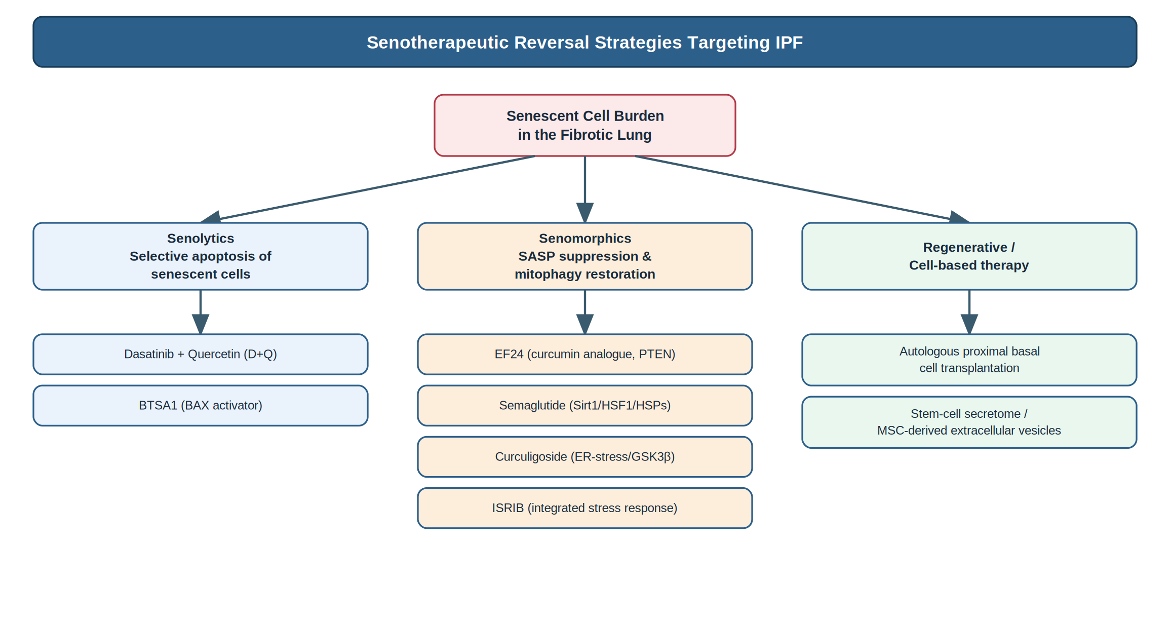
senescence as a central organizing mechanism in the pathogenesis of IPF has shifted the therapeutic focus from broad, non-selective anti-fibrotic agents toward pathway-directed molecular interventions and cellular therapies (Baurzhan et al., 2026; Deng et al., 2026). The clinical and preclinical landscape of these reversal strategies can be organized into three distinct therapeutic classes: senolytics, senomorphics, and regenerative progenitor transplantations (Baurzhan et al., 2026; Deng et al., 2026). Figure 4 organizes this therapeutic landscape into its three constituent arms — senolytics, senomorphics, and regenerative cell-based approaches
2.5. Senolytics: Selective Elimination of Senescent Cells
Senolytics are small molecules designed to selectively eliminate senescent cells by transiently disabling the pro-survival and anti-apoptotic networks (SCAPs) that protect them from their own pro-inflammatory secretome (Baurzhan et al., 2026).
- Dasatinib + Quercetin (D+Q): The combination of the tyrosine kinase inhibitor dasatinib and the natural flavonoid quercetin targets key pro-survival signaling pathways, including the BCL-2 family and PI3K/Akt signaling, thereby selectively inducing apoptosis in senescent cells (Baurzhan et al., 2026). In preclinical models of bleomycin-induced pulmonary fibrosis, the administration of D+Q successfully reduced the burden of senescent cells, attenuated collagen deposition, and restored lung compliance (Min et al., 2026). Crucially, a first-in-human, open-label, pilot clinical study evaluating intermittent oral administration of D+Q in 14 patients with IPF demonstrated that the treatment was well-tolerated and yielded clinically meaningful improvements in physical function---including six-minute walk distance and chair-stand times---although significant changes in forced vital capacity (FVC) were not observed over the brief treatment course (Baurzhan et al., 2026; Justice et al., 2019).
Targeting Myofibroblast Apoptosis: Emerging senolytic candidates, such as BTSA1, directly target senescent myofibroblasts by activating the pro-apoptotic protein BAX, effectively bypassing apoptosis resistance and reducing the established fibrotic burden in mouse models (Min et al., 2026).
2.6 Senomorphics: Modulating the Senescent Secretory Phenotype
Senomorphics are therapeutic agents that suppress or neutralize the SASP secretome and restore cellular quality-control systems without killing the senescent cells directly (Baurzhan et al., 2026).
Curcumin Analogue EF24: A highly bioactive monoketone derivative of curcumin, EF24 has emerged as a potent senomorphic candidate (Zhang et al., 2024). EF24 treatment significantly reduces senescence biomarkers (SA-β-gal, PAI-1, p21) and suppresses SASP secretion (IL-6, IL-1β) in bleomycin-induced senescent AECs (Zhang et al., 2024b). Mechanistically, EF24 up-regulates and stabilizes the tumor suppressor PTEN, which subsequently suppresses the hyperactive Akt/mTOR/NF-κB signaling cascade (Zhang et al., 2024). Furthermore, EF24 promotes mitophagy, facilitating the clearance of damaged mitochondria and reducing mitochondrial ROS production in senescent epithelial cells (Zhang et al., 2024). In vivo studies demonstrate that EF24 attenuates established fibrosis and improves survival in both acute and chronic phase mouse models of lung injury, highlighting its capacity to restore mitochondrial quality control (Zhang et al., 2024).
Semaglutide: A glucagon-like peptide-1 (GLP-1) receptor agonist, semaglutide has been shown to inhibit oxidative stress and cellular senescence in the lung (wei & Si-yuan, 2026). By suppressing the production of mitochondrial ROS and inflammatory cytokines, semaglutide limits bleomycin-induced fibrogenesis, representing a promising candidate for metabolic drug repurposing (wei & Si-yuan, 2026).
Curculigoside (CCG): A natural bioactive compound that targets cellular senescence by suppressing endoplasmic reticulum stress (ERS) and the unfolded protein response (UPR) (Tang & wei, 2024). CCG prevents crystalline silica- and bleomycin-induced senescence in AEC2s and lung fibroblasts by modulating the GSK3β/ERS pathway, thereby restoring cellular redox balance (Tang & wei, 2024).
ISRIB (Integrated Stress Response Inhibitor): An engineered small-molecule inhibitor of the integrated stress response that acts by rescuing translational initiation rates (Li et al., 2023). In models of silica-induced pulmonary fibrosis and ERS-stressed AECs, ISRIB administration effectively reverses cellular senescence, restores the regenerative capacity of epithelial progenitors, and attenuates downstream fibrosing cascades, highlighting the therapeutic potential of targeting proteostatic stress (Li et al., 2023).
2.7. Regenerative and Cell-Based Strategies
- Autologous Basal Cell (BC) Transplantation: A pioneering regenerative approach that aims to replace senescent, dysfunctional distal airway progenitors with healthy, non-senescent stem cells harvested from the patient\'s own proximal airways (Snyder et al., 2025). Clinical characterization reveals a striking spatial pattern of senescence along the proximal-distal axis of the IPF airway tree: while basal cells residing in the distal bronchioles demonstrate advanced functional impairment and high senescent marker expression, basal cells situated in the proximal small bronchi maintain a completely non-senescent phenotype (Snyder et al., 2025). These proximal BCs (Krt5+/p63+/ITGA6+/NGFR+) retain their self-renewal capacity, genomic stability, and multi-lineage differentiation potential (Snyder et al., 2025). In a landmark pilot clinical trial, three patients with advanced IPF received autologous proximal BCs transplanted via bronchoscopic instillation (Snyder et al., 2025). The therapy was exceptionally safe, with no severe adverse events reported, and yielded significant clinical improvements in lung volume (FVC increase), exercise capacity (6-minute walk distance), and small airway function over a 6-month follow-up period, demonstrating the translational feasibility of cellular replacement (Snyder et al., 2025).
Stem Cell Secretomes & Extracellular Vesicles: The nebulized delivery of lung stem cell-derived secretions (LSC-Sec) or mesenchymal stem cell-derived extracellular vesicles (MSC-EVs) represents a cell-free alternative (Gao et al., 2025). These extracellular vesicles deliver a specialized cargo of anti-fibrotic microRNAs, proteins, and mitochondrial-protective factors that modulate TGF-β signaling, enhance endogenous epithelial repair, and reduce the senescent secretome within the fibrotic niche (Baurzhan et al., 2026; Gao et al., 2025).
Cellular senescence is no longer viewed as a passive bystander of physiological aging, but as a central, highly integrated pathogenic network that actively coordinates the progressive, irreversible remodeling of the lung in IPF (Baurzhan et al., 2026; Deng et al., 2026). The complex crosstalk between senescent epithelial progenitors locked in regenerative arrest, highly glycolytic and mechanosensitive fibroblasts, and SPP1⁺ macrophages forms a self-reinforcing network that sustains the fibrotic microenvironment (Deng et al., 2026; Min et al., 2026). Traditional anti-fibrotic therapies, such as pirfenidone and nintedanib, fail to arrest or reverse this disease process because they target individual downstream pathways rather than the underlying cellular quality-control network (Baurzhan et al., 2026; Deng et al., 2026).
The emergence of senotherapeutic strategies represents a major conceptual shift in the clinical management of IPF (Baurzhan et al., 2026). By leveraging senolytics to selectively eliminate senescent cells, senomorphics (such as EF24, ISRIB, and CCG) to reprogram metabolic and proteostatic quality-control networks, and autologous basal cell transplantation to physically restore the injured epithelial stem cell niche, modern medicine is positioned to move beyond the palliative slowing of decline toward true structural and functional lung regeneration (Baurzhan et al., 2026; Snyder et al., 2025).