
Integrative Biomedical Research

Integrative Biomedical Research (Journal of Angiotherapy) | Online ISSN  3068-6326
463
Citations
1.8m
Views
746
Articles
Your new experience awaits. Try the new design now and help us make it even better
Switch to the new experience
Figures and Tables
REVIEWS   (Open Access)

Senescent Cell Heterogeneity and Precision Clearance Strategies: From Molecular Mechanism to Clinical Translation

Ragini Patel 1*, Yogendra Sahu 2

+ Author Affiliations

Integrative Biomedical Research 10 (1) 1-8 https://doi.org/10.25163/biomedical.10110880

Submitted: 04 November 2025 Revised: 20 December 2025  Published: 03 January 2026 


Abstract

Cellular senescence, an essentially irreversible growth arrest first described in cultured fibroblasts, is now recognized as a druggable driver of multiple age-related diseases, yet clinical progress has been slowed by the marked heterogeneity of senescent cells across tissues, inducing stressors, and disease states. We conducted a structured narrative synthesis of peer-reviewed literature senescence biology, single-cell senotyping, and senotherapeutic development, following informed search and appraisal strategy across PubMed/MEDLINE, Scopus, and Web of Science. Sixty-plus primary and review sources were synthesized into four evidence tables spanning first-generation agents, precision platforms, multi-tissue biomarkers, and human trials. First-generation senolytics (dasatinib plus quercetin, fisetin, navitoclax) demonstrate proof-of-concept efficacy but are constrained by dose-limiting toxicities, whereas next-generation platforms—PROTAC degraders, uPAR-directed CAR-T cells, senolytic vaccines, and galactose-caged prodrugs—achieve markedly higher predicted selectivity with lower off-target risk, operating through convergent mechanistic nodes upstream of tissue-level inflammaging. Early-phase human trials corroborate target engagement but remain underpowered for definitive efficacy claims. Precision senotherapy is mechanistically justified and clinically nascent; its translational success will depend on resolving the biomarker bottleneck and matching senotype-specific vulnerabilities to intervention class.

Keywords: cellular senescence; senolytics; senomorphics; SASP; single-cell senotyping; precision senotherapy; healthspan

1. Introduction

Aging is, if you look at it honestly, the single largest unaddressed risk factor in medicine — it rarely causes any one disease outright, yet it quietly loads the dice for nearly all of them (Sun, 2023). For much of the twentieth century, clinicians treated cardiovascular disease, neurodegeneration, cancer, and type 2 diabetes as though they told separate biological stories, each with its own textbook chapter and its own specialist (Paez-Ribes et al., 2019; Wyles et al., 2022). That separation is starting to look less like biology and more like convenience. The geroscience hypothesis — still argued over in places, but increasingly hard to dismiss — proposes that these seemingly unrelated conditions share upstream drivers, and that intervening on those shared drivers might blunt several diseases at once rather than chase them one at a time (Chaib et al., 2022; Cossarizza, 2026).

Among the candidate drivers, cellular senescence has probably drawn the most sustained attention, in part because it sits at an unusually convenient intersection: it is mechanistically tractable, and — unlike, say, epigenetic drift — it is a cell state, which means it can in principle be targeted and cleared (Chmielewski, 2026; Fu & Zhou, 2025). The phenomenon itself is not new. Leonard Hayflick and Paul Moorhead described it in 1961, watching human diploid fibroblasts simply stop dividing after a finite number of passages — the so-called Hayflick limit (Hayflick & Moorhead, 1961; Sun, 2023). For decades this was treated as a curiosity of the culture dish. It is now understood rather differently: as a stress-induced, essentially irreversible cell-cycle arrest that unfolds in vivo in response to a wide range of insults — telomere attrition, persistent DNA damage, oncogene activation, mitochondrial dysfunction, reactive oxygen species, and therapy-induced genotoxic stress among them (Chaib et al., 2022; Kusmanto, 2025; Ozdemir et al., 2025). Senescent cells do not die quietly; they resist apoptosis and remain, somewhat perversely, highly metabolically active (Liao et al., 2021).

There is, frustratingly, no single molecular flag that marks a senescent cell wherever it happens to be. Researchers instead lean on a multiparametric signature — elevated cyclin-dependent kinase inhibitors, lysosomal senescence-associated β-galactosidase activity at pH 6.0, cellular hypertrophy, loss of lamin B1 at the nuclear envelope, persistent γ-H2AX foci marking unresolved DNA damage, chromatin remodeling, and intracellular lipofuscin accumulation (Chmielewski, 2026; Cohn et al., 2023; Erusalimsky, 2021; Gasek et al., 2021). None of these, taken alone, is diagnostic. Together, they are workable.

It would be a mistake, though, to think of senescence as simply bad. Transient senescence does real physiological work — it helps pattern the embryo, closes wounds, and, notably, acts as an autonomous brake on tumor formation (Alum et al., 2025; Paez-Ribes et al., 2019). The trouble starts when senescent cells stop clearing on schedule and instead accumulate. This is driven largely by the senescence-associated secretory phenotype, or SASP — a loosely regulated, tissue- and context-dependent cocktail of pro-inflammatory cytokines (IL-6, IL-1β, TNF-α), chemokines such as MCP-1, matrix-degrading metalloproteinases, and extracellular vesicles (Chmielewski, 2026; Cossarizza, 2026; Erusalimsky, 2021). Persistent SASP output produces a low-grade, sterile inflammatory state that has come to be called “inflammaging” — a term that has aged well, so to speak — which erodes tissue architecture, exhausts local stem-cell niches, and pushes neighboring healthy cells into so-called bystander senescence (Cossarizza, 2026; Fu & Zhou, 2025).

This is where the field runs into its central complication, and arguably the reason this review exists at all: senescent cells are not one thing. They differ, sometimes dramatically, by tissue of origin, by the nature of the inciting stressor (replicative exhaustion versus oncogene-induced versus therapy-induced versus metabolic stress), by how long they have been senescent, and even by the sex of the organism (Mansfield et al., 2024; Ozdemir et al., 2025; Sun, 2023; Zhang et al., 2026). A senescent pre-adipocyte and a senescent endothelial cell do not share a transcriptome, a SASP composition, or, crucially, a dependency on the same survival pathway (Sun, 2023; Zhang et al., 2026). This variability sat largely unresolved until single-cell and single-nucleus RNA sequencing, spatial transcriptomics, and machine-learning classifiers such as SenCID, SenePy, and SenMayo made it possible to actually see it — to watch senescent cells move through heterogeneous trajectories rather than collapse into a single uniform cluster (Cohn et al., 2023; Fu & Zhou, 2025; Mansfield et al., 2024). The practical upshot is sobering: because a universal senescent marker does not exist, a clearance strategy that works beautifully against one subpopulation may do nothing — or, worse, cause harm — against another (Cohn et al., 2023; Mansfield et al., 2024).

In response, researchers have built senotherapeutics, broadly split into two families. Senolytics kill senescent cells outright by transiently disabling the senescent-cell anti-apoptotic pathways (SCAPs) — Bcl-2/Bcl-xL, PI3K/Akt, p53/p21, ephrins, HSP90 — that senescent cells rely on to survive their own toxic secretome (Chaib et al., 2022; Gasek et al., 2021). The first generation of these agents — dasatinib plus quercetin (D+Q), fisetin, and the Bcl-2-family inhibitor navitoclax (ABT-263) — supplied the field’s original proof of concept, improving physical function, easing fibrosis, and extending healthspan in animal models, with early human trials in idiopathic pulmonary fibrosis, diabetic kidney disease, and Alzheimer’s disease confirming at least safety and target engagement (Chaib et al., 2022; Fu & Zhou, 2025; Riessland et al., 2024; Zhang et al., 2026). But these agents are blunt instruments: navitoclax causes dose-limiting thrombocytopenia and neutropenia, and cell-type specificity remains incomplete across the class (Fu & Zhou, 2025; Zhang et al., 2026).

Senomorphics take a gentler route — dialing down the inflammatory output of the SASP through mTOR, NF-κB, JAK/STAT, or p38 MAPK inhibition without forcing the cell to die (Ozdemir et al., 2025; Wyles et al., 2022). Repurposed drugs like metformin and rapamycin fall into this camp (Cossarizza, 2026; Paez-Ribes et al., 2019). The trade-off is dosing: because the cells survive, senomorphics need continuous administration, which trades the intermittent “hit-and-run” convenience of senolytics for a longer exposure window and, presumably, more chances for something to go wrong (Cohn et al., 2023; Fu & Zhou, 2025).

These limitations have pushed the field toward precision senotherapy — immune-based senolysis using CAR-T cells against uPAR or senolytic vaccines against GPNMB and CD153; targeted proteolysis via PROTACs that route Bcl-xL or BRD4 to E3 ligases for degradation while sparing platelet-rich tissue; and galactose-caged prodrugs and nanoparticles that stay inert until cleaved by the high SA-β-Gal activity unique to senescent lysosomes (Kusmanto, 2025; Paez-Ribes et al., 2019; Riessland et al., 2024; Zhang et al., 2026).

To connect this mechanistic detail to something clinically useful, this review asks three questions: (1) how does in vivo transcriptomic heterogeneity dictate a senescent cell’s susceptibility to a given SCAP inhibitor, and can tissue-specific “senotypes” predict responsiveness; (2) can precision platforms — uPAR-CAR-T, Bcl-xL PROTACs, SA-β-Gal-responsive prodrugs — genuinely sidestep the toxicities and resistance mechanisms (e.g., Mcl-1 upregulation) that limit first-generation agents; and (3) what are the long-term safety implications of chronic SASP suppression relative to intermittent senolysis, particularly for regeneration, wound healing, and immune surveillance. Guided by these questions, we set out to (a) systematically map layers of senescent heterogeneity using recent multi-omics and spatial data, (b) critically compare first- and next-generation senotherapeutics on efficacy, pharmacokinetics, and safety, and (c) sketch a translational roadmap that confronts the biomarker bottleneck and the design of trials suited to geriatric and oncology populations.
2. Senescent Cell Heterogeneity: Mechanisms, Biomarkers, and Senotherapeutic Progress

2.1 Molecular Mechanisms and Biomarker Signatures of Cellular Senescence

At its core, cellular senescence is an end-stage change in cell fate — essentially irreversible cell-cycle arrest paired with macromolecular damage, metabolic reprogramming, and a stubborn resistance to apoptosis (Chmielewski, 2026; Riessland et al., 2024). It was first noticed, almost by accident, by Hayflick and Moorhead in 1961 as the finite replicative ceiling of primary human fibroblasts, and was initially dismissed as a culture-dish artifact tied to telomere erosion (Riessland et al., 2024; Sun, 2023). Modern biogerontology has since rehabilitated it into one of the central hallmarks of biological aging in vivo (Chmielewski, 2026) — a durable stress response triggered by persistent DNA-damage-response activation, oxidative stress, mitochondrial dysfunction, ROS accumulation, oncogenic activation, epigenetic remodeling, and therapy-induced genotoxic stress (Alum et al., 2025; Ozdemir et al., 2025).

It also is not a single event but a process, unfolding from early to full and, eventually, “deep” senescence (Liao et al., 2021). Mechanistically, this progression converges on two tumor-suppressor axes — p53/p21 and p16INK4a-Rb — that operate in both mitotic and post-mitotic cells (Gasek et al., 2021; Ozdemir et al., 2025). Telomere shortening or double-strand breaks activate ATM and ATR kinases, which stabilize p53 and upregulate p21, inhibiting CDK2 and driving Rb into its hypo-phosphorylated, cell-cycle-blocking state (Ozdemir et al., 2025; Wyles et al., 2022). A parallel route, more associated with chronological aging and cumulative stress, activates p16INK4a to inhibit CDK4/6 and reinforce the same Rb-mediated arrest (Liao et al., 2021; Wyles et al., 2022).

Given the field’s ongoing failure to land on one gold-standard biomarker, investigators have settled instead on a multiparametric signature (Cohn et al., 2023): elevated p16 and p21, high lysosomal SA-β-Gal activity at pH 6.0, senescence-associated heterochromatin foci, persistent γ-H2AX damage foci, and the enlarged, flattened cellular morphology that is, honestly, often the first visual clue a trained eye picks up (Alum et al., 2025; Chmielewski, 2026; Ozdemir et al., 2025). Senescent cells also lose lamin B1 at the nuclear envelope — degraded via LC3-mediated autophagy in oncogene-induced senescence, or simply transcriptionally silenced during replicative senescence (Liao et al., 2021) — and this nuclear disruption derepresses transposable elements like LINE-1, whose re-insertion into host DNA compounds genomic instability and feeds forward into the inflammatory cascade (Dey et al., 2023; Riessland et al., 2024; Wyles et al., 2022). Intracellularly, accumulating lipofuscin — an autofluorescent, essentially non-degradable lysosomal aggregate — has more recently emerged as a sensitive marker of cumulative cellular stress in both dividing and post-mitotic tissue (Chmielewski, 2026; Riessland et al., 2024).

2.2 The Senescence-Associated Secretory Phenotype and Tissue-Level Consequences

Senescent cells leave the cell cycle permanently, but they do not go quiet. If anything, they become secretorily hyperactive, releasing a dense, tissue-specific mixture of cytokines, chemokines, ECM-degrading proteases, growth factors, bioactive lipids, and extracellular vesicles — the SASP (Alum et al., 2025; Chmielewski, 2026; Ozdemir et al., 2025). Production of this secretome is orchestrated at multiple regulatory levels by NF-κB, the transcriptional co-factor C/EBPβ, and p38 MAPK (Liao et al., 2021; Ozdemir et al., 2025), and is further shaped by metabolic sensing through mTOR and by the cGAS-STING pathway, which is triggered when chromatin fragments or mitochondrial DNA leak into the cytosol (Fu & Zhou, 2025; Liao et al., 2021). Once engaged, these cascades drive robust output of IL-6, IL-1β, TNF-α, MCP-1, IL-8, and a suite of matrix metalloproteinases (MMP-1, -3, -9, -12) (Liao et al., 2021; Riessland et al., 2024; Syed et al., 2026) (Table 3).

The biological logic here is a textbook case of antagonistic pleiotropy (Chmielewski, 2026). In the right context — and only briefly — senescence is useful: it helps pattern developing limb buds, recruits immune cells to wounds via PDGF-AA secretion, restrains fibrosis, and functions as a cell-autonomous barrier against malignant transformation (Liao et al., 2021; Ozdemir et al., 2025). Under these acute, self-limiting conditions, senescent cells essentially arrange for their own removal, recruiting macrophages, NK cells, and T lymphocytes (Chmielewski, 2026; Erusalimsky, 2021). Persistent senescence is a different story. As organisms age, cumulative cellular damage collides with immunosenescence — the gradual decline of the immune system’s clearance capacity — and senescent cells begin to pile up across tissues rather than being cleared on schedule (Chmielewski, 2026; Ozdemir et al., 2025). Once entrenched, they exert wide non-cell-autonomous damage: SASP factors induce bystander senescence in neighboring healthy cells, deplete stem-cell niches, degrade ECM architecture, and sustain inflammaging (Chmielewski, 2026; Ozdemir et al., 2025; Sun, 2023). This accumulation has been causally implicated in idiopathic pulmonary fibrosis, COPD, atherosclerosis, chronic kidney disease, osteoarthritis, type 2 diabetes, and Alzheimer’s and Parkinson’s disease (Chmielewski, 2026; Ozdemir et al., 2025).

2.3 Deciphering Senescent Cell Heterogeneity: The Dawn of Single-Cell Senotyping

If there is one barrier standing most squarely between senescence biology and clinical translation, it is heterogeneity (Chmielewski, 2026; Sun, 2023). Senescent phenotype, transcriptomic identity, and secretome composition all vary — by tissue lineage, by inducing trigger (replicative, oncogene-induced, oxidative, therapy-induced), by how far along the senescence timeline a cell has traveled, and even by sex and genetic background (Cohn et al., 2023; Ozdemir et al., 2025; Sun, 2023). Single-cell work on human fibroblasts, for instance, shows that replicative senescence and oncogene-induced senescence produce recognizably different transcriptomic signatures and secretory outputs, despite superficially “looking” the same under a microscope (Liao et al., 2021; Sun, 2023). Even within a single tissue, senescent endothelial cells, fibroblasts, and resident macrophages diverge sharply in gene expression and in which survival pathway they depend on — evidence, cumulatively, that there is no such thing as a singular “senescent state” (Cohn et al., 2023; Ozdemir et al., 2025).

Resolving this diversity has taken high-resolution tooling: scRNA-seq, snRNA-seq, spatial transcriptomics, and high-throughput imaging flow cytometry have collectively let investigators map senescence at cellular resolution inside intact, complex tissue (Cohn et al., 2023; Sun, 2023). Several computational platforms have emerged to make sense of the resulting data. SenCID is a machine-learning classifier trained across species and tissues that sorts senescent cells into six senescence identities, or SIDs, based on shared molecular signatures (Cohn et al., 2023; Fu & Zhou, 2025). SenePy, trained on large mouse and human aging atlases spanning dozens of tissues, maps heterogeneous senescent populations and reconstructs tissue-specific trajectories (Cohn et al., 2023; Ozdemir et al., 2025). SenMayo, a 125-gene cross-species panel built from human bone biopsies and heavily weighted toward SASP genes, has proven useful for estimating senescent burden and tracking pharmacological target engagement

Table 1. Foundational First-Generation Small-Molecule Senotherapeutics. Summarizes the earliest clinically tested senolytic and senomorphic agents, including dasatinib, quercetin, fisetin, navitoclax, rapamycin, metformin, and ruxolitinib. For each agent, the table lists its pharmacological class, primary SCAP/SASP molecular target, dose-limiting toxicity, and supporting literature. It illustrates the proof-of-concept efficacy of first-generation compounds alongside the safety trade-offs that motivated next-generation drug design.

Agent

Class

SCAP/SASP Target

Dose-Limiting Toxicity

Reference(s)

Dasatinib

Tyrosine-kinase inhibitor

Ephrin-dependent SCAP node

Fluid retention, QTc prolongation, cytopenias

Chaib et al., 2022; Gasek et al., 2021

Quercetin

Bioflavonoid

PI3K/AKT survival network

Favorable safety; low bioavailability

Chaib et al., 2022; Gasek et al., 2021

Dasatinib + Quercetin

Combination senolytic

Multiple SCAP pathways

Mild GI distress, nausea, fatigue

Kirkland & Tchkonia, 2020; Hickson et al., 2019

Fisetin

Dietary flavonoid

PI3K/AKT, BCL-2, SIRT1

Low toxicity; limited bioavailability

Gasek et al., 2021; Yousefzadeh et al., 2018

Navitoclax (ABT-263)

BCL-2 family inhibitor

BCL-2/BCL-XL/BCL-W

Dose-limiting thrombocytopenia, neutropenia

Chang et al., 2016; Gasek et al., 2021

Rapamycin

Senomorphic (mTORC1)

IL-1/SASP translation

Hyperlipidemia, insulin resistance

Chmielewski, 2026; Gasek et al., 2021

Metformin

Senomorphic (AMPK)

NF-κB transcriptional axis

GI effects, lactic acidosis risk

Chmielewski, 2026; Fu & Zhou, 2025

Ruxolitinib

Senomorphic (JAK1/2)

JAK/STAT inflammatory axis

Myelosuppression, infection risk

Chaib et al., 2022; Chmielewski, 2026

Table 2. Next-Generation and Precision Senotherapeutic Platforms. Catalogues emerging precision strategies — PROTAC degraders (DT2216, PZ15227), immune-based approaches (uPAR CAR-T cells, CD153/GPNMB vaccines), and enzyme-activated prodrugs (NavGa, SSK1) — developed to overcome the selectivity limitations of earlier agents. Each entry specifies the molecular target, mechanism of action, and principal safety advantage. Collectively, the table demonstrates how tissue- or cell-restricted biology can be exploited to spare healthy bystander cells.

Strategy

Target/Antigen

Mechanism

Key Advantage

Reference(s)

DT2216

VHL E3 ligase / BCL-XL

PROTAC-mediated proteasomal degradation

Spares platelets (low VHL expression)

He et al., 2020; Zhang et al., 2026

PZ15227

CRBN E3 ligase / BCL-XL

PROTAC-mediated proteasomal degradation

Spares platelets (low CRBN expression)

He et al., 2020; Zhang et al., 2026

uPAR CAR-T cells

uPAR

T-cell-mediated cytotoxicity

Long-lasting, prophylactic clearance

Amor et al., 2020; Cohn et al., 2023

CD153 vaccine

CD153

Antibody-dependent clearance

No repeated dosing required

Suda et al., 2021; Zhang et al., 2026

GPNMB vaccine

GPNMB

Antibody-dependent cytotoxicity

High selectivity for endothelial senescence

Suda et al., 2021; Zhang et al., 2026

NavGa

Lysosomal SA-β-Gal

Enzyme-cleaved prodrug activation

Prevents thrombocytopenia

Paez-Ribes et al., 2019; Gonzalez-Gualda et al., 2020

SSK1

Lysosomal SA-β-Gal

Enzyme-cleaved prodrug activation

Lower systemic toxicity than parent drug

Cai et al., 2020; Gasek et al., 2021


after senolytic dosing (Cohn et al., 2023; Sun, 2023). Together, these platforms have demonstrated something that matters practically: senescent subpopulations are not static — they transition between metabolic, pro-inflammatory, and “deep-remodeling” phenotypes over time, which opens (and closes) different therapeutic windows depending on when you intervene (Cohn et al., 2023; Ozdemir et al., 2025).

2.4 Senotherapeutics: Pharmacological Modulation and Clinical Progress

Recognizing senescence as a druggable contributor to age-related multimorbidity has produced two broad pharmacological families — senolytics and senomorphics (Alum et al., 2025; Dey et al., 2023) — whose foundational representatives are catalogued in Table 1.

Senolytics. These compounds work by transiently disabling SCAPs — Bcl-2, Bcl-xL, Bcl-w, PI3K/Akt, p53/p21, and ephrin signaling — that senescent cells depend on to survive their own toxic secretome (Gasek et al., 2021; Syed et al., 2026; Liao et al., 2021; Riessland et al., 2024). Because senescent cells take weeks to re-accumulate once cleared, senolytics can be dosed intermittently — a “hit-and-run” schedule that meaningfully flattens the systemic toxicity curve relative to daily dosing (Gasek et al., 2021; Riessland et al., 2024).

Senomorphics. Rather than killing senescent cells, senomorphics such as rapamycin (mTORC1 inhibition), metformin (AMPK activation), and ruxolitinib (JAK/STAT inhibition) quiet the inflammatory secretome at the transcriptional or translational level (Liao et al., 2021; Ozdemir et al., 2025; Cossarizza, 2026). Because the senescent cells remain alive, though, this benefit is not free: chronic dosing is required, raising the odds of long-term off-target metabolic effects (Gasek et al., 2021; Fu & Zhou, 2025; Syed et al., 2026).

First-generation clinical experience. Dasatinib plus quercetin (D+Q) pairs a synthetic tyrosine-kinase inhibitor with a natural flavonoid to hit distinct SCAP nodes simultaneously (Dey et al., 2023; Riessland et al., 2024). In aged mice it reverses vascular stiffness and reduces fibrosis (Cossarizza, 2026; Liao et al., 2021); in humans, an open-label pilot in idiopathic pulmonary fibrosis reported improved six-minute walk distance (Gasek et al., 2021), and a phase I trial in diabetic kidney disease found reduced senescent-cell burden in adipose and skin biopsies alongside lower circulating SASP factors (Gasek et al., 2021; Syed et al., 2026). D+Q is now being tested for CNS penetrance in Alzheimer’s disease (the SToMP-AD pilot), with early signals of reduced CSF SASP markers (Dey et al., 2023). Fisetin, a naturally occurring flavonoid that inhibits PI3K/Akt and NF-κB, reduces senescent burden in adipose and immune compartments and extends both median and maximum lifespan in aged mice (Liao et al., 2021), and is now in trials for frailty, osteoporosis, and chronic kidney disease (Ozdemir et al., 2025; Wyles et al., 2022). Navitoclax (ABT-263), a potent pan-Bcl-2-family inhibitor, clears senescent hematopoietic stem cells, fibroblasts, and vascular cells preclinically but is limited clinically by dose-limiting thrombocytopenia, since circulating platelets depend on Bcl-xL for survival (Ozdemir et al., 2025; Riessland et al., 2024). Table 4 catalogues the corresponding human trial evidence in greater detail.

3. Methods

This review followed a structured, narrative-synthesis approach, adopted rather than a formal systematic review because the scope spans mechanistic biology, computational senotyping, and heterogeneous clinical-trial evidence that does not lend itself to strict quantitative pooling; the process is nonetheless documented here in sufficient detail to be reproducible by an independent reviewer.

3.1 Information sources and search strategy.

We searched PubMed/MEDLINE, Scopus, and Web of Science for records published between January 2011 and February 2026, reflecting the period since senolytics were first formally defined. Search strings combined controlled vocabulary and free-text terms in the following Boolean structure, adapted per database syntax: (“cellular senescence” OR “senescent cell” OR ”SASP” OR ”senescence-associated secretory phenotype”) AND (”senolytic” OR “senomorphic” OR ”senotherapeutic” OR “senotyping” OR “single-cell RNA sequencing” OR “PROTAC” OR “CAR-T” OR “clinical trial”). Reference lists of retrieved reviews were hand-searched for additional eligible primary studies (backward citation chasing), and forward citation chasing was performed for key foundational papers (e.g., Hayflick & Moorhead, 1961) using database “cited by” functions.

3.2 Eligibility criteria.

 We included peer-reviewed original research, mechanistic

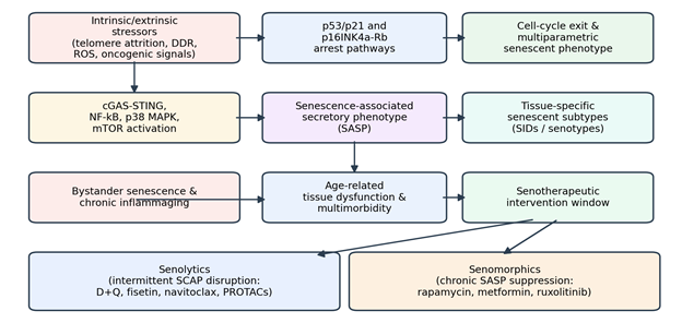
Figure 1. Conceptual Framework Linking Senescence Induction to Senotherapeutic Intervention. A mechanistic schematic tracing the pathway from intrinsic/extrinsic stressors through p53/p21 and p16INK4a-Rb arrest, SASP activation, and downstream tissue consequences (bystander senescence, inflammaging), culminating in the two major points of pharmacological intervention — senolytics and senomorphics. The diagram visually integrates the biology reviewed in Sections 2.1–2.2 into a single translational map.


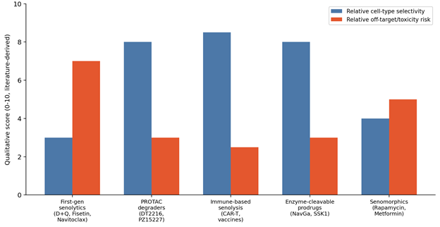
Figure 2. Qualitative Comparison of Selectivity and Off-Target Risk Across Senotherapeutic Classes.. A bar chart comparing five drug classes — first-generation senolytics, PROTAC degraders, immune-based senolysis, enzyme-cleavable prodrugs, and senomorphics — on two literature-derived qualitative dimensions: relative cell-type selectivity and relative off-target/toxicity risk. The figure visually summarizes the selectivity gains achieved by next-generation precision platforms relative to first-generation agents, as discussed in Sections 4.2 and 5.2.

Figure 1. Conceptual Framework Linking Senescence Induction to Senotherapeutic Intervention. A mechanistic schematic tracing the pathway from intrinsic/extrinsic stressors through p53/p21 and p16INK4a-Rb arrest, SASP activation, and downstream tissue consequences (bystander senescence, inflammaging), culminating in the two major points of pharmacological intervention — senolytics and senomorphics. The diagram visually integrates the biology reviewed in Sections 2.1–2.2 into a single translational map.


Figure 2. Qualitative Comparison of Selectivity and Off-Target Risk Across Senotherapeutic Classes.. A bar chart comparing five drug classes — first-generation senolytics, PROTAC degraders, immune-based senolysis, enzyme-cleavable prodrugs, and senomorphics — on two literature-derived qualitative dimensions: relative cell-type selectivity and relative off-target/toxicity risk. The figure visually summarizes the selectivity gains achieved by next-generation precision platforms relative to first-generation agents, as discussed in Sections 4.2 and 5.2.

Figure 1. Conceptual Framework Linking Senescence Induction to Senotherapeutic Intervention. A mechanistic schematic tracing the pathway from intrinsic/extrinsic stressors through p53/p21 and p16INK4a-Rb arrest, SASP activation, and downstream tissue consequences (bystander senescence, inflammaging), culminating in the two major points of pharmacological intervention — senolytics and senomorphics. The diagram visually integrates the biology reviewed in Sections 2.1–2.2 into a single translational map.


Figure 2. Qualitative Comparison of Selectivity and Off-Target Risk Across Senotherapeutic Classes.. A bar chart comparing five drug classes — first-generation senolytics, PROTAC degraders, immune-based senolysis, enzyme-cleavable prodrugs, and senomorphics — on two literature-derived qualitative dimensions: relative cell-type selectivity and relative off-target/toxicity risk. The figure visually summarizes the selectivity gains achieved by next-generation precision platforms relative to first-generation agents, as discussed in Sections 4.2 and 5.2.

Figure 1. Conceptual Framework Linking Senescence Induction to Senotherapeutic Intervention. A mechanistic schematic tracing the pathway from intrinsic/extrinsic stressors through p53/p21 and p16INK4a-Rb arrest, SASP activation, and downstream tissue consequences (bystander senescence, inflammaging), culminating in the two major points of pharmacological intervention — senolytics and senomorphics. The diagram visually integrates the biology reviewed in Sections 2.1–2.2 into a single translational map.


Figure 2. Qualitative Comparison of Selectivity and Off-Target Risk Across Senotherapeutic Classes.. A bar chart comparing five drug classes — first-generation senolytics, PROTAC degraders, immune-based senolysis, enzyme-cleavable prodrugs, and senomorphics — on two literature-derived qualitative dimensions: relative cell-type selectivity and relative off-target/toxicity risk. The figure visually summarizes the selectivity gains achieved by next-generation precision platforms relative to first-generation agents, as discussed in Sections 4.2 and 5.2.

Figure 1. Conceptual Framework Linking Senescence Induction to Senotherapeutic Intervention. A mechanistic schematic tracing the pathway from intrinsic/extrinsic stressors through p53/p21 and p16INK4a-Rb arrest, SASP activation, and downstream tissue consequences (bystander senescence, inflammaging), culminating in the two major points of pharmacological intervention — senolytics and senomorphics. The diagram visually integrates the biology reviewed in Sections 2.1–2.2 into a single translational map.


Figure 2. Qualitative Comparison of Selectivity and Off-Target Risk Across Senotherapeutic Classes.. A bar chart comparing five drug classes — first-generation senolytics, PROTAC degraders, immune-based senolysis, enzyme-cleavable prodrugs, and senomorphics — on two literature-derived qualitative dimensions: relative cell-type selectivity and relative off-target/toxicity risk. The figure visually summarizes the selectivity gains achieved by next-generation precision platforms relative to first-generation agents, as discussed in Sections 4.2 and 5.2.

Figure 1. Conceptual Framework Linking Senescence Induction to Senotherapeutic Intervention. A mechanistic schematic tracing the pathway from intrinsic/extrinsic stressors through p53/p21 and p16INK4a-Rb arrest, SASP activation, and downstream tissue consequences (bystander senescence, inflammaging), culminating in the two major points of pharmacological intervention — senolytics and senomorphics. The diagram visually integrates the biology reviewed in Sections 2.1–2.2 into a single translational map.


Figure 2. Qualitative Comparison of Selectivity and Off-Target Risk Across Senotherapeutic Classes.. A bar chart comparing five drug classes — first-generation senolytics, PROTAC degraders, immune-based senolysis, enzyme-cleavable prodrugs, and senomorphics — on two literature-derived qualitative dimensions: relative cell-type selectivity and relative off-target/toxicity risk. The figure visually summarizes the selectivity gains achieved by next-generation precision platforms relative to first-generation agents, as discussed in Sections 4.2 and 5.2.

Figure 1. Conceptual Framework Linking Senescence Induction to Senotherapeutic Intervention. A mechanistic schematic tracing the pathway from intrinsic/extrinsic stressors through p53/p21 and p16INK4a-Rb arrest, SASP activation, and downstream tissue consequences (bystander senescence, inflammaging), culminating in the two major points of pharmacological intervention — senolytics and senomorphics. The diagram visually integrates the biology reviewed in Sections 2.1–2.2 into a single translational map.

Figure 2. Qualitative Comparison of Selectivity and Off-Target Risk Across Senotherapeutic Classes.. A bar chart comparing five drug classes — first-generation senolytics, PROTAC degraders, immune-based senolysis, enzyme-cleavable prodrugs, and senomorphics — on two literature-derived qualitative dimensions: relative cell-type selectivity and relative off-target/toxicity risk. The figure visually summarizes the selectivity gains achieved by next-generation precision platforms relative to first-generation agents, as discussed in Sections 4.2 and 5.2.


studies, and review articles published in English that addressed (a) molecular mechanisms or biomarkers of cellular senescence, (b) single-cell or spatial technologies used to resolve senescent heterogeneity, (c) senolytic or senomorphic agents at the preclinical or clinical stage, or (d) registered human clinical trials of senotherapeutics. We excluded conference abstracts without full text, non-peer-reviewed preprints, and studies unrelated to mammalian senescence biology.

3.3 Study selection and data extraction.

Titles and abstracts were screened against the eligibility criteria, followed by full-text review of potentially relevant records. Data extracted from each included source comprised study design, model system (in vitro, murine, or human), therapeutic agent and mechanism, senescence biomarkers assessed, and reported efficacy or safety outcomes. Extracted data were organized into four evidence tables: foundational small-molecule senotherapeutics (Table 1), next-generation precision platforms (Table 2), multi-tissue senescence biomarkers (Table 3), and representative human clinical trials (Table 4), enabling structured cross-study comparison.

3.4 Quality appraisal.

Preclinical studies were appraised informally for model relevance (aged versus progeroid versus induced-senescence models) and for whether senescent-cell clearance was confirmed biochemically rather than inferred. Clinical trials were appraised for phase, sample size, and whether target engagement was measured directly (e.g., tissue biopsy) versus indirectly (e.g., circulating SASP factors), given that this distinction materially affects the strength of translational claims discussed later in this review.

3.5 Synthesis approach.

Given the methodological heterogeneity across included studies — spanning cell-culture mechanistic work, rodent healthspan studies, and early-phase human trials — findings were synthesized narratively rather than meta-analytically. Two conceptual figures were generated to summarize cross-cutting patterns: a mechanistic schema linking senescence induction to SASP output and points of therapeutic intervention (Figure 1), and a qualitative comparison of selectivity and off-target risk across senotherapeutic drug classes, derived from the safety and specificity data reported in Tables 1 and 2 (Figure 2).

4. Synthesis of the findings

4.1 Mechanistic Convergence Across Heterogeneous Senescent States

Despite substantial heterogeneity in senescent-cell phenotype, the included literature converges on a shared upstream architecture: intrinsic and extrinsic stressors funnel through p53/p21 or p16INK4a-Rb arrest pathways into a stereotyped multiparametric senescent phenotype, which then diversifies downstream into tissue- and trigger-specific senotypes via SASP-driven signaling (Figure 1) (Chmielewski, 2026; Cohn et al., 2023; Ozdemir et al., 2025). This convergent-then-divergent architecture helps explain a pattern visible across Table 3: biomarkers positioned early in the pathway (p16, p21, γ-H2AX) show broad but low-specificity expression across tissues, whereas later markers (lipofuscin, uPAR, LINE-1 derepression) are more tissue-restricted and, correspondingly, more diagnostically informative in specific contexts (Chmielewski, 2026; Gasek et al., 2021; Mansfield et al., 2024).

4.2 Comparative Selectivity of First- and Next-Generation Senotherapeutics

Synthesizing the safety and mechanistic data reported across Tables 1 and 2 into a qualitative comparison (Figure 2) highlights a consistent trade-off pattern: first-generation agents such as navitoclax achieve broad senolytic efficacy but at the cost of substantial off-target risk, driven mechanistically by shared dependence on Bcl-xL between senescent cells and circulating platelets (Chang et al., 2016; Gasek et al., 2021). Next-generation platforms — PROTAC degraders such as DT2216, uPAR-directed CAR-T cells, and galactose-caged prodrugs such as NavGa — cluster toward the opposite corner of this comparison, combining high predicted selectivity with markedly lower off-target risk, largely because each exploits a tissue- or cell-type-restricted determinant (VHL ligase expression, uPAR surface density, or lysosomal SA-β-Gal activity, respectively) rather than a pathway shared with healthy bystander cells (He et al., 2020; Amor et al., 2020; Paez-Ribes et al., 2019; Zhang et al., 2026).

4.3 Immune-Based and Enzyme-Activated Precision Strategies

Table 3. Multi-Tissue Biomarkers of Cellular Senescence. Profiles nine molecular, structural, and secretory markers used to identify senescent cells in vivo, including p16INK4a, p21, SA-β-Gal, lamin B1, lipofuscin, γ-H2AX, HMGB1, uPAR, and LINE-1 elements. For each biomarker, the detection method and a key diagnostic limitation are provided. The table underscores why no single "gold-standard" marker exists and why multiparametric panels remain necessary.


Biomarker

Category

Detection Method

Key Limitation

Reference(s)

p16INK4a

Cell-cycle inhibitor

IHC, qPCR, flow cytometry

Low specificity; transient in activated macrophages

Chmielewski, 2026; Gasek et al., 2021

p21

Cell-cycle inhibitor

IHC, IF, qPCR

Overlaps with quiescence; circadian variation

Gasek et al., 2021; Wyles et al., 2022

SA-β-Gal

Lysosomal enzyme

Chromogenic/fluorogenic, pH 6.0

High sensitivity, low specificity

Fu & Zhou, 2025; Gasek et al., 2021

Lamin B1

Nuclear envelope

Western blot, confocal IF

Loss is not universal across cell types

Gasek et al., 2021; Liao et al., 2021

Lipofuscin

Lysosomal aggregate

Histochemical staining

Robust but requires specialized staining

Chmielewski, 2026; Gasek et al., 2021

γ-H2AX

DNA-damage marker

Confocal IF foci counting

Not unique to senescence

Gasek et al., 2021; Wyles et al., 2022

HMGB1

Alarmin/DAMP

Western blot, IF

Also released during necrosis

Fu & Zhou, 2025; Gasek et al., 2021

uPAR

Surface receptor

Flow cytometry, immuno-PET

Also overexpressed in malignancy

Cohn et al., 2023; Zhang et al., 2026

LINE-1 elements

Retrotransposon

qPCR, RNA-seq

Late-stage marker; not senescence-exclusive

Gasek et al., 2021; Liao et al., 2021

Table 4. Representative Human Clinical Trials of Senotherapeutics. Lists seven registered clinical trials evaluating senolytics across idiopathic pulmonary fibrosis, diabetic kidney disease, Alzheimer's disease, bone health, frailty, diabetic macular edema, and osteoarthritis. Each row reports the trial identifier, condition, agent, phase/status, and headline finding. The table illustrates that biological target engagement has been consistently achievable in humans, though clinical benefit has been more variable.

Trial ID

Condition

Agent(s)

Phase/Status

Key Finding

Reference(s)

NCT02874989

Idiopathic pulmonary fibrosis

Dasatinib + Quercetin

Phase I pilot; completed

Improved walk distance and chair-stand times

Justice et al., 2019

NCT02848131

Diabetic kidney disease

Dasatinib + Quercetin

Phase I pilot; completed

Reduced senescent-cell density; lower serum SASP

Hickson et al., 2019

NCT04685590 (SToMP-AD)

Alzheimer’s disease

Dasatinib + Quercetin

Phase II; recruiting

Reduced CSF SASP factors; limited quercetin CNS penetrance

Riessland & Orr, 2024

NCT04313634

Bone health, postmenopausal women

D+Q vs. Fisetin vs. placebo

Phase II; completed

Safe; limited target engagement

Farr et al., 2024

NCT03430037 (AFFIRM)

Frailty in older women

Fisetin

Phase II; completed

High tolerability; reduced inflammaging markers

Kirkland & Tchkonia, 2020

NCT04537884

Diabetic macular edema

UBX1325

Phase I/II; completed

Improved visual acuity, reduced retinal thickness

Crespo-Garcia et al., 2024

NCT03513016

Knee osteoarthritis

UBX0101

Phase II; completed

Failed primary pain-reduction endpoint

Chaib et al., 2022


Across the included preclinical literature, immune-guided senolysis emerges as a particularly durable strategy. uPAR-targeted CAR-T cells achieve prophylactic, long-lasting clearance of senescent hepatocytes and fibroblasts, reversing liver fibrosis and improving metabolic parameters in aged mice (Amor et al., 2020; Cohn et al., 2023), while senolytic vaccines directed against GPNMB or CD153 stimulate endogenous antibody-mediated clearance and extend lifespan in progeroid models without requiring repeated dosing (Dey et al., 2023; Riessland et al., 2024). Enzyme-activated prodrugs (NavGa, SSK1) similarly demonstrate that exploiting a senescence-restricted enzymatic activity — SA-β-Gal cleavage in the lysosome — can markedly reduce systemic toxicity relative to the unmodified parent compound while preserving therapeutic efficacy against senescent tumor and cardiac cell populations (Paez-Ribes et al., 2019).

4.4 Human Clinical Trial Evidence

Representative trials summarized in Table 4 show a consistent, if still preliminary, translational signal. In diabetic kidney disease, a three-day course of D+Q reduced p16- and p21-expressing senescent-cell density in adipose and skin biopsies alongside falling circulating SASP factors (Hickson et al., 2019). In idiopathic pulmonary fibrosis, D+Q produced measurable gains in six-minute walk distance and gait speed (Justice et al., 2019). In the SToMP-AD trial, dasatinib—but only weakly quercetin—crossed the blood–brain barrier, yet combination dosing still reduced CSF SASP biomarkers in participants with mild cognitive impairment (Riessland & Orr, 2024). Organ-localized dosing has also shown promise: intravitreal UBX1325 improved visual acuity and reduced central subfield thickness in diabetic macular edema over 24 weeks (Crespo-Garcia et al., 2024), though a comparable intra-articular BCL-2/MDM2 inhibitor trial in knee osteoarthritis failed to meet its primary pain-reduction endpoint (Chaib et al., 2022), underscoring that target engagement does not automatically translate into a clinically meaningful outcome.

5. Translational Strategies, Therapeutic Precision, and Clinical Challenges in Senotherapy

5.1 From Intermittent Senolysis to Chronic Senomorphic Suppression

The translation of cellular senescence from an in vitro curiosity into a validated clinical target is, by most measures, one of the more significant reframings in recent geroscience (Chaib et al., 2022). Reading across the molecular, preclinical, and clinical evidence assembled here (Tables 1–4; Figures 1–2), a recurring theme is the trade-off between dosing convenience and durability of effect. Senolytics such as D+Q and fisetin exploit the fact that senescent cells take weeks to reaccumulate, permitting intermittent “hit-and-run” dosing that has repeatedly shown success in clearing senescent burden and extending healthy lifespan in aged and progeroid mice (Kirkland & Tchkonia, 2020; Yousefzadeh et al., 2018). Senomorphics such as rapamycin and metformin, by contrast, leave the cells alive and must therefore be given continuously, which — perhaps unsurprisingly — raises the cumulative risk of off-target effects such as hyperlipidemia, insulin resistance, or gastrointestinal intolerance over years of use (Gasek et al., 2021; Riessland & Orr, 2024).

5.2 Overcoming the Selectivity Barrier with Precision Platforms

The clinical ceiling for first-generation compounds is fairly well defined by now: navitoclax’s dose-limiting thrombocytopenia, rooted in platelets’ shared dependence on Bcl-xL, is probably the clearest illustration of why selectivity, not potency, has become the field’s central engineering problem (Chang et al., 2016; Gasek et al., 2021). PROTAC technology has made real headway here. DT2216 routes Bcl-xL to the VHL E3 ligase for degradation; because platelets express essentially no VHL, this design decouples senolysis from platelet toxicity almost by construction (He et al., 2020; Zhang et al., 2026) (Table 2; Figure 2). A parallel strategy exploits the senescent cell’s own lysosomal biology: galactose-caged prodrugs such as NavGa and SSK1 stay pharmacologically silent until cleaved by the high SA-β-Gal activity characteristic of senescent lysosomes, which meaningfully narrows the therapeutic window’s toxicity tail relative to the unmodified drug (Cai et al., 2020; Gonzalez-Gualda et al., 2020; Paez-Ribes et al., 2019).

5.3 Immune-Guided Senolysis: Durability Without Repeated Dosing

Immunotherapeutic approaches occupy a somewhat different niche in this landscape — not necessarily more selective in a molecular sense, but potentially more durable in a practical one. uPAR-directed CAR-T cells achieve long-lasting, arguably prophylactic clearance of senescent fibroblasts and hepatocytes, reversing fibrosis and restoring metabolic function in mouse models (Amor et al., 2020; Cohn et al., 2023), while senolytic vaccines against GPNMB or CD153 sidestep the adherence burden of repeated small-molecule dosing entirely by training the host’s own immune system to do the clearing (Suda et al., 2021; Zhang et al., 2026). Whether this durability holds up in aged human immune systems — which are, after all, themselves affected by immunosenescence — remains an open and clinically important question (Chmielewski, 2026; Ozdemir et al., 2025).

5.4 Translating Target Engagement into Clinical Benefit

Early-phase human data, summarized in Table 4, are encouraging but should be read with some caution. Hickson et al. (2019) and Justice et al. (2019) demonstrate that senolytic target engagement is achievable and measurable in humans — reduced senescent-cell density on biopsy, falling circulating SASP factors, improved six-minute walk distance — which is not a trivial finding. Yet the osteoarthritis trial of UBX0101 (Chaib et al., 2022) is a useful counterweight: biological target engagement did not translate into the primary clinical endpoint, a reminder that senescent-cell burden and patient-reported outcome are not automatically the same currency. The SToMP-AD findings (Riessland & Orr, 2024) add a further wrinkle specific to CNS-directed senotherapy — differential blood–brain-barrier penetrance between combination-drug components complicates dose optimization in ways that peripheral trials do not encounter.

5.5 Translational Hurdles and the Path Forward

Two structural problems seem to underlie most of the translational friction described above. First is what the field has taken to calling the biomarker bottleneck: without a validated, non-invasive, in vivo senescence marker, tracking real-time target engagement still generally requires biopsy, which limits both trial design and eventual clinical monitoring (Gasek et al., 2021). Second, because senescence retains beneficial, transient physiological roles, aggressive or poorly timed clearance risks impairing wound healing and tissue regeneration — an argument for careful patient stratification and genuinely intermittent, rather than maximal, dosing (Chmielewski, 2026; Paez-Ribes et al., 2019). Addressing both problems simultaneously, through senotype-informed biomarker panels (Table 3), precision delivery platforms (Table 2; Figure 2), and trial designs tailored to geriatric and oncology populations, represents what is probably the field’s most realistic near-term path toward extending human healthspan (Chaib et al., 2022; Sun, 2023).

5. Translational Strategies, Therapeutic Precision, and Clinical Challenges in Senotherapy

5.1 From Intermittent Senolysis to Chronic Senomorphic Suppression

The translation of cellular senescence from an in vitro curiosity into a validated clinical target is, by most measures, one of the more significant reframings in recent geroscience (Chaib et al., 2022). Reading across the molecular, preclinical, and clinical evidence assembled here (Tables 1–4; Figures 1–2), a recurring theme is the trade-off between dosing convenience and durability of effect. Senolytics such as D+Q and fisetin exploit the fact that senescent cells take weeks to reaccumulate, permitting intermittent “hit-and-run” dosing that has repeatedly shown success in clearing senescent burden and extending healthy lifespan in aged and progeroid mice (Kirkland & Tchkonia, 2020; Yousefzadeh et al., 2018). Senomorphics such as rapamycin and metformin, by contrast, leave the cells alive and must therefore be given continuously, which — perhaps unsurprisingly — raises the cumulative risk of off-target effects such as hyperlipidemia, insulin resistance, or gastrointestinal intolerance over years of use (Gasek et al., 2021; Riessland & Orr, 2024).

5.2 Overcoming the Selectivity Barrier with Precision Platforms

The clinical ceiling for first-generation compounds is fairly well defined by now: navitoclax’s dose-limiting thrombocytopenia, rooted in platelets’ shared dependence on Bcl-xL, is probably the clearest illustration of why selectivity, not potency, has become the field’s central engineering problem (Chang et al., 2016; Gasek et al., 2021). PROTAC technology has made real headway here. DT2216 routes Bcl-xL to the VHL E3 ligase for degradation; because platelets express essentially no VHL, this design decouples senolysis from platelet toxicity almost by construction (He et al., 2020; Zhang et al., 2026) (Table 2; Figure 2). A parallel strategy exploits the senescent cell’s own lysosomal biology: galactose-caged prodrugs such as NavGa and SSK1 stay pharmacologically silent until cleaved by the high SA-β-Gal activity characteristic of senescent lysosomes, which meaningfully narrows the therapeutic window’s toxicity tail relative to the unmodified drug (Cai et al., 2020; Gonzalez-Gualda et al., 2020; Paez-Ribes et al., 2019).

5.3 Immune-Guided Senolysis: Durability Without Repeated Dosing

Immunotherapeutic approaches occupy a somewhat different niche in this landscape — not necessarily more selective in a molecular sense, but potentially more durable in a practical one. uPAR-directed CAR-T cells achieve long-lasting, arguably prophylactic clearance of senescent fibroblasts and hepatocytes, reversing fibrosis and restoring metabolic function in mouse models (Amor et al., 2020; Cohn et al., 2023), while senolytic vaccines against GPNMB or CD153 sidestep the adherence burden of repeated small-molecule dosing entirely by training the host’s own immune system to do the clearing (Suda et al., 2021; Zhang et al., 2026). Whether this durability holds up in aged human immune systems — which are, after all, themselves affected by immunosenescence — remains an open and clinically important question (Chmielewski, 2026; Ozdemir et al., 2025).

5.4 Translating Target Engagement into Clinical Benefit

Early-phase human data, summarized in Table 4, are encouraging but should be read with some caution. Hickson et al. (2019) and Justice et al. (2019) demonstrate that senolytic target engagement is achievable and measurable in humans — reduced senescent-cell density on biopsy, falling circulating SASP factors, improved six-minute walk distance — which is not a trivial finding. Yet the osteoarthritis trial of UBX0101 (Chaib et al., 2022) is a useful counterweight: biological target engagement did not translate into the primary clinical endpoint, a reminder that senescent-cell burden and patient-reported outcome are not automatically the same currency. The SToMP-AD findings (Riessland & Orr, 2024) add a further wrinkle specific to CNS-directed senotherapy — differential blood–brain-barrier penetrance between combination-drug components complicates dose optimization in ways that peripheral trials do not encounter.

5.5 Translational Hurdles and the Path Forward

Two structural problems seem to underlie most of the translational friction described above. First is what the field has taken to calling the biomarker bottleneck: without a validated, non-invasive, in vivo senescence marker, tracking real-time target engagement still generally requires biopsy, which limits both trial design and eventual clinical monitoring (Gasek et al., 2021). Second, because senescence retains beneficial, transient physiological roles, aggressive or poorly timed clearance risks impairing wound healing and tissue regeneration — an argument for careful patient stratification and genuinely intermittent, rather than maximal, dosing (Chmielewski, 2026; Paez-Ribes et al., 2019). Addressing both problems simultaneously, through senotype-informed biomarker panels (Table 3), precision delivery platforms (Table 2; Figure 2), and trial designs tailored to geriatric and oncology populations, represents what is probably the field’s most realistic near-term path toward extending human healthspan (Chaib et al., 2022; Sun, 2023).

6. Conclusion

Cellular senescence has moved, over roughly two decades, from a laboratory curiosity to a mechanistically coherent and increasingly druggable target across age-related disease. The evidence synthesized here indicates that first-generation senolytics and senomorphics established genuine clinical proof-of-concept but remain constrained by incomplete selectivity and, for senomorphics, chronic dosing burden. Next-generation platforms — PROTAC degraders, uPAR-directed immunotherapies, senolytic vaccines, and enzyme-activated prodrugs — offer a mechanistically grounded route toward higher selectivity and lower off-target risk. Realizing this potential clinically will require resolving the biomarker bottleneck, matching senotype-specific vulnerabilities to intervention class, and designing trials capable of distinguishing biological target engagement from durable patient benefit.

References


Alum, E. U., Izah, S. C., Uti, D. E., Paul-Chima Ugwu, O., Betiang, P. A., Basajja, M., & Ejemot-Nwadiaro, R. I. (2025). Targeting cellular senescence for healthy aging: Advances in senolytics and senomorphics. Drug Design, Development and Therapy, 19, 8489–8522. https://doi.org/10.2147/DDDT.S543211     

Amor, C., Feucht, J., Leibold, J., Ho, Y. J., Zhu, C., Alonso-Curbelo, D., Mansilla-Soto, J., Boyer, J. A., Li, X., Giavridis, T., Kulick, A., Houlihan, S., Peerschke, E., Friedman, S. L., Ponomarev, V., Piersigilli, A., Sadelain, M., & Lowe, S. W. (2020). Senolytic CAR T cells reverse senescence-associated pathologies. Nature, 583(7814), 127–132. https://doi.org/10.1038/s41586-020-2403-9

Cai, Y., Zhou, H., Zhu, Y., Sun, Q., Ji, Y., Xie, C., & Muñoz-Espín, D. (2020). Elimination of senescent cells by β-galactosidase-targeted prodrug attenuates inflammation and restores physical function in aged mice. Cell Research, 30(7), 574–589. https://doi.org/10.1038/s41422-020-0314-z     

Chaib, S., Tchkonia, T., & Kirkland, J. L. (2022). Cellular senescence and senolytics: The path to the clinic. Nature Medicine, 28(8), 1556–1568. https://doi.org/10.1038/s41591-022-01923-y           

Chang, J., Wang, Y., Shao, L., Laberge, R. M., Demaria, M., Campisi, J., Janakiraman, K., Sharpless, N. E., Ding, S., Feng, W., Luo, Y., Wang, X., Aykin-Burns, N., Krager, K., Ponnappan, U., Hauer-Jensen, M., Meng, A., & Zhou, D. (2016). Clearance of senescent cells by ABT263 rejuvenates aged hematopoietic stem cells in mice. Nature Medicine, 22(1), 78–83. https://doi.org/10.1038/nm.4010        

Chmielewski, P. P. (2026). Cellular senescence and inflammageing: From mechanisms to senotherapeutic interventions. Biogerontology, 27, 129. https://doi.org/10.1007/s10522-026-10477-2     

Cohn, R. L., Gasek, N. S., Kuchel, G. A., & Xu, M. (2023). The heterogeneity of cellular senescence: Insights at the single-cell level. Trends in Cell Biology, 33(1), 9–17. https://doi.org/10.1016/j.tcb.2022.04.011          

Cossarizza, A. (2026). Inflammaging: Experimental insights and translational advances. European Journal of Immunology, 56(7), 70239. https://doi.org/10.1002/eji.202470239 

Crespo-Garcia, S., Fournier, F., Diaz-Marin, R., Klier, S., Ragusa, D., Masaki, L., & Khasanov, A. (2024). Therapeutic targeting of cellular senescence in diabetic macular edema: Preclinical and phase 1 trial results. Nature Medicine, 30, 443–454. https://doi.org/10.1038/s41591-024-02802-4  

Dey, A. K., Banarjee, R., Boroumand, M., Rutherford, D. V., Strassheim, Q., Nyunt, T., Olinger, B., & Basisty, N. (2023). Translating senotherapeutic interventions into the clinic with emerging proteomic technologies. Biology, 12(10), 1301. https://doi.org/10.3390/biology12090647        

Erusalimsky, J. D. (2021). The biological mechanisms of frailty: Focusing on cellular senescence. European Geriatric Medicine, 12, 1157. https://doi.org/10.1007/s41999-021-00481-z 

Farr, J. N., Atkinson, E. J., Achenbach, S. J., Volkman, T. L., Tweed, A. J., Vos, S. J., Tchkonia, T., Kirkland, J. L., & Khosla, S. (2024). Effects of intermittent senolytic therapy on bone metabolism in postmenopausal women: A phase 2 randomized controlled trial. Nature Medicine, 30(9), 2605–2612. https://doi.org/10.1038/s41591-024-03012-4

Fu, T. E., & Zhou, Z. (2025). Senescent cells as a target for anti-aging interventions: From senolytics to immune therapies. Journal of Translational Internal Medicine, 13(1), 33–47. https://doi.org/10.1515/jtim-2025-0005    

Gasek, N. S., Kuchel, G. A., Kirkland, J. L., & Xu, M. (2021). Strategies for targeting senescent cells in human disease. Nature Aging, 1(10), 870–879. https://doi.org/10.1038/s43587-021-00121-8    

Gonzalez-Gualda, E., Baker, A. G., Fruk, L., & Muñoz-Espín, D. (2020). Galacto-conjugation of Navitoclax as an efficient strategy to increase senolytic specificity and reduce platelet toxicity. Aging Cell, 19(4), e13142. https://doi.org/10.1111/acel.13142       

Hayflick, L., & Moorhead, P. S. (1961). The serial cultivation of human diploid cell strains. Experimental Cell Research, 25(3), 585–621. https://doi.org/10.1016/0014-4827(61)90192-6     

He, Y., Zhang, X., Chang, J., Kim, H. N., Zhang, P., Wang, Y., Khan, S., Liu, X., Zhang, X., Lv, D., & Zhou, D. (2020). Using proteolysis-targeting chimera technology to reduce navitoclax platelet toxicity and improve its senolytic activity. Nature Communications, 11, 1996. https://doi.org/10.1038/s41467-020-15838-0

Hickson, L. J., Langhi Prata, L. G. P., Bobart, S. A., Evans, T. K., Giorgadze, N., Hashmi, S. K., Herrmann, S. M., Jensen, M. D., Jia, Q., Jordan, K. L., Tchkonia, T., & Kirkland, J. L. (2019). Senolytics decrease senescent cells in humans: Preliminary report from a clinical trial of Dasatinib plus Quercetin in individuals with diabetic kidney disease. EBioMedicine, 47, 446–456. https://doi.org/10.1016/j.ebiom.2019.08.069   

Justice, J. N., Nambiar, A. M., Tchkonia, T., LeBrasseur, N. K., Pascual, R., Hashmi, S. K., Prata, L., Masternak, M. M., Kritchevsky, S. B., Musi, N., & Kirkland, J. L. (2019). Senolytics in idiopathic pulmonary fibrosis: Results from a first-in-human, open-label, pilot study. EBioMedicine, 40, 554–563. https://doi.org/10.1016/j.ebiom.2018.12.052     

Kirkland, J. L., & Tchkonia, T. (2020). Senolytic drugs: From discovery to translation. Journal of Internal Medicine, 288(5), 518–536. https://doi.org/10.1111/joim.13141           

Kusmanto, S. J. (2025). Senescence modulation: An applied science review of strategies in anti-aging, regenerative aesthetics, and oncology therapy. Current Issues in Molecular Biology, 47, 989. https://doi.org/10.3390/cimb47040989    

Liao, Z., Yeo, H. L., Wong, S. W., & Zhao, Y. (2021). Cellular senescence: Mechanisms and therapeutic potential. Biomedicines, 9(12), 1769. https://doi.org/10.3390/biomedicines9121769        

Mansfield, L., Ramponi, V., Gupta, K., Stevenson, T., Mathew, A. B., Barinda, A. J., Herbstein, F., & Morsli, S. (2024). Emerging insights in senescence: Pathways from preclinical models to therapeutic innovations. npj Aging, 10, 53. https://doi.org/10.1038/s41514-024-00181-1  

Ozdemir, S. A., Faizan, M. I., Kaur, G., Shaikh, S. B., Ul Islam, K., & Rahman, I. (2025). Heterogeneity of cellular senescence, senotyping, and targeting by senolytics and senomorphics in lung diseases. International Journal of Molecular Sciences, 26, 9687. https://doi.org/10.3390/ijms26199687              

Paez-Ribes, M., González-Gualda, E., Doherty, G. J., & Muñoz-Espín, D. (2019). Targeting senescent cells in translational medicine. EMBO Molecular Medicine, 11, e10234. https://doi.org/10.15252/emmm.201810234             

Riessland, M., & Orr, M. E. (2024). Therapeutic targeting of senescent cells in the CNS. Nature Reviews Drug Discovery, 23(11), 817–837. https://doi.org/10.1038/s41573-024-01033-z

Suda, M., Shimizu, I., Katsuumi, G., Yoshida, Y., Hayashi, Y., Ikegami, R., Matsumoto, N., Yoshida, Y., Mikawa, R., Katayama, A., & Minamino, T. (2021). Senolytic vaccination improves normal and pathological age-related phenotypes and increases lifespan in progeroid mice. Nature Aging, 1, 1117–1126. https://doi.org/10.1038/s43587-021-00151-2

Sun, Y. (2023). An updated landscape of cellular senescence heterogeneity: Mechanisms, technologies and senotherapies. Translational Medicine of Aging, 7, 46–51. https://doi.org/10.1016/j.tma.2023.06.001            

Syed, U., Aloti, S., Awad, M., Alghoul, A., Hassan, M., Kharoufeh, A., & Rangraze, O. (2026). Cellular senescence as a distinct contributor to metabolic dysfunction in type 2 diabetes. Frontiers in Endocrinology, 17, 1799261. https://doi.org/10.3389/fendo.2026.1799261   

Wyles, S. P., Tchkonia, T., & Kirkland, J. L. (2022). Targeting cellular senescence for age-related diseases: Path to clinical translation. Plastic and Reconstructive Surgery, 150(4), 686. https://doi.org/10.1097/PRS.0000000000009668

Yousefzadeh, M. J., Zhu, Y., McGowan, S. J., Angelini, L., Fuhrmann-Stroissnigg, H., Xu, M., Ling, Y. Y., Melos, K. I., Pirtskhalava, T., Inman, C. L., & Kirkland, J. L. (2018). Fisetin is a senotherapeutic that extends health and lifespan. EBioMedicine, 36, 18–28. https://doi.org/10.1016/j.ebiom.2018.09.015   

Zhang, W., Song, S., Zhang, Y., Pan, Y., Hu, D., & Wang, Y. (2026). Emerging strategies in senotherapeutics: From broad-spectrum senolysis to precision reprogramming. npj Aging, in press. https://doi.org/10.1038/s41514-026-00355-z


Article metrics
View details
0
Downloads
0
Citations
40
Views

View Dimensions


View Plumx


View Altmetric


0
Save
0
Citation
40
View
0
Share