Integrative Biomedical Research

Integrative Biomedical Research (Journal of Angiotherapy) | Online ISSN  3068-6326
463
Citations
1.8m
Views
768
Articles
Your new experience awaits. Try the new design now and help us make it even better
Switch to the new experience
Figures and Tables
REVIEWS   (Open Access)

Non-Coding RNA Networks in Pancreatic Ductal Adenocarcinoma: An Expanding Regulatory Landscape from the ceRNA TRIAD to Exosomal Immune Evasion and Ferroptosis Escape

Siti Fathiah Masre1,*, Eng Wee Chua2, Muhammad Asyaari Zakaria1, Amnani Aminuddin2, Nor Fadilah Rajab3

+ Author Affiliations

Integrative Biomedical Research 10 (1) 1-8 https://doi.org/10.25163/biomedical.10110921

Submitted: 19 January 2026 Revised: 07 March 2026  Published: 17 March 2026 


Abstract

Pancreatic ductal adenocarcinoma (PDAC) remains, even now, one of the few cancers where survival curves have barely moved in a generation, and that persistence is what motivated this review. Decades of work mapping driver mutations in KRAS, TP53, CDKN2A, and SMAD4 have not, on their own, translated into durable clinical benefit, which has pushed the field toward a broader, network-level view of the roughly 98% of the transcribed genome that never becomes protein — the non-coding RNAs (ncRNAs). we conducted a structured narrative synthesis of the peer-reviewed literature on lncRNAs, circular RNAs (circRNAs), microRNAs (miRNAs), and tRNA-derived small RNAs (tsRNAs) in PDAC, searching PubMed, Scopus, and Web of Science for studies published through 2026 and organizing the retrieved evidence around mechanistic themes rather than chronology, The evidence converges on an integrated lncRNA-miRNA-mRNA "TRIAD," in which oncogenic transcripts such as PVT1, UCA1, and MALAT1 sponge tumor-suppressive miRNAs while lncRNAs like GAS5 and MEG3 normally hold this network in check. Circular RNAs add a further, more stable layer — most strikingly exosomal circGANAB, which does not simply sponge miRNAs but physically degrades protective lncRNAs and cytokine transcripts, thereby upregulating GPX4-driven ferroptosis resistance and excluding cytotoxic T cells from the tumor core. Metabolic circuits (glutathione and riboflavin pathways) and stable serum tsRNA panels emerged as both mechanistic drivers and promising, minimally invasive biomarkers, while natural phytochemicals such as cordycepin and curcumin showed consistent, if pharmacokinetically limited, potential to re-sensitize resistant cells. Rather than isolated lesions, these ncRNA circuits behave as a coordinated regulatory system that shapes PDAC identity, stromal crosstalk, and therapeutic escape, and disrupting specific nodes within it — particularly the exosomal circGANAB axis — may offer a genuinely new route toward diagnosis and multi-target therapy in a disease that has resisted almost everything else.

Keywords: pancreatic ductal adenocarcinoma; non-coding RNA; competing endogenous RNA; circular RNA; exosomes; ferroptosis; tumor microenvironment.

1. Introduction

There is something almost stubborn about pancreatic ductal adenocarcinoma (PDAC). It accounts for the overwhelming majority — over 96% — of all pancreatic malignancies, and it remains one of the most lethal and aggressive diseases of the human digestive system (Jeong et al., 2026; Wong et al., 2026). Despite genuine advances elsewhere in oncology, the five-year overall survival rate for PDAC has stalled at somewhere between 10% and 13% across all stages combined (Wang et al., 2023; Wong et al., 2026) — a number that, frankly, has not moved much in years. This prognosis is not the product of any single failure. It reflects, instead, three reinforcing pathophysiological features: the absence of reliable early-stage diagnostic biomarkers, a striking propensity for early local invasion and distant metastasis, and a deep, systemic resistance to standard-of-care regimens such as gemcitabine, 5-fluorouracil (5-FU), and platinum-based chemotherapies (Eftekhari et al., 2026; Mathpal et al., 2025; Shi et al., 2025; Wang et al., 2023). Because the earliest stages of disease progress almost silently, more than 80% of patients are diagnosed only after the tumor has become advanced and non-resectable, leaving a modest 20% eligible for potentially curative surgery at the time of diagnosis (Limb et al., 2020; Wang et al., 2023). And even within that fortunate minority, recurrence after resection remains distressingly common — a reminder of just how infiltrative these cells are from the outset (Shi et al., 2025).

For a long time, the field's working assumption was that classical oncogenic driver mutations — in KRAS, TP53, CDKN2A, and SMAD4, most obviously — held the key. These genes have by now been characterized about as thoroughly as any in cancer biology. Yet therapeutic strategies built around targeting them individually have, on the whole, delivered disappointing clinical results (Jeong et al., 2026; Kan & Ayan, 2026). That gap between mechanistic understanding and clinical benefit is telling. It suggests that PDAC's molecular complexity — its capacity for tumor progression, metabolic remodeling, and therapeutic escape — is not really governed by isolated genetic lesions so much as by highly integrated regulatory networks that extend well beyond any single gene (Jeong et al., 2026; Luo et al., 2026). Which raises an obvious, if uncomfortable, question: if single-target strategies keep falling short, what would a network-level strategy even look like? Answering that has become something of an urgent priority, particularly for understanding cellular plasticity, epithelial-mesenchymal transition (EMT), cancer stem cell (CSC) maintenance, and immune evasion in this disease (Chen et al., 2025; Jeong et al., 2026).

One place this shift has been most visible is in the study of non-coding RNAs (ncRNAs). With the maturation of high-throughput RNA sequencing and increasingly sophisticated bioinformatic pipelines, it has become clear that non-coding RNAs are not simply transcriptional noise. They make up something on the order of 98% of the transcribed human genome, and rather than being an afterthought, they function as master coordinators of gene expression in their own right (Jeong et al., 2026; Long et al., 2026). Broadly, these regulatory molecules divide into small ncRNAs and long non-coding RNAs (lncRNAs) based on size and structure (Jeong et al., 2026). MicroRNAs (miRNAs) — typically around 22 nucleotides — classically act as post-transcriptional suppressors, binding the 3′ untranslated regions of target mRNAs to repress translation or trigger degradation (Limb et al., 2020; Son et al., 2021). Long non-coding RNAs, by contrast, exceed 200 nucleotides and take on a more architectural role, functioning as chromatin remodelers, transcriptional scaffolds, and cytoplasmic molecular sponges (Shi et al., 2025; Sun et al., 2024; Wang et al., 2023). Circular RNAs (circRNAs) form a third, unusually stable subtype, defined by a covalently closed-loop structure generated through back-splicing (Limb et al., 2020; Wong et al., 2026); lacking both a 5′ cap and a 3′ poly-A tail, they are essentially protected from exonuclease-mediated degradation, which makes them remarkably durable in tissue, plasma, and serum exosomes (Wong et al., 2026). More recently still, tRNA-derived small RNAs (tsRNAs) have emerged as another layer entirely — fine-tuning metabolic pathways such as glycolysis and ferroptosis under cellular stress (Pan et al., 2025).

What ties these molecules together, mechanistically, is the competing endogenous RNA (ceRNA) hypothesis: the idea that lncRNAs, circRNAs, and mRNAs share microRNA response elements (MREs) and, in effect, compete with one another for a finite pool of miRNAs (Jeong et al., 2026; Shi et al., 2025). This cross-talk forms what has come to be called the lncRNA-miRNA-mRNA "TRIAD," a structure that allows coordinated, network-level control of gene expression rather than the piecemeal regulation implied by studying any one transcript in isolation (Jeong et al., 2026). In PDAC, this normally homeostatic network is systematically re-wired to favor oncogenesis (Jeong et al., 2026). A particularly instructive example is the oncogenic circular RNA circGANAB (Wong et al., 2026), formed by back-splicing exons 2, 3, and 4 of the GANAB pre-mRNA and markedly overexpressed in both primary and metastatic PDAC tissue. What makes circGANAB interesting is that it does not behave simply as a miRNA sponge — the textbook circRNA function — but instead physically interacts with, and actively degrades, tumor-suppressive lncRNAs such as GAS5, lncLDAH3, and TMEM51-AS1, along with cytokine transcripts including IL-13 and IL-17D (Wong et al., 2026). Mechanistically, this happens because circGANAB blocks access of the RNA-stabilizing protein IGF2BP2 to these transcripts, leaving them exposed to degradation (Wong et al., 2026). Under normal conditions, GAS5 sponges the oncogenic miR-32-5p to upregulate PTEN, thereby restraining cell survival, migration, and EMT (Shi et al., 2025; Son et al., 2021); once circGANAB dismantles that protective barrier, downstream proliferative signaling, cell-cycle progression, and resistance to iron-dependent cell death (ferroptosis) all follow, in part through upregulation of the anti-ferroptosis protein GPX4 (Wong et al., 2026).

This kind of pathogenic ceRNA re-wiring is not always cell-intrinsic, either — it can be triggered externally. Somewhat unexpectedly, the swine hepatitis E virus (SHEV) ORF3 protein has been shown to act as a kind of metabolic master-switch, specifically upregulating hsa_circ_0077855 to sponge miR-181a-2-3p and miR-30b-3p (Luo et al., 2026). That competitive binding relieves the suppression of ENPP3, disrupting flavin adenine dinucleotide (FAD) hydrolysis, and simultaneously derepresses the proto-oncogene KRAS — a chain of events that links viral-induced riboflavin metabolic remodeling directly to oncogenic signaling activation (Luo et al., 2026). Beyond the tumor cell itself, non-coding RNA networks are also central to intercellular communication within the pancreatic tumor microenvironment (TME) (Chen et al., 2025; Shi et al., 2025), which in PDAC is notoriously dense and desmoplastic, built from cancer-associated fibroblasts (CAFs), pancreatic stellate cells (PSCs), and infiltrating immune cells that together form a formidable physical and immunological barrier to conventional therapy (Shi et al., 2025; Wang et al., 2023). Extracellular vesicles — exosomes, in particular — serve as the delivery vehicles here, packaging and protecting otherwise fragile ncRNAs so they can be horizontally transferred to neighboring cells (Limb et al., 2020; Wong et al., 2026). When neighboring pancreatic epithelial cells take up circGANAB-rich exosomes, tumor-suppressive lncRNAs are downregulated and invasive properties are, in effect, handed off to previously normal cells (Wong et al., 2026); at the same time, by suppressing IL-13 and IL-17D, circGANAB limits CD4+ and CD8+ T-cell infiltration and drives resistance to anti-PD-L1 immunotherapy (Wong et al., 2026). These interactions are further layered with epigenetic control — N6-methyladenosine (m6A) modification, for instance, regulates the stability, splicing, and translation of oncogenic and tumor-suppressive transcripts alike, including LINC00901, FOXD1-AS1, and circMYO1C (Rui et al., 2026). Taken together, these findings make a fairly compelling case that real therapeutic progress in PDAC will require a comprehensive understanding of the post-transcriptional networks orchestrated by the lncRNA-miRNA-mRNA TRIAD, in both tumor cells and their surrounding microenvironment (Jeong et al., 2026; Shi et al., 2025).

Against that backdrop, this review was built around four guiding research questions. First, how does the horizontal transfer of exosomal circRNAs such as circGANAB from PDAC cells to stromal CAFs and immune cells physically reshape the spatial landscape of cytotoxic T-cell infiltration, and to what extent does this drive adaptive resistance to anti-PD-L1 immunotherapy (Chen et al., 2025; Wong et al., 2026)? Second, to what degree does competitive binding between circular and long non-coding RNAs and specific microRNAs — the hsa_circ_0077855/miR-181a axis or the circ_0005397/PCBP2 axis, for example — coordinate metabolic reprogramming and ferroptosis resistance in drug-resistant PDAC cells (Chang et al., 2025; Wong et al., 2026; Luo et al., 2026)? Third, could a composite serum exosomal ncRNA panel, combining circGANAB, UCA1, and specific tRNA-derived fragments, meaningfully outperform conventional biomarkers like CA19-9 in distinguishing early-stage resectable PDAC from chronic pancreatitis (Limb et al., 2020; Mathpal et al., 2025; Wong et al., 2026)? And finally, what are the biophysical and kinetic constraints governing the circGANAB-IGF2BP2-lncRNA tripartite complex, and could disrupting this interaction — via antisense oligonucleotides or CRISPR-Cas13, say — rescue tumor-suppressive lncRNA expression in vivo (Chen & Gao, 2026; Shi et al., 2025; Wong et al., 2026)? These questions frame the objectives pursued throughout the remainder of this review: to characterize the spatiotemporal dynamics of exosomal ncRNA communication in the PDAC microenvironment, to evaluate the mechanistic basis of ferroptosis evasion, to identify non-invasive diagnostic ncRNA signatures, and to prioritize therapeutic strategies capable of disrupting these regulatory circuits at their source.

2. Non-Coding RNA Regulatory Networks in Pancreatic Cancer Mechanisms and Therapeutics

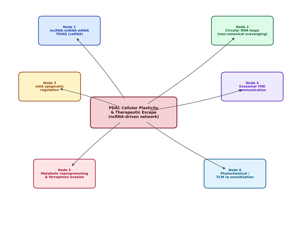
It helps, before going further, to say plainly what this section tries to do: rather than walking chronologically through individual studies, it organizes the evidence into a conceptual map — six interconnected regulatory nodes that, together, radiate outward from a single central process: the cellular plasticity and therapeutic escape that define PDAC biology (Figure 1). This is, admittedly, a simplification of what is a genuinely tangled web of interactions. But some simplification seems necessary if the literature is to be made legible at all.

2.1 The Paradigm Shift from Protein-Coding Drivers to ncRNA Networks

For decades, PDAC research was organized, more or less, around a handful of driver genes — KRAS, TP53, CDKN2A, and SMAD4 chief among them (Jeong et al., 2026; Wong et al., 2026). Targeting these individually, however, has not translated into the clinical gains that were once hoped for (Jeong et al., 2026; Kan & Ayan, 2026), and that disappointment has pushed the field toward a more systems-level view, in which tumor progression, metabolic remodeling, and therapeutic escape are understood as properties of integrated regulatory networks rather than of any single mutated gene (Jeong et al., 2026; Luo et al., 2026). RNA sequencing has been central to this shift: it is now reasonably well established that ncRNAs make up roughly 98% of the transcribed human genome (Jeong et al., 2026; Long et al., 2026), and that these molecules act, in their RNA form, as fine-tuners of gene expression across a wide range of pathological processes (Jeong et al., 2026; Rui et al., 2026). The conceptual map organized here (Figure 1) branches from that central hub into six regulatory nodes: the lncRNA-miRNA-mRNA TRIAD, circular-RNA loop mechanisms, epigenetic m6A modification, stromal exosomal communication, metabolic remodeling, and phytochemical re-sensitization.

2.2 The lncRNA-miRNA-mRNA TRIAD as a Post-Transcriptional Scaffold

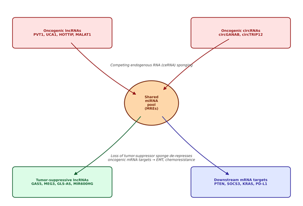
At the core of this network sits the competing endogenous RNA (ceRNA) hypothesis — the idea that lncRNAs, circRNAs, and mRNAs share microRNA response elements and, in a sense, compete for the same limited pool of miRNAs, forming the lncRNA-miRNA-mRNA TRIAD (Jeong et al., 2026; Shi et al., 2025; Figure 2). In PDAC, this cytoplasmic network is re-wired, fairly consistently across studies, to favor oncogenesis. PVT1, for instance, functions as a major oncogenic driver, activating Wnt/β-catenin and autophagic signaling through the miR-619-5p/Pygo2 and miR-619-5p/ATG14 axes, while also amplifying TGF-β signaling via upregulation of p-Smad2/3 and TGF-β1 to promote EMT and migration (Wang et al., 2023). The lncRNA UCA1 tells a similar story, promoting migration and invasion through the Hippo pathway (Jeong et al., 2026; Wang et al., 2023); notably, UCA1 correlates positively with the prognostic marker MEAK7 and negatively with miR-582-5p, a pattern associated with poorer overall survival (Kan & Ayan, 2026). Other oncogenic lncRNAs — HOTTIP and MALAT1 among them — sponge tumor-suppressive microRNAs (miR-137 and miR-217, respectively) to derepress oncogenes such as HOXA9/13 and KRAS, accelerating cancer stem cell (CSC) maintenance and inflammatory cytokine secretion along the way (Shi et al., 2025; Wang et al., 2023). On the opposite side of this ledger, tumor-suppressive lncRNAs such as Growth Arrest Specific 5 (GAS5) act as a kind of molecular checkpoint. Under ordinary conditions, GAS5 sponges the oncomiR miR-221 to upregulate SOCS3, which reverses EMT, stemness, and metastatic potential (Shi et al., 2025; Son et al., 2021) — and it is precisely the targeted degradation of GAS5, as discussed below, that appears to be a decisive event in aggressive PDAC progression (Wong et al., 2026).

2.3 Covalently Closed circRNAs: Loop Stability and Non-Canonical Scavenging

Circular RNAs occupy a genuinely distinct branch of this network. Their covalently closed structure protects them from exonuclease-mediated digestion (Limb et al., 2020; Wong et al., 2026), and while early work on circRNAs focused almost exclusively on their role as miRNA sponges, more recent studies have uncovered mechanisms that are considerably less straightforward (Wong et al., 2026). CircGANAB (hsa_circ_0008011), formed through back-splicing of exons 2, 3, and 4 of the GANAB pre-mRNA, is the clearest example (Wong et al., 2026). Overexpressed in both primary and metastatic PDAC, it does not primarily act as a sponge at all — instead, it physically interacts with, and degrades, tumor-suppressive lncRNAs (GAS5, lncLDAH3, TMEM51-AS1) and cytokine mRNAs (IL-13, IL-17D) (Wong et al., 2026).

Figure 1. Conceptual mind map of the six interconnected non-coding RNA regulatory nodes converging on pancreatic ductal adenocarcinoma (PDAC) cellular plasticity and therapeutic escape. The central hub represents the integrated phenotypic outcome — cellular plasticity, stromal communication, and drug resistance — while the six surrounding nodes summarize the major mechanistic themes synthesized in the Literature Review (Section 2): the lncRNA-miRNA-mRNA TRIAD, circular RNA loop mechanisms, m6A epigenetic regulation, exosomal tumor-microenvironment communication, metabolic reprogramming/ferroptosis evasion, and phytochemical re-sensitization strategies.

Figure 2. Schematic representation of the competing endogenous RNA (ceRNA) lncRNA-miRNA-mRNA TRIAD in pancreatic ductal adenocarcinoma. Oncogenic lncRNAs (e.g., PVT1, UCA1, HOTTIP, MALAT1) and oncogenic circRNAs (e.g., circGANAB, circTRIP12) compete for a shared pool of microRNAs via microRNA response elements (MREs), thereby limiting the sponge activity available to tumor-suppressive lncRNAs (e.g., GAS5, MEG3, GLS-AS, MIR600HG). The resulting shift de-represses downstream oncogenic mRNA targets (PTEN, SOCS3, KRAS, PD-L1), promoting epithelial-mesenchymal transition and chemoresistance, as detailed in Section 2.2.

The mechanism, as best understood, involves circGANAB forming stable RNA-RNA duplexes with these targets, which physically blocks access of the RNA-stabilizing protein IGF2BP2 (Wong et al., 2026). Deprived of that protective binding, the targeted transcripts undergo rapid degradation. What this shows, in effect, is that circRNAs can act as direct post-transcriptional regulators — not just decoys — capable of systematically dismantling endogenous tumor-suppressive pathways to promote invasion and metastasis (Wong et al., 2026).

2.4 Epigenetic and Epithelial Dynamics: m6A Methylation and EMT Regulation

Layered on top of these RNA-RNA interactions is an epigenetic regulatory system built around N6-methyladenosine (m6A) modification, maintained by "writer" enzymes (METTL3, METTL14), "eraser" enzymes (ALKBH5, FTO), and "reader" proteins (YTHDF1-3, IGF2BP2) (Rui et al., 2026; Shi et al., 2025). This m6A-ncRNA interaction drives EMT and metastasis fairly consistently across the studies reviewed here (Rui et al., 2026; Shi et al., 2025). METTL3, for example, mediates circularization and stabilization of circMYO1C, which complexes with IGF2BP2 on the m6A-modified 3′ UTR of PD-L1 mRNA to enhance its stability and promote immune evasion (Rui et al., 2026). METTL3, together with the reader YTHDF1, also promotes m6A-dependent upregulation of the oncogenic lncRNA FOXD1-AS1, which then sponges miR-570-3p to upregulate osteopontin (SPP1) and support CSC self-renewal (Rui et al., 2026), while METTL14 stabilizes the metastatic lncRNA LINC00941 via m6A-IGF2BP2 interaction to facilitate invasion (Rui et al., 2026). Not every arm of this system points toward malignancy, though: the eraser ALKBH5 acts as a tumor suppressor by demethylating the lncRNA KCNK15-AS1, which stabilizes PTEN expression, suppresses downstream PI3K/Akt signaling, and thereby halts EMT and inhibits invasion (Rui et al., 2026; Shi et al., 2025) — a useful reminder that this is a bidirectional regulatory layer, not a one-way path toward aggression.

2.5 Tumor Microenvironment Communication: Exosomes, Stromal Rewiring, and T-Cell Exclusion

PDAC's dense, desmoplastic stroma — built from CAFs, PSCs, and infiltrating immune cells — communicates internally largely through exosomes (Limb et al., 2020; Shi et al., 2025; Wong et al., 2026), which package and transport stable ncRNA cargo capable of remodeling recipient cells (Shi et al., 2025; Wong et al., 2026). Exosomal transfer of circGANAB from aggressive PDAC cells to neighboring epithelial cells downregulates GAS5, lncLDAH3, and TMEM51-AS1, in effect transferring metastatic and invasive properties horizontally (Wong et al., 2026). At the same time, these exosomal networks help build an immunosuppressive microenvironment (Chen et al., 2025; Wong et al., 2026): the cytokines IL-13 and IL-17D normally recruit CD4+ and CD8+ cytotoxic T cells, but circGANAB-mediated degradation of their transcripts within the TME prevents T-cell homing and infiltration, effectively converting PDAC into an immunologically "cold" tumor (Wong et al., 2026) — and this T-cell exclusion, in turn, drives resistance to anti-PD-L1 immunotherapy (Wong et al., 2026). What is encouraging, though, is that combining circGANAB knockdown with anti-PD-L1 antibodies synergistically restores T-cell infiltration, turning the TME from "cold" back to "hot" and halting tumor progression in vivo (Wong et al., 2026) — arguably one of the more actionable findings to emerge from this literature.

2.6 Metabolic Reprogramming, Ferroptosis Evasion, and Therapeutic Escape

A recurring downstream consequence of this ncRNA re-wiring is metabolic reprogramming coupled to evasion of cell death (Chang et al., 2025; Wong et al., 2026). A major mechanism of drug resistance in PDAC is evasion of ferroptosis, a form of regulated, iron-dependent cell death marked by the lethal accumulation of lipid hydroperoxides (Wong et al., 2026). GPX4 — which uses reduced glutathione (GSH) to detoxify lipid peroxides — sits at the center of ferroptosis defense (Chang et al., 2025), and the exosomal circGANAB axis inhibits ferroptosis directly by upregulating GPX4, protecting PDAC cells and xenografts from chemotherapy-induced lipid peroxidation and conferring resistance to gemcitabine, Nab-paclitaxel, and 5-FU (Wong et al., 2026). Other circRNAs converge on the same metabolic theme through different routes: cTRIP12 upregulates GSH metabolism by stabilizing ferritin heavy chain (FTH) through OGT-mediated O-GlcNAcylation while also promoting PD-L1-mediated immune evasion (Chang et al., 2025), and circ_0005397 recruits the acetyltransferase KAT6A to catalyze H3K9 acetylation at the PCBP2 promoter, enhancing GSH synthesis, activating GPX4, and driving chemoresistance (Chang et al., 2025). A somewhat different route to the same outcome appears under swine hepatitis E virus (SHEV) infection, where the viral ORF3 protein functions as a post-transcriptional metabolic master-switch, upregulating hsa_circ_0077855 to sponge miR-181a-2-3p and miR-30b-3p (Luo et al., 2026); this relieves repression of ENPP3, disrupts FAD hydrolysis and riboflavin metabolism, and derepresses the proto-oncogene KRAS — linking viral-induced metabolic stress directly to oncogenic signaling activation (Luo et al., 2026).

2.7 Therapeutic Re-Sensitization via Traditional Chinese Medicine and Phytochemicals

Because these ncRNA networks are so highly integrated and redundant, they turn out to be reasonably good targets for polypharmacological agents — traditional Chinese medicines (TCM) and natural phytochemicals among them (Chen et al., 2025). Preclinical work has shown that several bioactive compounds can reverse drug resistance and inhibit PDAC progression by modulating specific ncRNA circuits (Chen et al., 2025). Cordycepin, derived from Cordyceps sinensis, reduces viability, induces G2/M arrest, and promotes apoptosis by targeting CDK2 and suppressing the CDK2/Rb/E2F2/TNNI2 axis (He et al., 2026), synergizing with gemcitabine while also alleviating chemotherapy-associated hepatotoxicity (He et al., 2026). Curcumin, from Curcuma longa, suppresses EMT markers by inhibiting TGF-β/Smad, Hedgehog, and JNK signaling (Eftekhari et al., 2026); it upregulates miR-7 to target SET8, sensitizes cells to gemcitabine by downregulating EZH2 and PVT1 — disrupting the PRC2-PVT1-c-Myc axis — and modulates circ_0001535 and circ_0079440 to restrict malignant glycolysis (Eftekhari et al., 2026). Beyond these two, luteolin and kaempferol act as inhibitors of PDAC hub genes including AKT1, EGFR, MMP9, and SRC (Mathpal et al., 2025), the herbal formula Qingyihuaji (QYHJ) reverses gemcitabine resistance by upregulating lncRNA AB209630 and sponging miR-373 to derepress EphB2, downregulate NANOG, and deplete CSCs (Chen et al., 2025), and deoxyelephantopin downregulates circTNPO3 to suppress NF-κB signaling (Chen et al., 2025).

2.8 Clinical Translation Challenges: The Bioavailability Bottleneck

None of this preclinical promise translates automatically into clinical benefit, unfortunately. Clinical translation of natural compounds remains hindered by fairly conventional pharmacokinetic problems — poor solubility, low bioavailability, rapid metabolism, and a lack of tumor-specific targeting (Chen et al., 2025; Eftekhari et al., 2026). Standard oral decoctions, in particular, struggle to maintain therapeutically effective concentrations at the desmoplastic tumor site (Chen et al., 2025). To get around these delivery barriers, recent research has increasingly turned to nanotechnology-based delivery systems (Chen et al., 2025; Eftekhari et al., 2026): formulating curcumin into nanoparticles such as CurEm, or using polymer nanocarriers for co-delivering siRNAs, improves both stability and tumor accumulation via the enhanced permeability and retention effect (Chen et al., 2025; Eftekhari et al., 2026). Resolving these delivery challenges remains, arguably, one of the more consequential frontiers for translating ncRNA-targeted therapy into the clinic (Chen et al., 2025).

3. Methods

This review was conducted as a structured narrative synthesis rather than a formal systematic review, but the search and screening procedures below were designed, deliberately, to be as transparent and reproducible as PubMed-indexed methodological reporting standards would require, so that another investigator could retrace the same steps and arrive at a broadly similar evidence base.

3.1 Search Strategy and Information Sources

A structured literature search was performed across three electronic databases — PubMed/MEDLINE, Scopus, and Web of Science — covering records published from database inception through 2026, with particular emphasis on studies published in 2023 or later to capture the most recent mechanistic and translational advances (Jeong et al., 2026; Wong et al., 2026). Search strings combined controlled vocabulary (MeSH terms in PubMed) with free-text keywords using Boolean operators, structured broadly as: (“pancreatic ductal adenocarcinoma” OR “pancreatic cancer” OR “PDAC”) AND (“non-coding RNA” OR “lncRNA” OR “circular RNA” OR “circRNA” OR “microRNA” OR “miRNA” OR “tRNA-derived small RNA” OR “tsRNA” OR “competing endogenous RNA” OR “ceRNA” OR “exosome” OR “ferroptosis” OR “m6A”). Reference lists of key retrieved articles, including recent reviews (Jeong et al., 2026; Shi et al., 2025; Wang et al., 2023), were hand-searched to identify additional eligible primary studies not captured by the electronic search — a snowballing step that proved useful given how fast this subfield is moving.

3.2 Eligibility Criteria

Studies were included if they (a) were published in a peer-reviewed journal; (b) reported original mechanistic, bioinformatic, or translational data on at least one class of ncRNA (lncRNA, circRNA, miRNA, or tsRNA) in the context of pancreatic cancer, or provided a focused review synthesizing such data; and (c) were available in full text in English. Studies were excluded if they addressed non-pancreatic malignancies exclusively, lacked any mechanistic or functional data on ncRNA-target interactions, or were conference abstracts, preprints without peer review, or editorial commentaries without original synthesis. Where a veterinary or non-human model provided a mechanistically informative parallel to human PDAC biology — as with the swine hepatitis E virus ORF3-circRNA axis — that evidence was retained and explicitly flagged as originating from a non-human model (Luo et al., 2026).

3.3 Study Selection and Data Extraction

Titles and abstracts identified through the search strategy were screened for relevance against the eligibility criteria above, followed by full-text review of potentially eligible articles. For each included study, the following data were extracted into a structured table: the specific ncRNA(s) studied, its classification (lncRNA, circRNA, miRNA, or tsRNA), the reported molecular target(s) and mechanism of action, the functional consequence reported (oncogenic or tumor-suppressive), the experimental model used (cell line, xenograft, patient tissue, or serum/plasma cohort), and any reported diagnostic or prognostic performance metrics (e.g., area under the receiver operating characteristic curve, AUC), consistent with standard practice for narrative synthesis of mechanistic oncology literature (Pan et al., 2025; Shi et al., 2025).

3.4 Data Synthesis and Conceptual Framework

Given the heterogeneity of experimental designs across the included literature — spanning in vitro cell-line assays, in vivo xenograft models, and clinical serum-biomarker cohorts — a meta-analytic pooling of effect sizes was not appropriate, and a narrative, thematic synthesis approach was adopted instead. Extracted findings were organized around six recurring mechanistic themes, corresponding to the six regulatory nodes depicted in the conceptual mind map (Figure 1): the lncRNA-miRNA-mRNA ceRNA TRIAD, circRNA-mediated non-canonical RNA-RNA scavenging, m6A epigenetic regulation, exosome-mediated tumor microenvironment communication, metabolic reprogramming and ferroptosis evasion, and phytochemical re-sensitization strategies. This thematic structure follows the organizing logic used in recent comprehensive reviews of the PDAC ncRNA literature (Jeong et al., 2026; Shi et al., 2025) and allowed mechanistically related findings from independent studies to be cross-referenced and compared directly, as summarized in Tables 1 through 4.

3.5 Quality Considerations and Limitations of the Approach

Because this is a narrative rather than a systematic review, formal risk-of-bias scoring (e.g., using tools such as QUADAS-2 or ROBINS-I) was not applied uniformly across included studies; instead, methodological strengths and limitations of key studies are discussed qualitatively where relevant, particularly regarding sample size, validation cohort independence, and reproducibility of reported AUC values (Jin et al., 2021; Xue et al., 2021). This approach favors breadth and mechanistic integration over the quantitative rigor of a formal systematic review or meta-analysis, a trade-off that seemed reasonable given the review's aim — building a coherent, network-level map of a still-emerging field, rather than producing a pooled effect estimate for any single biomarker or intervention.

4. Non-Coding RNA Networks in PDAC: Mechanisms, Biomarkers, and Therapeutics

Pancreatic ductal adenocarcinoma progression, as the synthesized evidence makes clear, is governed to a striking degree by integrated non-coding RNA networks rather than isolated molecular lesions. This section works through how lncRNAs, circRNAs, and tsRNAs coordinate cellular plasticity, tumor microenvironment remodeling, and response to therapeutic compounds, with the full set of extracted findings summarized across Tables 1-4 and illustrated in Figures 3 and 4.

4.1 lncRNA ceRNA Networks Drive EMT and Chemoresistance

Long non-coding RNAs act as scaffolds within the lncRNA-miRNA-mRNA TRIAD, controlling cell plasticity and drug resistance throughout PDAC progression (Jeong et al., 2026; Wang et al., 2023). Under therapeutic stress specifically, this competing endogenous RNA network is systematically re-wired to drive epithelial-to-mesenchymal transition and drug escape (Wang et al., 2023; Table 1). The oncogene PVT1 is consistently upregulated in PDAC tissue, where it sponges miR-619-5p and miR-448 to derepress Pygo2 and ATG14, activating Wnt/β-catenin signaling and cytoprotective autophagy to drive gemcitabine resistance (Zhou et al., 2020); PVT1 further amplifies TGF-β signaling via p-Smad2/3 to facilitate EMT and metastasis (Zhang et al., 2018). Exosomal UCA1, secreted by hypoxic pancreatic stellate cells, recruits EZH2 to suppress the tumor suppressor SOCS3, again promoting gemcitabine resistance (Guo et al., 2020) — and, interestingly, UCA1 expression correlates positively with MEAK7, a prognostic indicator of poor overall survival (Kan & Ayan, 2026). HOTTIP, for its part, enhances cancer stem cell stemness through Wnt/β-catenin/HOXA9/HOXA13 signaling (Fu et al., 2017), while MALAT1 sponges miR-217 to upregulate KRAS expression and activate inflammatory cascades (Liu et al., 2017).

Set against these oncogenic drivers, tumor-suppressive lncRNAs function as molecular checkpoints that resist uncontrolled proliferation. Growth Arrest Specific 5 (GAS5) sponges miR-221-3p and miR-32-5p to derepress SOCS3 and PTEN, inactivating pro-survival PI3K/Akt and STAT3 signaling and, in doing so, reversing EMT, CSC properties, and gemcitabine/5-FU resistance (Gao et al., 2017; Liu et al., 2018). MEG3 induces G1 arrest and apoptosis (Ma et al., 2018), and GLS-AS binds GLS pre-mRNA under nutrient stress to destabilize c-Myc and impair glutamine metabolism (Deng et al., 2019). MIR600HG suppresses migration and EMT through the miR-1197/PITPNM3 axis (Yang et al., 2024), and LINC01963 sponges miR-641 to upregulate TMEFF2 and promote apoptosis (Li et al., 2020).

4.2 circRNA-Driven Regulation of Ferroptosis Evasion and “Cold” Immunological Microenvironments

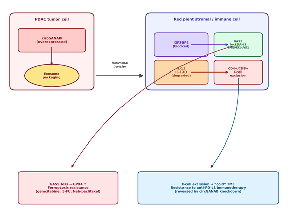
Circular RNAs act as unusually stable regulators within the pancreatic tumor microenvironment, owing to their covalently closed-loop structure (Limb et al., 2020; Wong et al., 2026; Table 2). CircGANAB, the most extensively characterized of these, is overexpressed in PDAC tissue and serum exosomes, achieving a reported area under the curve (AUC) of 0.822 for diagnostic discrimination (Wong et al., 2026). It physically binds tumor-suppressive lncRNAs (GAS5, lncLDAH3, TMEM51-AS1) and cytokine transcripts (IL-13, IL-17D), blocking the RNA stabilizer IGF2BP2 and systematically degrading these targets (Wong et al., 2026; Figure 3). Loss of GAS5 subsequently upregulates GPX4, protecting PDAC cells from lipid peroxidation and erastin-induced ferroptosis and thereby promoting chemoresistance (Wong et al., 2026), while exosomal transfer of circGANAB simultaneously downregulates IL-13 and IL-17D, limiting cytotoxic CD4+ and CD8+ T-cell infiltration and driving resistance to anti-PD-L1 immunotherapy (Wong et al., 2026).

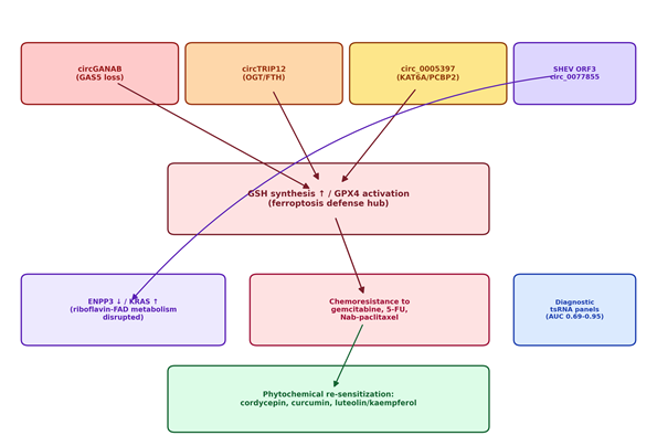
Ferroptosis resistance is reinforced by other circRNAs converging on the same metabolic endpoint (Figure 4): circTRIP12 stabilizes ferritin heavy chain (FTH) via OGT-mediated O-GlcNAcylation to limit the labile iron pool (Lin et al., 2025), and circ_0005397 recruits KAT6A to catalyze H3K9 acetylation at the PCBP2 promoter, enhancing glutathione synthesis and GPX4 activation (Qu et al., 2024). At the intercellular level, exosomal circ-IARS and circ-PDE8A sponge miR-122 and miR-338, respectively, to activate metastatic RhoA and MET signaling (Li et al., 2018a, 2018b). And, as noted above, viral SHEV ORF3 protein induces hsa_circ_0077855 to sponge miR-181a-2-3p and miR-30b-3p, derepressing ENPP3 and KRAS and linking riboflavin metabolic remodeling to oncogenesis (Luo et al., 2026).

4.3 tsRNAs as Sensitive Diagnostic and Prognostic Biomarkers

tRNA-derived small RNAs — including tRFs and tiRNAs — serve as stable, early diagnostic and prognostic biomarkers in PDAC (Pan et al., 2025; Table 3). Under cellular stress, specific tsRNAs are selectively dysregulated: tRF-Leu-AAG is upregulated in tumor tissue and promotes cell proliferation by targeting UPF1 (Sui et al., 2022), while pancreatic-stellate-cell-derived exosomal tRF-19-PNR8YPJZ downregulates AXIN2 to activate Wnt signaling and metastatic invasion (Cao et al., 2023a). By contrast, 5′-tRF-19-Q1Q89PJZ is downregulated in plasma, and restoring its expression represses hexokinase 1 (HK1) to inhibit glycolysis and cell mobility (Cao et al., 2023b); exosomal tRF-GluCTC-0005 promotes early hepatic metastasis by stabilizing WDR1 to activate Hippo/YAP signaling (Chen et al., 2024).

From a diagnostic standpoint, serum tsRNA-MetCAT-37 (AUC = 0.687) and tsRNA-ValTAC-41 (AUC = 0.793) function as reasonably powerful diagnostic panels on their own; combined with CA19-9, they achieve AUC values of 0.949 and 0.947, respectively (Xue et al., 2021). More strikingly still, the ratio of exosomal tRF-Leu-CAG-002 to exosome-free tRF-Pro-AGG-004 yields an AUC of 0.94

Table 1. Key lncRNAs within the lncRNA-miRNA-mRNA ceRNA TRIAD in pancreatic ductal adenocarcinoma. The table lists the principal oncogenic and tumor-suppressive lncRNAs discussed in this review, together with their validated miRNA/mRNA regulatory axis, reported functional consequence, and the primary supporting reference(s). Oncogenic lncRNAs predominantly act by sponging tumor-suppressive miRNAs to derepress downstream oncogenes, whereas tumor-suppressive lncRNAs restrain proliferative and invasive signaling.

ncRNA

Class

Type

Molecular Axis / Target

Functional Consequence

Key Reference(s)

PVT1

lncRNA

Oncogenic

miR-619-5p/Pygo2; miR-619-5p/ATG14; p-Smad2/3

Wnt/β-catenin activation, autophagy, EMT, gemcitabine resistance

Zhou et al., 2020; Wang et al., 2023

UCA1

lncRNA

Oncogenic

EZH2/SOCS3; correlates with MEAK7

Hippo pathway activation, gemcitabine resistance, poor survival

Guo et al., 2020; Kan & Ayan, 2026

HOTTIP

lncRNA

Oncogenic

Wnt/β-catenin/HOXA9/HOXA13

CSC stemness maintenance

Fu et al., 2017

MALAT1

lncRNA

Oncogenic

miR-217/KRAS

KRAS derepression, inflammatory signaling

Liu et al., 2017

GAS5

lncRNA

Tumor-suppressive

miR-221-3p/SOCS3; miR-32-5p/PTEN

PI3K/Akt & STAT3 inactivation, EMT reversal

Gao et al., 2017; Liu et al., 2018

MEG3

lncRNA

Tumor-suppressive

G1 cell-cycle checkpoint

G1 arrest, apoptosis induction

Ma et al., 2018

GLS-AS

lncRNA

Tumor-suppressive

GLS pre-mRNA / c-Myc

Impaired glutamine metabolism

Deng et al., 2019

MIR600HG

lncRNA

Tumor-suppressive

miR-1197/PITPNM3

Suppressed migration and EMT

Yang et al., 2024

LINC01963

lncRNA

Tumor-suppressive

miR-641/TMEFF2

Promotes apoptosis

Li et al., 2020

Table 2. Circular RNAs (circRNAs) implicated in ferroptosis evasion, immune exclusion, and metastatic signaling in pancreatic ductal adenocarcinoma. Entries summarize each circRNA's parent gene or inducing stimulus, its non-canonical or canonical molecular mechanism, and the resulting downstream phenotype, with emphasis on the convergent role of these circRNAs in glutathione/GPX4-mediated ferroptosis resistance.

circRNA

Parent Gene / Origin

Molecular Mechanism

Downstream Effect

Key Reference(s)

circGANAB

GANAB (exons 2-4)

Degrades GAS5/lncLDAH3/TMEM51-AS1 and IL-13/IL-17D by blocking IGF2BP2

GPX4 ↑ (ferroptosis resistance); T-cell exclusion; anti-PD-L1 resistance

Wong et al., 2026

circTRIP12

TRIP12

Stabilizes FTH via OGT-mediated O-GlcNAcylation

Limits labile iron pool; ferroptosis resistance; PD-L1-mediated immune evasion

Lin et al., 2025

circ_0005397

Recruits KAT6A; H3K9 acetylation at PCBP2 promoter

GSH synthesis ↑, GPX4 activation, chemoresistance

Qu et al., 2024

circ-IARS

IARS

Exosomal sponge of miR-122

Endothelial permeability ↑, metastasis

Li et al., 2018a

circ-PDE8A

PDE8A

Exosomal sponge of miR-338

MACC1/MET pathway activation, invasive growth

Li et al., 2018b

hsa_circ_0077855

— (SHEV ORF3-induced)

Sponges miR-181a-2-3p and miR-30b-3p

ENPP3 ↓, FAD hydrolysis disrupted, KRAS derepression

Luo et al., 2026


with 98.8% specificity for distinguishing early-stage resectable PDAC from chronic pancreatitis (Jin et al., 2021) — a level of performance that, if replicated in larger independent cohorts, would represent a meaningful advance over CA19-9 alone.

4.4 Multi-Target Therapeutic Resensitization by Bioactive Natural Compounds

Given the network-level complexity of ncRNA pathways in PDAC, multi-target natural compounds have shown a consistent ability to overcome chemoresistance across the studies reviewed here (Chen et al., 2025; Table 4). Cordycepin, from Cordyceps sinensis, directly binds the CDK2 pocket (binding energy: -7.4 kcal/mol, confirmed via cellular thermal shift assay), suppressing the downstream CDK2/E2F2/TNNI2 axis to induce G2/M arrest and apoptosis in PANC-1 and BxPC-3 models (He et al., 2026); because CDK2 promotes gemcitabine resistance, cordycepin synergistically enhances gemcitabine sensitivity in vitro and in vivo, while also protecting against gemcitabine-induced hepatotoxicity by reducing inflammatory sinusoidal congestion (He et al., 2026).

Curcumin, a polyphenol from Curcuma longa, downregulates EZH2 and the oncogenic lncRNA PVT1, dismantling the EZH2-PVT1-c-Myc axis to reverse gemcitabine resistance (Yoshida et al., 2017). Curcumin and its synthetic analogs (CDF, EF31, UBS109) further regulate miRNA networks — upregulating let-7, miR-26a, and miR-101 to target EZH2, Notch-1, and Nanog and suppress EMT and CSC stemness (Bao et al., 2012; Eftekhari et al., 2026). To circumvent curcumin's notoriously poor oral bioavailability, cationic lipid nanoparticles (CurEm) have been used to deliver curcumin directly, successfully upregulating p53-mediated apoptotic cascades (Demirci et al., 2025). Finally, luteolin and kaempferol from Annona muricata demonstrate exceptionally low binding free energies against key hub nodes — AKT1, EGFR, MMP9, and SRC — suppressing cell motility and downstream proliferative signaling in silico (Mathpal et al., 2025). Taken together, these findings suggest that natural compounds can simultaneously engage multiple, redundant ncRNA circuits, offering a genuinely multi-target strategy for overcoming drug resistance in pancreatic cancer.

5. Rewiring the Post-Transcriptional Landscape of PDAC — From Mechanistic Insight to Multi-Target Precision Oncology

5.1 A Network, Not a List: Reframing the Molecular Logic of PDAC

Taken as a whole, the evidence synthesized here suggests something that is, on reflection, fairly intuitive but easy to lose sight of in a literature organized around individual genes: PDAC is not usefully described as a collection of independent molecular lesions. It behaves, instead, like a coordinated system — one in which lncRNAs, circRNAs, miRNAs, and tsRNAs occupy specific nodes within an interconnected regulatory architecture (Table 1; Table 2; Figure 1). This reframing matters clinically, not just conceptually, because it may help explain why single-target strategies aimed at KRAS, TP53, or other classical drivers have so consistently underdelivered (Jeong et al., 2026; Kan & Ayan, 2026) — the network, in effect, routes around single-point interventions. If that is right, then durable therapeutic benefit will likely require disrupting entire circuits rather than isolated nodes, a principle that echoes across several of the mechanisms discussed above.

5.2 CircGANAB as a Convergent Node: Immune Evasion Meets Metabolic Escape

Among the mechanisms reviewed, the circGANAB axis stands out — not because it is necessarily the most prevalent, but because it appears to sit at the convergence of two otherwise distinct resistance pathways (Figure 3). By degrading GAS5 and thereby upregulating GPX4, circGANAB confers ferroptosis resistance and, with it, resistance to gemcitabine, 5-FU, and Nab-paclitaxel (Wong et al., 2026); by simultaneously degrading IL-13 and IL-17D transcripts, it excludes cytotoxic T cells and drives resistance to anti-PD-L1 checkpoint blockade (Wong et al., 2026). It is somewhat unusual, mechanistically, for a single circular RNA to bridge chemoresistance and immunotherapy resistance this directly, and it raises a fairly natural question: could disrupting circGANAB alone — through antisense oligonucleotides, small-molecule inhibitors of the back-splicing junction, or CRISPR-Cas13-based knockdown — restore sensitivity to both chemotherapy and immunotherapy simultaneously? The preclinical data combining circGANAB knockdown with anti-PD-L1 therapy (Wong et al., 2026) is suggestive here, though it remains, for now, a single-model observation rather than a validated clinical strategy.

Figure 3. Mechanistic model of exosomal circGANAB-mediated intercellular communication in the pancreatic ductal adenocarcinoma tumor microenvironment. CircGANAB, overexpressed in donor PDAC cells, is packaged into exosomes and horizontally transferred to recipient stromal and immune cells, where it blocks IGF2BP2-mediated stabilization of GAS5, lncLDAH3, and TMEM51-AS1, and independently promotes degradation of the cytokine transcripts IL-13 and IL-17D. These parallel events converge on two distinct resistance phenotypes: GPX4-driven ferroptosis resistance to chemotherapy, and CD4+/CD8+ T-cell exclusion driving resistance to anti-PD-L1 immunotherapy (Section 4.2).

Figure 4. Convergent metabolic reprogramming network linking circRNA-driven glutathione (GSH)/GPX4 activation to ferroptosis resistance and chemoresistance in pancreatic ductal adenocarcinoma. CircGANAB, circTRIP12, and circ_0005397 each independently enhance GSH synthesis or GPX4 activity through distinct molecular routes, converging on a shared ferroptosis-defense hub that confers resistance to gemcitabine, 5-fluorouracil, and Nab-paclitaxel. The SHEV ORF3-induced circRNA hsa_circ_0077855 additionally disrupts riboflavin/FAD metabolism via ENPP3 and KRAS. Diagnostic tsRNA panels and phytochemical re-sensitization strategies are shown as parallel diagnostic and therapeutic counterpoints to this resistance network (Section 4.3-4.4).


Table 3. tRNA-derived small RNAs (tsRNAs) as functional regulators and diagnostic biomarkers in pancreatic ductal adenocarcinoma. The table separates tsRNAs studied primarily for their functional molecular targets from those with reported diagnostic area-under-the-curve (AUC) performance, highlighting the superior discriminative value achieved when tsRNA panels are combined with the conventional biomarker CA19-9.

tsRNA

Source / Sample

Molecular Target

Reported Diagnostic Performance

Key Reference(s)

tRF-Leu-AAG

Tumor tissue

UPF1

Not reported (functional study)

Sui et al., 2022

tRF-19-PNR8YPJZ

PSC-derived exosomes

AXIN2

Not reported (functional study)

Cao et al., 2023a

5′-tRF-19-Q1Q89PJZ

Plasma

HK1

Not reported (functional study)

Cao et al., 2023b

tRF-GluCTC-0005

Exosomes

WDR1 / Hippo-YAP

Not reported (functional study)

Chen et al., 2024

tsRNA-MetCAT-37

Serum

AUC = 0.687 alone; 0.949 combined with CA19-9

Xue et al., 2021

tsRNA-ValTAC-41

Serum

AUC = 0.793 alone; 0.947 combined with CA19-9

Xue et al., 2021

tRF-Leu-CAG-002 / tRF-Pro-AGG-004 ratio

Exosomal / exosome-free serum

AUC = 0.94; specificity = 98.8%

Jin et al., 2021

Table 4. Natural phytochemicals and traditional Chinese medicine (TCM) compounds targeting non-coding RNA circuits in pancreatic ductal adenocarcinoma. Entries list each compound's botanical or formulation source, its principal molecular target(s) within the ncRNA network, and its reported preclinical effect, illustrating the shared polypharmacological logic underlying phytochemical re-sensitization strategies.

Compound

Natural Source

Molecular Target(s)

Reported Effect

Key Reference(s)

Cordycepin

Cordyceps sinensis

CDK2/E2F2/TNNI2 axis

G2/M arrest, apoptosis, synergy with gemcitabine, reduced hepatotoxicity

He et al., 2026

Curcumin

Curcuma longa

EZH2/PVT1/c-Myc; let-7, miR-26a, miR-101

EMT suppression, gemcitabine sensitization, CSC suppression

Yoshida et al., 2017; Eftekhari et al., 2026

CDF / EF31 / UBS109 (curcumin analogs)

Synthetic curcumin derivatives

EZH2, Notch-1, Nanog

Restored suppressor microRNAs, reduced tumor growth

Bao et al., 2012

CurEm (nanoparticle curcumin)

Emulsome-formulated curcumin

p53 apoptotic cascade

Improved bioavailability and apoptosis induction

Demirci et al., 2025

Luteolin / Kaempferol

Annona muricata

AKT1, EGFR, MMP9, SRC

Reduced cell motility (in silico network pharmacology)

Mathpal et al., 2025

Qingyihuaji (QYHJ)

Herbal formula (TCM)

lncRNA AB209630/miR-373/EphB2; NANOG

Reversed gemcitabine resistance, CSC depletion

Chen et al., 2025

Deoxyelephantopin

Elephantopus scaber

circTNPO3/NF-κB

Suppressed inflammatory signaling

Chen et al., 2025


5.3 Diagnostic Promise and Its Limits: Exosomal ncRNA Panels versus CA19-9

The diagnostic performance reported for several ncRNA panels is genuinely encouraging — the tRF-Leu-CAG-002/tRF-Pro-AGG-004 ratio, for instance, reaches an AUC of 0.94 with 98.8% specificity (Jin et al., 2021; Table 3), and combined tsRNA-CA19-9 panels approach AUC values near 0.95 (Xue et al., 2021). It would be premature, though, to treat these figures as settled. Most of the underlying cohorts are modest in size and drawn from single centers, and independent, multi-cohort validation — ideally with pre-specified cutoffs and blinded adjudication — has not yet been widely reported for these specific panels. This is not a criticism so much as an observation about where the field currently sits: the biology is compelling, but the path from a promising discovery cohort to a clinically deployable biomarker is a long one, and PDAC has seen biomarker candidates falter at exactly this stage before.

5.4 The Bioavailability Problem: Why Multi-Target Natural Compounds Have Not Yet Reached the Clinic

There is a certain irony in the phytochemical literature reviewed here (Table 4): compounds like curcumin and cordycepin appear to hit exactly the kind of redundant, multi-node targets that a network-level disease like PDAC arguably demands (Chen et al., 2025; Eftekhari et al., 2026; He et al., 2026), yet their clinical translation remains hindered by comparatively prosaic pharmacokinetic problems — poor solubility, rapid metabolism, and limited tumor accumulation (Chen et al., 2025; Eftekhari et al., 2026). Nanoparticle formulations such as CurEm represent one plausible way forward (Demirci et al., 2025), but it is worth being honest that delivery-system engineering, not molecular mechanism, may be the rate-limiting step for this entire therapeutic class going forward.

5.5 Limitations

Several limitations of this review are worth stating directly. First, as a narrative rather than systematic synthesis, it does not apply formal risk-of-bias scoring uniformly across included studies, and publication bias toward positive, mechanistically clean findings cannot be excluded. Second, much of the mechanistic evidence discussed here derives from cell-line and xenograft models, which may not fully recapitulate the heterogeneity of human PDAC, particularly its notoriously variable stromal composition. Third, one included mechanistic finding — the SHEV ORF3-circRNA-riboflavin axis — originates from a veterinary, non-human model (Luo et al., 2026), and while it offers a mechanistically instructive parallel, its direct applicability to human PDAC has not been established and should be interpreted cautiously. Finally, diagnostic AUC values reported across studies were not generated using harmonized assay platforms or cutoffs, which limits direct cross-study comparison.

5.6 Future Directions

Building on the gaps identified above, several directions seem particularly worth pursuing. Single-cell and spatial multi-omic profiling could help resolve how ncRNA network activity varies across the notoriously heterogeneous PDAC stroma, rather than treating the tumor as a single averaged entity. Multi-cohort, prospectively validated biomarker studies — ideally harmonizing assay platforms across centers — would help determine whether the diagnostic performance reported for panels like circGANAB or the tRF-Leu-CAG-002/tRF-Pro-AGG-004 ratio holds up outside the discovery cohort. And on the therapeutic side, combining circGANAB-targeted nucleic acid therapeutics with immune checkpoint blockade, alongside nanoparticle-based delivery of phytochemicals, represents a reasonably concrete near-term research agenda — one that follows fairly directly from the mechanistic convergence described in Section 5.2.

6. Conclusion

The expanding regulatory landscape of non-coding RNA networks in pancreatic cancer represents a genuine shift toward network-based precision oncology, not merely an addition to the existing driver-gene framework. As charted throughout this review, lncRNAs, circRNAs, miRNAs, and tsRNAs are not isolated transcriptional products but integrated components of dynamic, multi-layered circuits that jointly dictate cell identity, stromal communication, metabolic adaptation, and cell-death evasion. The exosomal circGANAB axis illustrates this integration especially clearly, linking ferroptosis resistance to T-cell exclusion and anti-PD-L1 resistance within a single molecular pathway, while stable serum tsRNA panels offer a genuinely promising, minimally invasive route toward earlier diagnosis. Natural phytochemicals and traditional Chinese medicine formulations, for their part, demonstrate that multi-target re-sensitization of resistant ncRNA circuits is mechanistically feasible, even if pharmacokinetic barriers still limit clinical translation. Going forward, priorities should include single-cell and spatial multi-omic profiling to capture intratumoral heterogeneity, optimized nanocarrier systems to overcome delivery limitations, and rigorous, multi-cohort validation of stable exosomal biomarker panels before any of these candidates can reasonably be considered for clinical deployment. None of this will be quick. But the evidence reviewed here suggests that disrupting these regulatory networks at their most convergent nodes — rather than at any single gene — offers a realistic, if still early-stage, path toward improving outcomes in a disease that has, for too long, resisted almost everything else.


References


Bao, B., Ali, S., Banerjee, S., Wang, Z., Logna, F., Azmi, A. S., Kong, D., Ahmad, A., Li, Y., Padhye, S., & Sarkar, F. H. (2012). Curcumin analogue CDF inhibits pancreatic tumor growth by switching on suppressor microRNAs and attenuating EZH2 expression. Cancer Research, 72(1), 335–345. https://doi.org/10.1158/0008-5472.CAN-11-2182   

Cao, W., Dai, S., Ruan, W., Long, T., Zeng, Z., & Lei, S. (2023a). Pancreatic stellate cell-derived exosomal tRF-19-PNR8YPJZ promotes proliferation and mobility of pancreatic cancer through AXIN2. Journal of Cellular and Molecular Medicine, 27(17), 2533–2546. https://doi.org/10.1111/jcmm.17838          

Cao, W., Zeng, Z., & Lei, S. (2023b). 5′-tRF-19-Q1Q89PJZ suppresses the proliferation and metastasis of pancreatic cancer cells via regulating hexokinase 1-mediated glycolysis. Biomolecules, 13(2), 253. https://doi.org/10.3390/biom13020253   

Chang, L., Qin, C., Wu, J., Jiang, H., Xu, Q., Chen, J., Xiao, X., Zhang, X., Guan, M., & Deng, X. (2025). The crosstalk between glutathione metabolism and non-coding RNAs in cancer progression and treatment resistance. Redox Biology, 84, 103689. https://doi.org/10.1016/j.redox.2025.103689           

Chen, G., He, Q., Xiao, Q., Ai, J., Qin, Z., Zhong, Z., Zou, J., Zhang, C., & Feng, Y. (2025). The role of Chinese medicines in targeting non-coding RNAs to overcome cancer drug resistance: Mechanisms and clinical translation challenges. Pharmacological Research, 222, 108049. https://doi.org/10.1016/j.phrs.2025.108049             

Chen, J., & Gao, J. (2026). The future of lncRNA in precision cancer therapy. Non-coding RNA Research, 17, 150–159. https://doi.org/10.1016/j.ncrna.2026.02.001            

Chen, W., Peng, W., Wang, R., Bai, S., Cao, M., Xiong, S., & Chen, Y. (2024). Exosome-derived tRNA fragments tRF-GluCTC-0005 promotes pancreatic cancer liver metastasis by activating hepatic stellate cells. Cell Death & Disease, 15(2), 102. https://doi.org/10.1038/s41419-024-06489-3          

Demirci, Z., Islek, Z., Siginc, H. I., Sahin, F., Ucisik, M. H., & Bolat, Z. B. (2025). Curcumin-loaded emulsome nanoparticles induces apoptosis through p53 signaling pathway in pancreatic cancer cell line PANC-1. Toxicology in Vitro, 102, 105958. https://doi.org/10.1016/j.tiv.2024.105958

Deng, S. J., Chen, H. Y., Zeng, Z., Deng, S., Zhu, S., Ye, Z., Liu, Y., He, C., Liu, X., Zhang, Q., Ai, Y., Guo, Q., Zhao, Y., Fang, X., & Tang, J. F. (2019). Nutrient stress–dysregulated antisense lncRNA GLS-AS impairs GLS-mediated metabolism and represses pancreatic cancer progression. Cancer Research, 79(7), 1398–1412. https://doi.org/10.1158/0008-5472.CAN-18-0710         

Eftekhari, H., Tajik, Z., & Ghafouri-Fard, S. (2026). Curcumin in pancreatic cancer: A comprehensive review of anticancer mechanisms, EMT modulation, and clinical challenges. Cancer Treatment and Research Communications, 49, 101414. https://doi.org/10.1016/j.ctarc.2026.101414            

Fu, Z., Chen, C., Zhou, Q., Wang, Y., Zhao, Y., Zhao, X., Li, W., Zheng, S., Wang, Q., Liu, Y., Ding, Q., & Wang, L. (2017). LncRNA HOTTIP modulates cancer stem cell properties in human pancreatic cancer by regulating HOXA9. Cancer Letters, 410, 68–81. https://doi.org/10.1016/j.canlet.2017.09.019            

Gao, Z. Q., Wang, J. F., Chen, D. H., Ma, X. S., Wu, Y., Tang, Z., & Dang, X. W. (2017). Long non-coding RNA GAS5 suppresses pancreatic cancer metastasis through modulating miR-32-5p/PTEN axis. Cell & Bioscience, 7, 66. https://doi.org/10.1186/s13578-017-0192-0             

Gao, Z. Q., Wang, J. F., Chen, D. H., Ma, X. S., Wu, Y., Tang, Z., Yang, T., & Dang, X. W. (2018). Long non-coding RNA GAS5 antagonizes the chemoresistance of pancreatic cancer cells through down-regulation of miR-181c-5p. Biomedicine & Pharmacotherapy, 97, 809–817. https://doi.org/10.1016/j.biopha.2017.10.157 

Guo, Z., Wang, X., Yang, Y., Chen, W., Zhang, K., Teng, B., Xu, L., Wang, K., & Zhao, Q. (2020). Hypoxic tumor-derived exosomal long noncoding RNA UCA1 promotes angiogenesis via miR-96-5p/AMOTL2 in pancreatic cancer. Molecular Therapy – Nucleic Acids, 22, 179–195. https://doi.org/10.1016/j.omtn.2020.08.021           

He, R., Du, Y., Jiang, W., & Zhou, W. (2026). Cordycepin inhibits pancreatic cancer progression and enhances gemcitabine sensitivity by targeting the CDK2/E2F2 axis to modulate TNNI2 expression. Biochemical Pharmacology, 253, 118299. https://doi.org/10.1016/j.bcp.2026.118299

Jeong, H. S., Lee, Y. J., Lee, D. H., Roh, H. Y., Jeong, G. R., & Kim, H. S. (2026). The multi-target lncRNA–miRNA–mRNA TRIAD in pancreatic cancer diagnosis and therapy. International Journal of Molecular Sciences, 27, 1400. https://doi.org/10.3390/ijms27031400      

Jin, F., Yang, L., Wang, W., Yuan, N., Zhan, S., Yang, P., Chen, J., Zhong, L., Ma, F., & To, K. F. (2021). A novel class of tsRNA signatures as biomarkers for diagnosis and prognosis of pancreatic cancer. Molecular Cancer, 20, 95. https://doi.org/10.1186/s12943-021-01389-5

Kan, M. U., & Ayan, D. (2026). Exploratory multi-platform bioinformatic analysis of MEAK7 and its molecular, survival, and immune associations in pancreatic ductal adenocarcinoma. International Journal of Molecular Sciences, 27(17), 7610. https://doi.org/10.3390/ijms27177610      

Li, J., Han, H., Gu, W., Cao, C., & Zheng, P. (2020). Long non-coding RNA LINC01963 inhibits progression of pancreatic carcinoma by targeting miR-641/TMEFF2. Biomedicine & Pharmacotherapy, 129, 110346. https://doi.org/10.1016/j.biopha.2020.110346               

Li, J., Li, Z., Jiang, P., Peng, M., Zhang, X., Chen, K., Liu, H., Bi, H., Liu, X., & Li, X. (2018a). Circular RNA IARS (circ-IARS) secreted by pancreatic cancer cells and located within exosomes regulates endothelial monolayer permeability to promote tumor metastasis. Journal of Experimental & Clinical Cancer Research, 37, 177. https://doi.org/10.1186/s13046-018-0822-3

Li, Z., Yanfang, W., Li, J., Jiang, P., Peng, T., Chen, K., Zhao, X., Zhang, Y., Zhen, P., Zhu, J., & Li, X. (2018b). Tumor-released exosomal circular RNA PDE8A promotes invasive growth via the miR-338/MACC1/MET pathway in pancreatic cancer. Cancer Letters, 432, 237–250. https://doi.org/10.1016/j.canlet.2018.04.035          

Limb, C., Liu, D. S. K., Veno, M. T., Rees, E., Krell, J., Bagwan, I. N., Giovannetti, E., Pandha, H., Strobel, O., Rockall, T. A., & Frampton, A. E. (2020). The role of circular RNAs in pancreatic ductal adenocarcinoma and biliary-tract cancers. Cancers, 12(11), 3250. https://doi.org/10.3390/cancers12113250

Lin, H., Zhu, S., Chen, Y., Lu, J., Xie, C., Liao, C., & Wang, Z. (2025). Targeting cTRIP12 counteracts ferroptosis resistance and augments sensitivity to immunotherapy in pancreatic cancer. Drug Resistance Updates, 81, 101240. https://doi.org/10.1016/j.drup.2025.101240             

Liu, L., Liu, F. B., Huang, M., Xie, K., Xie, Q. S., & Huang, Q. (2019). Circular RNA ciRS-7 promotes the proliferation and metastasis of pancreatic cancer by regulating miR-7-mediated EGFR/STAT3 signaling pathway. Hepatobiliary & Pancreatic Diseases International, 18(6), 546–552. https://doi.org/10.1016/j.hbpd.2019.03.003

Liu, P., Yang, H., Zhang, J., Peng, X., Lu, Z., & Chen, J. (2017). The lncRNA MALAT1 acts as a competing endogenous RNA to regulate KRAS expression by sponging miR-217 in pancreatic ductal adenocarcinoma. Scientific Reports, 7, 44122. https://doi.org/10.1038/srep44122             

Long, M., Liu, H., Yuan, M., Zhou, X., Zhang, T., Wang, Q., & Jiang, W. (2026). ncFN: A comprehensive non-coding RNA function annotation framework based on a global and heterogeneous biomolecular network. Non-coding RNA Research, 16, 70–78. https://doi.org/10.1016/j.ncrna.2025.09.007            

Luo, W., Li, J., Wu, S., Wang, L., Yin, Y., Cao, X., Wang, L., & Jiao, H. (2026). Integrated transcriptomics reveals a SHEV ORF3-mediated circRNA network that disrupts riboflavin metabolism and activates the ko05212 pathway. Veterinary Sciences, 13, 253. https://doi.org/10.3390/vetsci13010253   

Ma, L., Wang, F., Du, C., Zhang, Z., Guo, H., Xie, X., Wang, T., Chen, T., & Gao, H. (2018). Long non-coding RNA MEG3 functions as a tumour suppressor and has prognostic predictive value in human pancreatic cancer. Oncology Reports, 39(3), 1132–1140. https://doi.org/10.3892/or.2018.6182        

Mathpal, S., Priyamvada, P., Ashok, G., Joshi, T., Saha, D., Mukherjee, A., Roy, B., Ramaiah, S., & Anbarasu, A. (2025). Network pharmacology-driven investigation of luteolin from Annona muricata as a promising multi-target inhibitor for pancreatic cancer. Results in Chemistry, 16, 102506. https://doi.org/10.1016/j.rechem.2025.102506          

Pan, Y., Ying, X., Zhang, X., Jiang, H., Yan, J., & Duan, S. (2025). The role of tRNA-derived small RNAs (tsRNAs) in pancreatic cancer and acute pancreatitis. Non-coding RNA Research, 11(2), 200–208. https://doi.org/10.1016/j.ncrna.2025.01.004           

Qu, T., Cha, L., Liu, H., Tian, L., & Zhou, B. (2024). Circ_0005397 inhibits ferroptosis of pancreatic cancer cells by up-regulating PCBP2 through KAT6A/H3K9Ac. FASEB Journal, 38(7), e70028. https://doi.org/10.1096/fj.202400327R

Rui, X., Gao, J., Bi, Y., Han, Z., Zhang, X., Hao, Y., Yang, C., Peng, Y., Dai, X., Xia, Y., & Jin, Y. (2026). Emerging role of interaction between m6A and main ncRNAs in pancreatic cancer. Non-coding RNA Research, 19, 93–107. https://doi.org/10.1016/j.ncrna.2026.01.007               

Safarzadeh, A., Jamil, M. U., Saddozai, U. A. K., Zhang, L., Ma, F., Ji, X. Y., & Khawar, M. B. (2025). Clinical significance of lncRNA polymorphisms and associations with tumor-node-metastasis staging in cancer. Biochemistry and Biophysics Reports, 44, 102283. https://doi.org/10.1016/j.bbrep.2025.102283           

Sayed, U., Abdulsahib, W. K., Jyothi, S. R., Nayak, P. P., Chauhan, A. S., Singla, S., Polatova, D., Sead, F. F., & Akhavan-Sigari, R. (2026). Non-coding RNAs at the intersection of interferon signaling and cancer: Mechanistic insights and clinical prospects. Cancer Treatment and Research Communications, 47, 101155. https://doi.org/10.1016/j.ctarc.2025.101155        

Shi, M., Zhang, R., Lyu, H., Xiao, S., Guo, D., Zhang, Q., Chen, X. Z., Tang, J., & Zhou, C. (2025). Long non-coding RNAs: Emerging regulators of invasion and metastasis in pancreatic cancer. Journal of Advanced Research, 78, 285–306. https://doi.org/10.1016/j.jare.2025.02.001

Son, S. W., Song, M. G., Yun, B. D., & Park, J. K. (2021). Noncoding RNAs associated with therapeutic resistance in pancreatic cancer. Biomedicines, 9, 263. https://doi.org/10.3390/biomedicines9030263

Sui, S., Wang, Z., Cui, X., Jin, L., & Zhu, C. (2022). The biological behavior of tRNA-derived fragment tRF-Leu-AAG in pancreatic cancer cells. Bioengineered, 13(4), 10617–10628. https://doi.org/10.1080/21655979.2022.2064612     

Sun, Q., Lei, X., & Yang, X. (2024). The crosstalk between non-coding RNAs and oxidative stress in cancer progression. Genes & Diseases, 12, 101286. https://doi.org/10.1016/j.gendis.2024.101286 

Wang, B., Yuan, C., Qie, Y., & Dang, S. (2023). Long non-coding RNAs and pancreatic cancer: A multifaceted view. Biomedicine & Pharmacotherapy, 167, 115601. https://doi.org/10.1016/j.biopha.2023.115601              

Wong, C. H., Peña-Paladines, J. J., Luo, Z., Ma, C. Y., Lee, Y. L., Lin, Z. X., Xian, Y. F., Hollingsworth, M. A., To, K. F., & Chen, Y. (2026). Exosomal circGANAB promotes cancer progression and immunotherapy resistance by degrading interacting RNAs and limiting T cell infiltration in pancreatic ductal adenocarcinoma. Drug Resistance Updates, 87, 101416. https://doi.org/10.1016/j.drup.2026.101416             

Xue, M., Shi, M., Xie, J., Zhang, J., & Deng, X. (2021). Serum tRNA-derived small RNAs as potential novel diagnostic biomarkers for pancreatic ductal adenocarcinoma. American Journal of Cancer Research, 11(3), 837–848.

Yang, B., Jiao, Z., Feng, N., Zhang, Y., & Wang, S. (2024). Long non-coding RNA MIR600HG as a ceRNA inhibits the pancreatic cancer progression through regulating the miR-1197/PITPNM3 axis. Heliyon, 10, e24546. https://doi.org/10.1016/j.heliyon.2024.e24546               

Yoshida, K., Toden, S., Ravindranathan, P., Han, H., & Goel, A. (2017). Curcumin sensitizes pancreatic cancer cells to gemcitabine by attenuating PRC2 subunit EZH2, and the lncRNA PVT1 expression. Carcinogenesis, 38(10), 1036–1046. https://doi.org/10.1093/carcin/bgx085      

Zhang, Z., Zhou, C., Chang, Y., Zhang, Z., Hu, Y., Zhang, F., Lu, Y., Zheng, L., Zhang, W., Li, X., & Yang, Q. (2018). Long non-coding RNA CASC9 promotes tumorigenesis of oesophageal squamous cell carcinoma via the increased expression of LY6K. American Journal of Cancer Research, 8(5), 762–776.

Zhou, C., Yi, C., Yi, Y., Qin, W., Yan, Y., Dong, X., Zhang, X., Huang, Y., Zhang, R., Wei, J., Xu, J., & Zhou, J. (2020). LncRNA PVT1 promotes gemcitabine resistance of pancreatic cancer via activating Wnt/beta-catenin and autophagy pathway through modulating the miR-619-5p/Pygo2 and miR-619-5p/ATG14 axes. Molecular Cancer, 19, 118. https://doi.org/10.1186/s12943-020-01237-y       


Article metrics
View details
0
Downloads
0
Citations
28
Views

View Dimensions


View Plumx


View Altmetric


0
Save
0
Citation
28
View
0
Share