Preclinical science, it could fairly be said, has spent the last several decades caught between two imperfect worlds: the simplicity of flat, two-dimensional cultures and the translational unreliability of animal models (Chauhdari et al., 2025; Mirlohi et al., 2026). Monolayer systems scale beautifully for high-throughput screening, but they cannot reproduce the three-dimensional architecture, spatial polarity, or cell-matrix signaling that actually determine how human tissue responds to a drug (Castiglione et al., 2022; Papamichail et al., 2025). Animal models, for their part, carry their own species-specific baggage — discrepancies in pharmacokinetics, metabolism, and toxicity that routinely fail to predict what happens in a human trial, at real financial and ethical cost (Fanizza et al., 2022; Patra et al., 2026). Nowhere is this more visible than in oncology and neurology, fields in which roughly 90% of animal-validated candidates still fail in human testing (Mirlohi et al., 2026; Geng et al., 2024).
Two complementary technologies have grown up in response: three-dimensional organoids and organ-on-a-chip (OoC) platforms (Chauhdari et al., 2025; Leung et al., 2025). Organoids — self-organizing, multicellular structures derived from embryonic stem cells (ESCs), induced pluripotent stem cells (iPSCs), or primary biopsies — reproduce much of the cellular diversity and functional histopathology of native organs (Carvalho et al., 2023; Yao et al., 2024). But cultured statically, off-chip, they remain developmentally limited: no vasculature, no convective perfusion, no biomechanical stimulation to speak of (Chauhdari et al., 2025; Papamichail et al., 2025). Fusing the two paradigms — organoid and chip — gives rise to organoid-on-chip technology, embedding biologically authentic tissue within a microdevice that offers precise control over the physical, mechanical, and biochemical microenvironment (Li & Zhong, 2026; Saini et al., 2026). The distinction from conventional organ chips is mostly cellular: where organ chips typically rely on established cell lines or primary cells arranged into simplified interfaces, organoid chips leverage stem-cell-derived tissue that self-organizes into genuinely multi-lineage structures (Carvalho et al., 2023). Momentum here has been considerable since the FDA Modernization Act 2.0 (2022) formally sanctioned human-relevant in vitro and AI-based systems for de-risking candidates ahead of clinical trials (Patra et al., 2026; Leung et al., 2025).
2.1 Biophysical and Mechanobiological Engineering of the Microenvironment
Cells in the body rarely sit still, mechanically speaking. They exist within a constantly active physical environment where forces continuously shape morphology, gene expression, and maturation (Li & Zhong, 2026). Static culture removes almost all of that, and the result — perhaps unsurprisingly — is de-differentiation and gradual functional decline (Castiglione et al., 2022). Microfluidic organoid-chip platforms attempt to put those cues back, exposing tissue to a controlled suite of biophysical forces: fluid shear stress (FSS), cyclic tensile strain, hydrostatic pressure, and tunable viscoelastic matrix properties (Li & Zhong, 2026; Papamichail et al., 2025).
2.2 Fluid Perfusion and Shear Stress Dynamics
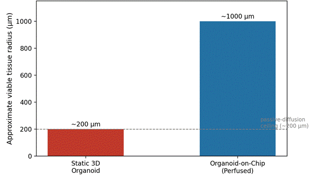
Perhaps the single most immediate benefit of microfluidic integration is simply this: diffusion stops being passive. Convective perfusion takes over (Chauhdari et al., 2025; Saini et al., 2026). Static organoids, recall, are capped by an oxygen and nutrient diffusion limit of roughly 100–200 micrometers in dense tissue (Zhang et al., 2025; Chauhdari et al., 2025); beyond that threshold, the core starves, waste accumulates, and necrosis follows (Chauhdari et al., 2025; Papamichail et al., 2025). Microfluidic channels recreate convective flow around and through the tissue, continuously delivering growth factors, fresh media, and dissolved gases while carrying toxic byproducts away (Papamichail et al., 2025; Geng et al., 2024). This flow imparts a tangential fluid shear stress on the cell surface (Li & Zhong, 2026). Physiological FSS in human capillaries sits between roughly 0.1 and 2.0 Pa (up to 2.0 Pa in larger arteries), but chip designers deliberately keep perfusion much lower — on the order of 10⁻³ to 10⁻² Pa — to sustain mass transport without shredding delicate epithelial or progenitor structures (Li & Zhong, 2026). Even at these modest levels, shear acts as a genuine morphogenetic signal, engaging mechanoreceptors such as Piezo1 to drive cytoskeletal remodeling, cellular alignment, and barrier polarization (Li & Zhong, 2026; Geng et al., 2024).
2.3 Tensile Strain, Cyclic Stretch, and Viscoelasticity
Shear stress is only part of the mechanical story. Native organs also undergo cyclic deformation — the expansion of the lungs with each breath, the rhythmic contraction of the heart, the peristaltic wave of the gut (Li & Zhong, 2026). Chips built from flexible elastomers such as polydimethylsiloxane (PDMS) reproduce this through pneumatic or vacuum actuation, applying strains of roughly 5–15% at frequencies between 0.1 and 1.0 Hz across thin, porous membranes (Li & Zhong, 2026; Geng et al., 2024). The effect is not merely cosmetic: cyclic deformation stretches the cytoskeleton, strengthens cell-matrix adhesion, and appears to accelerate functional maturation (Li & Zhong, 2026). In cardiac-organoid chips, for example, periodic mechanical pacing coaxes cardiomyocytes into coordinated electrophysiological rhythms and more mature sarcomeric structure (Li & Zhong, 2026).
Native tissue is also, of course, viscoelastic — it relaxes under stress and dissipates energy in ways purely elastic materials do not (Li & Zhong, 2026). Researchers have accordingly begun moving away from rigid elastomer substrates toward tunable hydrogels — polyethylene glycol (PEG), gelatin methacryloyl (GelMA), alginate — engineered to match the native stiffness of the organ in question, from around 100 Pa in brain tissue to well over 10 kPa in muscle (Li & Zhong, 2026). These viscoelastic matrices allow organoids to remodel and expand their internal lumens without accumulating residual stress, which in turn seems to improve long-term viability and reproducibility (Li & Zhong, 2026; Geng et al., 2024).
2.4 Advanced Biofabrication, Seeding, and Vascularization Strategies
Building a reproducible organoid-chip system is, at bottom, an exercise in careful placement: how does one get living tissue to sit, self-assemble, and eventually vascularize inside a microdevice (Chauhdari et al., 2025; Geng et al., 2024)? Three seeding strategies dominate the literature. In direct organoid seeding, organoids are pre-cultured off-chip in suspension plates or ECM domes and then mechanically transferred into the chip once sufficiently differentiated (Chauhdari et al., 2025). This approach exploits the parallel-generation advantages of multi-well plates, though manual transfer inevitably introduces size heterogeneity and occasional structural damage (Chauhdari et al., 2025; Papamichail et al., 2025). Cell suspension seeding instead introduces dissociated single cells directly into hydrogel-coated channels or microwells, allowing aggregation and differentiation to occur in situ under flow — a route that tends to yield more consistent size and alignment (Chauhdari et al., 2025). Cell suspension droplet seeding, meanwhile, encapsulates cells in Matrigel or synthetic hydrogel microdroplets via droplet microfluidics, producing highly uniform microenvironments well suited to high-throughput, automated screening (Chauhdari et al., 2025; Papamichail et al., 2025).
2.5 Promoting Maturation through Vascularization
Vascularization remains, arguably, the single most stubborn obstacle to long-term organoid survival (Zhang et al., 2025; Papamichail et al., 2025). Without a perfusable vessel network, organoid size and complexity are inherently capped (Papamichail et al., 2025). Three engineering strategies attempt to solve this: co-culture, in which endothelial cells are mixed directly with tissue progenitors; co-differentiation, in which iPSCs are directed toward both vascular and tissue-specific lineages simultaneously; and assembly, in which preformed tissue organoids are physically fused with vascular spheroids (Geng et al., 2024). In a top-down approach, endothelial cells line the microchannel walls, forming vessel-like tubes around the organoid chamber; in a bottom-up approach, they are embedded in fibrin-collagen hydrogels adjacent to the tissue, where they spontaneously sprout capillaries that penetrate the organoid (Geng et al., 2024; Papamichail et al., 2025). Connecting these vessels to a pump system allows circulation of media, plasma, or even whole blood under defined flow — a physiologically grounded route for both drug delivery and immune cell trafficking (Leung et al., 2025; Papamichail et al., 2025).
2.6 High-Fidelity Disease Modeling Across Diverse Organ Systems
Taken together, these engineering advances have enabled unusually detailed disease modeling across a range of organ systems (Yao et al., 2024; Geng et al., 2024). The subsections that follow work through several of the more mature examples.
2.7 Brain Organoids-on-Chip: Modeling Neurodevelopment and Neurodegeneration
Modeling the human brain in vitro is a notoriously hard problem, thanks to its intricate circuitry, its blood-brain barrier (BBB), and its markedly species-specific development (Castiglione et al., 2022). Brain organoids derived from patient iPSCs can replicate early neurogenesis and cortical layering reasonably well, and when combined with microfluidic chips they show greater structural complexity, longer survival, and more advanced neural differentiation than static well-plate cultures (Castiglione et al., 2022; Papamichail et al., 2025). These systems have proven useful for studying developmental toxicity: brain-organoid chips exposed to constant prenatal nicotine perfusion, for instance, show disrupted forebrain regionalization and premature neuronal differentiation, mirroring cognitive deficits observed in exposed human fetuses (Chauhdari et al., 2025; Castiglione et al., 2022). Similar platforms modeling prenatal exposure to valproic acid or cadmium reveal altered progenitor proliferation and disrupted region-specific gene expression (Castiglione et al., 2022). For neurodegeneration, midbrain organoid chips derived from Parkinson's patients carrying the LRRK2-G2019S mutation recapitulate dopaminergic apoptosis, mitochondrial dysfunction, and abnormal α-synuclein aggregation (Zhang et al., 2025; Fanizza et al., 2022), while Alzheimer's-disease chips reproduce senile plaque deposition, neurofibrillary tangles, and BBB leakage (Zhang et al., 2025).
2.8 Kidney Organoids-on-Chip: Nephrotoxicity and Hereditary Cystogenesis
The kidney sits at the center of drug excretion and is, correspondingly, quite vulnerable to drug-induced injury (Yao et al., 2024). Static kidney organoids, lacking perfusable vasculature and structural maturity, are of limited use for pharmacodynamic work (Papamichail et al., 2025). Subjecting them to controlled flow on-chip promotes proximal tubule polarization, primary cilia enrichment, and upregulation of functional transporters such as organic cation transporter 2 (OCT2) (Chauhdari et al., 2025; Papamichail et al., 2025); the resulting tissue is noticeably more sensitive to nephrotoxic compounds, including tacrolimus-induced cell death, and tracks clinical outcomes more closely than static culture does (Chauhdari et al., 2025; Papamichail et al., 2025). Kidney organoid chips have also reshaped how researchers think about Autosomal Dominant Polycystic Kidney Disease (ADPKD): PKD-edited organoids cultured under tubular flow spontaneously form large cystic outgrowths, and mechanistic work suggests that fluid shear alters membrane tension in ways that drive cystogenesis, with cysts remaining actively absorptive and transporting glucose into the lumen to fuel expansion — a finding later validated in mouse models (Chauhdari et al., 2025; Zhang et al., 2025; Geng et al., 2024).
2.9 Tumor-on-Chip: Immunotherapy, CAR-T Cells, and the Microenvironment
Oncology carries some of the field's highest preclinical failure rates, in large part because animal models simply cannot reproduce the human tumor microenvironment (TME) or its immune interactions (Leung et al., 2025; Mirlohi et al., 2026). Tumor-organoid chips address this by co-culturing patient-derived tumor organoids with stromal fibroblasts, ECM components, and immune populations — T cells, natural killer cells, macrophages — under continuous perfusion (Leung et al., 2025; Geng et al., 2024). These immunocompetent platforms have become genuinely useful tools for evaluating checkpoint blockade and CAR-T therapies (Leung et al., 2025; Geng et al., 2024). An endothelialized channel, acting as a physical vessel-like barrier, lets investigators watch immune cell extravasation and cytolytic contact happen in real time and in three dimensions (Leung et al., 2025). Work using colorectal and breast cancer organoids has shown that CAR-T cytotoxicity depends heavily on antigen density, tumor subtype, and TME-driven immunosuppression — cancer-associated fibroblasts depositing dense collagen, secreting TGF-β or IL-10 (Leung et al., 2025).
2.10 Multi-Organ Platforms and Systemic Pharmacokinetics
Because most human disease unfolds across, not within, single organs, the field also needs systemic models — platforms capable of simulating absorption, distribution, metabolism, excretion, and toxicity (ADME-Tox) (Carvalho et al., 2023; Geng et al., 2024). Multi-organoid, "body-on-a-chip" systems address this by physically or fluidically interconnecting multiple organoid chambers (Carvalho et al., 2023; Geng et al., 2024). A representative example is a liver-islet axis model of Type 2 diabetes: pancreatic islet spheroids and liver organoids, linked within a single platform, together reconstruct a functional metabolic loop in which glucose-stimulated insulin secretion travels via microperfusion to the liver and promotes glucose uptake and glycogen synthesis there (Chauhdari et al., 2025). Tripartite systems combining stomach, small intestine, and liver organoids have similarly modeled first-pass drug metabolism (Carvalho et al., 2023; Geng et al., 2024), while lung-liver-cardiac platforms have been used to screen for systemic chemotherapy toxicity; bleomycin, for instance, produced cardiac organoid arrhythmia and arrest only in the multi-organ configuration, not when cardiac organoids were dosed in isolation — a finding that underscores just how much inter-organ communication matters for accurate toxicity prediction (Chauhdari et al., 2025).
2.11 Integrating Artificial Intelligence, Biosensors, and Digital Twins
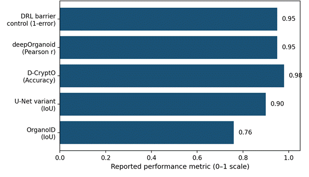
As these platforms scale, they generate a genuinely large volume of high-dimensional data — confocal imaging, biosensor streams, spatial transcriptomics and metabolomics — more than any human team can reasonably parse by hand (Mirlohi et al., 2026; Saini et al., 2026). Artificial intelligence has, somewhat inevitably, moved in to fill that gap.
2.11.1 Real-Time Biosensing of Physiology and Function
Embedding physical, chemical, and electrochemical biosensors directly onto the chip allows continuous, non-invasive monitoring of tissue state (Chauhdari et al., 2025; Geng et al., 2024). Transepithelial electrical resistance (TEER) sensors track barrier function and polarization under shear in real time (Li & Zhong, 2026; Geng et al., 2024); electrochemical and amperometric sensors monitor neurotransmitter, cytokine, or metabolite release — amperometric dopamine sensors integrated into midbrain chips, for instance, allow longitudinal tracking of dopaminergic activity in Parkinson's models (Zhang et al., 2025). Optical lipid tension reporters such as FliptR add another layer, capturing membrane tension changes caused by shear and thereby directly visualizing mechanosensitive pathway activation (Geng et al., 2024).
2.11.2 AI-Driven Adaptive Control and the "Digital Twin" Concept
Conventional proportional-integral-derivative (PID) controllers tend to struggle in microfluidic environments — pump drift, bubble nucleation, and nonlinear biological delays all conspire against them (Geng et al., 2024). Deep reinforcement learning (DRL) algorithms, particularly Proximal Policy Optimization (PPO), offer a more robust alternative, treating the chip as a partially observable Markov decision process and adjusting pump speed and media exchange based on multimodal sensor input (Geng et al., 2024). This real-time feedback loop between physical chip and computational model has given rise to the idea of an "organoid digital twin" (Saini et al., 2026): longitudinal imaging and sensor data feed a mathematical model that simulates drug-tumor interactions and, in principle, predicts patient-specific responses before a single dose is administered in the clinic (Mirlohi et al., 2026; Saini et al., 2026). Fusing spatial omics with generative AI and pharmacokinetic/pharmacodynamic modeling is, at least on paper, a plausible path toward translating chip-level signals into genuinely actionable clinical insight (Mirlohi et al., 2026; Saini et al., 2026).
2.12 Technical Bottlenecks and Clinical Translation Hurdles
None of this comes without cost. From an engineering standpoint, microfluidic devices remain vulnerable to pump drift and bubble nucleation, which can introduce shear-stress errors of 10–20% and compromise barrier-integrity readouts (Geng et al., 2024). PDMS fabrication is labor-intensive, and the material's tendency to absorb small hydrophobic molecules can bias drug efficacy and toxicity results — a real problem for anyone trying to run a clean pharmacokinetic study (Chauhdari et al., 2025; Geng et al., 2024). The field is, in response, gradually shifting toward injection-molded thermoplastics such as PMMA or cyclic olefin polymers, and toward standardized 3D printing for higher-throughput, more uniform fabrication (Chauhdari et al., 2025; Geng et al., 2024). Culture-medium standardization remains an unresolved problem too: interconnecting different organoid types on a single chip is complicated by the fact that each tissue wants its own, sometimes incompatible, mix of growth factors and hormones, and a genuinely universal medium has yet to be formulated (Chauhdari et al., 2025).
Regulatory and ethical uncertainty add a further layer of friction. Because organoid-chip systems combine microfluidics, biomaterials, and living human tissue, they do not map cleanly onto existing validation frameworks (Leung et al., 2025; Geng et al., 2024). Still, the trend line is encouraging: in 2022, the FDA authorized a clinical trial built on human-on-a-chip preclinical data alone, without any accompanying animal testing — a genuinely notable shift in regulatory posture (Leung et al., 2025). Ethically, the use of primary patient stem cells and iPSCs demands rigorous biospecimen traceability, informed consent, and data-privacy governance (Leung et al., 2025; Geng et al., 2024), and the emerging field of "organoid intelligence" — neural organoids coupled to microelectrode arrays functioning as biological computing units — has opened a genuinely difficult debate about the possibility of consciousness in laboratory-grown tissue, one the field will need to navigate with considerable care (Zhang et al., 2025; Saini et al., 2026).