2.1 Molecular, cellular and microenvironmental dimensions of heterogeneity
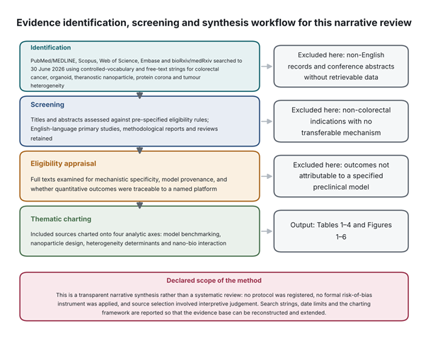
Although early-stage localised CRC is routinely managed by resection and adjuvant chemotherapy, a substantial proportion of patients experience local recurrence or distant metastasis, particularly to liver and peritoneum (Li et al., 2024; S. Wang et al., 2026). The principal obstacle to durable control is biological heterogeneity expressed at interpatient, intralesional and subclonal levels (Li et al., 2024; Roerink et al., 2018).
At the molecular level, the CIN, MSI and CIMP cascades establish distinct evolutionary trajectories, with somatic drivers in APC, KRAS, TP53, BRAF and PIK3CA defining the dominant oncogenic circuitry (S. Wang et al., 2026; Zhang et al., 2026). Transcriptomic classification into CMS1 (immune/MSI-hypermutated), CMS2 (canonical, WNT-activated), CMS3 (metabolic) and CMS4 (mesenchymal, stroma-rich) adds a layer with direct therapeutic consequence (Guinney et al., 2015). CMS4 tumours in particular combine stromal invasion, TGF-β pathway activation and marked resistance to standard cytotoxic therapy (S. Wang et al., 2026). Beyond interpatient variation, multi-region sequencing and single-cell multi-omic profiling demonstrate branched clonal evolution within individual lesions (Roerink et al., 2018; Zhang et al., 2026). Under selective pressure from chemotherapy or anti-EGFR biologics, dominant sensitive clones are removed and minor resistant subclones expand (Li et al., 2024; S. Wang et al., 2026). Cancer stem cells carrying CD44 and the CD44v9 variant isoform are central to this resistant fraction, sustaining self-renewal and distant colonisation (Li et al., 2024; Resmi et al., 2026). Multi-omic comparison of primary lesions with matched peritoneal metastases has further identified glycosylphosphatidylinositol anchor attachment 1 (GPAA1) as a metastasis-associated protein whose contribution arises through protein-level dysregulation rather than genomic amplification (Zhang et al., 2026). The determinants reviewed here, together with the model-selection strategy each implies, are set out in Table 3 and depicted schematically in Figure 2.
2.2 From monolayers and xenografts to patient-derived organoids
Preclinical oncology has long relied on immortalised two-dimensional lines (Li et al., 2024; X. Wang et al., 2026). These do not reproduce three-dimensional architecture,
Table 1: Comparative evaluation of preclinical colorectal cancer platforms for drug and nanomedicine screening. Four platform classes are assessed across architectural complexity, retention of inter- and intratumoural heterogeneity, fidelity of nanomedicine transport and uptake, throughput, and principal translational barriers. The comparison establishes why monolayer potency data transfer poorly to patients, why patient-derived xenografts cannot support screening at scale, and why patient-derived organoids occupy the position from which subclone-resolved nanoparticle behaviour becomes observable. Tumour organoid-on-a-chip systems are included as the emerging configuration that restores vascular and mechanical variables absent from static organoid culture. Compiled from Dhanapal et al. (2026), Drost and Clevers (2018), Li et al. (2024), Resmi et al. (2026), Sato et al. (2011), Vlachogiannis et al. (2018), S. Wang et al. (2026), X. Wang et al. (2026) and Younas et al. (2026).
|
Platform
|
Architecture and cell–matrix interaction
|
Retention of heterogeneity
|
Nanomedicine transport fidelity
|
Throughput and turnaround
|
Key limitations
|
References
|
|
2D monolayer cell lines (HT29, HCT116, Caco-2, COLO201)
|
No 3D organisation, ECM barrier, shear stress or polarisation; flat growth on rigid plastic
|
Minimal; serial passage drives clonal selection, genetic drift and loss of parental subclonal diversity
|
Poor; overestimates uptake ~2-fold through unimpeded surface exposure and lacks endosomal–lysosomal trafficking barriers
|
High; low cost, 96/384-well automation, 1–3 days per assay
|
Overstates clinical potency; models neither ECM diffusion, corona modulation nor subpopulation tropism
|
Li et al. (2024); Resmi et al. (2026); S. Wang et al. (2026)
|
|
Patient-derived xenografts (subcutaneous or orthotopic implantation in mice)
|
High architectural fidelity in vivo; recreates vascular supply, desmoplastic stroma and tissue architecture
|
High; retains parental mutations (APC, KRAS, TP53), copy-number variation and branched clonal structure
|
Moderate to high; captures physiological EPR, though human stroma is progressively replaced by murine elements
|
Low; prohibitive cost, labour-intensive husbandry, 4–8 months to engraft
|
Immunodeficient hosts preclude evaluation of immunomodulatory agents; engraftment varies across CMS subtypes
|
S. Wang et al. (2026); X. Wang et al. (2026)
|
|
Patient-derived organoids (3D Matrigel/BME epithelial biobanks)
|
High epithelial 3D fidelity; self-organises into polarised crypt–villus architecture with stem, proliferative and differentiated compartments
|
Superior; mirrors interpatient, intralesional and subclonal heterogeneity, retaining CMS identity and parental drivers
|
High; reproduces subpopulation-specific uptake, TME-driven corona formation and endocytic routing (clathrin versus caveolae)
|
Medium to high; scalable living biobanks, 2–4 weeks to actionable result
|
Lacks functional vasculature, haemodynamic flow and resident immune cells unless co-cultured with autologous TILs or PBMCs
|
Drost and Clevers (2018); Li et al. (2024); Resmi et al. (2026); Sato et al. (2011); Vlachogiannis et al. (2018)
|
|
Tumour organoid-on-a-chip (microfluidic biomimetic biochips)
|
Highest physiological relevance; couples 3D organoids to perfusable endothelial channels, flow and controlled shear
|
High; maintains multi-lineage heterogeneity while enabling spatial mapping of invasive margins and intravasation
|
Exceptional; recreates extravasation, shear stress, interstitial pressure gradients and dynamic stimuli-responsive release
|
Moderate; complex fabrication with specialised optical or electrochemical sensors
|
High technical complexity; flow rates and matrix hydrogels require standardisation for cross-laboratory reproducibility
|
Dhanapal et al. (2026); Li et al. (2024); Younas et al. (2026)
|
Table 2: Classification, design principles and theranostic functionality of nanoparticle platforms in colorectal cancer. Five material classes are organised by their diagnostic imaging modality, therapeutic payload and mechanism of action, targeting and tumour microenvironment responsiveness, physiological advantages, and outstanding safety or manufacturing concerns. The table shows how imaging and therapy are integrated within single constructs and how acidic pH, glutathione gradients and protease overexpression are exploited as release triggers. It also makes visible the trade-off recurring across classes, in which the features conferring targeting precision — surface functionalisation, membrane camouflage, multi-step synthesis — are the same features that complicate reproducible manufacture and regulatory characterisation. Compiled from Dhanapal et al. (2026), Guo et al. (2026), Qu et al. (2022), Resmi et al. (2026) and Younas et al. (2026).
|
Nanoparticle class
|
Imaging modality
|
Therapeutic payload and action
|
Targeting and TME responsiveness
|
Physiological advantages
|
Translational and safety concerns
|
References
|
|
Lipid nanoparticles and liposomes (PEGylated liposomes, PFP@PDM-PEG)
|
NIR fluorescence dyes, photoacoustic contrast agents, ultrasound microbubbles
|
5-Fluorouracil, oxaliplatin, paclitaxel, disulfiram, siRNA and mRNA payloads
|
Dual pH/MMP-2 with low-intensity focused ultrasound; acidic TME (pH 6.5–6.8) and MMP-2 cleavage de-shield the PEG layer
|
High biocompatibility, encapsulation efficiency above 85%, prolonged circulation via PEGylation
|
Corona formation in blood compromises targeting; potential complement activation-related pseudoallergy
|
Guo et al. (2026); Younas et al. (2026)
|
|
Polymeric nanoparticles and micelles (PLGA, poly(β-L-malic acid), PEG-PCL)
|
Fluorescent probes (Ce6, BODIPY), radiolabels, MRI contrast agents
|
Doxorubicin, camptothecin, STING agonists (diABZI), MEK and KRAS inhibitors
|
pH- and enzyme-responsive disassembly; endolysosomal acidity or cathepsin B/MMP-2 cleavage triggers charge reversal or core collapse
|
Tunable degradation kinetics, sustained release, surface functionalisation with hyaluronic acid or peptide ligands
|
Multi-step synthesis increases batch-to-batch variability and reduces long-term colloidal stability
|
Dhanapal et al. (2026); Guo et al. (2026); Younas et al. (2026)
|
|
Superparamagnetic iron oxide nanoparticles (SPIONs)
|
T₂-weighted MRI, magnetic particle imaging
|
Magnetic hyperthermia, Fenton-reaction chemodynamic therapy, melittin (MNPs-Mel)
|
Active ligand decoration and magnetic guidance; CD44 or EGFR aptamer coating plus external alternating magnetic fields
|
Combined MR diagnosis and thermal ablation; magnetic guidance directs particles to deep tumour niches
|
Long-term iron accumulation in liver and spleen; reticuloendothelial clearance
|
Dhanapal et al. (2026); Guo et al. (2026); Qu et al. (2022); Younas et al. (2026)
|
|
Semiconductor and carbon quantum dots (SiQDs, GQDs, carbon dots)
|
Photostable multicolour fluorescence; NIR intraoperative visualisation
|
Pre-incubated TME signalling proteins (WNT, EGF) for targeted subclone delivery
|
Protein corona pre-engineering; WNT/EGF-functionalised coronas target CD44v9-positive chemoresistant stem cells
|
Ultra-small size (<10 nm), high photostability, tunable emission, resistance to photobleaching
|
Heavy-metal toxicity for cadmium-based dots; strict surface passivation required for silicon and carbon types
|
Dhanapal et al. (2026); Resmi et al. (2026)
|
|
Biomimetic and core–satellite nanocomposites (cell-membrane wrapped, MMFn, MnO₂R@FePDAc)
|
Multimodal fluorescence/MRI/PET imaging, photoacoustic tomography
|
Photothermal ablation, ROS amplification, GSH depletion, ferroptosis induction (RSL3)
|
Homotypic homing and enzyme cascades; cancer cell or macrophage membrane camouflage plus MMP-2/9-cleavable GPLGVR linkers
|
Prolonged circulation via self markers, immune evasion, site-specific catalytic activation
|
Complex membrane extraction and coating protocols; regulatory difficulty in characterising membrane proteins
|
Guo et al. (2026); Qu et al. (2022); Younas et al. (2026)
|
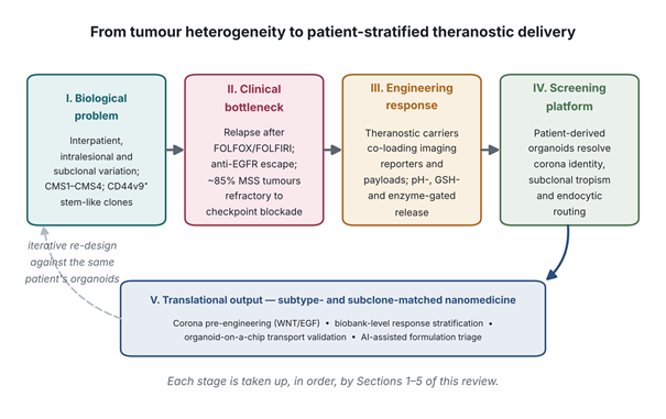
Figure 1. Conceptual architecture of the review: from tumour heterogeneity to patient-stratified theranostic delivery. The schematic traces the logical sequence connecting the five stages of the argument developed across Sections 1 to 5. Intrinsic biological variation in colorectal cancer (stage I) produces the clinical bottleneck of relapse and immunotherapy refractoriness (stage II), to which theranostic nanoparticle engineering is the designed response (stage III). Patient-derived organoid screening (stage IV) supplies the resolution at which corona identity, subclonal tropism and endocytic routing become measurable, yielding subtype- and subclone-matched nanomedicine (stage V). The dashed return path indicates the iterative loop in which screening outcomes feed back into formulation design against organoids from the same patient.
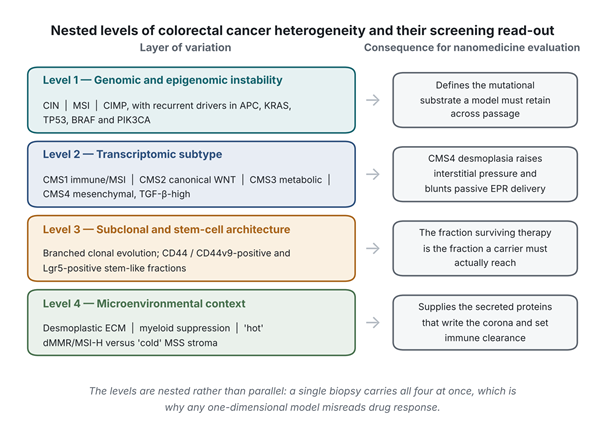
Figure 2. Nested levels of colorectal cancer heterogeneity and their consequences for nanomedicine evaluation. Four hierarchically nested layers of biological variation are shown alongside the specific methodological demand each imposes on preclinical screening. Level 1 establishes the mutational substrate through chromosomal instability, microsatellite instability and CpG island methylator phenotype pathways; level 2 resolves transcriptomic subtype; level 3 captures branched subclonal and stem-cell architecture; and level 4 supplies the microenvironmental context that determines protein corona composition. The levels are nested rather than parallel, since a single biopsy carries all four simultaneously, which is the reason any one-dimensional model systematically misreads drug response. Synthesised from Guinney et al. (2015), Roerink et al. (2018), S. Wang et al. (2026) and Zhang et al. (2026).
cell–cell contact, extracellular matrix (ECM) barriers or microenvironmental gradients, and decades of passage on rigid plastic produce genetic drift, loss of parental subclonal diversity and altered phenotype (Li et al., 2024; Resmi et al., 2026). Candidates potent in monolayer assays therefore fail disproportionately in trials, in part because such assays overestimate internalisation and omit physiologically relevant transport barriers (Resmi et al., 2026; Younas et al., 2026).
PDX models, generated by implanting patient tumour fragments into immunodeficient mice, address several of these deficits, maintaining parental histology, genotype, copy-number profile and stromal architecture in vivo, and permitting study of tumour–stroma crosstalk and metastatic behaviour (S. Wang et al., 2026; X. Wang et al., 2026). Their utility for screening is nonetheless limited by establishment times of four to eight months, high cost, variable engraftment across molecular subtypes, progressive replacement of human stroma by murine elements, and an immunodeficient host that precludes evaluation of human-specific immunotherapies (S. Wang et al., 2026; X. Wang et al., 2026).
PDO technology represents the substantive methodological advance (Drost & Clevers, 2018; Sato et al., 2011). Derived from primary or metastatic resections, biopsies or circulating tumour cells, PDOs self-organise within basement membrane extract into three-dimensional epithelial structures that retain histopathological differentiation, driver mutations, epigenetic landscape and subclonal heterogeneity across long-term culture (Li et al., 2024; Vlachogiannis et al., 2018; S. Wang et al., 2026). Living biobanks can be expanded for medium-to-high-throughput assays within clinically actionable windows of two to four weeks, with demonstrated concordance against cytotoxic, targeted and chemoradiotherapeutic responses (Vlachogiannis et al., 2018), and medium-throughput screening of peritoneal metastasis organoids has been used to direct individualised therapy (Narasimhan et al., 2020). A direct comparison across all four platform classes is provided in Table 1, and their relative positioning in Figure 3.
2.3 Theranostic nanoparticle engineering and TME-responsive release
In parallel, nanomedicine has produced platforms intended to overcome non-specific biodistribution, poor aqueous solubility and systemic toxicity (Dhanapal et al., 2026; Younas et al., 2026). Theranostic constructs co-encapsulate therapeutic agents — 5-fluorouracil, paclitaxel, doxorubicin, siRNA — with diagnostic reporters including magnetic resonance contrast agents, near-infrared dyes and photoacoustic probes, enabling real-time visualisation of accumulation, distribution, release kinetics and efficacy (Dhanapal et al., 2026; Guo et al., 2026; Younas et al., 2026).
Material classes span lipid nanoparticles and liposomes, polymeric micelles such as PLGA and poly-L-glutamic acid, inorganic SPIONs, gold nanostructures, mesoporous silica, and semiconductor quantum dots including silicon (SiQDs) and graphene quantum dots (GQDs) (Dhanapal et al., 2026; Resmi et al., 2026; Younas et al., 2026). Passive targeting via EPR is unreliable where vascularisation is irregular and interstitial fluid pressure is elevated, as in dense desmoplastic stroma (Guo et al., 2026; X. Wang et al., 2026). Active targeting therefore employs surface ligands — antibodies, aptamers, hyaluronic acid, peptides — directed at CD44, EGFR, VEGFR and integrins (Guo et al., 2026; Younas et al., 2026).
The most consequential design advance is stimuli-responsiveness (Guo et al., 2026; Younas et al., 2026). Such carriers remain stable at physiological pH 7.4 yet undergo disassembly, charge reversal or bond cleavage on encountering tumour-specific triggers. Three dominate. Mildly acidic pH (6.2–6.8 extracellularly, 4.5–5.5 in endolysosomes) cleaves hydrazone, acetal or maleic acid amide linkages and drives protonation-dependent disassembly (Guo et al., 2026; Younas et al., 2026). Elevated intracellular glutathione, 100- to 1000-fold above extracellular concentrations, reduces disulfide bonds to release payload intracellularly (Guo et al., 2026; Younas et al., 2026). Overexpressed proteases including MMP-2/MMP-9, legumain and hyaluronidases cleave peptide motifs such as PLGVRG or AAN, stripping PEG shielding, altering particle morphology or liberating drug at the tumour margin (Guo et al., 2026). These mechanisms are illustrated in Figure 4; the corresponding platform classes, payloads, advantages and safety concerns appear in Table 2.
2.4 Nano-bio interactions: protein corona dynamics and subpopulation-specific delivery
Nanoparticles do not meet cells as pristine synthetic objects. On entering biological fluid they adsorb a dynamic
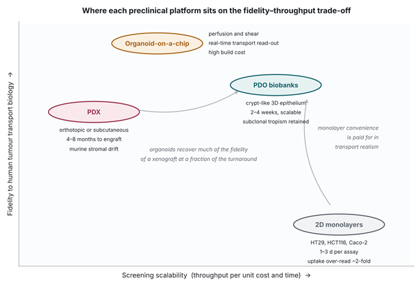
Figure 3. Positioning of preclinical platforms on the fidelity–throughput trade-off. Four model classes are plotted against screening scalability on the horizontal axis and fidelity to human tumour transport biology on the vertical axis, with annotations giving characteristic turnaround and principal limitation. Two-dimensional monolayers occupy the high-throughput, low-fidelity corner and overestimate nanoparticle uptake approximately two-fold; patient-derived xenografts recover physiological fidelity but require four to eight months per model; patient-derived organoid biobanks recover much of that fidelity at a fraction of the turnaround; and organoid-on-a-chip systems achieve the highest transport realism at substantial cost in throughput. The figure illustrates why a tiered configuration, rather than substitution of one platform for another, is the configuration this review recommends.
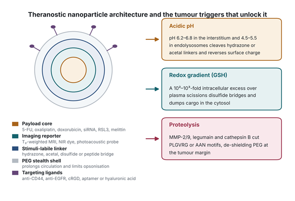
Figure 4. Theranostic nanoparticle architecture and the tumour microenvironment triggers that unlock payload release. The concentric construct on the left shows the canonical theranostic design, comprising a therapeutic payload core, a co-encapsulated imaging reporter, a stimuli-labile linker layer, a polyethylene glycol stealth shell and surface targeting ligands directed at receptors overexpressed on colorectal epithelium. The three panels on the right summarise the endogenous triggers exploited for site-specific release: mildly acidic interstitial and endolysosomal pH, the steep intracellular glutathione gradient, and overexpressed tumour proteases including matrix metalloproteinases, legumain and cathepsin B. External stimuli such as near-infrared irradiation, focused ultrasound and alternating magnetic fields may be layered onto these endogenous triggers. Synthesised from Dhanapal et al. (2026), Guo et al. (2026) and Younas et al. (2026).
protein layer — the corona — that alters hydrodynamic diameter, surface charge, colloidal stability and receptor affinity, conferring a biological identity distinct from the synthetic one and governing endocytosis, immune clearance and tissue accumulation (Resmi et al., 2026; Younas et al., 2026). Corona research historically concentrated on abundant serum proteins such as albumin, immunoglobulins, transferrin and apolipoproteins, which left unexamined the question of how proteins secreted within a patient’s own tumour microenvironment shape particle behaviour (Resmi et al., 2026).
Work in patient-derived colonic organoids (PDCOs) has begun to close that gap (Resmi et al., 2026). Using material-distinct nanoprobes, specifically SiQDs and GQDs, Resmi et al. (2026) reported that conventional monolayers overestimate uptake approximately two-fold relative to ex vivo PDCOs. PDCO monolayers preserve crypt-like organisation, self-assembling into node and non-node regions that contain stem-like, proliferative and differentiated compartments (Resmi et al., 2026). High-dimensional spectral flow cytometry with single-cell dimensionality reduction showed that uptake kinetics, cellular tropism and endocytic mechanism differ substantially between cell lines, fibroblasts and organoid subpopulations (Resmi et al., 2026).
Critically, the surrounding protein environment determines whether carriers accumulate in particular subclones. Pre-incubating SiQDs in organoid growth medium containing TME signalling factors produced a distinct, highly crosslinked corona unlike that formed in serum-rich medium (Resmi et al., 2026). Pre-engineering the corona with WNT, a secreted glycoprotein driving stemness, or with EGF, raised uptake in chemoresistant CD44v9-positive epithelial subpopulations from 3.8% in non-engineered particles to 70% and 66.85% respectively — an approximately 18-fold enhancement in delivery to the fraction associated with relapse (Resmi et al., 2026).
PDO screening additionally revealed that intracellular fate depends on cell-type-specific endocytic routing (Resmi et al., 2026). Primary organoid epithelial cells and normal fibroblasts internalise via clathrin- and dynamin-1-dependent endocytosis, directing carriers to late endosomes and lysosomes for acidic degradation. Malignant epithelial cells instead use caveolin-1-mediated uptake, bypassing lysosomal fusion and routing particles through caveosomes toward the endoplasmic reticulum and cytosol (Resmi et al., 2026). This divergence offers a mechanistic explanation for why carriers degraded in normal tissue may still deliver intact payload within malignant cells (Guo et al., 2026; Resmi et al., 2026). Figure 5 summarises these relationships, and the underlying experimental studies are compiled in Table 4.
2.5 Methodological challenges and emerging platforms
Several obstacles stand between this rationale and clinical translation (Li et al., 2024; S. Wang et al., 2026; Younas et al., 2026). Standard epithelial PDOs cultured in Matrigel or basement membrane extract lack functional vasculature, shear stress, interstitial flow and resident immune populations including T lymphocytes, tumour-associated macrophages and cancer-associated fibroblasts (Li et al., 2024; S. Wang et al., 2026). Non-epithelial components are progressively lost across passage, which may lead to underestimation of physical transport barriers and immune-mediated clearance (Li et al., 2024; Younas et al., 2026). Batch-to-batch variability in naturally derived hydrogels, labour-intensive protocols and the absence of standardised high-content imaging further complicate cross-laboratory reproducibility (Li et al., 2024; S. Wang et al., 2026).
Bioengineering responses are emerging. Tumour organoid-on-a-chip (ToOC) microfluidic systems combine three-dimensional organoids with perfusable endothelial channels, biomimetic matrices and controlled flow, reproducing extravasation, haemodynamic shear and tissue penetration while permitting real-time optical and electrochemical monitoring of carrier assembly and disassembly (S. Wang et al., 2026; Younas et al., 2026). Heterotypic co-cultures combining epithelial organoids with autologous peripheral blood mononuclear cells, tumour-infiltrating lymphocytes or cancer-associated fibroblasts enable functional assessment of immunomodulatory nanomedicines, tumour–immune crosstalk and macrophage repolarisation (Li et al., 2024; S. Wang et al., 2026). Finally, artificial intelligence and machine learning trained on high-content organoid imaging, single-cell transcriptomes and physicochemical descriptors can predict corona composition, uptake efficiency and patient-specific sensitivity in silico, allowing formulation design to be triaged before experimental validation (Dhanapal et al., 2026; Younas et al., 2026). Coupling AI-driven design with PDO validation offers a plausible route to systematic optimisation and patient stratification (Dhanapal et al., 2026; Li et al., 2024; S. Wang et al., 2026). The comparative strengths and remaining limitations of each platform, including ToOC, are detailed in Table 1.

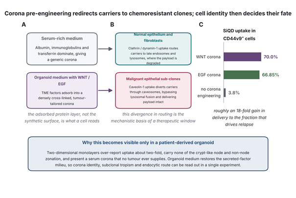 Figure 5. Protein corona pre-engineering redirects nanocarriers to chemoresistant subclones, and cell identity determines their intracellular fate. Panel A contrasts the generic corona formed in serum-rich medium with the densely crosslinked, tumour-tailored corona formed when silicon quantum dots are pre-incubated in organoid medium containing WNT or EGF. Panel B shows the divergence in endocytic routing, in which normal epithelium and fibroblasts internalise carriers via clathrin and dynamin-1 into degradative lysosomes while malignant subclones use caveolin-1 entry that bypasses lysosomal fusion and preserves payload. Panel C quantifies the consequence, with uptake in CD44v9-positive chemoresistant cells rising from 3.8% without corona engineering to 66.85% and 70% with EGF and WNT coronas respectively. These relationships are not resolvable in monolayer culture. Data from Resmi et al. (2026).