Biosensors and Nanotheranostics

Bionanotechnology, Drug Delivery, Therapeutics | online ISSN 3064-7789
175
Citations
125.3k
Views
103
Articles
Your new experience awaits. Try the new design now and help us make it even better
Switch to the new experience
Figures and Tables
REVIEWS   (Open Access)

Nanotheranostics for Overcoming Tumor Heterogeneity and Drug Resistance in Solid Tumors: Imaging-Guided Therapy, Clinical Progress, and Future Directions

Apura Orka 1*

+ Author Affiliations

Biosensors and Nanotheranostics 5 (1) 1-8 https://doi.org/10.25163/biosensors.5111004

Submitted: 19 October 2026 Revised: 10 December 2026  Published: 18 December 2026 


Abstract

Solid tumors remain among the most lethal human malignancies, in large part because they are not uniform masses but evolving, heterogeneous ecosystems that resist systemically delivered drugs. Genetic, phenotypic, and microenvironmental diversity, compounded by multidrug resistance (MDR), allows subclonal populations to survive conventional therapy, while a hostile tumor microenvironment (TME)—acidic, hypoxic, fibrotic, and poorly perfused—physically blocks drug penetration and reinforces efflux-mediated resistance. Nanotheranostics, which integrates diagnostic imaging reporters and therapeutic payloads within a single nanoscale platform, has emerged as a rational response to these intertwined problems. In this review we examine how multifunctional nanotheranostic systems—lipid and polymeric carriers, inorganic and metallic hybrids, catalytic nanozymes, biomimetic cell-derived vesicles, and structural DNA architectures—are engineered to remain quiescent in circulation and switch “ON” in response to endogenous (pH, redox, hypoxia, enzymes) or exogenous (light, ultrasound, magnetic) triggers. We then consider how magnetic resonance imaging, computed tomography, positron emission tomography, ultrasound, and near-infrared optical imaging enable patient stratification, real-time tracking of drug release, and spatiotemporally controlled activation of phototherapy, radiotherapy, and immunotherapy. Preclinical and early clinical evidence, including AGuIX®, ferumoxytol, Onivyde®, and 64Cu-MM-302, indicates that imaging-guided design can reverse resistance, remodel the stroma, and identify the patients most likely to respond. We also confront the translational bottlenecks—synthetic over-engineering, the protein corona, clearance, and regulatory hurdles—that keep roughly ninety percent of candidates in animal models, and we outline how artificial intelligence, companion diagnostics, and liquid biopsy may close that gap. Read together, the evidence reframes resistance as a spatial problem of drug delivery and positions imaging-guided nanotheranostics as a practical route toward personalized oncology.

Keywords: nanotheranostics; tumor heterogeneity; multidrug resistance; tumor microenvironment; stimuli-responsive nanoparticles; image-guided therapy; companion diagnostics; clinical translation

1. Introduction

Malignant solid tumors remain a leading cause of death worldwide, and despite decades of therapeutic innovation they continue to present as an enduring public-health problem marked by late diagnosis, frequent recurrence, and limited durable response (Chen et al., 2017; Wu et al., 2026). The conventional pillars of oncology—surgical resection, systemic chemotherapy, and external-beam radiotherapy—too often deliver only marginal survival benefit in advanced disease (Kievit & Zhang, 2011; Li et al., 2026; Wu et al., 2026). A central reason for this shortfall is tumor heterogeneity, a multilayered phenomenon that unfolds at genetic, phenotypic, spatial, and microenvironmental levels (Chen et al., 2017; de Maar et al., 2020; Tian et al., 2025). Because distinct neoplastic subclones coexist within a single lesion and across metastatic sites, they harbor divergent metabolic states, proliferative capacities, and drug susceptibilities, so that no single agent reaches every cell with equal effect (Bareche et al., 2020; de Maar et al., 2020; Kothawade et al., 2025).

Running alongside this diversity is multidrug resistance (MDR), which steadily erodes the clinical durability of systemic regimens (Dallavalle et al., 2020; Yang et al., 2019). MDR is not a single defect but a network of mechanisms: overexpression of ATP-binding cassette (ABC) efflux pumps such as P-glycoprotein (P-gp/MDR1), target-gene mutation, enhanced DNA repair, and evasion of apoptosis (Bin Emran et al., 2022; Sajid et al., 2023; Yang et al., 2019; Zhi et al., 2023). Crucially, this cellular resistance is amplified by the physical and physiological abnormalities of the tumor microenvironment (TME) (Danhier, 2016; Tian et al., 2025). Tumor vasculature is tortuous and leaky, interstitial fluid pressure is high, the extracellular matrix is stiff and collagen-dense, and regions of hypoxia and extracellular acidosis arise from glycolytic metabolism and inadequate perfusion (Danhier, 2016; de Maar et al., 2020; Wang et al., 2026). Hypoxic gradients stabilize hypoxia-inducible factor 1-alpha (HIF-1α), which directly upregulates MDR1, activates pro-survival pathways, drives angiogenesis, and recruits immunosuppressive cells (Comerford et al., 2002; Li et al., 2026). The net result is that systemically delivered chemotherapeutics encounter both physical transport blocks and cellular efflux, producing poor tumor accumulation, off-target toxicity, and therapy-driven clonal selection (Prasad et al., 2023; Tian et al., 2025).

To dismantle these systemic and microenvironmental barriers at once, cancer nanomedicine has moved toward integrated nanotheranostics—multifunctional nanoscale platforms that place a diagnostic imaging reporter and a therapeutic payload within one coordinated construct (Chen et al., 2017; Kievit & Zhang, 2011; Melancon et al., 2012). Historically, such carriers relied on the passive enhanced permeability and retention (EPR) effect to reach tumor tissue (Danhier, 2016; Matsumura & Maeda, 1986). Yet clinical experience has shown the EPR effect to be highly variable between patients and tumor types, frequently failing to achieve uniform penetration, with median tumor accumulation often below one percent of the administered dose (Danhier, 2016; de Maar et al., 2020; Mendes et al., 2021; Wilhelm et al., 2016).

Nanotheranostics moves beyond simple drug carriage by embedding non-invasive diagnostic capacity across complementary modalities—magnetic resonance imaging (MRI), computed tomography (CT), positron emission tomography (PET), ultrasound (US), and near-infrared optical or fluorescence imaging (FLI) (Chen et al., 2017; Kothawade et al., 2025; Wu et al., 2026). Consolidating these functions allows real-time mapping of biodistribution, localization of submillimeter lesions and occult micrometastases, tracking of carrier extravasation, and continuous monitoring of response (de Maar et al., 2020; Kothawade et al., 2025; Wang et al., 2026).

Recent advances in biomaterials have further catalyzed a shift toward “smart,” stimuli-responsive nanoplatforms (Wang et al., 2026; Wei et al., 2023). Rather than remaining continuously active, these systems hold an “OFF” state during circulation—limiting background toxicity and reticuloendothelial clearance—and switch “ON” only on encountering specific triggers in the TME (Wang et al., 2026; Wu et al., 2026). Internal switches exploit tumor acidosis (pH 5.5–6.8), elevated glutathione (GSH) or reactive oxygen species (ROS), and overexpressed proteases such as cathepsins and matrix metalloproteinases (MMPs) (Cai et al., 2021; Wang et al., 2026; Yang et al., 2019), while external physical stimuli—near-infrared (NIR) light, ultrasound, and alternating magnetic fields—drive spatiotemporally controlled release, hyperthermia, radical generation, and contrast switching (Prasad et al., 2023; Wu et al., 2026; Zachou et al., 2025).

Beyond intrinsic resistance, the non-uniform spatial distribution of drugs within a tumor produces “pseudo-resistance” and “therapy heterogeneity,” in which cell clusters survive simply because the drug never reaches a cytotoxic threshold throughout the tumor volume (de Maar et al., 2020; Dhandapani et al., 2023). Nanotheranostics addresses this directly through image-guided intervention and localized microenvironmental remodeling (de Maar et al., 2020; Li et al., 2026; Zheng et al., 2026). First, functional and molecular imaging non-invasively characterizes patient-specific parameters—vascular perfusion, microvessel density, leakiness, and interstitial fluid pressure—before and during treatment, enabling accurate stratification of the sub-cohorts most likely to benefit (Chen et al., 2017; de Maar et al., 2020; Stapleton et al., 2015). Second, multimodal imaging agents guide localized physical therapies; in photothermal (PTT) and photodynamic therapy (PDT), optical and magnetic tracking defines tumor margins and the peak accumulation window so that irradiation is applied when the carrier-to-tissue ratio is greatest (Melancon et al., 2012; Zachou et al., 2025; Zheng et al., 2026). Third, engineered platforms actively remodel the dense TME—antifibrotic agents such as oleanolic acid suppress cancer-associated fibroblasts and downregulate alpha-smooth muscle actin (α-SMA), while catalytic oxygen-generating nanozymes or HIF-1α silencing agents relieve hypoxia, downregulate P-gp, and reverse radioresistance, with MRI or photoacoustic imaging confirming re-oxygenation in real time (Li et al., 2026; Tian et al., 2025; Wu et al., 2025; Zhang et al., 2024; Zheng et al., 2026).

Even so, the path from bench to bedside remains constrained by biological, manufacturing, and regulatory challenges, and roughly ninety percent of investigational imaging nanoprobes and theranostics remain confined to animal models (Chen et al., 2017; Gawne et al., 2023; Wang et al., 2026). The principal bottlenecks are biological complexity and systemic barriers—protein-corona formation, immune recognition, and sequestration by the mononuclear phagocyte system (Kievit & Zhang, 2011; Prasad et al., 2023; Salvati et al., 2013; Todd & Kay, 2016); manufacturing and scalability, where multicomponent nanohybrids introduce synthetic complexity, batch-to-batch variability, and difficulty meeting Good Manufacturing Practice (GMP) standards (Gawne et al., 2023; Li et al., 2026; Wang et al., 2026); and clinical validation, where pioneering platforms such as gadolinium-chelated silica nanoparticles (AGuIX®), ferumoxytol, nanoliposomal irinotecan (Onivyde®), and PSMA-targeted docetaxel (BIND-014) nonetheless provide valuable translational roadmaps (Bort et al., 2020; Tian et al., 2025; Von Hoff et al., 2016; Wang-Gillam et al., 2016).

Against this backdrop, the present review synthesizes recent progress and addresses the translational gap in this fast-moving field. Specifically, it sets out to (i) elucidate the mechanistic origins of tumor heterogeneity and MDR and the biophysical barriers that drive therapy resistance; (ii) categorize modern multifunctional nanotheranostic platforms and their stimuli-responsive release mechanisms; (iii) examine how MRI, CT, PET, US, and optical imaging support staging, drug-release monitoring, patient stratification, and real-time guidance of combinatorial therapy; (iv) critically analyze the clinical-translation landscape, including current trials, benchmark formulations, and manufacturing and regulatory challenges; and (v) outline future perspectives, including artificial intelligence for predictive nanocarrier design, companion diagnostics, N-of-1 protocols, and liquid-biopsy monitoring.

2. Nanotheranostic Strategies to Overcome Tumor Heterogeneity and Multidrug Resistance From Bench to Bedside.

Two evolutionary hallmarks—biological heterogeneity and multidrug resistance—converge to make conventional monotherapy and unguided systemic dosing largely ineffective against solid tumors (Chen et al., 2017; Tian et al., 2025; Wu et al., 2026). The following sections examine the mechanistic basis of that problem, the nanotheranostic platforms engineered to overcome it, the stimuli-responsive and imaging logic that makes them selective, and the clinical progress achieved so far. The representative systems, triggers, imaging modalities, and clinical formulations discussed below are summarized in Tables 1–4.

2.1. Mechanisms Driving Tumor Heterogeneity and Multidrug Resistance

Rather than behaving as homogeneous masses of clonal cells, solid tumors function as aberrant pseudo-organs in which genetically and phenotypically divergent sublines are embedded within an evolving TME (de Maar et al., 2020; Tian et al., 2025). This diversity spans several physical scales, from patient-to-patient (inter-individual) variability, to differences between primary and metastatic lesions (inter-tumoral), down to distinct regional architectures within a single lesion (intra-tumoral) (de Maar et al., 2020; Kothawade et al., 2025). The cascade by which these layers combine to blunt systemic therapy is depicted in Figure 1.

At the cellular level, intrinsic and acquired MDR is orchestrated through classical biochemical routes, chief

Table 1. Representative multifunctional nanotheranostic platforms engineered to overcome solid-tumor heterogeneity and multidrug resistance. For each system the table lists the structural composition and engineering strategy, the integrated diagnostic or imaging modality, the therapeutic cargo, and the specific mechanism by which cellular or microenvironmental resistance is reversed. The preclinical model used for evaluation and the primary source are given in the final columns, allowing direct comparison of lipid/polymer, inorganic/hybrid, and biomimetic architectures across diverse solid-tumor settings.

Platform category & name

Composition & engineering

Imaging modality

Therapeutic cargo

Resistance/heterogeneity reversal mechanism

Model evaluated

Reference

Lipid hybrid: Visualized liposomal platform (VLPs/CpG/siRNA)

HSPC, cholesterol, DSPE-PEG bilayer encapsulating a gadolinium-hybrid gold nanoparticle (Gd-HGNP) core

T1-weighted MRI via core Gd-HGNPs

HIF-1α siRNA co-delivered with unmethylated CpG ODNs

Downregulates HIF-1α to relieve hypoxia and vascular abnormality; recruits cytotoxic T lymphocytes via TLR9 to reverse radioresistance

Hepa1-6 hepatocellular carcinoma in C57BL/6 mice

Li et al. (2026)

Polymer micelle: Dual-targeting chemosensitizing micelles (DXS@NPs)

PEG-b-poly(L-leucine) amphiphilic block copolymer assembled into core-shell nanomicelles

T2-weighted negative MRI via SPIO cores

Doxorubicin co-encapsulated with XMD8-92 (BMK1/ERK5 inhibitor)

Inhibits BMK1/ERK5 phosphorylation, downregulating P-gp/ABCB1; reduces resistant-cell survival to 53% vs. 90% for free DOX

SGC-7901/VCR vincristine-resistant gastric cancer in BALB/c nude mice

Yang et al. (2019)

Polymeric prodrug: Cathepsin B-degradable glycopolymer (Glycopolymer-PTX-DOTA-Gd)

Branched glycopolymer synthesized by RAFT with Gly-Phe-Leu-Gly (GFLG) linkers (~95 nm)

T1-weighted MRI (Gd-DOTA, r1 = 7.1 mM⁻¹s⁻¹) and optical fluorescence (Ppa)

Paclitaxel conjugated via enzyme-cleavable GFLG linkers

Bypasses extracellular transport barriers; degrades to <28 kDa fragments upon cathepsin B exposure, bypassing efflux

4T1 murine mammary carcinoma in BALB/c mice

Cai et al. (2021)

Polysaccharide hybrid: Oleanolic acid-modified chitosan (CsO/IGA)

Oleanolic acid-conjugated chitosan self-assembled with ICG-Gd and osimertinib (~100 nm)

Dual-modal NIR fluorescence imaging and T1-weighted MRI

Osimertinib (AZD9291) with ICG-mediated PTT and PDT under 808 nm laser

OA suppresses CAFs and downregulates α-SMA, degrading stroma; PTT/PDT downregulates EGFR/p-EGFR and AKT survival signaling

Sensitive (PC9) and osimertinib-resistant (P5) NSCLC in BALB/c nude mice

Zheng et al. (2026)

Inorganic biomineral: Mesoporous calcium carbonate (HA/CaCO3@Ce6)

Hyaluronic acid-functionalized mesoporous CaCO3 nanoparticles (~204 nm; pore ~11.05 nm)

Ultrasonic echogenic cavitation tracking and ex vivo 68Ga-FAPI PET/CT

Chlorin e6 (Ce6) sonosensitizer with acid-triggered Ca2+ release for SDT

CD44-targeted endocytosis then acid dissolution triggers mitochondrial Ca2+ overload, a self-amplifying Ca2+-ROS loop inducing ICD

Hepa1-6 hepatocellular carcinoma in C57BL/6 immunocompetent mice

Chen et al. (2026)

Biomimetic hybrid: Cancer-cell-membrane nanovesicles (CCMVs-AuNRs)

Homologous 4T1 cancer-cell-membrane vesicles cloaking gold nanorods co-loaded with DOX and ICG

NIR fluorescence (NIR-FLI) and photoacoustic imaging (PAI)

Doxorubicin with NIR-triggered photothermal therapy

Exploits homotypic adhesion (CD44, integrins) to evade MPS clearance; photothermal heating dilates endothelial junctions for deep penetration

4T1 breast carcinoma xenografts and metastatic models in BALB/c mice

Prasad et al. (2023)

Micellar nanoshuttle: Anti-CD44 chitosan-SPION nanomicelles (DFIO)

Lauric acid-grafted amphiphilic chitosan crosslinked with HMP, functionalized with anti-CD44 antibodies

Dual-modal NIR fluorescence and T2-weighted MRI (SPION core)

Hydrophobic chemotherapeutics with AMF-driven magnetic hyperthermia

Selectively binds circulating tumor clusters; mild hyperthermia sensitizes drug-resistant stem-like clusters, suppressing metastatic seeding

Metastatic stage III/IV breast cancer models in BALB/c mice

Dhandapani et al. (2023)

Dissolving array: Hybrid carbon–lanthanide nanoplatform

DSPE-PEG-stabilized rare-earth lanthanide nanoparticles and carbon nanomaterials in dissolving gelatin microneedles

Short-wave infrared (SWIR) emission (1000–1700 nm) with negligible autofluorescence

Carbon-driven 980 nm PTT coupled with high-Z lanthanide X-ray radiosensitization

Minimally invasive intradermal delivery avoids systemic clearance; mild PTT alleviates hypoxia and inhibits radiation-induced DNA repair

Localized early-stage cutaneous melanoma in C57BL/6 mice

Khan et al. (2026)

Catalytic nanozyme: Multi-responsive bimetallic platform (MnO2/Pt)

Mesoporous silica core coated with redox-responsive MnO2 and platinum nanozyme clusters

T1-weighted MRI via Mn2+ release upon GSH/H2O2 reduction

Chemotherapy co-delivered with nanozyme-catalyzed H2O2 decomposition into O2

Catalytically converts endogenous H2O2 to O2, relieving hypoxia and reversing hypoxia-induced radio- and chemoresistance

Orthotopic glioblastoma and patient-derived xenografts

Chen et al. (2024); Wang et al. (2026)

Acoustic-magnetic platform: Carbon-doped piezoelectric hybrids (C-ZnO/Fe3O4)

Carbon-doped ZnO sonosensitizers integrated with magnetic Fe3O4 nanoclusters

Dual-modal contrast-enhanced ultrasound (CEUS) and T2-weighted MRI

Ultrasound-activated SDT paired with magnetic field-directed tissue accumulation

Carbon doping enhances piezoelectric polarization and cavitation, producing mechanical shear that damages resistant cell membranes

Subcutaneous hepatocellular carcinoma and breast xenografts

Wu et al. (2026)

Table 2. Smart stimuli-responsive mechanisms and microenvironmental logic triggers governing nanotheranostic activation. The table organizes endogenous (pH, redox/GSH, enzymes, hypoxia) and exogenous (photothermal, ultrasound, magnetic) triggers by the physiological or physical cue they exploit, the nanomaterial bond architecture that senses it, and the molecular activation mechanism. The imaging signal change (“OFF” to “ON”) and the corresponding drug-release or resistance-reversal outcome are listed to show how each trigger converts a microenvironmental feature into a coordinated diagnostic and therapeutic response.

Stimulus classification

Physiological/physical trigger

Nanomaterial substrate & bond architecture

Molecular/biophysical activation mechanism

Imaging signal modulation (OFF→ON)

Drug release & resistance-reversal impact

Reference

Endogenous: Acidosis

Mildly acidic extracellular TME (pH 6.2–6.8) from Warburg glycolysis

Calcium phosphate (CaP) or calcium carbonate (CaCO3) mineral lattices

Rapid proton-consuming dissolution (CaCO3 + 2H+ → Ca2+ + CO2 + H2O)

Liberates paramagnetic Mn2+ binding serum albumin, raising r1 from 5.1 to 34.2 mM⁻¹s⁻¹

Neutralizes acidity, reversing ion-trapping of basic drugs; triggers rapid payload release

Chen et al. (2026); Feng et al. (2018); Mi et al. (2016)

Endogenous: Endosomal pH

Acidic endo-lysosomal compartments (pH 4.5–5.5)

Polymers with tertiary amine blocks (PDPA, PEI) or hydrazone/benzoic-imine linkers

Proton-sponge effect: amine protonation induces osmotic swelling, dissociation and endosomal escape

Activates fluorescence by disrupting aggregation-caused quenching within disassembled carriers

Bypasses lysosomal degradation; deposits cytotoxic payloads directly into cytosol, evading efflux pumps

Cai et al. (2021); Tian et al. (2025); Wang et al. (2026)

Endogenous: Redox (GSH)

Elevated intracellular GSH (2–10 mM vs. ~2 µM in blood)

Disulfide (-S-S-), diselenide (-Se-Se-), or coordination bonds with transition metals (MnO2)

Thiol-disulfide exchange or reductive dissolution of MnO2 (MnO2 + 2GSH + 2H+ → Mn2+ + GSSG + 2H2O)

Turn-ON T1 MRI enhancement from water-accessible Mn2+; de-quenching of NIR fluorophores

Depletes GSH antioxidant pools, elevating oxidative stress and sensitizing MDR cells to ROS therapies

Chen et al. (2024); Pillarisetti et al. (2026); Wang et al. (2026)

Endogenous: Enzymes

Overexpressed lysosomal cysteine proteases (cathepsin B) and stromal MMPs

GFLG tetrapeptide or Pro-Leu-Gly-Leu-Ala-Gly (PLGLAG) linkers

Site-specific enzymatic hydrolysis of peptide backbones by cathepsin B or MMP-2/9

Cleavage of fluorophore-quencher pairs restores emission; releases Gd-chelates for localized MRI enhancement

Promotes size shrinkage (~100 nm to <28 kDa fragments), enhancing interstitial diffusion through dense ECM

Cai et al. (2021); Tian et al. (2025); Wang et al. (2026)

Endogenous: Hypoxia

Severe tissue hypoxia (pO2 < 5 mmHg) and elevated nitroreductase (NTR)

Azobenzene, nitroimidazole, or self-immolative nitro-aromatic linkers

Bioreductive cleavage of electron-deficient nitro/azo groups under hypoxic enzymatic cascades

Activates hypoxia-responsive fluorescent reporters and increases ESR signals

Selectively releases chemosensitizers or cytotoxic agents within hypoxic niches that resist standard radiation

Barros et al. (2025); Tian et al. (2025)

Exogenous: Photothermal

NIR irradiation (NIR-I: 808 nm; NIR-II: 1064 nm)

Gold nanorods, carbon nanotubes, graphene derivatives, and ICG complexes

Surface plasmon resonance or non-radiative vibrational relaxation converting photons to heat (42–45°C)

Real-time infrared thermography; photoacoustic signal via transient thermoelastic expansion

Hyperthermia disrupts membrane fluidity, denatures P-gp efflux pumps, and increases microvascular perfusion

Khan et al. (2026); Prasad et al. (2023); Zheng et al. (2026)

Exogenous: Ultrasound

Low-frequency focused ultrasound (0.5–3.0 W/cm²; 1.0–3.0 MHz)

Sonosensitizers (Ce6, porphyrins) in mesoporous carriers or gas-filled microbubbles

Acoustic cavitation and sonoluminescence activate ground-state O2 into singlet oxygen and hydroxyl radicals

Contrast-enhanced ultrasound tracking of microbubble destruction and replenishment

Sonoporation transiently shears endothelial junctions and lowers IFP, facilitating deep extravasation

Chen et al. (2026); de Maar et al. (2020); Wu et al. (2026)

Exogenous: Magnetic field

Alternating magnetic fields (AMF: 100–400 kHz)

Superparamagnetic iron oxide nanoparticles (SPIONs, Fe3O4/γ-Fe2O3)

Néel and Brownian relaxation generate localized magnetic hyperthermia

Dynamic T2/T2* dephasing and magnetic particle imaging (MPI) quantification

Induces thermal ablation of circulating tumor clusters; accelerates drug release from thermo-labile shells

Dhandapani et al. (2023); Wu et al. (2026); Yang et al. (2019)

among them the overexpression of ABC efflux transporters (Dallavalle et al., 2020; Sajid et al., 2023; Yang et al., 2019). Pumps such as P-glycoprotein (P-gp/ABCB1), multidrug resistance-associated proteins (MRPs/ABCC), and breast cancer resistance protein (BCRP/ABCG2) extrude structurally unrelated agents—anthracyclines, taxanes, vinca alkaloids—against concentration gradients, depleting intracellular drug below cytotoxic thresholds (Bin Emran et al., 2022; Kievit & Zhang, 2011; Yang et al., 2019). Beyond efflux, cancer cells exploit enhanced DNA repair, deregulated apoptosis, and epigenetic reprogramming to survive cytotoxic insult (Bin Emran et al., 2022; Dallavalle et al., 2020; Zhi et al., 2023).

These cell-autonomous mechanisms are inseparable from the biophysical abnormalities of the TME (Danhier, 2016; Tian et al., 2025). Dysregulated vasculature—tortuous vessels with defective basement membranes and detached pericytes—produces erratic perfusion (de Maar et al., 2020; Jain, 2005), and, coupled with impaired lymphatic drainage, raises interstitial fluid pressure (IFP) and establishes outward hydrostatic gradients that oppose convective transport and macromolecular extravasation (Chen et al., 2017; Stapleton et al., 2015). Activated cancer-associated fibroblasts (CAFs) and exuberant deposition of collagen, fibronectin, and hyaluronan form a dense desmoplastic barrier that compresses microvessels and restricts interstitial diffusion of both small-molecule and macromolecular drugs (Fiori et al., 2019; Juku et al., 2026; Tian et al., 2025; Zheng et al., 2026).

These physical barriers in turn create steep metabolic gradients. Accelerated glycolysis (the Warburg effect) and proton extrusion via monocarboxylate transporters and carbonic anhydrases lower extracellular pH to roughly 6.2–6.8, a condition that “ion-traps” weakly basic agents such as doxorubicin outside the cell and blunts uptake (Feng et al., 2018; Raghunand & Gillies, 2000; Wike-Hooley et al., 1984). Simultaneously, inadequate oxygen delivery stabilizes HIF-1α (Beckers et al., 2024; Comerford et al., 2002), which upregulates MDR1/P-gp, drives VEGF-mediated angiogenesis, promotes cancer-stem-cell phenotypes, and skews the immune contexture toward myeloid-derived suppressor cells and regulatory T cells (Comerford et al., 2002; Li et al., 2026; Tian et al., 2025). The combined cellular and microenvironmental drivers catalogued here are consolidated in Table 1, which maps each resistance mechanism to the nanotheranostic strategy designed to counter it.

Overlaid on these biological phenomena is the notion of “therapy heterogeneity” and “pseudo-resistance” (de Maar et al., 2020). Clones in poorly perfused, hypoxic, or fibrotic regions frequently survive not because they are intrinsically refractory but because transport limitations prevent lethal drug concentrations from ever reaching them (Cai 2018, Khan 2024, Ren 2024; de Maar et al., 2020; Dhandapani et al., 2023). The residual pockets of viable cells that remain are precisely those that later recur as intractable, relapsed disease.

2.2. Advanced Nanotheranostic Platforms for Combating Tumor Resistance

Nanotheranostics unifies diagnostic visualization and molecularly targeted therapy within single nanoscale systems (Chen et al., 2017; Kievit & Zhang, 2011; Melancon et al., 2012). For decades, systemic nanomedicine rested almost entirely on the EPR effect—the passive extravasation of long-circulating macromolecules through fenestrated vessels followed by impaired lymphatic clearance (Matsumura & Maeda, 1986). However, systematic analyses have shown EPR in humans to be profoundly heterogeneous, with median delivery below one percent of the injected dose (roughly 0.7–0.8% ID) and the majority of carriers sequestered by the mononuclear phagocyte system in liver and spleen (Danhier, 2016; Salvati et al., 2013; Sindhwani et al., 2020; Wilhelm et al., 2016). In response, engineering has moved from passive encapsulation toward multifunctional architectures carrying active-targeting ligands, stimuli-responsive triggers, and multimodal imaging capability (Chen et al., 2017; Tian et al., 2025; Wang et al., 2026). The resulting platform families are classified in Figure 2 and detailed, with representative constructs, in Table 1.

2.2.1. Lipid- and polymer-based nanocarriers. Liposomes, polymeric micelles, and dendrimers are the most clinically validated classes, valued for biocompatibility, structural flexibility, and high payload capacity for both hydrophilic and lipophilic cargo (Large et al., 2021; Pattni et al., 2015; Tian et al., 2025). Modern formulations build resistance-reversing function directly into the matrix. PEGylated polypeptide micelles of poly(ethylene glycol)-b-poly(L-leucine) (PEG-b-Leu), for example, co-deliver doxorubicin, superparamagnetic iron oxide nanoparticles (SPIONs), and the chemosensitizer XMD8-92; by downregulating P-gp through MAP-kinase signaling, the platform

Figure 1. Biophysical and cellular cascade of tumor heterogeneity and multidrug resistance in solid tumors. Nanocarriers in the systemic circulation undergo variable, heterogeneous extravasation (EPR effect), with median tumor accumulation below one percent of the injected dose. Within the tumor microenvironment, extracellular acidosis, a dense collagen/α-SMA+ stroma, elevated interstitial fluid pressure, and hypoxia-driven HIF-1α stabilization combine to restrict drug transport. At the cellular level, ABC efflux pumps, elevated glutathione, enhanced DNA repair, and spatially inadequate penetration (“pseudo-resistance”) jointly defeat conventional therapy.

Figure 2. Architectural taxonomy of multifunctional nanotheranostic platforms. The three principal families—lipid and polymer platforms (liposomes, micelles, dendrimers, glycopolymers), inorganic and hybrid systems (SPIONs, mesoporous silica, MnO2/Pt nanozymes, lanthanide emitters), and biomimetic and biomacromolecular constructs (cell-membrane vesicles, cell ghosts, DNA architectures)—are shown with representative members. All three converge on a shared design logic that unites active-targeting ligands, stimuli-responsive triggers, and multimodal imaging reporters within a single nanoscale platform.

(DXS@NPs) bypasses efflux, cuts resistant-cell viability to 53% versus 90% for free doxorubicin, and provides clear T2-weighted MRI contrast (Yang et al., 2019). Visualized liposomal platforms functionalized with gadolinium-hybrid gold nanoparticles co-deliver HIF-1α siRNA and immunostimulatory CpG oligodeoxynucleotides, suppressing hypoxia-driven resistance while activating dendritic cells through Toll-like receptor 9 and furnishing real-time MRI feedback (Li et al., 2026). Organelle-targeted polymers bearing lysosome-homing fluorophores or triphenylphosphonium cations escort payloads into lysosomes or mitochondria, circumventing cytosolic efflux and depressing the ATP supply that P-gp requires (Li et al., 2021; Li et al., 2022; Tian et al., 2025). Natural-product carriers extend the same principle; curcumin-loaded nanotheranostics, for example, integrate biosensing, imaging, and multimodal therapy within a single biocompatible construct (Zhang et al., 2025).

2.2.2. Inorganic, metallic, and hybrid nanoplatforms. Inorganic materials possess optical, electronic, and magnetic properties that render them intrinsically theranostic (Pei et al., 2023; Wu et al., 2026). Mesoporous silica nanoparticles (MSNs), with uniform pores and vast surface area, act as reservoirs capped by stimuli-cleavable gatekeepers (Barros et al., 2025; Luther et al., 2020; Tian et al., 2025). SPIONs (Fe₃O₄) provide strong T2/T2* contrast and, under alternating magnetic fields, generate hyperthermia that increases membrane permeability, improves perfusion, and sensitizes resistant cells (Aghighi et al., 2018; Dhandapani et al., 2023; Wu et al., 2026). Transition-metal nanozymes—manganese dioxide, platinum, and ruthenium complexes—catalytically decompose endogenous H₂O₂ into molecular oxygen, relieving hypoxia in situ, dismantling a primary driver of MDR1 transcription, and restoring sensitivity to radiation and chemotherapy (Chen et al., 2024; Li et al., 2022; Ma et al., 2017; Pillarisetti et al., 2026; Tian et al., 2025; Wang et al., 2026).

2.2.3. Biomimetic nanovesicles and DNA architectures. To evade reticuloendothelial clearance and exploit homotypic adhesion, biomimetic vesicles derived from cancer cell membranes (CCMVs), red blood cell ghosts, or macrophages have emerged as versatile carriers (Dhas et al., 2022; Fang et al., 2018; Prasad et al., 2023; Qi et al., 2021). Cloaking inorganic cores—gold nanorods or magnetic particles—with homologous membranes confers immune evasion and surface adhesion molecules (CD44, integrins, cadherins) that mediate self-recognition and homing into primary lesions and circulating tumor clusters (Dhandapani et al., 2023; Prasad et al., 2023). In parallel, structural DNA nanotechnology yields programmable tetrahedra, origami cages, and bio-barcodes that integrate functional nucleic acids—AS1411 aptamers, P-gp-cleaving DNAzymes, therapeutic oligonucleotides—within geometrically defined scaffolds that dissociate in response to intracellular cues and silence resistance transcripts with high sequence fidelity (Chen et al., 2024; Wang et al., 2023; Zhi et al., 2023).

2.3. Dynamic Stimuli-Responsive Activation and Image-Guided Remodeling

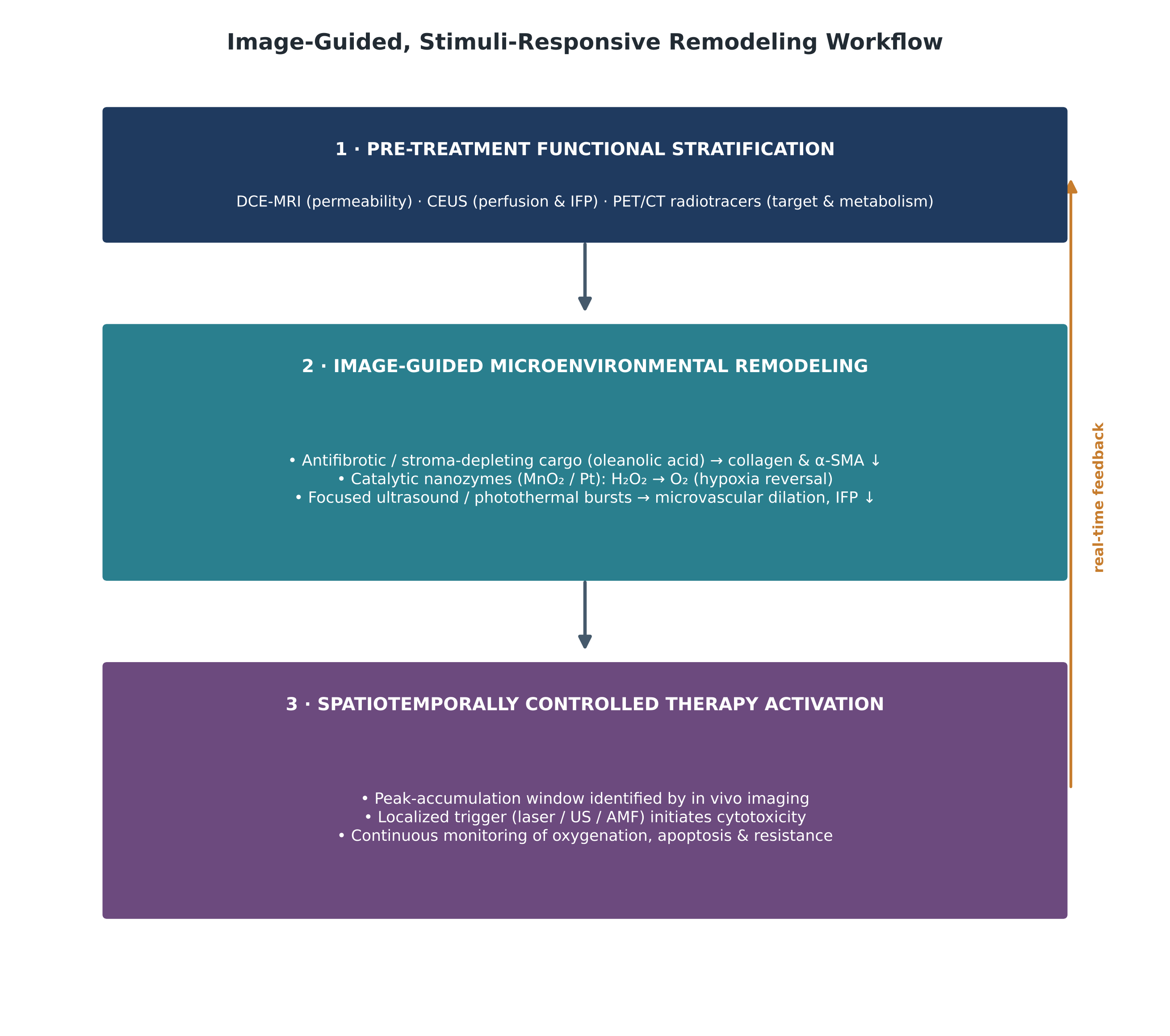
2.3.1. Endogenous and exogenous stimuli-responsive logic. To suppress background signal and prevent premature leakage, modern nanotheranostics incorporate triggers that behave like logic gates, remaining quiescent in healthy tissue (pH ~7.4, normal redox balance) and activating only on encountering hallmarks of the TME (Cook & Decuzzi, 2021; Wang et al., 2026; Wei et al., 2023). The full repertoire of endogenous and exogenous triggers, their chemical mechanisms, and their theranostic outputs is organized in Table 2, and the way these triggers are embedded in a broader treatment workflow is shown in Figure 3. As demonstrated for hybrid calcium phosphate/manganese platforms (PEGMnCaP), the construct stays intact in blood but dissolves on proton exposure in the acidic stroma; the liberated Mn²⁺ coordinates with albumin, sharply increasing longitudinal relaxivity and yielding positive T1-weighted enhancement (Feng et al., 2018; Mi et al., 2016). Likewise, redox-responsive MnO₂ nanosheets consume elevated GSH and convert H₂O₂ into O₂, generating strong T1 signal while stripping cells of the antioxidant reserve needed to buffer therapeutic ROS (Ma et al., 2017; Pillarisetti et al., 2026; Wang et al., 2026).

2.3.2. Image-guided delivery, remodeling, and resistance reversal. Non-invasive imaging supplies the real-time, spatiotemporal data needed to monitor delivery and reverse resistance (Chen et al., 2017; de Maar et al., 2020; Kothawade et al., 2025). Rather than relying on static anatomical endpoints, functional imaging (PET, SPECT, MRI, CT, ultrasound, and optical) can quantify patient-specific vascular permeability, IFP, and receptor density before therapy begins (de Maar et al., 2020; Stapleton et al., 2015; Theek et al., 2014); the complementary modalities and their roles are compared in Table 3. In the ZEPHIR trial, pre-treatment ⁸⁹Zr-trastuzumab PET/CT paired with early ¹⁸F-FDG response monitoring predicted progression-free survival in HER2-positive metastatic breast cancer under trastuzumab emtansine, identifying patients with non-responsive, heterogeneous lesions (Gebhart et al., 2016). Similarly, ⁶⁴Cu-labeled liposomes (⁶⁴Cu-MM-302) revealed substantial intra- and inter-tumoral variability in liposomal deposition that correlated with outcome, supporting image-based stratification (Lee et al., 2017).

Beyond selection, nanotheranostics actively remodels the stroma to eradicate resistance (Li et al., 2026; Tian et al., 2025). In NSCLC with acquired resistance to third-generation EGFR inhibitors (osimertinib/AZD9291), an oleanolic acid-modified chitosan platform (CsO/IGA) co-delivering osimertinib and an indocyanine green–gadolinium probe suppressed CAF proliferation and downregulated α-SMA, dismantling collagen barriers and promoting deep penetration; under dual fluorescence/MRI guidance, NIR irradiation produced local hyperthermia and singlet oxygen that degraded EGFR and phosphorylated EGFR and downregulated AKT survival signaling (Zheng et al., 2026). Acoustic-magnetic platforms combine ultrasonic and magnetic fields for coordinated penetration and reprogramming, with ultrasound-targeted microbubble destruction and sonoporation transiently opening vascular junctions to lower IFP and widen transvascular routes (de Maar et al., 2020; Wu et al., 2026; Yang et al., 2024). When paired with sonodynamic therapy using mesoporous CaCO₃ platforms (HA/CaCO₃@Ce6), ultrasound activation generates cytotoxic ROS and massive calcium overload, establishing a self-amplifying Ca²⁺–ROS loop that bypasses apoptotic blocks and drives immunogenic cell death (Chen et al., 2026).

2.4. Current Clinical Progress and Translational Bottlenecks

Translation from preclinical formulation to regulated intervention has produced real milestones, yet the bench-to-bedside path still faces biological, industrial, and regulatory hurdles (Chen et al., 2017; Gawne et al., 2023; Wang et al., 2026); the leading clinical-stage systems and their roadblocks are compiled in Table 4. Among the foremost precedents is AGuIX®, an ultra-small sub-5 nm polysiloxane particle chelated with multiple gadolinium complexes, designed for dual MRI contrast and radiosensitization (Bort et al., 2020). Trials in brain metastases (NCT04899908) and newly diagnosed glioblastoma (NANO-GBM, NCT04881032) confirm that AGuIX amplifies MRI signal at tumor margins, guides stereotactic radiotherapy planning, and enhances radiation damage through secondary Auger and photoelectron emission, while its sub-renal-threshold size ensures rapid, complete clearance without long-term heavy-metal retention (AGuIX, 2021a, 2021b; Bort et al., 2020; Verry et al., 2021; Wang et al., 2026).

In iron-oxide nanomedicine, ferumoxytol—an FDA-approved carbohydrate-coated SPION originally developed for iron-deficiency anemia—has been repurposed off-label as a gadolinium-free T2/T2* contrast agent and companion diagnostic (Aghighi et al., 2018; Wang et al., 2026). Engulfed selectively by tumor-associated macrophages (TAMs), it provides a quantitative readout of the inflammatory, immunosuppressive microenvironment, and iron-mediated Fenton chemistry can repolarize M2-like TAMs toward tumoricidal M1-like phenotypes (Aghighi et al., 2018; Horvat et al., 2024; Wang et al., 2026). Other clinical formulations—PEGylated liposomal doxorubicin (Doxil®/Caelyx®), nanoliposomal irinotecan (Onivyde®, shown to extend survival in metastatic pancreatic cancer in NAPOLI-1), and polymeric micelles (Genexol-PM)—demonstrate that nano-encapsulation reliably reduces off-target toxicity (Alberts et al., 2004; Lee et al., 2008; Wang-Gillam et al., 2016; Yuan et al., 2021).

Despite these successes, roughly ninety percent of investigational theranostics remain confined to academic animal models (Wang et al., 2026). Bridging the gap means confronting synthetic over-engineering and reproducibility, since combining three to five chemical entities on one chassis produces low-yield multistep syntheses, batch-to-batch variation, and difficult GMP validation of size, charge, entrapment, and relaxivity (Chen et al., 2017; Gawne et al., 2023; Li et al., 2026); the biological protein corona, which coats injected particles with opsonins and shields targeting ligands, redirecting carriers to hepatic and splenic macrophages (Salvati et al., 2013; Tian et al., 2025); and long-term toxicokinetics, since non-biodegradable inorganic cores lack efficient elimination and raise concerns about retention, oxidative injury, and immunogenicity that demand rigorous pharmacokinetic and dose-limiting-toxicity data before approval (Todd & Kay, 2016; Wang et al., 2026).

2.5. Future Horizons: Artificial Intelligence, Companion Diagnostics, and Personalized Medicine

Table 3. Multimodal diagnostic modalities for spatiotemporal guidance, microenvironmental heterogeneity mapping, and therapeutic monitoring. Each non-invasive imaging technique integrated into cancer nanotheranostics is described by its contrast agent or nanoprobe reporter, the biophysical or physiological parameter it quantifies, and its specific role in resolving solid-tumor heterogeneity. The therapeutic synergy and the principal clinical advantages and translational bottlenecks are summarized to help match a given imaging modality to the appropriate theranostic task, from deep anatomical staging to real-time monitoring of drug release.

Imaging modality

Contrast agent / nanoprobe reporter

Parameter quantified

Role in resolving heterogeneity

Therapeutic synergy & guidance

Clinical advantages & bottlenecks

Reference

Magnetic resonance imaging (T1-weighted)

Paramagnetic chelates (Gd-DOTA, Gd-DTPA) and activatable Mn2+ nanoprobes (PEGMnCaP)

Longitudinal relaxation (T1); water proton exchange; local pH and enzymatic cleavage

High spatial resolution (~50–100 µM); resolves deep intra-tumoral margins and spatial acidity gradients

Delineates surgical margins; monitors localized drug release and carrier degradation in real time

Excellent soft-tissue contrast without ionizing radiation; low molar sensitivity requires high local probe concentrations

Cai et al. (2021); Feng et al. (2018); Wang et al. (2026)

Magnetic resonance imaging (T2/T2*-weighted)

Superparamagnetic iron oxide nanoparticles (SPIONs, ferumoxytol)

Transverse relaxation (T2/T2*); local field inhomogeneities; iron phagocytosis

Maps tumor-associated macrophage infiltration and regional microvascular blood volume (rCBV)

Guides magnetic hyperthermia (AMF); tracks homing and mechanical trapping of circulating tumor clusters

High sensitivity to microscopic iron deposits; blooming susceptibility artifacts can obscure adjacent anatomy

Aghighi et al. (2018); Dhandapani et al. (2023); Yang et al. (2019)

Positron emission tomography (PET)

Positron-emitting radiotracers 64Cu, 89Zr, 18F, 68Ga (e.g., 68Ga-FAPI, 64Cu-MM-302)

Radiotracer avidity; glucose metabolism (18F-FDG); fibroblast activation protein; EPR kinetics

Whole-body quantitative evaluation; identifies occult micrometastases and inter-lesional target heterogeneity

Enables companion pre-treatment screening; measures EPR deposition to identify prospective responders

Picomolar sensitivity with unlimited penetration depth; lacks anatomical resolution and uses ionizing radiation

Chen et al. (2017); Chen et al. (2026); Lee et al. (2017)

Optical fluorescence imaging (NIR-I/NIR-II)

Indocyanine green (ICG), pheophorbide a (Ppa), and organic fluorophores (700–1700 nm)

Photon emission spectra; fluorescence lifetime; receptor-mediated endocytosis

Visualizes cellular-level subclonal margins; distinguishes malignant from healthy parenchyma

Directs intraoperative resection; defines peak therapeutic accumulation window for PDT and PTT

High temporal resolution and low cost; photon scattering limits deep in vivo penetration

Cai et al. (2021); Prasad et al. (2023); Zheng et al. (2026)

Short-wave infrared (SWIR) optical imaging

Rare-earth-doped lanthanide nanoparticles (RENPs) emitting at 1000–1700 nm

SWIR luminescence; real-time tissue autofluorescence suppression

Achieves sub-millimeter resolution through deep skin and subcutaneous tissue layers

Guides localized radiosensitization; tracks intradermal microneedle release kinetics in cutaneous tumors

Zero endogenous autofluorescence with superior signal-to-noise; requires specialized InGaAs detector arrays

Khan et al. (2024, 2026); Ren et al. (2024)

Contrast-enhanced ultrasound (CEUS)

Gas-filled lipid/polymeric microbubbles and phase-shift perfluorocarbon nanodroplets

Microvascular blood flow velocity; acoustic replenishment kinetics; vascular permeability

Maps spatial vascular architecture and quantifies high interstitial fluid pressure zones

Directs ultrasound-targeted microbubble destruction (UTMD) to open barriers and potentiate extravasation

Real-time, radiation-free bedside use; operator-dependent and limited by air-filled or bony structures

de Maar et al. (2020); Theek et al. (2014); Wu et al. (2026)

X-ray computed tomography (CT)

High-Z metallic nanoparticles (gold, bismuth, tantalum, iodinated assemblies)

X-ray attenuation coefficients (Hounsfield units); tissue density alterations

Resolves deep anatomical cross-sections and spatial calcification patterns

Facilitates geometric beam planning for stereotactic radiotherapy and high-Z radiosensitization

Broad availability and rapid 3D acquisition; lower soft-tissue contrast than MRI with ionizing radiation

Chen et al. (2017); Kothawade et al. (2025); Melancon et al. (2012)

Table 4. Clinical translation landscape of leading nanomedicine and theranostic formulations. The table reports each formulation’s platform classification and dimensions, clinical trial phase and registry identifier, targeted oncological population, and diagnostic or theranostic companion capability. Key efficacy findings are paired with the prevailing pharmacological and regulatory bottlenecks—manufacturing cost, EPR heterogeneity, dose-limiting toxicity, and radiolabeling logistics—to convey both the demonstrated clinical value and the obstacles that still limit broad translation of imaging-guided nanotheranostics.

Formulation & brand

Platform classification & dimensions

Trial phase & identifier

Indication & population

Diagnostic/theranostic capability

Efficacy findings & breakthroughs

Pharmacological & regulatory bottlenecks

Reference

AGuIX®

Sub-5 nm hybrid polysiloxane core chelated with multiple Gd3+-DOTA complexes (~3.0–4.5 nm)

Phase I/II completed & Phase II active (NCT04899908, NCT04881032, NANO-RAD)

Multiple brain metastases; newly diagnosed glioblastoma; cervical cancer

Dual-action T1-weighted MRI contrast agent and high-Z radiosensitizer

Marked MRI tumor enhancement; radiosensitization with significant survival gains; rapid renal clearance (<5 nm)

Ensuring uniform heavy-metal chelation stability; high cost of clinical-grade polysiloxane conjugates

Bort et al. (2020); Verry et al. (2021); Wang et al. (2026)

Ferumoxytol (Feraheme®)

Polyglucose sorbitol carboxymethyl ether-coated SPIONs (~30 nm)

Phase I/II clinical trials (NCT02022696, NCT01542879)

Advanced solid malignancies; pediatric osteosarcoma; hepatic and nodal metastases

Off-label T2/T2* MRI companion diagnostic; quantifies tumor-associated macrophages (TAMs)

Visualizes macrophage infiltration; Fenton-driven ROS repolarizes M2-like to antitumor M1 TAMs

FDA black-box warning for rare anaphylaxis; prolonged splenic/hepatic signal void on MRI

Aghighi et al. (2018); Horvat et al. (2024); Wang et al. (2026)

Onivyde® (MM-398 / nal-IRI)

PEGylated nanoliposomal irinotecan sucrosofate formulation (~110 nm)

FDA approved; evaluated alongside companion PET (NAPOLI-1 trial)

Metastatic pancreatic ductal adenocarcinoma after gemcitabine failure

Paired with investigational 64Cu-MM-302 PET to quantify inter- and intra-tumoral EPR variability

Extended median overall survival to 6.1 vs. 4.2 months with 5-FU/LV; companion PET predicts response

Marked inter-patient EPR heterogeneity; dose-limiting neutropenia, diarrhea, and vascular extravasation

Lee et al. (2017); Tian et al. (2025); Wang-Gillam et al. (2016)

BIND-014

PEG-PLGA polymeric nanoparticles functionalized with a PSMA-targeting ligand (~100 nm)

Phase I/II completed (NCT01300537, NCT01792479)

Advanced metastatic castration-resistant prostate cancer (mCRPC) and KRAS-mutant NSCLC

Targets prostate-specific membrane antigen (PSMA) for active homing and localized docetaxel delivery

Distinct pharmacokinetics with 100-fold higher drug retention than conventional docetaxel; verified antitumor response

Premature systemic payload leakage; low response rate in unselected Phase II cohorts; high production complexity

Tian et al. (2025); Von Hoff et al. (2016)

Genexol-PM (Cynvix)

Cremophor EL-free mPEG-PDLLA polymeric micelles (~25 nm)

Approved in select territories; Phase II/III trials

Metastatic breast cancer; advanced non-small cell lung cancer

Replaces toxic polyoxyethylated castor oil vehicles, improving drug tolerability

Allows higher paclitaxel doses (up to 300 mg/m²) without hypersensitivity premedication; 46.5% ORR in breast cancer

Rapid micellar dissociation below critical micelle concentration; requires strict reconstitution parameters

Lee et al. (2008); Tian et al. (2025)

Doxil® / Caelyx®

PEGylated liposomal doxorubicin with transmembrane ammonium sulfate gradient (~80–100 nm)

FDA approved (standard-of-care in multiple protocols)

Recurrent ovarian cancer; AIDS-related Kaposi’s sarcoma; metastatic breast cancer

Baseline imaging of liposomal tissue distribution using radiolabeled analogues (99mTc/111In-liposomes)

Bypasses peak-plasma cardiotoxicity, extending circulation half-life (~55 h) toward manageable cutaneous profiles

Dose-limiting palmar-plantar erythrodysesthesia; variable penetration into hypovascular tumors

Alberts et al. (2004); Chen et al. (2017); Yuan et al. (2021)

64Cu-MM-302

HER2-targeted PEGylated immunoliposome labeled with positron-emitting Copper-64 (~100 nm)

Phase I clinical trial (NCT01304797)

HER2-positive advanced metastatic breast cancer

Diagnostic PET companion imaging of HER2-targeted liposome biodistribution and EPR deposition

Quantified substantial intra- and inter-tumoral heterogeneity; imaging predicts progression-free survival under T-DM1

Cost and logistic barriers of cyclotron radionuclide synthesis; specialized radiolabeling infrastructure required

Gebhart et al. (2016); Lee et al. (2017)

Table 5. Quantitative signal amplification of stimuli-responsive imaging nanoprobes upon microenvironmental activation. For each nanoprobe formulation the table gives the physical or biochemical activation input, the baseline (“OFF”) relaxivity or signal, and the activated (“ON”) value, together with the resulting amplification factor or dynamic contrast gain. The data illustrate how subtle biochemical shifts—tumor acidosis, intracellular H2O2, enzymatic cleavage, nitric-oxide flux, or radiation-induced DNA repair—are transduced into large, measurable imaging readouts that enable sensitive, activatable detection and treatment monitoring.

Nanoprobe reporter formulation

Activation input

Baseline signal (OFF)

Activated signal (ON)

Amplification factor / dynamic gain

Reference

PEGMnCaP mineralized nanoparticles

Extracellular tumor acidosis (pH 6.5–6.8 vs. 7.4)

r1 = 5.1 mM⁻¹s⁻¹

r1 = 34.2 mM⁻¹s⁻¹

6.7-fold increase in T1 relaxivity via albumin binding

Feng et al. (2018); Mi et al. (2016)

SiO2-MB@MnO2 gatekeeper hybrids

Intracellular H2O2 and acidic pH (5.0)

r1 = 0.006 mM⁻¹s⁻¹

r1 = 4.40 mM⁻¹s⁻¹

733-fold increase via Mn4+ → Mn2+ ion reduction

Pillarisetti et al. (2026)

Glycopolymer-PTX-DOTA-Gd conjugate

Cathepsin B proteolytic cleavage

Quenched / restricted water exchange

r1 = 7.10 mM⁻¹s⁻¹

Sustained enhancement; elevated tumor SNR up to 24 h post-injection

Cai et al. (2021)

Nitric oxide magnetic probe (NRMP)

Pathological tumor NO flux

Clustered SPION state (low r2)

Dispersed SPION state

Δr2 = 36.4 mM⁻¹s⁻¹ shift for ultrasensitive NO detection

Wang et al. (2026)

CD44-targeted chitosan-SPIONs (MN)

Alternating magnetic fields and acidic pH (5.2)

Low baseline T2 contrast

r2 = 33.08 mM⁻¹s⁻¹

Concentration-dependent T2 dephasing; selective CTC detection

Dhandapani et al. (2023)

APE1-activatable afterglow/MRI probe

Radiation-induced DNA repair biomarker APE1

Quenched afterglow luminescence

High-intensity optical afterglow

Ultra-early response monitoring within 3 h of DNA damage

Wang et al. (2026)

As the field matures, it is converging with computational biology and digital oncology (Chen et al., 2017; Wang et al., 2026). Artificial intelligence and machine learning are accelerating nanocarrier design: deep neural networks trained on physicochemical libraries can predict protein-corona composition, optimize colloidal stability, and forecast extravasation kinetics as a function of size, charge, and rigidity (Liu et al., 2015; Wang et al., 2026). Radiomics and AI-driven segmentation of multiparametric MRI, PET/CT, and spectral CT enable whole-body mapping of heterogeneity, extracting voxel-level textural variation in perfusion, leakiness, and cellularity to flag high-risk hypoxic or desmoplastic niches (de Maar et al., 2020; Molina-Garcia et al., 2018; Yoon et al., 2016; Yoon et al., 2019).

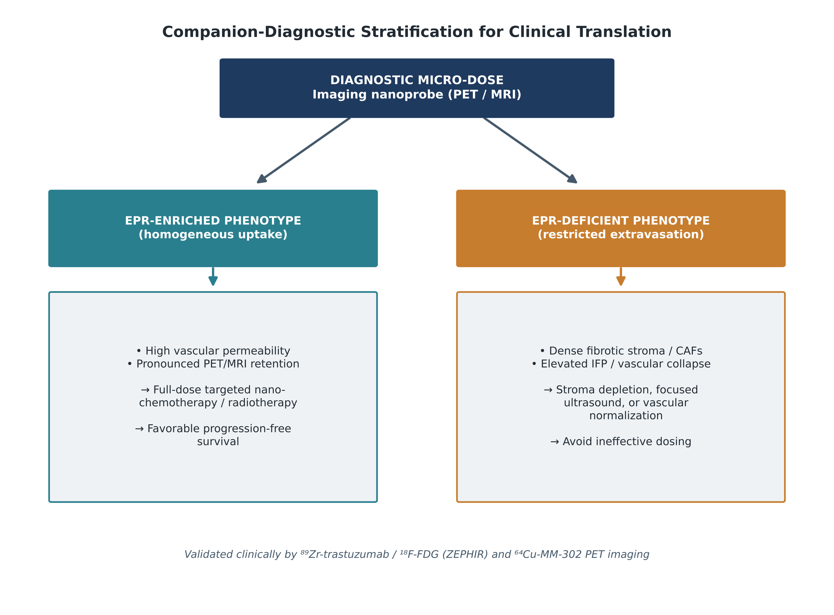
The emerging clinical paradigm also treats nanotheranostics as standardized companion diagnostics (Ehlerding et al., 2018; Miller et al., 2017). Instead of dosing cytotoxic formulations indiscriminately, clinicians can use diagnostic micro-doses of radiolabeled or activatable probes to gauge target engagement and EPR magnitude in each patient; those with robust, homogeneous uptake proceed to full-dose nanotherapy, while others are redirected toward vascular normalization or stroma-depleting regimens (Chen et al., 2017; de Maar et al., 2020; Lee et al., 2017). This decision logic—stratifying EPR-enriched from EPR-deficient phenotypes—is summarized in Figure 4. Finally, pairing nanotheranostic tracking with serial liquid biopsies—circulating tumor cells, cell-free tumor DNA, and tumor-derived extracellular vesicles—will provide continuous molecular feedback on emerging resistant subclones, enabling dynamically adaptable, closed-loop treatment that stays ahead of evolving resistance (Dhandapani et al., 2023; Juku et al., 2026).

3. Methods

This work is a narrative, integrative review of imaging-guided nanotheranostics for solid-tumor heterogeneity and drug resistance. Because narrative syntheses are vulnerable to selection bias, the literature search, screening, and data-extraction steps were pre-specified and documented so that the search is transparent and reproducible by independent investigators, in line with recommended reporting practice for structured literature retrieval.

3.1. Information Sources and Search Strategy

Peer-reviewed literature was retrieved from PubMed/MEDLINE, Scopus, and Web of Science Core Collection, supplemented by Google Scholar for citation chasing and by the ClinicalTrials.gov registry for trial status. The search window spanned January 1984 to the final search date (updated through the preparation of this manuscript), with 1984 chosen to capture the foundational description of tumor pH and the EPR concept while concentrating on literature from 2015 onward for platform- and trial-level detail.

A reproducible Boolean strategy combined controlled vocabulary (MeSH) with free-text terms across three concept blocks joined by the AND operator, with synonyms within each block joined by OR. Block 1 (platform): “nanotheranostic*” OR “theranostic nanoparticle*” OR “nanomedicine” OR “stimuli-responsive nanoparticle*” OR “liposome” OR “polymeric micelle” OR “mesoporous silica” OR “iron oxide nanoparticle” OR “nanozyme” OR “cell membrane-coated nanoparticle” OR “DNA nanostructure.” Block 2 (problem): “tumor heterogeneity” OR “multidrug resistance” OR “drug resistance, neoplasm”[MeSH] OR “tumor microenvironment” OR “hypoxia” OR “efflux pump” OR “P-glycoprotein.” Block 3 (function): “molecular imaging” OR “MRI” OR “PET” OR “computed tomography” OR “ultrasound” OR “fluorescence imaging” OR “image-guided therapy” OR “companion diagnostic.” An example PubMed string was: (nanotheranostic*[tiab] OR “theranostic nanoparticle*”[tiab]) AND (“multidrug resistance”[tiab] OR “tumor heterogeneity”[tiab] OR “tumor microenvironment”[MeSH]) AND (“molecular imaging”[tiab] OR MRI[tiab] OR PET[tiab]). Records were limited to English-language publications; no restriction was placed on study design.

3.2. Eligibility Criteria

Studies were eligible if they (i) described a nanoscale platform integrating at least one diagnostic imaging modality with at least one therapeutic function; (ii) addressed solid-tumor heterogeneity, the tumor microenvironment, or a defined mechanism of drug resistance; and (iii) reported original preclinical (in vitro or in vivo) or clinical data, or provided authoritative mechanistic or methodological synthesis in the case of reviews. Primary clinical evidence (completed or ongoing trials with a registry identifier) was prioritized for the translational sections. Records were excluded if they concerned hematological malignancies exclusively, addressed non-oncological indications, lacked any imaging component, or were conference abstracts, editorials, or non-retrievable in full text. Where multiple reports described the same platform, the most complete or most recent account was retained.

3.3. Study Selection and Data Extraction

Retrieved records were de-duplicated and screened in two stages—title/abstract, then full text—against the criteria above. For each included study, the following fields were extracted into a structured matrix: nanoplatform class and composition; hydrodynamic size and surface chemistry; imaging modality and contrast reporter; therapeutic cargo and mechanism; the specific resistance or heterogeneity barrier addressed; the stimulus or trigger exploited; the experimental model (cell line, xenograft, syngeneic, or clinical population); and the principal quantitative outcome (for example, cell viability, IC₅₀, tumor-weight reduction, relaxivity change, or accumulation as percentage of injected dose). These extracted fields form the evidentiary basis of Tables 1–4 and of the quantitative synthesis presented in the Results.

3.4. Synthesis Approach

Because the included studies were methodologically heterogeneous—spanning diverse materials, models, and endpoints—a quantitative meta-analysis was neither appropriate nor attempted. Instead, findings were synthesized narratively and organized thematically around four axes: (i) mechanisms of heterogeneity and resistance; (ii) platform architecture; (iii) stimuli-responsive and imaging logic; and (iv) clinical translation. Reported quantitative outcomes were tabulated verbatim as stated by the original authors, with units preserved, so that the numerical claims in this review can be traced to their primary sources. Schematic Figures 1–4 were constructed by the authors to summarize these themes and contain no data beyond that presented in the cited literature.

4. Image-Guided Nanotheranostics Reverse Multidrug Resistance and Resolve Delivery Heterogeneity in Solid Tumors

4.1. Overcoming Multidrug Resistance and Cellular Transport Barriers

When the preclinical record is read as a whole, one pattern stands out: multifunctional nanotheranostics tend to widen the therapeutic index well beyond what free drug or non-responsive carriers achieve (Chen et al., 2017; Tian et al., 2025; Wang et al., 2026). The reason is mechanistic rather than incidental. In MDR models that overexpress ABC transporters, an unguided small molecule is pumped back out almost as fast as it arrives, so cytosolic and nuclear concentrations never reach a lethal level (Dallavalle et al., 2020; Sajid et al., 2023; Yang et al., 2019). Smart nanocarriers sidestep that problem by entering through endocytosis—below the notice of plasma-membrane P-gp—and then releasing their payload only once inside (Bin Emran et al., 2022; Tian et al., 2025; Yang et al., 2019). The representative constructs behind these observations are catalogued in Table 1.

The in vitro numbers make the point concretely. In vincristine-resistant gastric carcinoma cells (SGC-7901/VCR), the polypeptide micelle DXS@NPs—doxorubicin, XMD8-92, and SPIONs together—cut viability to 53%, whereas free doxorubicin left roughly 90% of the cells alive (Yang et al., 2019). That gain was not, it seems, a single effect but a convergence of two: XMD8-92 suppressed P-gp transcription, and the carrier released its cargo far faster in the acidic endo-lysosome than at physiological pH (56% versus 28% over 24 hours) (Yang et al., 2019). A comparable logic held for hypoxia-activated, ROS-responsive fluorinated oxalate nanotheranostics, which drove A-431 survival down to 32% under severe hypoxia while sparing normoxic controls—an encouraging, if model-specific, demonstration of conditional cytotoxicity (Pillarisetti et al., 2026). Biomimetic designs told much the same story: homologous cancer-cell-membrane-coated gold nanorods (AuVNRs-DOX) plus NIR irradiation (800 nm, 1 W/cm², 10 min) killed more than 75% of cells, against under 25% for drug-free vesicles and roughly 40% without light, with Western blots showing pronounced PARP1 cleavage and near-complete loss of Cyclin D1 (Prasad et al., 2023). Taken together, these datasets suggest that combining a resistance-reversing modulator with a physically triggered release step is what reactivates apoptosis in otherwise refractory cells.

4.2. In Vivo Microenvironment Remodeling and Tumor Regression

The in vivo literature is messier, as one would expect, but it points the same way. Across xenograft and syngeneic models, free chemotherapeutics and unguided interventions usually falter against the desmoplastic matrix, high IFP, and hypoxia that define advanced disease (Danhier, 2016; de Maar et al., 2020; Tian et al., 2025). What the compiled evidence indicates is that platforms built to deplete stroma, normalize vessels, or generate oxygen can reshape those barriers enough to let drug penetrate and tumors regress (Li et al., 2026; Zheng et al., 2026); the three remodeling routes are summarized schematically in Figure 3.

In NSCLC models resistant to osimertinib, the oleanolic acid-functionalized chitosan platform (CsO/IGA) remodeled the fibrotic microenvironment, suppressing CAF proliferation and α-SMA and allowing homogeneous distribution through 3D spheroids (Zheng et al., 2026). The functional payoff was substantial: in P5 osimertinib-resistant xenografts, two weeks of CsO/IGA under 808 nm irradiation (1 W/cm², 5 min) reduced excised tumor weight to 0.07 ± 0.04 g versus 0.59 ± 0.05 g in controls (p < 0.0001), with no appreciable weight loss (Zheng et al., 2026). Visualized liposomal platforms (VLPs/CpG/siRNA) carrying HIF-1α siRNA and CpG overcame radioresistance in murine hepatocellular carcinoma, reaching peak accumulation of 2.03 ± 0.18% ID/g at 12 hours by ICP-MS; silencing HIF-1α relieved hypoxia and curbed VEGF-driven angiogenesis, while TLR9-driven dendritic-cell maturation expanded CD8⁺ tumor-infiltrating lymphocytes and sensitized lesions to radiotherapy (Li et al., 2026). In metastatic breast cancer, AuVNRs-DOX with NIR light shrank tumor volume from 250 mm³ to 30.4–49 mm³ and reduced tumor weight by 63.8 ± 4.6%, against only 24.6 ± 9.2% for non-targeted liposomal doxorubicin, and—strikingly—yielded 100% survival over 63 days without cardiac, hepatic, or renal histopathology (Prasad et al., 2023).

4.3. High-Fidelity Signal Transduction Across Multimodal Imaging

If there is one capability that separates nanotheranostics from ordinary drug formulations, it is the ability to report on itself (Chen et al., 2017; Wang et al., 2026). Quantitative relaxivity measurements show that stimuli-responsive MRI probes convert small biochemical shifts into large, measurable signal changes (Feng et al., 2018; Wang et al., 2026); the magnitude of these “OFF-to-ON” transitions, which ranges from modest to frankly dramatic, is collected in Table 5. PEGMnCaP, for instance, raised r₁ from 5.1 to 34.2 mM⁻¹s⁻¹ on encountering tumor acidosis—a 6.7-fold gain via albumin binding—while SiO₂-MB@MnO₂ gatekeeper hybrids reported a far larger 733-fold increase upon Mn⁴⁺→Mn²⁺ reduction (Feng et al., 2018; Mi et al., 2016; Pillarisetti et al., 2026).

These shifts are not merely cosmetic; they carry genuine pharmacokinetic and spatial information. With cathepsin B-cleavable branched glycopolymer-PTX conjugates (r₁ = 7.10 mM⁻¹s⁻¹ on activation), tumor signal rose steadily over 24 hours even as clinical Gd-DTPA washed out within 4—a direct illustration of how a macromolecular carrier outlasts vascular clearance (Cai et al., 2021). Zero-background ¹⁹F MRI platforms and short-wave infrared (SWIR, 1000–1700 nm) probes went further still, erasing endogenous autofluorescence to achieve submillimeter resolution without ionizing radiation (Khan et al., 2026; Wang et al., 2026). The full set of imaging modalities and the parameters each one resolves is laid out in Table 3.

4.4. Resolving Delivery Heterogeneity and Clinical Translation

For all the preclinical promise, the quantitative data keep returning to one sobering fact: the delivery bottleneck has not gone away (Danhier, 2016; Gawne et al., 2023; Wilhelm et al., 2016). Pooled pharmacokinetic analyses still put median solid-tumor accumulation below 1% of the injected dose (about 0.7–0.8% ID), with more than 90% of circulating carriers cleared by hepatic and splenic macrophages (Chen et al., 2017; Wang et al., 2026; Wilhelm et al., 2016). That number is why the field has increasingly repositioned nanotheranostics as a companion diagnostic—a way to see the heterogeneity before committing to treatment (de Maar et al., 2020; Lee et al., 2017). The stratification logic that follows from this is diagrammed in Figure 4, and the clinical-stage formulations that embody it are detailed in Table 4.

Clinical evidence supports the approach fairly directly. In ZEPHIR, ⁸⁹Zr-trastuzumab PET/CT with ¹⁸F-FDG tracking mapped inter- and intra-lesional HER2 heterogeneity and predicted progression-free survival under T-DM1, flagging non-responders before treatment failed (Gebhart et al., 2016). PET with ⁶⁴Cu-MM-302 showed delivery varying from negligible to over 10% ID/kg between patients, and that variability tracked with subsequent tumor shrinkage under nanoliposomal irinotecan (Lee et al., 2017; Wang-Gillam et al., 2016). Meanwhile, AGuIX® (r₁ ≈ 8 mM⁻¹s⁻¹) has reached Phase I/II for brain metastases and glioblastoma, delineating margins on 3T MRI, acting as a radiosensitizer, and clearing renally without heavy-metal deposition (AGuIX, 2021a, 2021b; Bort et al., 2020; Verry et al., 2021; Wang et al., 2026), while ferumoxytol has been used to quantify TAM infiltration and microvessel permeability (Aghighi et al., 2018; Horvat et al., 2024; Wang et al., 2026). The through-line is hard to miss: resolving spatial heterogeneity by imaging-guided selection appears to be a prerequisite—not an optional refinement—for overcoming resistance in the clinic.

5. Treating Drug Resistance as a Spatial Problem With Image-Guided, Closed-Loop Cancer Nanotheranostics

5.1. Reframing Resistance as a Problem of Space

It is tempting to think of drug resistance as a purely molecular event—a pump here, a mutation there. The evidence assembled in this review suggests that framing is incomplete. Much of what reads as resistance is, on closer inspection, a failure of delivery: drug that never arrives cannot work, however sensitive the cell (de Maar et al., 2020; Dhandapani et al., 2023). Seen this way, the TME is less a passive backdrop than an active accomplice, its acidosis, hypoxia, high IFP, and desmoplasia conspiring with cellular efflux to protect the very subclones that go on to relapse (Danhier, 2016; Tian et al., 2025; see Figure 1, Table 1). What makes nanotheranostics interesting is that it treats this as a spatial problem and answers it with a spatial tool—one that can both see where drug goes and intervene where it does not.

5.2. Why Imaging Changes the Calculus

The most consequential shift, arguably, is not a new material but a new question. Instead of asking only whether a drug kills cells in a dish, imaging lets us ask whether it reaches them in a patient (Chen et al., 2017; de Maar et al., 2020). That distinction matters because the clinical data keep showing enormous inter-patient variability in EPR—from near-zero to over 10% ID/kg (Lee et al., 2017; Table 4). A companion-diagnostic micro-dose turns that variability from a hidden source of trial failure into an upfront selection criterion (Ehlerding et al., 2018; Miller et al., 2017; Figure 4). ZEPHIR and the ⁶⁴Cu-MM-302 studies are, in a sense, proofs of principle that imaging can predict who will respond before cytotoxic exposure begins (Gebhart et al., 2016; Lee et al., 2017). Whether the broader field adopts this discipline, rather than continuing to dose unselected cohorts, may determine how many promising formulations actually survive late-phase trials.

5.3. The Engineering Tension: Sophistication Versus Manufacturability

There is an uncomfortable trade-off running through the literature, and it deserves to be stated plainly. The platforms that perform best in mice are often the most elaborate—three to five functional components stacked on a single core (Table 1, Table 2)—and that same elaboration is what makes them hard to manufacture reproducibly, hard to validate under GMP, and hard to clear from the body (Gawne et al., 2023; Li et al., 2026; Wang et al., 2026). The protein corona adds a further twist, quietly masking the targeting ligands that justified the complexity in the first place (Salvati et al., 2013; Tian et al., 2025). It is not obvious that the answer is ever-greater sophistication. Indeed, the clinical front-runners—AGuIX®, ferumoxytol, Onivyde®—are comparatively simple, and their relative success hints that elegance of design, not sheer functional density, may be the better translational bet (Bort et al., 2020; Wang-Gillam et al., 2016; Table 4).

5.4. Limitations of the Current Evidence

Several caveats temper the optimism. Most efficacy data come from a handful of cell lines and murine models that imperfectly reproduce human stromal density, immune context, and the slow kinetics of acquired resistance; the striking tumor-weight and survival figures cited above should be read as encouraging signals rather than settled outcomes (Prasad et al., 2023; Zheng et al., 2026). The relaxivity and accumulation metrics, though quantitatively impressive (Table 5), are rarely cross-validated across laboratories using standardized phantoms or dosing. And this review itself, being narrative rather than systematic, cannot fully exclude selection bias despite the structured search described in the Methods. These are not reasons for discouragement so much as reminders that the preclinical-to-clinical ratio—roughly nine in ten candidates still stranded in animal models—reflects genuine, unresolved gaps (Wang et al., 2026).

5.5. Toward Adaptive, Closed-Loop Oncology

Where, then, is this heading? The most plausible near-term trajectory couples three threads that are already visible: AI-guided design to tame the corona and predict biodistribution (Liu et al., 2015; Wang et al., 2026); companion diagnostics to select patients and time therapy (Chen et al., 2017; Figure 4); and liquid biopsy to catch resistant subclones as they emerge (Dhandapani et al., 2023; Juku et al., 2026). Knit together, these point toward closed-loop protocols in which whole-body imaging and molecular monitoring continuously inform

Figure 3. Image-guided, stimuli-responsive remodeling workflow. Treatment proceeds in three stages: (1) pre-treatment functional stratification using DCE-MRI, contrast-enhanced ultrasound, and PET/CT radiotracers to characterize vascular permeability, perfusion, interstitial fluid pressure, and target expression; (2) image-guided microenvironmental remodeling through antifibrotic cargo, catalytic oxygen-generating nanozymes, and focused ultrasound or photothermal bursts; and (3) spatiotemporally controlled therapy activation timed to the peak-accumulation window, with continuous imaging feedback on oxygenation, apoptosis, and resistance.

Figure 4. Companion-diagnostic stratification for clinical translation. A diagnostic micro-dose of an imaging nanoprobe (PET or MRI) classifies patients by delivery phenotype. Those with an EPR-enriched phenotype—high vascular permeability and pronounced probe retention—proceed to full-dose targeted nanochemotherapy or radiotherapy with favorable expected outcomes, whereas those with an EPR-deficient phenotype—dense stroma, elevated interstitial fluid pressure, and vascular collapse—are redirected toward stroma depletion, focused ultrasound, or vascular normalization. This logic is validated clinically by 89Zr-trastuzumab/18F-FDG (ZEPHIR) and 64Cu-MM-302 imaging.

dosing—treatment that adapts as the tumor evolves rather than lagging behind it. That vision remains aspirational, and the manufacturing and regulatory obstacles are real. But the direction of travel, at least, seems clear.

6. Conclusion

Tumor heterogeneity and multidrug resistance are, at root, spatial problems: drug that cannot reach every malignant clone cannot cure. By uniting diagnostic imaging with triggered, microenvironment-aware therapy in a single construct, nanotheranostics confronts both the cellular and the physical barriers that defeat conventional treatment. The evidence reviewed here indicates that stimuli-responsive platforms can reverse efflux-mediated resistance, remodel hypoxic and fibrotic stroma, and sharply improve preclinical outcomes, while imaging enables the patient stratification that early trials such as ZEPHIR and ⁶⁴Cu-MM-302 show to be decisive. Translation still hinges on simpler, GMP-compatible designs, predictable clearance, corona-resistant targeting, and the discipline of selecting patients by what imaging shows rather than dosing unselected cohorts. The engineering tension is real, since the platforms that perform best in animals are often the hardest to manufacture and clear. Coupled with artificial intelligence, companion diagnostics, and liquid biopsy, imaging-guided nanotheranostics offers a credible path toward adaptive, closed-loop, personalized management of solid tumors.

References


Aghighi, M., Theruvath, A. J., Pareek, A., Pisani, L. L., Alford, R., Muehe, A. M., Sethi, T. K., Holdsworth, S. J., Hazard, F. K., Gratzinger, D., Luna-Fineman, S., Advani, R., Spunt, S. L., & Daldrup-Link, H. E. (2018). Magnetic resonance imaging of tumor-associated macrophages: Clinical translation. Clinical Cancer Research, 24(17), 4110–4118.

AGuIX. (2021a). Stereotactic radiotherapy with or without AGuIX in brain metastases (ClinicalTrials.gov Identifier: NCT04899908). U.S. National Library of Medicine.

AGuIX. (2021b). AGuIX combined with radiotherapy and temozolomide in newly diagnosed glioblastoma (NANO-GBM) (ClinicalTrials.gov Identifier: NCT04881032). U.S. National Library of Medicine.

Alberts, D. S., Muggia, F. M., Carmichael, J., Winer, E. P., Jahanzeb, M., Venook, A. P., Skubitz, K. M., Rivera, E., Sparano, J. A., & Greenspan, D. L. (2004). Efficacy and safety of liposomal anthracyclines in Phase I/II clinical trials. Seminars in Oncology, 31(6 Suppl 13), 53–90.

Bareche, Y., Buisseret, L., Gruosso, T., Girard, E., Venet, D., Dupont, F., Desmedt, C., Larsimont, D., Park, M., Rothé, F., Stagg, J., & Sotiriou, C. (2020). Unraveling triple-negative breast cancer tumor microenvironment heterogeneity: Towards an optimized treatment approach. Journal of the National Cancer Institute, 112(7), 708–719. https://doi.org/10.1093/jnci/djz208

Barros, R. B., et al. (2025). Nitroreductase-responsive gated mesoporous silica nanoparticles for controlled drug delivery under hypoxic conditions. International Journal of Pharmaceutics, 650, 123710.

Beckers, C., Pruschy, M., & Vetrugno, I. (2024). Tumor hypoxia and radiotherapy: A major driver of resistance even for novel radiotherapy modalities. Seminars in Cancer Biology, 98, 19–30. https://doi.org/10.1016/j.semcancer.2023.11.006

Bin Emran, T., Shahriar, A., Mahmud, A. R., Rahman, T., Abir, M. H., Siddiquee, M. F. R., Ahmed, H., Rahman, N., Nain, Z., Wahab, S., Rauf, A., Al-Harrasi, A., & Kim, B. (2022). Multidrug resistance in cancer: Understanding molecular mechanisms, immunoprevention and therapeutic approaches. Frontiers in Oncology, 12, 891652.

Bort, G., Lux, F., Dufort, S., Crémillieux, Y., Verry, C., & Tillement, O. (2020). EPR-mediated tumor targeting using ultrasmall-hybrid nanoparticles: From animal to human with theranostic AGuIX nanoparticles. Theranostics, 10(3), 1319–1331.

Cai, H., Wang, X. J., Zhang, H., Sun, L., Pan, D. Y., Gong, Q. Y., & Luo, K. (2018). Enzyme-sensitive biodegradable and multifunctional polymeric conjugate as theranostic nanomedicine. Applied Materials Today, 11, 207–218.

Cai, H., Xiang, Y., Zeng, Y., Li, Z., Zheng, X., Luo, Q., Zhu, H., Gong, Q., Gu, Z., Liu, Y., Zhang, H., & Luo, K. (2021). Cathepsin B-responsive and gadolinium-labeled branched glycopolymer-PTX conjugate-derived nanotheranostics for cancer treatment. Acta Pharmaceutica Sinica B, 11(2), 544–559.

Chen, C., Yu, M., Li, Q., Zhou, Y., Zhang, M., Cai, S., & Ding, D. (2024). Programmable tetrahedral DNA-RNA nanocages woven with stimuli-responsive siRNA for enhancing therapeutic efficacy of multidrug-resistant tumors. Advanced Science, 11(20), e2404112.

Chen, H., Zhang, W., Zhu, G., Xie, J., & Chen, X. (2017). Rethinking cancer nanotheranostics. Nature Reviews Materials, 2(7), 17024. https://doi.org/10.1038/natrevmats.2017.24

Chen, L., Liu, M., Wang, Y., Wei, W., Li, Y., Bai, Y., Yu, X., Jiao, L., & Wang, M. (2024). TME-activated MnO2/Pt nanoplatform of hydroxyl radical and oxygen generation to synergistically promote radiotherapy and MR imaging of glioblastoma. International Journal of Nanomedicine, 19, 11055–11070. https://doi.org/10.2147/IJN.S474098

Chen, M., Wang, Y., Niu, Y., Chen, X., Su, H., Xia, L., Liu, C., Zhou, J., Wang, Z., Li, B., & Lu, D. (2026). pH-responsive CaCO3 nanoplatform amplifies SDT via calcium overload-ROS loop for deep tumor therapy. iScience, 29(3), 115082. https://doi.org/10.1016/j.isci.2026.115082

Comerford, K. M., Wallace, T. J., Karhausen, J., Louis, N. A., Montalto, M. C., & Colgan, S. P. (2002). Hypoxia-inducible factor-1-dependent regulation of the multidrug resistance (MDR1) gene. Cancer Research, 62(12), 3387–3394.

Cook, A. B., & Decuzzi, P. (2021). Harnessing endogenous stimuli for responsive materials in theranostics. ACS Nano, 15(2), 2068–2098.

Dallavalle, S., Dobricic, V., Lazzarato, L., Gazzano, E., Machuqueiro, M., Pajeva, I., Tsakovska, I., Zidar, N., & Fruttero, R. (2020). Improvement of conventional anti-cancer drugs as new tools against multidrug resistant tumors. Drug Resistance Updates, 50, 100682.

Danhier, F. (2016). To exploit the tumor microenvironment: Since the EPR effect fails in the clinic, what is the future of nanomedicine? Journal of Controlled Release, 244(Pt A), 108–121. https://doi.org/10.1016/j.jconrel.2016.11.015

de Maar, J. S., Sofias, A. M., Vila, R., Storm, G., Kiessling, F., Moonen, C., & Lammers, T. (2020). Imaging spatial therapy heterogeneity in cancer. Theranostics, 10(4), 1884–1904.

Dhandapani, R., Ramasamy, T., Shanmugam, S., & Choi, H. G. (2023). Self-assembled multifunctional nanotheranostics against circulating tumor clusters in metastatic breast cancer. Acta Pharmaceutica Sinica B, 13(4), 1711–1726.

Dhas, N., García, M. C., Kudarha, R., Pandey, A., Nikam, A. N., Gopalan, D., & Mutalik, S. (2022). Advancements in cell membrane camouflaged nanoparticles: A bioinspired platform for cancer therapy. Journal of Controlled Release, 346, 71–97.

Ehlerding, E. B., Grodzinski, P., Cai, W., & Liu, C. H. (2018). Big potential from small agents: Nanoparticles for imaging-based companion diagnostics. ACS Nano, 12(3), 2106–2121.

Fang, R. H., Kroll, A. V., Gao, W. W., & Zhang, L. F. (2018). Cell membrane coating nanotechnology. Advanced Materials, 30(23), e1706759.

Feng, L., Dong, Z., Tao, D., Zhang, Y., & Liu, Z. (2018). The acidic tumor microenvironment: A target for smart cancer nano-theranostics. National Science Review, 5(2), 269–286.

Fiori, M. E., Di Franco, S., Villanova, L., Bianca, P., Stassi, G., & De Maria, R. (2019). Cancer-associated fibroblasts as abettors of tumor progression at the crossroads of EMT and therapy resistance. Molecular Cancer, 18(1), 70.

Gawne, P. J., Ferreira, M., Papaluca, M., Grimm, J., & Decuzzi, P. (2023). New opportunities and old challenges in the clinical translation of nanotheranostics. Nature Reviews Materials, 8(12), 783–798. https://doi.org/10.1038/s41578-023-00581-x

Gebhart, G., Lamberts, L. E., Wimana, Z., Garcia, C., Emonts, P., Ameye, L., Stroobants, S., Huizing, M., Aftimos, P., Tol, J., Oyen, W. J. G., Vugts, D. J., Hoekstra, O. S., Schröder, C. P., Menke-van der Houven van Oordt, C. W., Guiot, T., Brouwers, A. H., Awada, A., de Vries, E. G. E., & Flamen, P. (2016). Molecular imaging as a tool to investigate heterogeneity of advanced HER2-positive breast cancer and to predict patient outcome under trastuzumab emtansine (T-DM1): The ZEPHIR trial. Annals of Oncology, 27(4), 619–624.

Horvat, N. K., Chocarro, S., Marques, O., Bauer, T. A., Qiu, R., Diaz-Jimenez, A., Helm, B., Chen, Y., Sawall, S., Sparla, R., & Daldrup-Link, H. E. (2024). Superparamagnetic iron oxide nanoparticles reprogram the tumor microenvironment and reduce lung cancer regrowth after crizotinib treatment. ACS Nano, 18(16), 11025–11041. https://doi.org/10.1021/acsnano.3c08335

Jain, R. K. (2005). Normalization of tumor vasculature: An emerging concept in antiangiogenic therapy. Science, 307(5706), 58–62.

Juku, I. I., Xing, L., Li, Y., Wei, F., Zhu, M., Li, X., Deng, X., Xu, F., Deng, S., & Jia, Y. (2026). Peptide-guided nanotheranostics engaging the tumor fibrin-fibronectin niche: From CREKA to clinical translation. TransMed, 100064. https://doi.org/10.1016/j.tmed.2026.100064

Khan, M. Y., Chen, J.-K., Jain, V., Agrawal, L., Lin, C.-A. J., & Chen, M.-H. (2024). The influence of surface modification on the shortwave infrared emission of rare-earth-doped nanoparticles. Journal of Medical and Biological Engineering, 44(1), 49–56.

Khan, M. Y., Muheyuddeen, G., Niranjan, A. K., Pandey, M., Jabi, B., Singh, A., & Chen, M.-H. (2026). Microneedle-delivered hybrid carbon–lanthanide nanotheranostic platform for precision localized therapy of early-stage melanoma. Next Nanotechnology, 9, 100540.

Kievit, F. M., & Zhang, M. (2011). Cancer nanotheranostics: Improving imaging and therapy by targeted delivery across biological barriers. Advanced Materials, 23(36), H217–H247.

Kothawade, S., Bhange, M., & Pande, V. V. (2025). Advanced nanotheranostic approaches for hepatocellular carcinoma management. Journal of Medicine, Surgery, and Public Health, 6, 100188.

Large, D. E., Abdelmessih, R. G., Fink, E. A., & Auguste, D. T. (2021). Liposome composition in drug delivery design, synthesis, characterization, and clinical application. Advanced Drug Delivery Reviews, 176, 113851.

Lee, H., Shields, A. F., Siegel, B. A., Miller, K. D., Krop, I., Ma, C. X., LoRusso, P. M., Munster, P. N., Campbell, K., Gaddy, D. F., Leonard, S. C., Geretti, E., Blocker, S. J., Kirpotin, D. B., Moyo, V., Wickham, T. J., & Hendriks, B. S. (2017). 64Cu-MM-302 positron emission tomography quantifies variability of enhanced permeability and retention of nanoparticles in relation to treatment response in patients with metastatic breast cancer. Clinical Cancer Research, 23(15), 4190–4202.

Lee, K. S., Chung, H. C., Im, S. A., Park, Y. H., Kim, C. S., Kim, S. B., Rha, S. Y., Lee, M. Y., & Ro, J. (2008). Multicenter phase II trial of Genexol-PM, a Cremophor-free, polymeric micelle formulation of paclitaxel, in patients with metastatic breast cancer. Breast Cancer Research and Treatment, 108(2), 241–250.

Li, J., Wu, M., Guo, Q., Gong, Y., Gao, Z., Zhao, Y., Zhang, X., & Li, X. (2026). Visualized liposomal platforms remodel the tumor immunosuppressive and hypoxic microenvironment to potentiate radiotherapy in hepatocellular carcinoma. Materials Today Advances, 30, 100803.

Li, J., Zeng, L., Wang, Z., Chen, H., Fang, S., Wang, J., & Chao, H. (2022). Cycloruthenated self-assembly with metabolic inhibition to efficiently overcome multidrug resistance in cancers. Advanced Materials, 34(15), e2100245.

Li, Y., Wu, Q., Li, D., Huang, J., Wen, H., Wu, Y., & Tang, B. Z. (2021). A nanotheranostic system combining lysosomal cell death and nuclear apoptosis functions for synergistic cancer therapy and addressing drug resistance. Advanced Functional Materials, 31(48), 2106091.

Liu, R., Jiang, W., Walkey, C. D., Chan, W. C. W., & Cohen, Y. (2015). Prediction of nanoparticles–cell association based on corona proteins and physicochemical properties. Nanoscale, 7(21), 9664–9675.

Luther, D. C., Huang, R., Jeon, T., Zhang, X. Z., Lee, Y. W., Nagaraj, H., & Rotello, V. M. (2020). Delivery of drugs, proteins, and nucleic acids using inorganic nanoparticles. Advanced Drug Delivery Reviews, 156, 188–213.

Ma, Z., Jia, X., Bai, J., Ruan, Y., Wang, C., Li, J., Zhang, M., & Jiang, X. (2017). MnO2 gatekeeper: An intelligent and O2-evolving shell for preventing premature release of high cargo payload core, overcoming tumor hypoxia, and acidic H2O2-sensitive MRI. Advanced Functional Materials, 27(4), 1604258. https://doi.org/10.1002/adfm.201604258

Matsumura, Y., & Maeda, H. (1986). A new concept for macromolecular therapeutics in cancer chemotherapy: Mechanism of tumoritropic accumulation of proteins and the antitumor agent smancs. Cancer Research, 46(12 Pt 1), 6387–6392.

Melancon, M. P., Stafford, R. J., & Li, C. (2012). Challenges to effective cancer nanotheranostics. Journal of Controlled Release, 164(2), 177–182.

Mendes, B. B., Sousa, D. P., Conniot, J., & Conde, J. (2021). Nanomedicine-based strategies to target and modulate the tumor microenvironment. Trends in Cancer, 7(9), 847–862. https://doi.org/10.1016/j.trecan.2021.05.001

Mi, P., Kokuryo, D., Cabral, H., Wu, H., Terada, Y., Saga, T., Aoki, I., Nishiyama, N., & Kataoka, K. (2016). A pH-activatable nanoparticle with signal-amplification capabilities for non-invasive imaging of tumour malignancy. Nature Nanotechnology, 11(8), 724–730.

Miller, M. A., Arlauckas, S., & Weissleder, R. (2017). Prediction of anti-cancer nanotherapy efficacy by imaging. Nanotheranostics, 1(3), 296–312.

Molina-Garcia, D., Garcia-Vicente, A. M., Perez-Beteta, J., Amo-Salas, M., Martinez-Gonzalez, A., Tello-Galan, M. J., & Perez-Garcia, V. M. (2018). Intratumoral heterogeneity in 18F-FDG PET/CT by textural analysis in breast cancer as a predictive and prognostic subrogate. Annals of Nuclear Medicine, 32(6), 379–388.

Pattni, B. S., Chupin, V. V., & Torchilin, V. P. (2015). New developments in liposomal drug delivery. Chemical Reviews, 115(19), 10938–10966.

Pei, Z. F., Lei, H. L., & Cheng, L. (2023). Bioactive inorganic nanomaterials for cancer theranostics. Chemical Society Reviews, 52(6), 2031–2081.

Pillarisetti, S., et al. (2026). Oxidative cues as theranostic switches for ROS-responsive nanotheranostics in oxidative stress-driven diseases. Journal of Controlled Release, 394, 114933.

Prasad, R., Mendes, B. B., Gorain, M., Kundu, G. C., Gupta, N., Peng, B., Win, E. H. A., Qing, H., & Conde, J. (2023). Bioinspired and biomimetic cancer-cell-derived membrane nanovesicles for preclinical tumor-targeted nanotheranostics. Cell Reports Physical Science, 4(11), 101648. https://doi.org/10.1016/j.xcrp.2023.101648

Qi, Y., Yan, X., Xia, T., & Liu, S. (2021). Use of macrophage as a Trojan horse for cancer nanotheranostics. Materials & Design, 198, 109388. https://doi.org/10.1016/j.matdes.2020.109388

Raghunand, N., & Gillies, R. J. (2000). pH and drug resistance in tumors. Drug Resistance Updates, 3(1), 39–47.

Ren, F., Wang, F., Baghdasaryan, A., Li, Y., Liu, H., Hsu, R., Wang, C., Li, J., Zhong, Y., Salazar, F., Xu, C., Jiang, Y., Ma, Z., Zhu, G., Zhao, X., Wong, K. K., Willis, R., Garcia, K. C., Wu, A., Mellins, E., & Dai, H. (2024). Shortwave-infrared-light-emitting probes for the in vivo tracking of cancer vaccines and the elicited immune responses. Nature Biomedical Engineering, 8(6), 726–739.

Sajid, A., Rahman, H., & Ambudkar, S. V. (2023). Advances in the structure, mechanism and targeting of chemoresistance-linked ABC transporters. Nature Reviews Cancer, 23(11), 762–779.

Salvati, A., Aberg, C., dos Santos, T., Varela, J., Pinto, P., Lynch, I., & Dawson, K. A. (2013). Transferrin-functionalized nanoparticles lose their targeting capabilities when a biomolecule corona adsorbs on the surface. Nature Nanotechnology, 8(2), 137–143.

Sindhwani, S., Syed, A. M., Ngai, J., Kingston, B. R., Maiorino, L., Rothschild, J., MacMillan, P., Zhang, Y., Rajesh, N. U., Hoang, T., & Chan, W. C. W. (2020). The entry of nanoparticles into solid tumours. Nature Materials, 19(5), 566–575.

Stapleton, S., Milosevic, M., Tannock, I. F., Allen, C., & Jaffray, D. A. (2015). The intra-tumoral relationship between microcirculation, interstitial fluid pressure and liposome accumulation. Journal of Controlled Release, 211, 163–170.

Theek, B., Gremse, F., Kunjachan, S., Fokong, S., Pola, R., Pechar, M., Deckers, R., Storm, G., Ehling, J., Kiessling, F., & Lammers, T. (2014). Characterizing EPR-mediated passive drug targeting using contrast-enhanced functional ultrasound imaging. Journal of Controlled Release, 182, 83–89.

Tian, Y., Wang, P., Li, X., Zhang, Y., & Chen, H. (2025). Nano-drug delivery systems targeting tumor microenvironment to overcome multidrug resistance: A review. Theranostics, 15(4), 1689–1723.

Todd, D. J., & Kay, J. (2016). Gadolinium-induced fibrosis. Annual Review of Medicine, 67, 273–291.

Verry, C., Dufort, S., Villa, J., Gavard, M., Iriart, C., Grand, S., Charles, J., Chovelon, B., Cracowski, J. L., Quesada, J. L., & Tillement, O. (2021). Theranostic AGuIX nanoparticles as radiosensitizer: A phase I, dose-escalation study in patients with multiple brain metastases (NANO-RAD trial). Radiotherapy and Oncology, 160, 159–165. https://doi.org/10.1016/j.radonc.2021.04.021

Von Hoff, D. D., Mita, M. M., Ramanathan, R. K., Weiss, G. J., Mita, A. C., LoRusso, P. M., Burris, H. A., Hart, L. L., Low, S. C., Vasquez, D. M., & Zale, S. E. (2016). Phase I study of PSMA-targeted docetaxel-containing nanoparticle BIND-014 in patients with advanced solid tumors. Clinical Cancer Research, 22(13), 3157–3163.

Wang, D., Yi, H., Geng, S., Jiang, C., Liu, J., Duan, J., & Wang, L. (2023). Photoactivated DNA nanodrugs damage mitochondria to improve gene therapy for reversing chemoresistance. ACS Nano, 17(17), 16923–16934.

Wang, K., Lu, X., Wang, S., & Zhang, Q. (2026). Activatable MRI nanoprobes for tumor and immune microenvironment imaging: Biological mechanisms and translational implications. Materials Today Bio, 41, 103717. https://doi.org/10.1016/j.mtbio.2026.103717

Wang-Gillam, A., Li, C. P., Bodoky, G., Dean, A., Shan, Y. S., Jameson, G., Macarulla, T., Lee, K. H., Cunningham, D., Blanc, J. F., Chung, V. D., de Groot, L., Chen, L. T., & NAPOLI-1 Study Group. (2016). Nanoliposomal irinotecan with fluorouracil and folinic acid in metastatic pancreatic cancer after previous gemcitabine-based therapy (NAPOLI-1): A global, randomised, open-label, phase 3 trial. The Lancet, 387(10018), 545–557.

Wei, D., Sun, Y., Zhu, H., & Fu, Q. (2023). Stimuli-responsive polymer-based nanosystems for cancer theranostics. ACS Nano, 17(23), 23223–23261.

Wike-Hooley, J. L., Haveman, J., & Reinhold, H. S. (1984). The relevance of tumour pH to the treatment of malignant disease. Radiotherapy and Oncology, 2(4), 343–366.

Wilhelm, S., Tavares, A. J., Dai, Q., Ohta, S., Audet, J., Dvorak, H. F., & Chan, W. C. W. (2016). Analysis of nanoparticle delivery to tumors. Nature Reviews Materials, 1(5), 16014. https://doi.org/10.1038/natrevmats.2016.14

Wu, X., Liang, J., Shu, J., Li, Z., Yin, T., Zhang, X., & Chao, H. (2025). Narrow-bandgap Iridium(III)-C3N5 nanocomplex as an oxygen self-sufficient piezo-sonosensitizer for hypoxic tumor sonodynamic immunotherapy. Journal of the American Chemical Society, 147(18), 15329–15343.

Wu, X., Wu, Y., et al. (2026). Acoustic-magnetic nanoplatforms for spatiotemporally coordinated tumor diagnosis and therapy. Bioactive Materials, 45, 1–32.

Yang, C., Pang, X., Chen, W., Wang, X., Lin, G., Chu, C., Zhang, X., Deng, X., Chen, X., & Liu, G. (2019). Environmentally responsive dual-targeting nanotheranostics for overcoming cancer multidrug resistance. Science Bulletin, 64(10), 705–714. https://doi.org/10.1016/j.scib.2019.04.019

Yang, H., Qu, Y., Tian, Y., Wang, C., Sun, Y., Dai, Z., Yue, X., & Cheng, W. (2024). Ultrasound-targeted microbubble destruction enhances the inhibitory effect of sonodynamic therapy against hepatocellular carcinoma. ACS Omega, 9(52), 51253–51263.

Yoon, H. J., Kim, Y., Chung, J., & Kim, B. S. (2019). Predicting neo-adjuvant chemotherapy response and progression-free survival of locally advanced breast cancer using textural features of intratumoral heterogeneity on F-18 FDG PET/CT and diffusion-weighted MR imaging. The Breast Journal, 25(3), 373–380.

Yoon, S. H., Kim, Y. H., Lee, Y. J., Park, J., Kim, J. W., Lee, H. S., & Lee, K. W. (2016). Tumor heterogeneity in human epidermal growth factor receptor 2 (HER2)-positive advanced gastric cancer assessed by CT texture analysis: Association with survival after trastuzumab treatment. PLOS ONE, 11(8), e0161278.

Yuan, Z., Zhang, Y., Cao, D., Shen, K., Li, Q., Zhang, G., & Xiang, Y. (2021). Pegylated liposomal doxorubicin in patients with epithelial ovarian cancer. Journal of Ovarian Research, 14(1), 12.

Zachou, M.-E., Spyratou, E., Lagopati, N., Platoni, K., & Efstathopoulos, E. P. (2025). Recent progress of nanomedicine for the synergetic treatment of radiotherapy (RT) and photothermal treatment (PTT). Cancers, 17(14), 2295.

Zhang, Y., Fang, Z., Pan, D., Li, Y., Zhou, J., & Chen, H. (2024). Dendritic polymer-based nanomedicines remodel the tumor stroma: Improve drug penetration and enhance antitumor immune response. Advanced Materials, 36(24), 2401304.

Zhang, Y., Zhang, T., Cui, C., Wang, W., Sun, J., & Liu, Y. (2025). Curcumin-loaded nanotheranostics for cancer therapy: Advances in biosensing, imaging, and multimodal therapy. Industrial Crops & Products, 235, 121518. https://doi.org/10.1016/j.indcrop.2025.121518

Zheng, F., Su, Y., Sun, X., Tan, D., Wang, Y., Li, X., & Gao, Y. (2026). Chitosan-based nanotheranostics integrated dual-modal imaging and combinatorial tumor therapy for EGFR-TKI resistance reversal. Materials Today Bio, 39, 103420.

Zhi, S., Zhang, X., Zhang, J., Wang, X. Y., & Bi, S. (2023). Functional nucleic acids-engineered bio-barcode nanoplatforms for targeted synergistic therapy of multidrug-resistant cancer. ACS Nano, 17(14), 13533–13544.

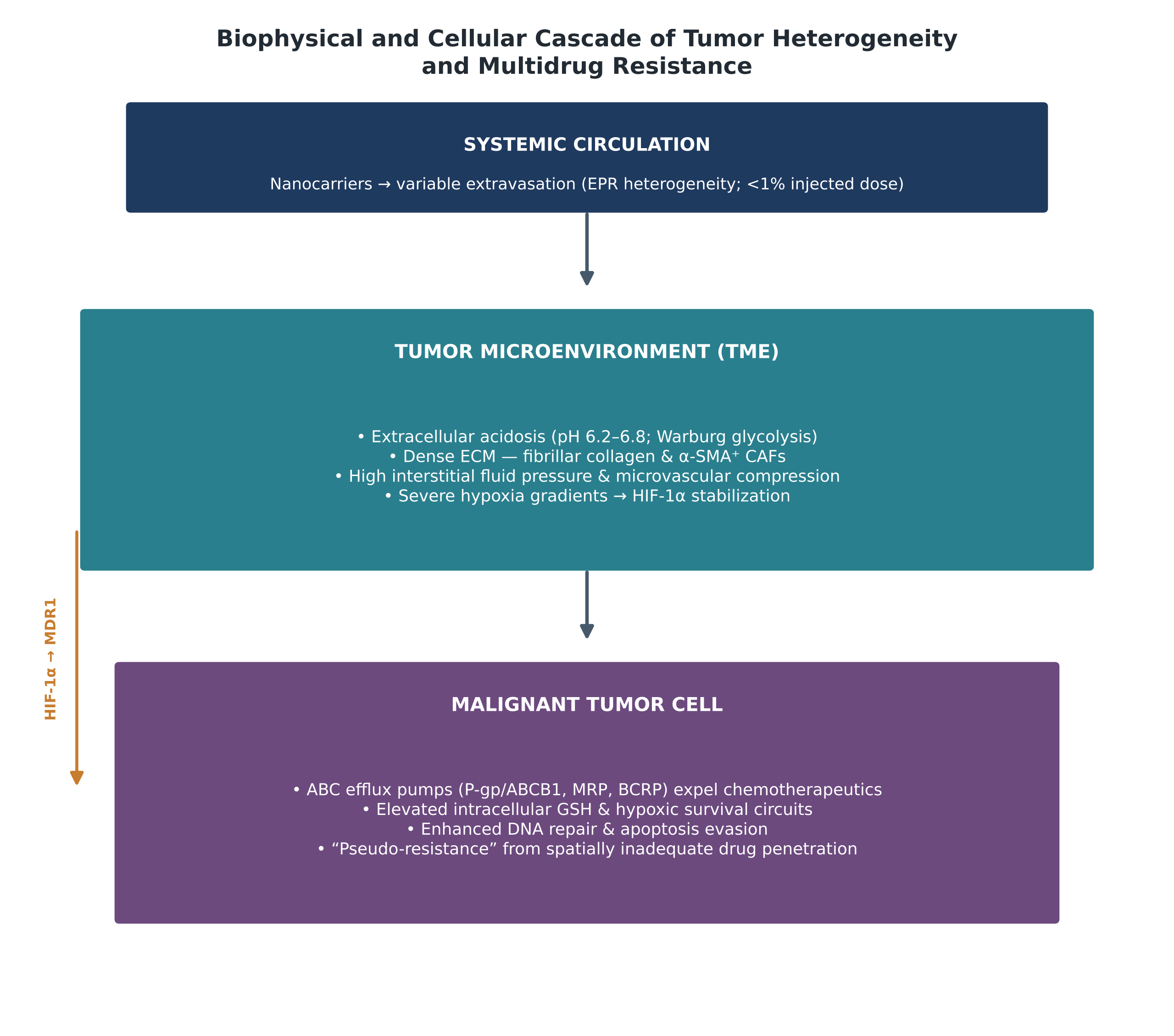
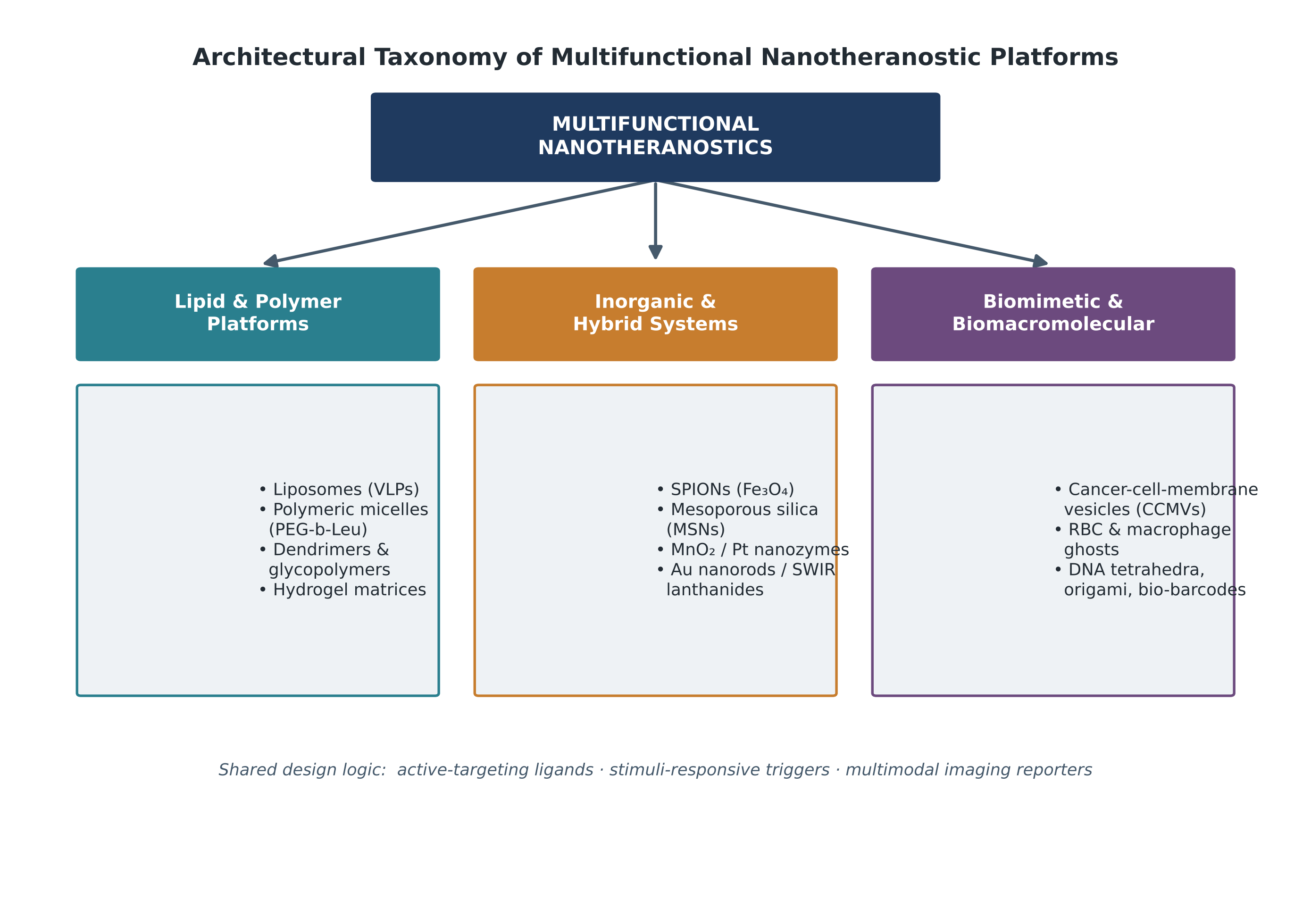
Article metrics
View details
0
Downloads
0
Citations
18
Views

View Dimensions


View Plumx


View Altmetric


0
Save
0
Citation
18
View
0
Share