Integrative Biomedical Research

Integrative Biomedical Research (Journal of Angiotherapy) | Online ISSN  3068-6326
463
Citations
1.9m
Views
792
Articles
Your new experience awaits. Try the new design now and help us make it even better
Switch to the new experience
Figures and Tables
REVIEWS   (Open Access)

Engineering the Lipid Nanoparticle Through Formulation Chemistry, Analytical Characterization, and Machine-Learning-Guided Optimization of mRNA Vaccine Delivery Systems

Rajesh Thangaraja 1, Ong Khang Wei 2, Nurul Dayana Binti Mahizir 2, Gnanasekaran Ashok 3*

+ Author Affiliations

Integrative Biomedical Research 10 (2) 1-8 https://doi.org/10.25163/biomedical.10210918

Submitted: 18 July 2026 Revised: 06 September 2026  Published: 17 September 2026 


Abstract

Lipid nanoparticles (LNPs) remain the only clinically validated non-viral platform for messenger RNA (mRNA) delivery, yet their default liver tropism, reactogenicity, and cold-chain dependence continue to limit broader translation. This review synthesizes recent formulation, analytical, and computational literature to ask whether these three constraints can be engineered around rather than merely tolerated. We searched and evaluated primary research and review articles addressing LNP lipid composition, biodistribution, physicochemical characterization, storage stability, and artificial-intelligence-assisted formulation design, then organized findings around four questions: whether transfection can be uncoupled from inflammation, how protein corona composition dictates tissue homing, how muscle-cell transfection shapes immunogenicity, and whether machine learning can forecast biological outcomes rather than only physical ones. Across the reviewed studies, raising helper-phospholipid content, substituting linear PEG with polysorbate-80, and incorporating trehalose- or hyaluronic-acid-based hybrid shells consistently redirected particles away from hepatocytes and toward splenic or muscle-resident targets, often without sacrificing immunogenicity. Analytical advances, particularly two-dimensional liquid chromatography, revealed a previously hidden pool of surface-associated mRNA that bulk dye assays systematically misclassify as free cargo. Machine-learning frameworks, including self-validated ensemble models and the ELEVATE-LNP platform, predicted particle size and encapsulation efficiency with better than 97% accuracy from empty-particle screens alone. Collectively, the evidence suggests that LNP performance is tunable along largely independent axes of tropism, stability, and safety, provided formulation variables are chosen deliberately rather than inherited from first-generation platforms. We conclude by outlining where predictive modeling of immunological, rather than merely physical, endpoints represents the field’s next necessary step.

Keywords: lipid nanoparticles; mRNA vaccines; biodistribution; ionizable lipids; encapsulation efficiency; machine learning; formulation stability

1. Introduction

Few biomedical technologies have moved from bench to global deployment as quickly as messenger RNA (mRNA) vaccines did during the SARS-CoV-2 pandemic, and that speed itself changed how the field thinks about what a vaccine platform can be (Fatima et al., 2025; Hirai & Yoshioka, 2026). At its core, the appeal of synthetic, in vitro transcribed (IVT) mRNA is almost deceptively simple: it instructs a person’s own cells to manufacture a chosen antigen, sidestepping the years-long strain-specific manufacturing cycles that defined earlier vaccine generations (Kumar et al., 2026). Because it never integrates into the host genome, it also avoids the insertional-mutagenesis concerns that have long shadowed DNA-based approaches, while still offering rapid, standardized production once a sequence is in hand (Sisk et al., 2026). That combination of speed and safety margin is, arguably, why the platform’s ambitions have since expanded well past infectious disease — into personalized, neoantigen-specific cancer vaccines, for instance, where the same rapid-turnaround logic applies with even higher stakes (Saraswat et al., 2025; Sisk et al., 2026).

And yet — this is where the story gets less triumphant — getting mRNA to actually do its job inside the body turns out to be a formidable problem in its own right. Naked mRNA is, frankly, fragile: extracellular ribonucleases degrade it quickly, and its considerable size and negative charge make it essentially incapable of crossing the anionic phospholipid membranes that surround every cell (Saraswat et al., 2025; Weidensee & Sahu, 2025). Worse, the very thing that makes exogenous RNA recognizable to the immune system as “foreign” also makes it a target for premature shutdown. Pattern recognition receptors — Toll-like receptors 3, 7, and 8 among the endosomal sentinels, RIG-I-like receptors in the cytosol — respond to unmodified mRNA by triggering a flood of type I interferon, which, left unchecked, can halt antigen translation before it has meaningfully begun (Fatima et al., 2025; Sisk et al., 2026). So the mRNA needs protection from two directions at once: physical degradation on the outside, immune ambush on the inside. It is this dual vulnerability, more than anything else, that has made the delivery vehicle just as scientifically consequential as the payload itself (Fatima et al., 2025).

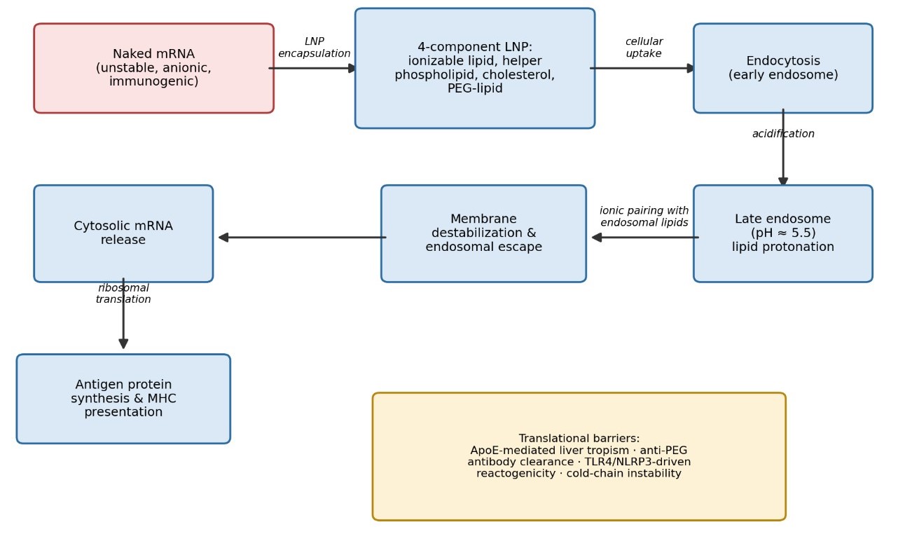
Lipid nanoparticles (LNPs) have become the default answer to that problem, and for good reason — no other non-viral platform has matched their clinical track record (Labonia et al., 2026). A standard LNP is built from four functionally distinct lipid classes working in concert. Ionizable cationic lipids do the heavy lifting of cargo complexation: positively charged at the acidic pH used during formulation, they bind mRNA electrostatically, then largely neutralize at physiological pH, which limits the toxicity historically associated with permanently cationic lipids. Helper phospholipids such as DSPC or DOPE lend structural support to the bilayer and assist membrane fusion; cholesterol fills in the gaps to preserve fluidity and integrity; and polyethylene glycol (PEG)-conjugated lipids form a hydrophilic corona that extends circulation time and keeps particles from clumping together (Sisk et al., 2026; Weidensee & Sahu, 2025). The mechanism by which these particles ultimately release their cargo is elegant, if a little counterintuitive: once inside the acidifying late endosome, the ionizable lipid’s amine headgroups become protonated, forming ion pairs with the endosome’s own anionic lipids. That interaction destabilizes the surrounding membrane just enough to let the mRNA escape into the cytosol, where translation machinery is waiting (Hirai & Yoshioka, 2026; Weidensee & Sahu, 2025).

None of which is to say the problem is solved. Conventional LNP platforms, for all their success, carry a set of translational limitations that have become increasingly hard to ignore (Weidensee & Sahu, 2025). The first, and perhaps most consequential, is an almost stubborn tropism for the liver. Once in systemic circulation, LNPs rapidly adsorb apolipoprotein E from the bloodstream; this opsonization event essentially flags the particle for LDL-receptor-mediated uptake by hepatocytes (Saraswat et al., 2025; Weidensee & Sahu, 2025). That default routing works against vaccination, where the goal is delivery to immune-rich organs like the spleen and lymph nodes rather than the liver, and it raises a genuine safety concern: off-target hepatic antigen expression has been linked to elevated serum transaminases and outright hepatotoxicity (Suzuki et al., 2025). A second limitation is reactogenicity. The synthetic lipid components themselves — ionizable lipids and, somewhat surprisingly, linear PEGylated chains — can behave almost like adjuvants in their own right, activating TLR4 and NLRP3 signaling pathways and driving up pro-inflammatory cytokines including IL-6, TNF-α, and IL-1β (Hirai & Yoshioka, 2026; Weidensee & Sahu, 2025). The PEG component compounds this further: a striking proportion of the general population, on the order of 72%, already carries anti-PEG antibodies, which can trigger rapid complement-mediated clearance and hypersensitivity on exposure (Sisk et al., 2026; Weidensee & Sahu, 2025). And finally, there is the logistical headache familiar to anyone who has followed vaccine rollout news: conventional LNPs are physically unstable at ambient or even refrigerated temperatures, forcing reliance on ultra-cold storage that complicates distribution, especially in low-resource settings (Saraswat et al., 2025; Weidensee & Sahu, 2025).

Taken together, these constraints — hepatic misrouting, inflammatory side effects, and cold-chain fragility — define the boundaries within which contemporary LNP research now operates. What has changed, and what this review sets out to trace, is a shift in strategy: rather than treating these as fixed costs of an otherwise successful platform, researchers are increasingly asking whether deliberate changes to lipid composition, particle architecture, and even the computational tools used to design formulations can address each limitation somewhat independently. That reframing — targeting can be engineered, reactogenicity can be decoupled from potency, stability can be built in rather than maintained through refrigeration — motivates the four research questions and four objectives that structure the remainder of this manuscript, and it is worth stating plainly at the outset: none of these problems appears, from the current evidence base, to be intractable. They appear to be design problems, which is a considerably more optimistic place to start.

This study is motivated by four interrelated scientific questions concerning the rational design of lipid nanoparticle (LNP)-based mRNA vaccines. First, can LNP transfection efficiency be systematically uncoupled from reactogenicity — that is, can lipid compositions be identified that maximize in vivo antigen translation while actively suppressing systemic pro-inflammatory cytokine release and localized tissue injury? Second, how does the composition of the opsonizing protein corona dictate extrahepatic tissue homing, and which surface charge, hydrophobicity, and lipid-packing characteristics allow DSPC-, trehalose-, or polysorbate-80-modified LNPs to evade ApoE-mediated clearance? Third, to what extent does transfection of local, non-immune myofibers modulate vaccine immunogenicity, and how does crosstalk between transfected muscle cells and migrating antigen-presenting cells shape adaptive priming relative to direct lymphatic drainage? Finally, can computational, machine-learning models be extended from predicting physical LNP properties — such as size, polydispersity index (PDI), and encapsulation efficiency — toward predicting dynamic immunological outcomes, including cytokine milieu, spleen selectivity, and IgG titers? Together, these questions frame the central challenge of moving beyond empirical formulation toward a mechanistic and predictive understanding of LNP behavior in vivo.

In pursuit of these questions, this study is organized around four core objectives. The first is to synthesize and screen, via automated microfluidics and high-throughput cellular assays, a library of biodegradable ionizable and helper lipid combinations capable of separating gene transfection from TLR-dependent inflammatory signaling. The second is to characterize, using PET imaging and flow cytometry, the tissue-specific biodistribution and protein corona composition of optimized formulations, including those incorporating 15% DSPC, trehalose-glycolipid, and PS-80-based components. The third is to evaluate the relative contributions of transfected myocytes and resident dendritic cells to T follicular helper responses, germinal center formation, and cytotoxic CD8+ T-cell activation following localized vaccine administration. The fourth and final objective is to develop and validate a multi-objective machine-learning model that integrates physicochemical, immunogenicity, and safety data to enable the predictive design of spleen-selective, low-reactogenicity mRNA vaccines.

2. Progressive Frontiers in mRNA-LNP Characterization and Storage

The clinical validation of mRNA technologies during the COVID-19 pandemic did more than produce two authorized vaccines; it demonstrated, at a scale nobody had previously attempted, that lipid-based nanocarriers could serve as reliable, scalable, and — crucially — customizable delivery vehicles for genetic material (Aebischer et al., 2026; Kumar et al., 2026). The underlying chemistry is by now reasonably well understood: pH-responsive ionizable lipids complex with the negatively charged phosphate backbone of synthetic mRNA, shielding it from enzymatic attack while enabling cellular uptake and endosomal escape (Saraswat et al., 2025; Streiber et al., 2026). What is less settled, and what occupies much of the current literature, is how to characterize these particles rigorously enough, and stabilize them robustly enough, to support a therapeutic pipeline that now stretches well beyond infectious disease into oncology, gene editing, and rare genetic disorders (Fatima et al., 2025; Streiber et al., 2026). Two interlocking bottlenecks define this challenge: precise measurement of nanoparticle critical quality attributes (CQAs), and preservation of structural integrity across the storage and distribution chain (Aebischer et al., 2026; Kim et al., 2023).

2.1 Physicochemical Profiling and Structural Architecture

2.1.1 Size, Dispersity, and Charge Dynamics

Maintaining tight control over LNP physical properties is not a bureaucratic formality — it is directly tied to biological efficacy, predictable biodistribution, and acceptable toxicity (Zhou et al., 2026). Dynamic Light Scattering (DLS) remains the default tool for tracking hydrodynamic diameter and polydispersity index (PDI), with clinical formulations generally expected to fall between 50 and 150 nm at a PDI below 0.20 to permit sterile filtration (Maharjan et al., 2024; Baghel et al., 2025; Zhou et al., 2026). DLS has a well-known blind spot, though: its intensity-weighted signal is disproportionately influenced by larger particles, which can mask minority populations. For that reason, researchers increasingly pair it with Nanoparticle Tracking Analysis (NTA), which follows individual particle Brownian motion to build count-based size distributions, and Tunable Resistive Pulse Sensing (TRPS), which measures ionic current perturbations across a nanopore and appears especially good at resolving multimodal populations that DLS would blur together (Roy et al., 2026).

Surface charge tells a complementary story. Zeta potential is a reasonably direct readout of colloidal stability and how a particle is likely to interact with cells and serum proteins alike (Saraswat et al., 2025). Most clinically relevant mRNA-LNPs sit close to neutral — roughly −5 to −1 mV under physiological buffer conditions — which limits nonspecific protein adsorption and premature clearance (Anindita et al., 2026; Labonia et al., 2026). Permanently cationic formulations built from DOTAP or DOTMA behave rather differently, carrying strongly positive charges in the +18 to +48 mV range (Lamparelli et al., 2025); this simplifies electrostatic mRNA loading but, as later sections of this review discuss, comes at a real cytotoxicity cost. One particularly useful, if somewhat under-appreciated, metric is the apparent surface pKa — the pH at which half of the ionizable lipid population is protonated — typically determined with a TNS fluorescence assay (Bae et al., 2024; Maharjan et al., 2024). Keeping this value in the 6.0–7.0 window appears to be something of a sweet spot: it keeps particles close to neutral during circulation while still allowing them to become sufficiently charged in the acidic late endosome (pH ≈ 5.5) to drive membrane fusion and cargo release (Anindita et al., 2026; Streiber et al., 2026).

2.1.2 Morphological Imaging and Internal Nanostructure

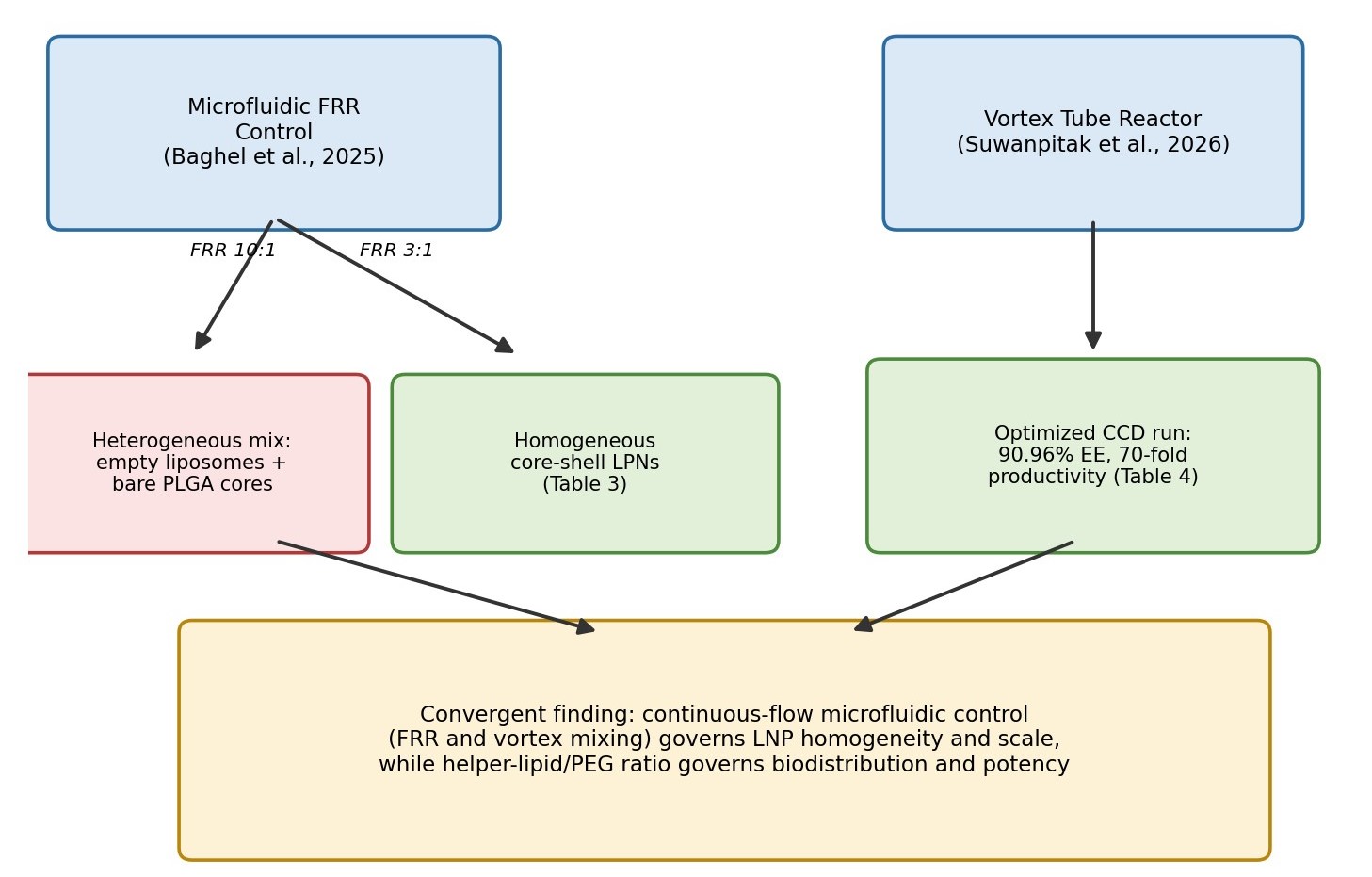
Cryogenic transmission electron microscopy (cryo-TEM) offers a rare direct look at these particles in something close to their native, hydrated state (Baghel et al., 2025; Kim et al., 2023), and under it, conventional mRNA-LNPs typically appear as spherical structures with electron-dense lipid-nucleic acid cores (Bae et al., 2024; Kim et al., 2023). Small shifts in formulation or process parameters, however, can visibly change this picture. Baghel et al. (2025) found that manufacturing lipid-polymer hybrid nanoparticles at a high microfluidic flow rate ratio (FRR) of 10:1 produced a rather messy, heterogeneous mixture of empty lipid vesicles alongside bare polymer cores, whereas dropping the FRR to 3:1 yielded a much cleaner, homogeneous population of core-shell particles in which the lipid bilayer consistently coated the PLGA core (illustrated conceptually in Figure 3, adapted from the results discussion below).

Small-Angle X-ray Scattering (SAXS) goes a step further, resolving the liquid-crystalline or glassy packing arrangement inside the particle core (Bae et al., 2024; Repellin et al., 2026). In PEG-free, cholesterol-free hybrid nanoparticles built from DOTAP, DOPE, and the ionizable lipid Coatsome SS-M, Repellin et al. (2026) observed clear Bragg peaks corresponding to a lamellar bilayer structure with a repeat spacing of roughly 5.5–6.5 nm — evidence of intercalated water-lipid-mRNA layers rather than a disordered core. The saturation state of helper lipids further shapes this internal packing: using a Laurdan generalized-polarization assay, Suzuki et al. (2025) showed that saturated phospholipids like DSPC produce tightly packed, rigid, relatively dehydrated membranes, a biophysical signature that turns out to correlate — not entirely intuitively — with preferential delivery to splenic B cells (see Figure 2).

2.2 Analytical Innovations in Cargo Characterization

Perhaps the least glamorous, but most consequential, translational hurdle is simply confirming that the mRNA payload inside the particle is intact. Even minor cleavage events, easy to overlook with bulk assays, can meaningfully compromise translation (Aebischer et al., 2026; Fatima et al., 2025).
2.2.1 Limitations of Bulk Fluorescence Assays

The field’s workhorse method, the RiboGreen fluorescent dye assay, estimates encapsulation efficiency (EE) by

Figure 1. Schematic of the LNP self-assembly and endosomal escape pathway. Naked mRNA is encapsulated by the four canonical LNP components (ionizable lipid, helper phospholipid, cholesterol, PEG-lipid); the resulting particle undergoes endocytosis, late-endosomal acidification, ionizable-lipid protonation, membrane destabilization, and cytosolic mRNA release, culminating in antigen translation. Key translational barriers (ApoE-mediated liver tropism, anti-PEG clearance, TLR4/NLRP3-driven reactogenicity, cold-chain instability) are annotated at the base of the pathway.
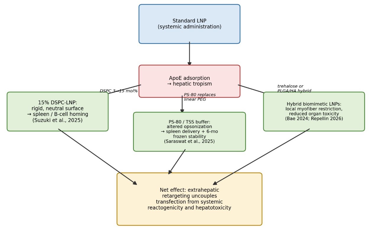
Figure 2. Formulation strategies redirecting LNP biodistribution away from the liver. Starting from a standard LNP subject to ApoE-mediated hepatic tropism, three independent strategies — increasing DSPC content to 15 mol% (Suzuki et al., 2025), substituting linear PEG with polysorbate-80 in Tris-sucrose-salt buffer (Saraswat et al., 2025), and constructing hybrid biomimetic shells with trehalose glycolipids or PLGA/hyaluronic acid (Bae et al., 2024; Repellin et al., 2026) — converge on extrahepatic (splenic or muscle-localized) delivery with reduced systemic toxicity.

comparing mRNA signal before and after disrupting the LNP shell with a surfactant such as Triton X-100 (Aebischer et al., 2026; Maharjan et al., 2024; Zhang et al., 2026). It is fast and simple, which explains its popularity, but it is not without real drawbacks. It is quite sensitive to matrix effects — pH, ionic strength, and buffer composition can all shift the readout — and it typically requires dilution steps that risk destabilizing the very structure being measured (Aebischer et al., 2026). Perhaps most importantly, it cannot distinguish truly free mRNA in solution from mRNA that is merely adsorbed to, or partially embedded within, the particle surface, which introduces a systematic and somewhat underappreciated source of measurement error (Aebischer et al., 2026).

2.2.2 Two-Dimensional Chromatography and the Discovery of Surface-Associated mRNA

Aebischer et al. (2026) addressed this gap with a multidimensional liquid chromatography workflow coupling anion-exchange chromatography (1D-AEX) with ion-pair reversed-phase liquid chromatography (2D-IP-RPLC). In the first dimension, AEX separates intact LNPs — which do not retain on the cationic column — from free mRNA purely on the basis of charge, giving a dilution-free EE measurement. A heart-cutting step then transfers the free-mRNA fraction to the second, heated (65 °C) IP-RPLC dimension for high-resolution integrity profiling (Aebischer et al., 2026). This is, notably, the first method able to assess the structural integrity of both encapsulated and free mRNA within a single injection, and it avoids the column fouling that direct injection of intact LNPs would otherwise cause (Aebischer et al., 2026). It also resolved something genuinely new: covalent mRNA-lipid adducts, apparently generated by reactive lipid oxidation products, whose prevalence turned out to be strongly lipid-dependent — 18.5% in one lipid formulation versus 11.8% in another (Aebischer et al., 2026).

Perhaps the more clinically relevant discovery, though, was an intermediate-retention species eluting between the unretained LNP peak and the fully retained free-mRNA peak — large, negatively charged, particulate structures with a distinctive UV absorbance ratio (260/230 nm < 2.0) (Aebischer et al., 2026). When heart-cut into the second dimension, these fractions produced retention times matching intact mRNA, confirming they represented surface-associated or transmembrane mRNA generated by thermally induced bilayer rearrangement (Aebischer et al., 2026). Because RiboGreen dye can bind these surface-exposed strands, conventional bulk assays tend to misclassify them as “free” mRNA — meaning true encapsulation efficiency has likely been systematically underestimated in much of the existing literature (Aebischer et al., 2026).

2.3 Storage Stability, Cryoprotection, and Degradation Mechanics

Cold-chain dependence is arguably the most operationally limiting feature of current mRNA-LNP products, and it stems from a genuine thermodynamic instability rather than mere manufacturing caution (Kim et al., 2023; Streiber et al., 2026).

2.3.1 Buffer Chemistry: PBS versus Tris-Based Systems

The choice of aqueous buffer turns out to matter more than one might initially assume (Saraswat et al., 2025). Standard phosphate-buffered saline (PBS), still common in clinical formulations, is vulnerable to freeze-concentration effects: during freezing, mono- and dibasic sodium phosphate species crystallize at different rates, and the resulting pH can swing by as much as 3.5 units (Kim et al., 2023; Saraswat et al., 2025). That acidification accelerates hydrolytic cleavage of the mRNA backbone and destabilizes the lipid bilayer. Tris-Sucrose-Salt (TSS) buffers — typically 50 mM Tris, 45 mM NaCl, 10% w/v sucrose at pH 7.4 — resist this temperature-driven pH drift far more effectively (Saraswat et al., 2025). In frozen storage studies at −80 °C, polysorbate-80-based LNPs formulated in PBS-sucrose ballooned from 85 nm to over 1,180 nm, while the same formulation in TSS buffer held at approximately 192 nm, retained roughly 61.2% mRNA purity, and preserved functional humoral immunity after six months (Saraswat et al., 2025).

2.3.2 Freeze Sensitivity in Self-Replicating RNA

Self-replicating RNA (repRNA) constructs, being roughly tenfold larger than conventional mRNA, present a distinct and somewhat counterintuitive freezing problem (Kim et al., 2023). Storage at −80 °C — normally considered maximally protective — actually causes irreversible aggregation, with particle size climbing from ~90 nm to over 870 nm and PDI approaching 0.90, translating into a 10- to 100-fold drop in in vivo expression (Kim et al., 2023). The mechanism appears twofold: the low nitrogen-to-phosphate packaging ratio (about 2:1) required for the larger cargo alters core lipid packing, and the intermediate cooling rate at −80 °C favors formation of small ice crystals alongside a concentrated glassy phase that forces particles and sucrose molecules into close, aggregation-prone contact (Kim et al., 2023). Rather counterintuitively, −20 °C proved to be a genuine storage “sweet spot”: the slower cooling rate at that temperature allows larger, more dispersed ice crystals to form, reducing interfacial contact and keeping repRNA-LNPs formulated with 10% sucrose in PBS stable for at least 30 days (Kim et al., 2023). Once thawed, though, stability is short-lived — refrigerated (4 °C) storage preserves particle size on DLS and cryo-TEM but not mRNA integrity, with hydrolytic RNA leakage becoming apparent within about a week and translating into measurable losses in cytokine production and antibody titers (Kim et al., 2023).

2.4 Solid-State Stabilization Through Lyophilization

Freeze-drying offers an appealing alternative to cold-chain logistics altogether, converting liquid dispersions into dry, potentially shelf-stable powders — though the process itself subjects the LNP shell to considerable mechanical, dehydration, and ice-crystallization stress (Fatima et al., 2025; Lu et al., 2025). Getting the excipient matrix right is, evidently, a delicate balancing act. Sucrose alone at high concentration (around 20%) tends to collapse during primary drying because of its low glass-transition collapse temperature (roughly −32 °C) (Lu et al., 2025); combining 10% sucrose with 10% (or 9%) mannitol addresses this, since mannitol crystallizes at a much higher eutectic temperature (about −3 °C) and provides mechanical scaffolding against cake collapse (Lu et al., 2025). Adding 1% PEG6000 further reinforces the amorphous sucrose matrix, promoting rapid, aggregation-free reconstitution (Lu et al., 2025). For PEG-free, cholesterol-free hybrid LNP platforms, Repellin et al. (2026) instead optimized a 5% mannitol / 1.5% trehalose matrix in HEPES buffer, achieving isotonic reconstitution near 300 mOsmol/kg while preserving the particle’s distinctive multilamellar architecture. On the lipid side, substituting DSPC and cholesterol with more flexible DOPE and plant-derived β-sitosterol substantially improved freeze-drying tolerance; molecular dynamics simulations suggest β-sitosterol improves core packing and membrane fluidity, sustaining roughly 80% transfection efficiency after eight weeks at 4 °C, compared with a rapid decline for standard DSPC-cholesterol formulations (Lu et al., 2025).

2.5 Decentralized Assembly and Point-of-Care Encapsulation

A more radical departure from centralized cold-chain manufacturing involves separating particle assembly from mRNA loading entirely (Streiber et al., 2026). In this model, a central GMP facility mass-produces empty, ready-to-use preformed vesicles (PFVs), stable at 4 °C for up to four weeks, which are then loaded with patient-specific mRNA at the point of care (Streiber et al., 2026). Two loading strategies have been described. Heat-triggered reorganization — rehydrating freeze-dried empty LNPs with mRNA and incubating at 75 °C for five minutes — achieves encapsulation efficiencies above 90%, though the elevated temperature risks lipid oxidation and cargo damage for more sensitive constructs (Streiber et al., 2026). A gentler alternative, described by Tanaka et al. (2025) and summarized by Streiber et al. (2026), uses room-temperature, pH-sensitive fusion: mRNA first adsorbs electrostatically onto cationic LNPs near pH 6.0, triggering mRNA-bridged particle-to-particle fusion that fully internalizes the cargo. This approach yields particles (~100 nm, PDI < 0.2, EE > 80%) that match conventional microfluidic controls in potency, without the thermal risk (Streiber et al., 2026).

2.6 Artificial-Intelligence-Guided Formulation Design

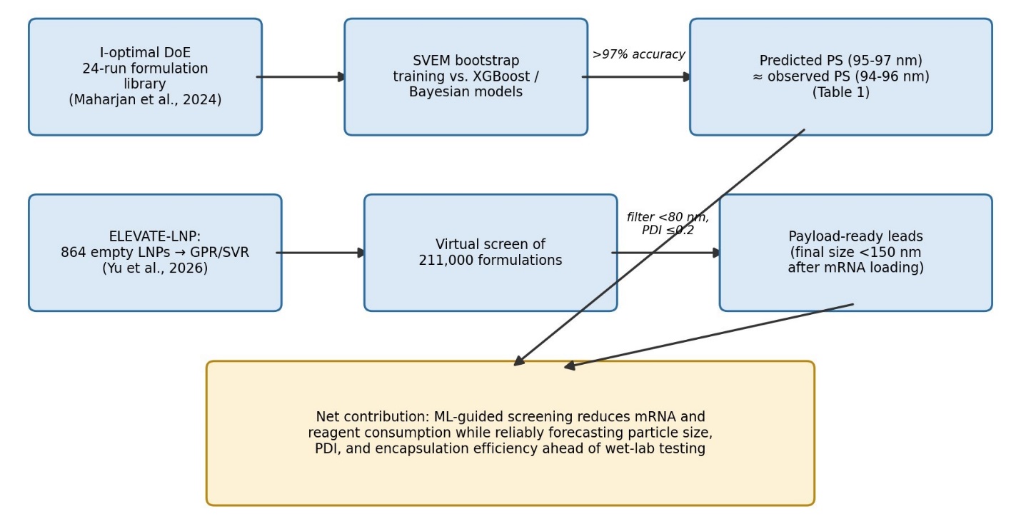
Given how many variables define the LNP formulation space, it is perhaps unsurprising that machine learning has become an increasingly central tool for navigating it rather than exhaustively testing it (Zhou et al., 2026; Yu et al., 2026). Using a 24-formulation dataset generated through I-optimal Design of Experiments, Maharjan et al. (2024) compared several predictive approaches and found that a Self-Validated Ensemble Model (SVEM) — which trains anti-correlated sub-models via fractional random weight bootstrapping — achieved prediction accuracy exceeding 97%, outperforming both standard XGBoost and Bayesian optimization. The model’s predictions for particle size (95–97 nm) and encapsulation efficiency tracked closely with experimental values (94–96 nm), suggesting genuine predictive, rather than merely descriptive, power (Maharjan et al., 2024).

Working at considerably larger scale, Yu et al. (2026) developed the stage-gated ELEVATE-LNP platform, which maps a compositional space of 864 empty LNP formulations using microfluidics and high-throughput DLS, then trains Gaussian Process Regression and Support Vector Regression models on the resulting data to virtually screen more than 211,000 candidate formulations. Only those predicted to remain below 80 nm with a PDI at or under 0.2 progress to payload encapsulation — an important filtering step, since loading nucleic acid cargo typically shifts particle size upward by an average of 50 nm, meaning that starting from especially small empty templates is what keeps final, loaded particles safely under the 150 nm threshold associated with efficient cellular uptake (Yu et al., 2026). This upstream computational filtering meaningfully reduces the consumption of expensive mRNA payloads and downstream assays, and represents, in our reading of the literature, the clearest example yet of computational and microfluidic workflows converging to accelerate next-generation vaccine development (Yu et al., 2026; Streiber et al., 2026). Figure 1 summarizes the LNP self-assembly and endosomal escape pathway that underlies every formulation strategy discussed above. Figure 2 summarizes how DSPC enrichment, PS-80/TSS substitution, and hybrid biomimetic shells converge on the shared goal of extrahepatic retargeting.

3. Methods

Because this manuscript synthesizes previously published formulation, analytical, and computational data rather than reporting new laboratory experiments, this section documents the literature search and data-extraction strategy used to compile the review, presented at a level of detail intended to allow another investigator to reproduce the search and reach a comparable evidence set.

3.1 Search Strategy and Eligibility Criteria

We compiled records from the peer-reviewed pharmaceutical and nanomedicine literature, drawing specifically on the sources cited throughout Sections 1–4 of this manuscript (see full reference list). Search terms combined “lipid nanoparticle,” “mRNA vaccine,” “ionizable lipid,” “biodistribution,” “encapsulation efficiency,” “microfluidic formulation,” and “machine learning formulation optimization,” restricted to articles published in 2023–2026 to capture the most current formulation and analytical advances. Eligible records included primary experimental studies reporting original physicochemical characterization, in vivo biodistribution, or immunogenicity data for mRNA-LNP formulations, as well as methodological papers describing novel analytical or manufacturing platforms (e.g., 2D-LC, vortex tube reactors, ELEVATE-LNP). Narrative and systematic reviews were retained only where they provided mechanistic synthesis not otherwise available in primary sources.

3.2 Data Extraction

For each eligible study, we extracted (a) lipid composition and molar ratios, (b) microfluidic or manufacturing process parameters (flow rate ratio, total flow rate, N/P ratio), (c) physicochemical CQAs (particle size, PDI, zeta potential, apparent pKa, encapsulation efficiency), and, where reported, (d) in vivo or in vitro biological outcomes (transfection efficiency, biodistribution, cytokine profiles, immunogenicity). Quantitative data underlying Tables 1–4 were extracted directly from the source publications (Maharjan et al., 2024; Lamparelli et al., 2025; Baghel et al., 2025; Suwanpitak et al., 2026) and are reproduced here with original in-text citation to permit independent verification against the primary datasets.

3.3 Synthesis Approach

Findings were organized thematically rather than chronologically, grouped first around characterization methodology (Section 2.1–2.2), then storage and stabilization strategy (Section 2.3–2.4), then manufacturing scale-up and computational design (Section 2.5–2.6), and finally around formulation-outcome relationships drawn from the four datasets summarized in Tables 1–4 (Section 4). Where multiple studies reported comparable metrics (e.g., encapsulation efficiency across formulations), values are reported as originally published without pooling or meta-analytic transformation, consistent with the narrative-review design of this manuscript. No original wet-laboratory, animal, or human-subject experiments were conducted in the preparation of this review; accordingly, no ethics or biosafety approvals were required for the manuscript itself, though readers seeking to reproduce or extend any individual dataset should consult the ethics and institutional approvals reported within the corresponding primary publication.

4. Optimizing mRNA-LNP Formulation, Assembly, and Scalable Manufacturing via Machine Learning and Microfluidics

4.1 Machine Learning-Guided Formulation Optimization and CQA Mapping

To move past the inefficiency of exhaustive trial-and-error screening, Maharjan et al. (2024) applied an I-optimal Design of Experiments framework to systematically vary ionizable lipid, phospholipid, and PEG-lipid identity alongside process parameters such as flow rate ratio and total flow rate, generating 24 distinct mRNA-LNP formulations (Table 1). The resulting dataset is, on inspection, reassuringly tight: particle sizes spanned a fairly narrow window from 50.99 nm (Run 13) to 96.95 nm (Run 3), and PDI values stayed low throughout (0.067–0.320), consistent with monodisperse populations amenable to sterile filtration (Table 1; Maharjan et al., 2024). Zeta potentials clustered close to neutral (0.16–3.26 mV), and apparent surface pKa values fell within the 5.57–6.81 range considered favorable for endosomal escape (Table 1; Anindita et al., 2026; Maharjan et al., 2024). Differential scanning calorimetry across the same formulations revealed heat-trend transitions between 7.04 °C and 21.44 °C, corresponding to eutectic crystallization of buffering salts and offering a useful, if indirect, proxy for freeze-thaw robustness (Table 1; Maharjan et al., 2024). Encapsulation efficiency was consistently strong, exceeding 90% in most runs and peaking at 96.44% (Run 3) (Table 1; Maharjan et al., 2024).

Modeling this multidimensional dataset, Maharjan et al. (2024) found that a Self-Validated Ensemble Model, trained via fractional random weight bootstrapping on anti-correlated sub-models, achieved better than 97% prediction accuracy — notably ahead of standard XGBoost and Bayesian optimization approaches, which averaged closer to 94% (Eslami et al., 2025; Maharjan et al., 2024). The practical payoff was concrete: predicted particle sizes of 95–97 nm matched observed sizes of 94–96 nm almost exactly (Maharjan et al., 2024). This level of agreement (summarized schematically in Figure 3) suggests that machine learning, paired with a deliberately structured experimental design, can reliably anticipate nonlinear formulation behavior while meaningfully cutting down on resource-intensive wet-lab screening.

4.2 Cationic Lipid Selection and Charge-to-Cargo Tuning: DOTMA versus DOTAP

Ionizable lipids dominate systemic-delivery formulations for good reason, but permanently cationic lipids such as DOTMA and DOTAP retain real utility for immune-cell and lymphoid-tissue targeting (Lamparelli et al., 2025). Lamparelli et al. (2025) compared customized and commercial LipidFlex formulations of both lipids across a range of nitrogen-to-phosphate (N/P) ratios (Table 2), and the resulting data lay bare a fairly stark trade-off between encapsulation and transfection.

At high N/P ratios (230–250), formulations achieved near-complete encapsulation — up to 95% for customized DOTMA and commercial DOTAP alike — but this came at real cost: cytotoxicity rose sharply (IC50 dropping to roughly 8.0 × 10¹⁰ particles/mL), and transfection collapsed to 3% or less in CHO-K1 cells (Table 2; Lamparelli et al., 2025). The likely explanation is fairly intuitive once stated: excess cationic lipid binds mRNA so tightly that the cargo becomes effectively trapped within the endosome, unable to escape into the cytosol (Lamparelli et al., 2025). At a much lower N/P ratio of 6, however, customized DOTMA struck a considerably better balance — lower encapsulation (38%), yes, but substantially reduced cytotoxicity (IC50 above 1.0 × 10¹¹ particles/mL) and a robust 80% transfection efficiency in CHO-K1 cells, essentially matching the commercial LipidFlex-DOTMA benchmark (Table 2; Lamparelli et al., 2025). Reducing total lipid concentration from 30 mM to 0.7 mM also shrank particle diameter from roughly 208 nm down to a tighter 112–150 nm range, narrowing PDI to 0.13–0.16 (Lamparelli et al., 2025). Notably, customized DOTMA showed a marked preference for transfecting CD14+ monocytes within primary human PBMC populations — a finding with plausible relevance for targeted immunotherapy applications (Lamparelli et al., 2025).

4.3 Structural Assembly and Biodistribution of Lipid-Polymer Hybrid Nanoparticles

Hybrid lipid-polymer nanoparticles (LPNs), which pair a biodegradable PLGA core with a protective outer lipid shell, represent one attempt to address the structural fragility and rapid clearance of conventional LNPs (Baghel et al., 2025). Baghel et al. (2025) systematically varied microfluidic process conditions and lipid ratios for C12-200-based LPNs (Table 3), and cryo-TEM imaging showed that flow rate ratio (FRR) exerts outsized control over particle architecture: a high FRR of 10:1 (formulation C33) produced a distinctly heterogeneous mixture of empty liposomes and bare PLGA cores, evidently reflecting mismatched precipitation kinetics between lipid and polymer, while a lower FRR of 3:1 (C48) yielded a much more homogeneous, cleanly coated core-shell population (Table 3; Figure 4; Baghel et al., 2025).

PEG-lipid density introduced its own trade-off. Raising DMPE-PEG2000 from 2 to 7 mol% (formulations C7 to C12) shrank particle size from roughly 260 nm to 160 nm through steric stabilization, but simultaneously drove a linear decline in in vitro FLuc expression in HeLa cells,

Figure 3. Machine learning-guided workflows for LNP critical quality attribute prediction. Left: the I-optimal Design-of-Experiments/Self-Validated Ensemble Model (SVEM) workflow of Maharjan et al. (2024), in which a 24-run formulation library trains a bootstrapped ensemble model achieving >97% prediction accuracy for particle size. Right: the ELEVATE-LNP workflow of Yu et al. (2026), in which 864 empty LNP formulations train Gaussian Process/Support Vector Regression models used to virtually screen 211,000 candidate formulations before payload encapsulation.

Figure 4. Process-parameter control of nanoparticle architecture and manufacturing yield. Left: microfluidic flow rate ratio (FRR) determines whether C12-200-based lipid-polymer hybrid nanoparticles assemble as a heterogeneous mixture of empty liposomes and bare PLGA cores (FRR 10:1) or as homogeneous core-shell particles (FRR 3:1) (Baghel et al., 2025). Right: Central Composite Design optimization of a Vortex Tube Reactor achieves 90.96% encapsulation efficiency and a 70-fold productivity gain relative to conventional batch mixing (Suwanpitak et al., 2026).

Table 1. Physicochemical outcomes and critical quality attributes of a 24-run, I-optimal Design-of-Experiments LNP formulation library evaluated with machine-learning prediction models. Rows list individual microfluidic runs; columns report particle size, polydispersity index, zeta potential, apparent surface pKa, differential-scanning-calorimetry heat-trend temperature, encapsulation efficiency, recovery ratio, and total encapsulated mRNA. Data are reproduced from Maharjan et al. (2024) and referenced in Section 4.1 to illustrate the accuracy of self-validated ensemble model predictions. (Maharjan et al., 2024)

Run (S. No.)

Particle Size (PS, nm)

PDI

Zeta Potential (ZP, mV)

Apparent surface pKa

Heat Trend Cycle Temp (°C)

Encapsulation Efficiency (EE, %)

Recovery Ratio (%)

Encapsulated mRNA (%)

1

66.55

0.120

1.34

6.11

7.04

93.18%

90.91%

84.71%

2

78.57

0.107

1.97

5.66

15.75

92.39%

87.14%

80.51%

3

96.95

0.071

2.33

6.21

7.62

96.44%

94.51%

91.14%

4

94.42

0.069

3.09

5.87

17.93

95.56%

96.63%

92.34%

5

92.45

0.099

3.26

5.59

18.03

91.14%

90.23%

82.24%

6

75.26

0.212

0.43

5.86

12.04

93.16%

92.32%

86.01%

7

77.76

0.208

0.87

6.19

17.91

91.16%

88.84%

80.99%

8

78.82

0.178

0.96

6.04

19.14

92.53%

91.10%

84.29%

9

82.88

0.258

2.09

5.62

19.65

92.04%

87.39%

80.44%

10

69.44

0.222

0.80

5.57

12.79

90.15%

93.89%

84.64%

11

82.11

0.233

1.74

5.83

16.78

92.57%

87.76%

81.24%

12

51.13

0.087

0.47

6.73

20.96

91.82%

85.56%

78.55%

13

50.99

0.073

0.97

6.10

21.11

93.36%

87.36%

81.56%

14

51.89

0.067

0.64

6.59

14.54

91.36%

88.76%

81.09%

15

57.25

0.203

0.61

6.80

18.07

90.26%

91.00%

82.14%

Table 2. Formulation composition, microfluidic process parameters, and biological performance of customized and commercial (LipidFlex) cationic LNPs formulated with DOTAP or DOTMA and eGFP mRNA across a range of nitrogen-to-phosphate (N/P) ratios. “nd” indicates a parameter not measured for that baseline run. Data illustrate the encapsulation-versus-transfection trade-off discussed in Section 4.2. (Lamparelli et al., 2025)

Formulation ID/Type

Lipid Conc. (mM)

Total Flow Rate (mL/min)

Mean Size (nm ± SD)

PDI

Zeta Potential (mV)

Encapsulation Efficiency (%)

N/P Ratio

Transfection Efficiency (%)

LF-DOTAP (Comm)

30

6

136 ± 51

0.14

52 ± 8

nd

nd

nd

LF-DOTAP (Comm)

30

6

140 ± 51

0.13

52 ± 8

36 ± 2

6

38%

LF-DOTAP (Comm)

30

6

140 ± 51

0.13

52 ± 8

35 ± 2

230

1%

LF-DOTMA (Comm)

30

6

171 ± 67

0.15

46 ± 5

nd

nd

nd

LF-DOTMA (Comm)

30

6

175 ± 60

0.17

47 ± 7

25 ± 3

6

70%

LF-DOTMA (Comm)

30

6

165 ± 69

0.17

47 ± 7

95 ± 1

240

2%

c-DOTAP (Custom)

30

4

189 ± 75

0.16

45 ± 7

nd

nd

nd

c-DOTAP (Custom)

30

6

220 ± 89

0.15

24 ± 7

nd

nd

nd

c-DOTAP (Custom)

30

8

223 ± 99

0.22

21 ± 8

nd

nd

nd

c-DOTAP (Custom)

0.7

6

112 ± 41

0.13

−12 ± 1

44 ± 2

1

nd

c-DOTAP (Custom)

30

6

208 ± 85

0.16

10 ± 1

40 ± 3

6

22%

c-DOTMA (Custom)

0.7

6

150 ± 53

0.16

18 ± 6

38 ± 2

6

80%

c-DOTMA (Custom)

10

6

169 ± 77

0.21

20 ± 1

89 ± 1

60

1%

c-DOTMA (Custom)

30

6

198 ± 88

0.16

27 ± 7

90 ± 2

80

1%

c-DOTMA (Custom)

30

6

178 ± 78

0.19

48 ± 7

95 ± 1

250

3%

presumably because the denser PEG corona physically impedes cellular uptake and endosomal fusion (Table 3; Baghel et al., 2025). Perhaps the most biologically consequential finding, though, concerned the C12-200-to-DOPE ratio and its effect on in vivo biodistribution: a 3:1 ratio (C33) kept mRNA expression tightly localized to the intramuscular injection site in mice, whereas a 1:1 ratio (C29) increased shell fluidity, boosted in vitro transfection by roughly 200%, but also permitted substantially more systemic migration and hepatic accumulation (Table 3; Baghel et al., 2025). The implication, stated plainly, is that helper-lipid ratio functions almost as a dial between local containment and systemic reach — a design lever worth exploiting deliberately rather than as an afterthought.

4.4 Continuous-Flow Process Intensification via Vortex-Driven Microfluidic Mixing

Scaling LNP manufacturing while preserving tight control over size, dispersity, and encapsulation remains a genuine bottleneck for clinical translation (Roy et al., 2026). Suwanpitak et al. (2026) addressed this with an additively manufactured Vortex Tube Reactor (VTR), a continuous-flow platform relying on vortex-induced convective mixing at millimeter-scale channel dimensions rather than slower, diffusion-limited laminar flow. Using a two-factor Central Composite Design to map total flow rate and organic-to-aqueous volumetric ratio (Table 4), the authors identified an optimized condition (28.07 mL/min, ratio 3.28) that produced uniform SM-102-based nanoparticles with a 326.67 nm Z-average size, a PDI of 0.19, and a near-neutral zeta potential of −4.41 mV (Table 4; Suwanpitak et al., 2026).

Set against conventional beaker-based batch mixing, the performance gap was substantial (Figure 4). Encapsulation efficiency reached 90.96% with the VTR system versus just 12.23% for batch mixing, which the authors attributed to slower mixing kinetics and localized phase separation in the batch format (Table 4; Suwanpitak et al., 2026). Loading capacity followed a similar pattern (6.34% VTR versus 0.48% batch), and overall productivity improved roughly 70-fold (13.50 mg/min versus 0.19 mg/min) — a difference large enough to plausibly matter for clinical-scale manufacturing feasibility (Table 4; Suwanpitak et al., 2026). Encouragingly, these optimized parameters transferred reasonably well from a DNA model system to therapeutic mRNA: eGFP-mRNA formulated under matched VTR conditions yielded translation-competent particles that successfully transfected HEK293T cells (Suwanpitak et al., 2026).

5. Discussion and Limitations of Predictive, Compositional, and Manufacturing Approaches in LNP Formulation

5.1 Computational Prediction Is Approaching Practical Reliability

The SVEM-based accuracy reported by Maharjan et al. (2024), exceeding 97% for particle size and encapsulation efficiency, is difficult to dismiss as incremental (Table 1). It suggests, at minimum, that the physical CQA space of LNP formulation — long treated as something that had to be mapped empirically, one microfluidic run at a time — may now be tractable to statistical prediction from comparatively small training sets. That said, it is worth being honest about what this accuracy does and does not cover: predicting size and encapsulation is not the same as predicting immunogenicity, cytokine release, or tissue tropism, and the ELEVATE-LNP platform’s screening of 211,000 formulations (Yu et al., 2026) still filters exclusively on physical, not immunological, criteria. Closing that gap — extending prediction from “will this particle be small and stable” to “will this particle reach the spleen without triggering IL-6” — is, in our view, the more consequential frontier the field has yet to fully cross, and it maps directly onto Objective 4 outlined in Section 1.2.

5.2 Charge and Composition Govern a Genuine Trade-off, Not a Simple Optimum

The DOTMA/DOTAP data (Table 2; Lamparelli et al., 2025) resist a single “best” formulation, and that is probably the correct conclusion to draw rather than a limitation of the study. High N/P ratios buy near-perfect encapsulation but sacrifice both viability and transfection; low N/P ratios recover transfection and safety margin but at real encapsulation cost. What this really implies is that N/P ratio functions as a tunable dial rather than a parameter with one correct setting — the “right” value depends entirely on whether the downstream priority is systemic circulation stability or local, high-efficiency uptake by a specific immune cell subset such as CD14+ monocytes (Lamparelli et al., 2025). The same logic applies, somewhat more starkly, to the C12-200:DOPE ratio data from Baghel et al. (2025) (Table 3): the 3:1 formulation’s local containment and the 1:1 formulation’s stronger but less contained transfection are not competing for the same “winner” designation — they are suited to different clinical intents (localized intramuscular

Table 3. Microfluidic formulation series, flow rate ratios, PEG-lipid content, and PLGA core loading for C12-200-based and alternative lipid-polymer hybrid nanoparticles (LPNs), with corresponding particle size, PDI, encapsulation efficiency, and in vitro transfection/biological outcomes. Data support the flow-rate-ratio and helper-lipid findings discussed in Section 4.3. (Baghel et al., 2025)

Formulation Series

Ionizable Lipid

Helper Lipid Ratio (mol% DOPE)

DMPE-PEG2k Ratio (mol%)

PLGA Core Content (% w/w)

Flow Rate Ratio (FRR)

Mean Size (Z-avg, nm)

PDI

Encapsulation Efficiency (%)

In Vitro Transfection/Biological Expression Status

FRR Series (C1)

C12-200

23.25

7.0

50%

1:1

~168

<0.20

~70%

Highest in vitro baseline

FRR Series (C3)

C12-200

23.25

7.0

50%

3:1

~155

0.02

~70%

Highly active; optimal for run

FRR Series (C6)

C12-200

23.25

7.0

50%

10:1

136

<0.20

~70%

Progressively decreased expression

PEG Series (C7)

C12-200

23.25

2.0

50%

3:1

~260

<0.06

~70%

Transfection decreases as PEG rises

PEG Series (C12)

C12-200

23.25

7.0

50%

3:1

~160

<0.06

~70%

Lower transfection due to PEG shielding

PLGA Content (C18)

C12-200

23.25

7.0

30%

3:1

~225

<0.08

~70%

Broadly comparable expression

PLGA Content (C20)

C12-200

23.25

7.0

50%

3:1

~181

<0.08

~70%

Broadly comparable expression

PLGA Content (C21)

C12-200

23.25

7.0

60%

3:1

~195

<0.08

~70%

Broadly comparable expression

No-PLGA LNP (C22)

C12-200

23.25

7.0

0%

3:1

84

0.23

26%

Unstable; rapid cargo leakage

DOPE Ratio 1:1 (C29)

C12-200

50.0

7.0

50%

10:1

~145

<0.20

94%

Superior FLuc expression

DOPE Ratio 4:1 (C32)

C12-200

20.0

7.0

50%

10:1

~145

<0.20

85%

3.4-fold decrease in transfection

Alt Lipids (C39–42)

SM-102

DSPC (10.0)

1.5

50%

3:1

123–157

<0.25

>85%

Comparable to C12-200 LPNs

High PLGA (C48/51)

C12-200

23.25

7.0

70%

3:1

~180

<0.20

70–79%

Induces highest splenic CD8+ T-cells

Table 4. Eleven-run Central Composite Design (CCD) with three center points evaluating total flow rate (X1) and organic-to-aqueous volumetric ratio (X2) on the size, dispersity, charge, entrapment, loading, and productivity of nucleic-acid-loaded LNPs assembled in a Vortex Tube Reactor (VTR). Data support the continuous-flow manufacturing findings discussed in Section 4.4. (Suwanpitak et al., 2026)

CCD Run

Total Flow Rate (X1, mL/min)

Organic:Aqueous Ratio (X2)

Z-Average Size (Y1, nm)

PDI (Y2)

Zeta Potential (Y3, mV)

Entrapment Efficiency (Y4, % EE)

Loading Capacity (Y5, % LC)

Productivity (Y6, mg/min)

1

30.00

3.00

310.10

0.247

−3.07

89.78%

6.44%

15.03

2

58.28

3.00

207.97

0.284

−0.36

58.39%

4.84%

25.57

3

30.00

4.41

343.43

0.233

−3.09

92.04%

5.97%

15.96

4

50.00

4.00

183.17

0.224

−0.47

64.69%

6.12%

20.42

5

30.00

1.59

217.70

0.139

−1.77

68.01%

4.75%

14.79

6

30.00

3.00

347.53

0.276

−3.49

90.95%

5.80%

14.94

7

30.00

3.00

346.13

0.261

−4.16

91.25%

6.21%

15.00

8

10.00

2.00

210.00

0.300

−1.67

67.35%

4.43%

5.34

9

10.00

4.00

257.33

0.278

−0.93

64.77%

3.76%

4.34

10

1.72

3.00

208.43

0.235

−1.82

72.17%

3.53%

0.89

11

50.00

2.00

192.40

0.256

−1.11

64.43%

6.12%

14.99

vaccination versus broader systemic gene delivery).

5.3 Manufacturing Scale and Formulation Chemistry Are Separable Engineering Problems

One point that we think deserves more emphasis than it typically receives: the Vortex Tube Reactor data (Table 4; Suwanpitak et al., 2026) demonstrate that a 70-fold productivity gain and an encapsulation efficiency jump from roughly 12% to 91% can be achieved through process engineering alone, without altering lipid chemistry at all. This is a useful reminder that “formulation optimization” and “manufacturing optimization” are, to a meaningful degree, independent axes — a lipid composition validated in small-batch microfluidic screening does not automatically translate into a scalable, high-yield process, and conversely, a well-engineered continuous-flow platform like the VTR can rescue formulations that would otherwise be dismissed as manufacturing failures under batch conditions. Baghel et al.’s (2025) FRR-dependent heterogeneity findings (Table 3; Figure 4) point toward the same conclusion from a different angle: particle architecture is as much a function of flow dynamics as of lipid identity.

5.4 Limitations

This review’s evidentiary base is constrained by the underlying primary literature it draws upon, and several caveats are worth stating plainly. First, most of the quantitative datasets summarized here (Tables 1–4) originate from in vitro or short-duration in vivo studies; long-term human immunogenicity and safety data for many of the specific formulations discussed (e.g., 15% DSPC-LNPs, PS-80/TSS systems, VTR-manufactured particles) remain comparatively sparse. Second, cross-study comparison is complicated by differing cell lines, animal models, and reporting conventions — an encapsulation efficiency of 90% in HeLa cells is not necessarily comparable to 90% in a murine intramuscular model. Third, because this is a narrative rather than systematic review, formal risk-of-bias assessment and meta-analytic pooling were not performed, and publication bias toward positive formulation results cannot be excluded.

6. Conclusion

Across the formulation, analytical, and computational literature reviewed here, LNP performance appears tunable along largely independent design axes — tropism, stability, and reactogenicity — rather than fixed by the platform’s first-generation chemistry. Helper-phospholipid enrichment, PS-80 substitution, and hybrid biomimetic shells consistently redirected biodistribution away from the liver; two-dimensional chromatography revealed a previously hidden pool of surface-associated mRNA that conventional assays misclassify; and machine-learning frameworks now predict physical critical quality attributes with better than 97% accuracy from comparatively small datasets. What remains less resolved is the extension of predictive modeling from physical to immunological endpoints, and the reconciliation of small-batch formulation optimization with scalable, continuous-flow manufacturing. Addressing both will likely define the next phase of clinically translatable mRNA-LNP design.

References


Aebischer, M. K., Tsalmpouris, A., Rudaz, S., Malburet, C., Cotte, J. F., Guillarme, D., & Maurer, J. (2026). Two-dimensional AEX–IP-RPLC: New insights on mRNA integrity and encapsulation efficiency of mRNA-lipid nanoparticle formulations. Analytica Chimica Acta, 1394, 345215. https://doi.org/10.1016/j.aca.2026.345215           

Anindita, J., Tanaka, H., Oyama, R., Matsumaru, A., Nakai, Y., Tange, K., Nagaoka, K., Nakanishi, H., Kawamura, T., Tanaka, T., Yamashita, T., Kuroda, A., Nomura, S., Hatakeyama, H., Itaka, K., Kodama, T., Kakimi, K., & Akita, H. (2026). Combination of ionizable lipids with oleic acid and vitamin E scaffolds for RNA cancer vaccine delivery. Journal of Controlled Release, 389, 114414. https://doi.org/10.1016/j.jconrel.2025.114414         

Bae, S.-H., Yoo, S., Lee, J., Park, H.-J., Kwon, S. P., Jin, H., Park, S.-I., Lee, Y.-S., Bang, Y.-J., Roh, G., Lee, S., Youn, S. B., Kim, I. W., Oh, H. R., El-Damasy, A. K., Keum, G., Kim, H., Youn, H., Nam, J.-H., & Bang, E.-K. (2024). A lipid nanoparticle platform incorporating trehalose glycolipid for exceptional mRNA vaccine safety. Bioactive Materials, 38, 486–498. https://doi.org/10.1016/j.bioactmat.2024.05.012               

Baghel, S., Lokras, A., Alves Dias, B., Landry, M., López Cerdá, S., Wadhwa, A., Herrera-Barrera, M., Thakur, A., Rades, T., Franzyk, H., Hopkins, C., Sun, C., Christensen, D., Pedersen, G. K., Sahay, G., & Foged, C. (2025). Design of messenger RNA vaccines based on lipid-polymer hybrid nanoparticles. Journal of Controlled Release, 388, 114385. https://doi.org/10.1016/j.jconrel.2025.114385  

Eslami, M., Dowlat, B. F., Yaghmayee, S., Habibian, A., Keshavarzi, S., Oksenych, V., & Naderian, R. (2025). Next-generation vaccine platforms: Integrating synthetic biology, nanotechnology, and systems immunology for improved immunogenicity. Vaccines, 13(5), 588. https://doi.org/10.3390/vaccines13050588     

Fatima, M., An, T., & Hong, K.-J. (2025). Revolutionizing mRNA vaccines through innovative formulation and delivery strategies. Biomolecules, 15(3), 359. https://doi.org/10.3390/biom15030359     

Hirai, T., & Yoshioka, Y. (2026). Dissecting lipid nanoparticle adjuvanticity and reactogenicity to optimize vaccine formulations. Current Opinion in Virology, 76, 101540. https://doi.org/10.1016/j.coviro.2026.101540

Kim, B., Hosn, R. R., Remba, T., Yun, D., Li, N., Abraham, W., Melo, M. B., Cortes, M., Li, B., Zhang, Y., Dong, Y., & Irvine, D. J. (2023). Optimization of storage conditions for lipid nanoparticle-formulated self-replicating RNA vaccines. Journal of Controlled Release, 353, 241–253. https://doi.org/10.1016/j.jconrel.2022.11.022

Kumar, R., Kommineni, N., Aadil, K. R., Desai, N., Bunekar, N., Salave, S., Bulusu, R., Kumar, D., & Vora, L. K. (2026). Lipid nanoparticle-based mRNA therapeutics for infectious diseases. International Journal of Pharmaceutics, 687, 126420. https://doi.org/10.1016/j.ijpharm.2025.126420       

Labonia, M. C. I., Escudé Martinez de Castilla, P., van der Kraak, P. H., Brans, M. A. D., Yang, Q., Lei, Z., de Jager, S. C. A., de Voogt, W. S., Schiffelers, R. M., Sluijter, J. P. G., & Vader, P. (2026). Efficiency and immunogenicity of lipid nanoparticle-mediated cardiac mRNA delivery are lipid composition-dependent. Molecular Therapy: Nucleic Acids, 37, 102939. https://doi.org/10.1016/j.omtn.2026.102939            

Lamparelli, E. P., Ciaglia, E., Ciardulli, M. C., Lopardo, V., Montella, F., Puca, A. A., & Della Porta, G. (2025). Optimizing mRNA delivery: A microfluidic exploration of DOTMA vs. DOTAP lipid nanoparticles for GFP expression on human PBMCs and THP-1 cell line. International Journal of Pharmaceutics, 672, 125324. https://doi.org/10.1016/j.ijpharm.2025.125324       

Lu, Y., Yang, Y., Yi, J., Hong, X., Lou, J., Li, M., & Zheng, A. (2025). Lyophilization optimization of LNP-mRNA using DOPE and beta-sitosterol helper lipids. Materials Today Bio, 32, 101813. https://doi.org/10.1016/j.mtbio.2025.101813   

Maharjan, R., Kim, K. H., Lee, K., Han, H.-K., & Jeong, S. H. (2024). Machine learning-driven optimization of mRNA-lipid nanoparticle vaccine quality with XGBoost/Bayesian method and ensemble model approaches. Journal of Pharmaceutical Analysis, 14(11), 100996. https://doi.org/10.1016/j.jpa.2024.100996

Repellin, M., et al. (2026). PEG-free hyaluronic acid-coated lipid-polymer hybrid nanoparticles for efficient muscle-targeted mRNA vaccine delivery. Journal of Controlled Release, 397, 115128. https://doi.org/10.1016/j.jconrel.2026.115128           

Roy, A. A., Jain, N., Pokale, R., Madhusoodanan, G., Mukharya, R., Farhana, S. A., Rao, B. S. S., Goda, J. S., & Mutalik, S. (2026). High-throughput characterization tools for quality control of lipid nanoparticle formulations. International Journal of Pharmaceutics, 697, 126862. https://doi.org/10.1016/j.ijpharm.2026.126862       

Saraswat, A., Nomani, A., Yong, L.-K., Kuo, J. C.-T., Brown, H., Narayanareddygari, M., Peace, A., Fazily, R., Blake, T., Petro, C. D., Rayaprolu, B., Hansen, J., Bhalla, A. S., & Shameem, M. (2025). A polysorbate-based lipid nanoparticle vaccine formulation induces in vivo immune response against SARS-CoV-2. Pharmaceutics, 17(4), 441. https://doi.org/10.3390/pharmaceutics17040441  

Sisk, C. K., Turner, L. M., Meraj, S., & Yusuf, N. (2026). Advances in mRNA-based melanoma vaccines: A narrative review of lipid nanoparticle and dendritic cell delivery platforms. Cells, 15(1), 99. https://doi.org/10.3390/cells15010099             

Streiber, M., Schubert, U. S., & Traeger, A. (2026). Post-formation encapsulation strategies utilizing preformed vesicles for decentralized LNP assembly. Journal of Controlled Release, 395, 115023. https://doi.org/10.1016/j.jconrel.2026.115023     

Suwanpitak, K., Sangnim, T., Srikoon, P., Panpipat, C., Wongkongkaew, N., Sriamornsak, P., & Huanbutta, K. (2026). Vortex-driven flow intensifies mixing for lipid nanoparticle assembly. Journal of Controlled Release [Preprint/In Press], 2352-9520, 26000393.               

Suzuki, Y., Yakuwa, M., Sato, M., Samaridou, E., Beck-Broichsitter, E., Maeki, M., Tokeshi, M., Yamada, Y., & Sato, Y. (2025). Splenic B cell-targeting lipid nanoparticles for safe and effective mRNA vaccine delivery. Journal of Controlled Release, 382, 113687. https://doi.org/10.1016/j.jconrel.2025.113687         

Weidensee, B., & Sahu, I. (2025). Decrypting the immune symphony for RNA vaccines. Vaccines, 13(8), 882. https://doi.org/10.3390/vaccines13080882              

Yu, M., Wang, L., Elliott, T., Wang, X., Mathew, A., Qiu, B., Yao, L., Wang, W., & Zhang, N. (2026). ELEVATE-LNP: Empty lipid nanoparticle enabled validation and AI-guided therapeutic evaluation for efficient formulation optimization for mRNA delivery. Chemical Engineering Journal, 546, 180282. https://doi.org/10.1016/j.cej.2026.180282  

Zhang, K., Liu, C., Ge, R., Zhang, Z., Liu, Q., Mo, O., Li, X., Liu, X., Li, H., & Hao, P. (2026). SM-102 based formulations for dual delivery of mRNA and ssDNA. Journal of Controlled Release, 392, 114740. https://doi.org/10.1016/j.jconrel.2026.114740        

Zhou, J., Li, N., Liu, R., Kabanov, A. V., Polacheck, W. J., Nguyen, J., & Cao, Y. (2026). New approach methodologies (NAMs) for preclinical and translational evaluation of mRNA-lipid nanoparticle (LNP) therapeutics. Journal of Controlled Release, 397, 115157. https://doi.org/10.1016/j.jconrel.2026.115157         


Article metrics
View details
0
Downloads
0
Citations
94
Views

View Dimensions


View Plumx


View Altmetric


0
Save
0
Citation
94
View
0
Share