2.1 Eubiosis, Dysbiosis, and the Dynamics of Intestinal Permeability
It helps, before going further, to be precise about what a “healthy” gut microbiome actually looks like — not as a static inventory of species, but as a functioning ecology. The human gastrointestinal tract houses an estimated 3.8 × 10^13 microbial cells, and under ordinary physiological conditions this community exists in a state researchers call eubiosis: high taxonomic diversity, a resilient interaction network, and, crucially, substantial functional redundancy (Abeltino et al., 2024). That last property is easy to overlook but matters enormously — it refers to the capacity of taxonomically distinct organisms to perform overlapping metabolic jobs, so that the loss of any single species does not necessarily collapse the system's overall function (Abeltino et al., 2024). It is, in a sense, a built-in redundancy that buys the ecosystem some tolerance for minor perturbation.
Mechanistically, this stability rests on a fairly delicate physical arrangement: a single layer of intestinal epithelial cells, tight-junction proteins — zonula occludens-1 and -2, occludins, claudins — and a thick mucus layer secreted by goblet cells (Salem et al., 2018; Munteanu et al., 2025). Together these form a mucosal barrier that keeps luminal microbes physically and immunologically separated from the gut-associated lymphoid tissue (GALT), which happens to be the body's largest reservoir of immune cells (Salem et al., 2018). When environmental, pharmacological, or lifestyle stressors disturb this balance — and antibiotics are a particularly blunt disturbance — the system tips into dysbiosis (Socałą et al., 2021). What follows is fairly characteristic: a marked drop in microbial diversity, depletion of protective keystone taxa such as Faecalibacterium prausnitzii and Akkermansia muciniphila, and an overgrowth of opportunistic pathobionts, often within the Enterobacteriaceae family (Rampanelli & Nieuwdorp, 2023; Tan et al., 2026).
At the barrier level, dysbiosis compromises tight-junction assembly, increasing paracellular permeability in what is colloquially, if a bit imprecisely, called “leaky gut” (Salem et al., 2018). This breach permits translocation of viable bacteria, pathobionts, and pro-inflammatory microbial fragments — lipopolysaccharide (LPS) chief among them — across the epithelial monolayer into portal and systemic circulation, where they can trigger both local and systemic inflammatory cascades (Salem et al., 2018; Munteanu et al., 2025) (Figure 2).
This ecological breakdown is inseparable from shifts in microbial metabolism. Beneficial commensals ferment dietary fiber into short-chain fatty acids (SCFAs) — acetate, propionate, and butyrate being the dominant three — which function as critical regulatory signals rather than mere metabolic byproducts (Salem et al., 2018). Butyrate, in particular, serves as the primary energy substrate for colonic epithelial cells and reinforces tight-junction integrity by modulating occludin, claudin, and zonulin expression (Tan et al., 2026). SCFAs additionally act through G-protein-coupled receptors — GPR41, GPR43, GPR109a — to favor differentiation of anti-inflammatory regulatory T cells while suppressing pro-inflammatory Th1 and Th17 populations (Salem et al., 2018). In parallel, a healthy microbiota converts primary bile acids into secondary bile acids, such as deoxycholic and lithocholic acid, via specialized gene clusters like the bile acid-inducible (bai) operon found in certain Clostridiales (Suresh Kumar et al., 2026). When dysbiosis depletes the commensals responsible for these conversions, primary bile acids accumulate, SCFA output collapses, and the host's broader immunological competence takes a hit that extends well beyond the gut itself (Tan et al., 2026; Rampanelli & Nieuwdorp, 2023).
2.2 Systemic Inter-Organ Communication: Decoding the Physiological Axes
Bidirectional communication between the intestinal tract and the skin runs largely through the systemic circulation of immune and metabolic signals (Salem et al., 2018; Munteanu et al., 2025). Gut dysbiosis raises mucosal permeability, letting translocated bacterial DNA, LPS, and active metabolites reach the blood and lymphatic circulation (Munteanu et al., 2025). Once in cutaneous tissue, these mediators activate keratinocytes and recruit pro-inflammatory T cells, reducing synthesis of cutaneous antimicrobial peptides and precipitating inflammatory skin disorders such as atopic dermatitis (Munteanu et al., 2025; Lagoa et al., 2025). The relationship runs both ways, too: skin exposures — UV-stimulated vitamin D synthesis, topical treatments — can shift the composition of the intestinal microbiome in turn (Lagoa et al., 2025).
The gut-brain axis links the central nervous system to enteric metabolic outputs in a similarly bidirectional fashion (Socałą et al., 2021). Commensal gut bacteria synthesize a surprisingly diverse set of neuroactive molecules; Lactobacillus and Bifidobacterium produce acetylcholine and GABA, while Streptococcus, Enterococcus, and Escherichia contribute serotonin, dopamine, and norepinephrine (Socałą et al., 2021). Gut microbes also regulate tryptophan metabolism, controlling systemic availability of serotonin, melatonin, and indoles, while microbiota-derived SCFAs support neural plasticity, help maintain blood-brain barrier integrity, and modulate neuroinflammation (Socałą et al., 2021).
In veterinary medicine, the enteromammary pathway offers a particularly direct illustration of gut-distant organ trafficking (Li et al., 2025). Dendritic cells in the gut lamina propria sample luminal microorganisms, ferry them through the lymphatic system to mesenteric lymph nodes, and ultimately traffic them to mammary-associated lymph nodes, where they help shape the milk microbiota (Li et al., 2025). Intestinal dysbiosis — frequently driven by subacute ruminal acidosis in dairy cattle — compromises gut barriers, allowing gut-derived LPS and pathobionts such as Stenotrophomonas to enter the bloodstream, localize in the mammary gland, and trigger mastitis (Zhao et al., 2022a; Li et al., 2025). Neural-microbial interactions compound this further: vagotomy alters gut microbial structure, reduces anti-inflammatory tryptophan metabolites such as 5-hydroxyindole acetic acid, disrupts the blood-milk barrier, and worsens mammary inflammation (Zhao et al., 2022b; Zhao et al., 2023).
In the pathogenesis of Type 1 Diabetes Mellitus, gut dysbiosis and mucosal barrier failure appear to consistently precede clinical pancreatic autoimmunity (Tan et al., 2026; Rampanelli & Nieuwdorp, 2023). Depletion of butyrate-producing commensals impairs tight junctions and disrupts GPCR-mediated signaling in GALT, which in turn impairs regulatory T-cell differentiation — normally promoted by taxa like Prevotella histicola — while accelerating differentiation of pro-inflammatory Th1 and Th17 subsets, a shift associated with Akkermansia muciniphila overrepresentation in this particular context (Tan et al., 2026). The resulting translocation of trans-epithelial endotoxins activates toll-like receptor 4 pathways, escalating systemic inflammation and accelerating autoimmune destruction of insulin-producing pancreatic β-cells (Tan et al., 2026; Rampanelli & Nieuwdorp, 2023).
2.3 The Intestinal Resistome: Molecular Mechanisms of Horizontal Gene Transfer
The human gastrointestinal tract, then, is best understood not merely as a digestive organ but as an active ecological crucible for the selection and dissemination of antimicrobial resistance genes — a collective genetic repository often termed the gut resistome (Idakwoji et al., 2026; Michaelis & Grohmann, 2023). This resistome contains both intrinsic resistance factors, such as cell-wall impermeability or efflux pumps in dominant Gram-negative anaerobes like Bacteroides, and acquired resistance determinants sitting on mobilizable genetic elements (Michaelis & Grohmann, 2023). Under selective antibiotic pressure, resident microbes effectively behave as silent incubators, accumulating and mobilizing resistance genes well before transferring them to clinically significant pathobionts (Idakwoji et al., 2026).
Horizontal gene transfer remains the principal mechanism behind resistome expansion, moving resistance traits rapidly across phylogenetically distant taxa (Idakwoji et al., 2026; Niculescu et al., 2026). Of its three canonical routes — conjugation, transformation, and transduction — plasmid-mediated conjugation is consistently identified as the dominant pathway in vivo, largely a function of the sheer cellular density and physical contact within the intestinal lumen (Idakwoji et al., 2026; Neil et al., 2020). Genomic surveys point to specific conjugative plasmid families — IncX4, IncI2, and IncHI2 — as unusually efficient vectors for global ARG dissemination (Niculescu et al., 2026; Zeb et al., 2026) (Figure 1).
The physical architecture of biofilms only intensifies this process (Costea et al., 2026). Their extracellular polymeric substance (EPS) matrix acts as a diffusion barrier that limits antibiotic penetration, allowing persister cells to remain in close proximity and exchange conjugative plasmids — the highly transferrable IncI2 plasmid TP114 being a well-documented example — at rates substantially higher than those observed in free-living, planktonic populations (Neil et al., 2020; Costea et al., 2026). In agricultural settings, subtherapeutic antibiotic administration in poultry and livestock has, in effect, converted the avian and mammalian gut into an active evolutionary crucible for resistome expansion, with zoonotic transmission pathways subsequently carrying resistant pathobionts to humans via contaminated food chains (Mayyas et al., 2026; Chen et al., 2025).
Of particular clinical concern is the zoonotic transmission of extended-spectrum β-lactamase genes — bla_CTX-M, bla_TEM, bla_SHV — which compromise third-generation cephalosporins, and mobilized colistin resistance genes, mcr-1 through mcr-10, which compromise polymyxins, the last-resort antibiotic class for treating multidrug-resistant Gram-negative infections (Mayyas et al., 2026; Zeb et al., 2026).
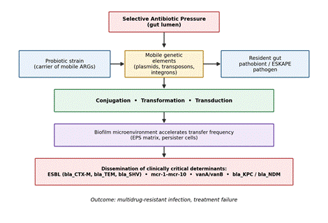
Figure 1. Mechanistic pathway from antibiotic selective pressure to multidrug-resistant infection via horizontal gene transfer (HGT). Antibiotic exposure selects for probiotic and commensal strains harboring mobile antimicrobial resistance genes (ARGs); these genes are transferred to resident pathobionts through conjugation, transformation, or transduction, a process markedly accelerated within biofilm microenvironments, ultimately disseminating clinically critical resistance determinants such as ESBLs, mcr genes, vanA/vanB, and carbapenemases (Idakwoji et al., 2026; Niculescu et al., 2026).
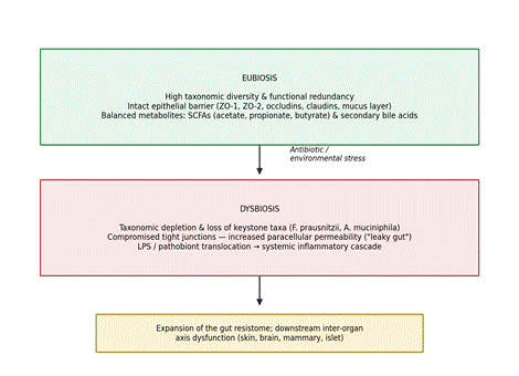
Figure 2. Structural and functional transition from eubiosis to dysbiosis. Under stable conditions (top), high microbial diversity, an intact epithelial barrier, and balanced short-chain fatty acid and bile-acid metabolism maintain host-microbiome homeostasis; antibiotic or environmental stress collapses this balance (bottom), depleting keystone taxa, compromising tight junctions, and permitting lipopolysaccharide translocation, which expands the gut resistome and disrupts downstream inter-organ signaling axes (Salem et al., 2018; Tan et al., 2026).
2.4 Therapeutic Revolutions: Next-Generation Probiotics, CRISPR Engineering, and Tri-Modal Integration
Faced with these risks, microbial therapeutics research has been shifting — not without some growing pains — from empirical, broad-spectrum pathogen eradication toward target-specific, mechanism-driven precision medicine (Jadhav et al., 2026; Hussain et al., 2026b).
Rather than relying on empirical donor fecal microbiota transplantation (FMT), which carries its own risks of pathogen transmission and considerable batch-to-batch variability, research has been moving toward standardized, multi-strain bacterial consortia manufactured from clonal cell banks (Suresh Kumar et al., 2026). Formulations such as SER-109 (Vowst) and VE303, developed under Good Manufacturing Practice conditions, contain purified Bacillota (Firmicutes) spores designed to restore specific metabolic fluxes (Suresh Kumar et al., 2026). These defined consortia re-establish primary-to-secondary bile acid conversion via the bai operon and stimulate SCFA production, which appears sufficient to suppress germination and colonization of Clostridioides difficile in several reported settings (Nogueira et al., 2026; Suresh Kumar et al., 2026).
Bacteriocins offer a conceptually different strategy: sequence-specific, ribosome-synthesized peptides with a narrow spectrum of activity, in contrast to the indiscriminate commensal die-off caused by conventional antibiotics (Tyagi et al., 2026). They recognize target receptors on specific pathogens and execute rapid bactericidal activity through membrane depolarization and pore formation, largely sparing bystander commensals (Tyagi et al., 2026). Synthetic biology is now leveraging these traits directly — a TAD1-deficient knockout mutant of Saccharomyces cerevisiae, for instance, has been shown to secrete cell-free supernatants capable of disrupting pathogenic biofilms and eradicating multidrug-resistant E. coli, S. aureus, and K. pneumoniae in vivo by triggering intracellular reactive oxygen species accumulation and compromising envelope integrity (Tyagi et al., 2026).
CRISPR-Cas genome editing and CRISPR interference have, meanwhile, enabled site-specific modification of probiotic chassis organisms such as Lactobacillus, Bifidobacterium, and Enterococcus (Hussain et al., 2026a; Meng et al., 2025). This programmable technology works through two broadly complementary routes. Subtractive editing, or pathogenicity curing, programs CRISPR systems to selectively target and cleave plasmids or chromosomal regions harboring specific ARGs — bla_NDM-1, bla_KPC, mcr-1 — within pathobionts, clearing resistance traits and resensitizing bacteria to conventional antibiotics without necessarily lysing the cell (Kuo et al., 2026; Allemailem, 2024). Additive engineering, by contrast, equips probiotics with defined expression cassettes that respond to local biochemical cues, allowing them to overexpress tight-junction proteins, synthesize immunomodulatory cytokines, or secrete quorum-quenching enzymes — lactonases and acylases — that degrade interspecies signaling autoinducers such as AI-2 and AHL, silencing pathobiont virulence networks without applying direct selective killing pressure (Hussain et al., 2026a; Gholizadeh et al., 2020). To keep these engineered strains from persisting or spreading unintentionally in the environment, robust biocontainment safeguards — synthetic auxotrophy, context-dependent genetic kill switches — are generally built in alongside the therapeutic payload (Hussain et al., 2026a; Wan et al., 2021).
To address the persistent challenge of probiotic gastric viability and delivery precision, a tripartite therapeutic framework has emerged, weaving together three previously separate disciplines (Hussain et al., 2026b) (Figure 4). Artificial intelligence and machine learning platforms — iProbiotics and ProbML among them — use Support Vector Machines, Random Forest classifiers, and deep learning to mine clinical, genomic, and metagenomic datasets, predicting probiotic-pathogen interactions and optimizing multi-strain consortium design (Jeyavelkumaran et al., 2026). Systems-biology approaches, including joint flux balance analysis and generalized Lotka-Volterra models, simulate metabolic cross-feeding and competition to forecast probiotic engraftment (Jeyavelkumaran et al., 2026). In parallel, nanotechnology-based delivery addresses the very real problem of probiotic viability loss under gastric acidity and bile-salt exposure; encapsulating cells or their postbiotics within pH-responsive nanocarriers — alginate-chitosan nanoparticles, liposomes, hydrogels — protects them during gastric transit and enables controlled, targeted release at intestinal, urogenital, or wound-associated sites (Hussain et al., 2026b; Gholamian et al., 2025). Once delivered, the selected or bioengineered probiotics restore the mucosal barrier, downregulate inflammatory markers, and deploy localized metabolites and bacteriocins, establishing conditions that
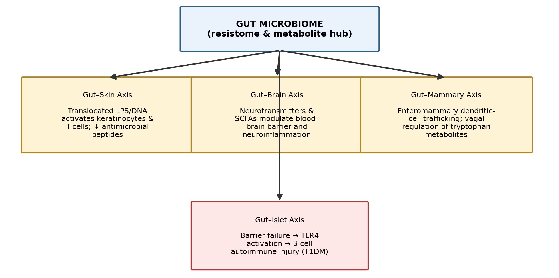
Figure 3. Inter-organ communication axes originating from the gut microbiome. Translocated microbial metabolites and immune mediators connect gut dysbiosis to cutaneous inflammation (gut-skin axis), neuroinflammatory changes (gut-brain axis), mastitis risk (gut-mammary axis), and pancreatic β-cell autoimmunity (gut-islet axis), illustrating how a single ecological disturbance can propagate into multiple, clinically distinct disease phenotypes (Munteanu et al., 2025; Socałą et al., 2021; Li et al., 2025; Tan et al., 2026).
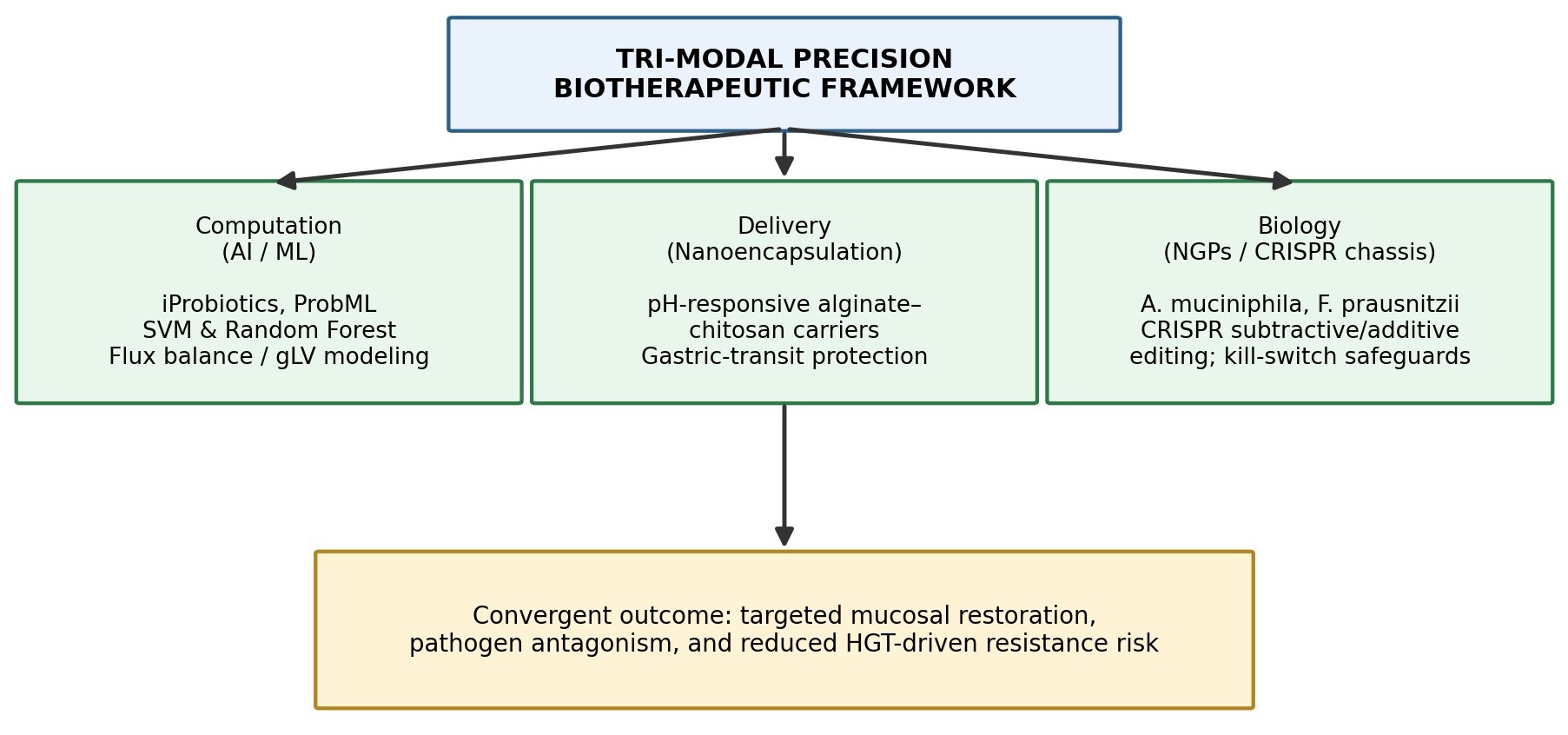
Figure 4. Tri-modal convergence framework for next-generation probiotic therapeutics. Computational modeling (AI/ML strain selection and metabolic simulation), nanoencapsulation-based delivery (pH-responsive gastric protection), and CRISPR-engineered biological chassis (next-generation probiotics with biocontainment safeguards) act in concert to restore mucosal homeostasis while constraining the horizontal-gene-transfer risk associated with conventional probiotic strains (Hussain et al., 2026b; Jeyavelkumaran et al., 2026).
are, at least in principle, resistant to reinfection (Hussain et al., 2026b; Jeyavelkumaran et al., 2026).
