Autoimmune diseases, considered collectively, represent a heterogeneous group of chronic inflammatory conditions unified less by their clinical presentation than by a shared underlying failure: the breakdown of immunological self-tolerance, which leads to targeted, immune-mediated destruction of host tissue (Steri et al., 2026). Genetic susceptibility, encoded predominantly within the human leukocyte antigen (HLA) complex, establishes the groundwork for this vulnerability, but it is the interaction between that genetic backdrop and modifiable environmental triggers that appears to determine both disease onset and the timing of subsequent flares (Cichocka et al., 2025; Izzo et al., 2026). What follows is an attempt to draw these threads - genetic architecture, environmental breach, pathogen-driven mimicry, gut-organ signaling, and emerging computational diagnostics - into something resembling a coherent whole, imperfect as that whole inevitably remains.
2.1 The Genetic Landscape of Autoimmunity: Shared Predispositions and Points of Divergence
It would be an oversimplification, though a tempting one, to treat the HLA class II region as though it told the whole genetic story. It remains the single strongest determinant of risk (Kotsiri et al., 2025; Steri et al., 2026), yet the details differ meaningfully by disease. In T1DM, susceptibility clusters around the HLA-DR3-DQ2 and HLA-DR4-DQ8 haplotypes, which present beta-cell autoantigens such as insulin B:9-23 and GAD65 to autoreactive T lymphocytes (Asamoah & Ahima, 2026; Cichocka et al., 2025). In MS, the analogous role falls to the HLA-DRB1*15:01 allele, implicated in driving CNS demyelination (Frohman & Tsirka, 2026; Steri et al., 2026). Beyond HLA, a long tail of non-HLA loci - CTLA4, PTPN22, FOXO3, TYK2, and CLEC16A among them - contribute smaller, but cumulatively meaningful, effects on immune tolerance, lymphocyte activation, and cytokine signaling (Cichocka et al., 2025; Steri et al., 2026).
A particularly illuminating, if somewhat counterintuitive, body of work has come from large-scale genome-wide association studies (GWAS) paired with statistical colocalization analysis. Steri et al. (2026), applying a Bayesian colocalization framework, identified 26 signals shared between T1DM and MS, the majority (17 of 26) reflecting general autoimmune susceptibility rather than disease-specific variants. What stands out, though, is that nine of these signals showed opposite directions of effect between the two conditions - a pattern termed antagonistic pleiotropy (Steri et al., 2026). The variant rs3806624A near the eomesodermin (EOMES) gene illustrates this well: it raises T1DM risk while apparently lowering MS risk, plausibly because EOMES governs CD8+ T-cell and natural killer cell cytotoxicity, and reduced cytotoxic capacity might protect pancreatic tissue while simultaneously weakening surveillance against neurotropic pathogens (Steri et al., 2026). A comparable divergence appears at the IRF8 locus, where the variant rs13330176 downregulates HLA class II-mediated antigen presentation - lowering MS risk but, somewhat paradoxically, raising T1DM susceptibility, perhaps by impairing viral clearance (Steri et al., 2026). Genetic risk further intersects with metabolic phenotype: in SLE, polygenic risk scores correlate with specific metabolic sub-phenotypes, including reduced ferritin and platelet counts, even when they fail to predict acute inflammatory markers directly (Alonso-Bernáldez et al., 2026).
2.2 The Environmental Breach: Barrier Dysfunction and the HPA Axis under Stress
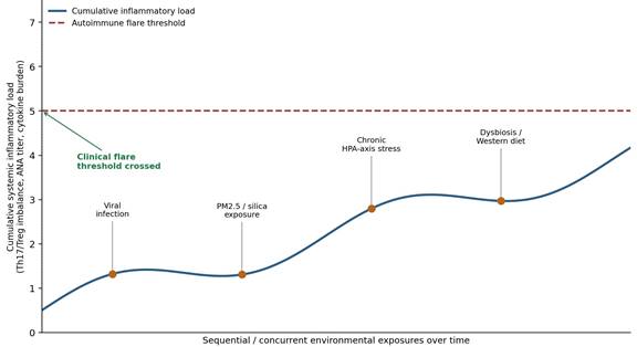
If genetics sets the threshold, it is the environment that seems to push people across it. The Epithelial Barrier Hypothesis holds that modern pollutants, toxicants, and lifestyle exposures compromise the physiological barriers of skin, gut, and airway, permitting antigen translocation and igniting local and systemic inflammatory cascades (Mpakosi et al., 2024; Triggianese et al., 2026). Climate disasters exemplify this rather starkly: PM2.5 and crystalline silica exposure follow wildfires, earthquakes, and building collapses, generating free radicals, degrading tight-junction proteins (claudin-1, occludin, E-cadherin), and activating TLR2/TLR4-NF-κB-MAP kinase signaling that skews T-cell differentiation toward Th17 at the expense of Treg populations (Mpakosi et al., 2024). Silica, meanwhile, behaves almost like a deliberately engineered adjuvant - stimulating alveolar macrophages, activating PRRs and the NLRP3 inflammasome, and driving IL-1β and TNF-α secretion, ultimately correlating with antinuclear antibodies and heightened RA, SLE, and systemic sclerosis risk (Mak & Tay, 2014; Mpakosi et al., 2024).
Layered on top of these physical insults is chronic psychological stress, which functions less as an independent trigger than as an amplifier of neuroendocrine-immune dysfunction (Gutierrez Nunez et al., 2025). Acute stress restrains inflammation through HPA-driven cortisol release; chronic stress does the opposite, producing glucocorticoid receptor resistance, HPA feedback failure, and unchecked elevation of IL-6, TNF-α, and IL-17 (Gutierrez Nunez et al., 2025). The clinical consequences of this shift are not merely theoretical - lupus-prone mouse models exposed to predator-induced anxiety show accelerated anti-dsDNA antibody production and renal injury, a fairly direct demonstration that psychological state and disease trajectory are mechanistically entangled (Gutierrez Nunez et al., 2025). Figure 1 attempts to render this converging architecture visually, situating viral, toxicant, stress-related, and dietary triggers as parallel inputs that funnel toward a common endpoint of tolerance breakdown and clinical flare (Figure 1).
2.3 Pathogen-Induced Autoimmunity: Molecular Mimicry and Viral Catalysts
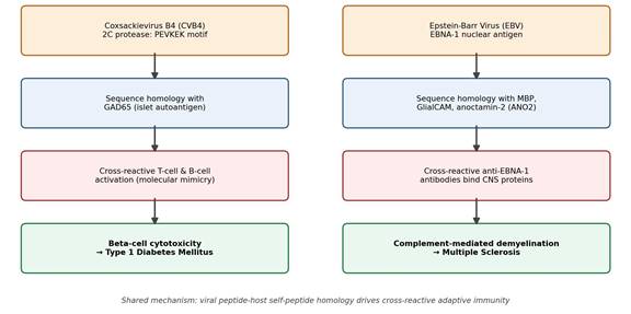
Among the modifiable triggers reviewed here, viral pathogens are arguably the best characterized (Kotsiri et al., 2025). They act through several overlapping routes - bystander activation, epitope spreading, and molecular mimicry - the last of which occurs when structural or sequence homology between a viral antigen and a host self-protein provokes a cross-reactive B- and T-cell response (Frohman & Tsirka, 2026; Kotsiri et al., 2025). In T1DM, enteroviruses, particularly CVB4, display marked tropism for pancreatic beta cells, and longitudinal cohort studies (DiViD, TEDDY, DAISY) have repeatedly linked enteroviral RNA detection to islet autoimmunity initiation and progression (Kotsiri et al., 2025; Steri et al., 2026). The CVB4 2C protease's PEVKEK hexapeptide sequence shares enough structural similarity with GAD65 to drive cross-reactive autoantibody formation and T-cell activation, contributing directly to islet destruction (Kotsiri et al., 2025; Steri et al., 2026). Rotavirus VP7 proteins add a second, related route toward the same outcome, triggering islet autoantibody seroconversion through homology with GAD65 and IA-2 (Kotsiri et al., 2025).
In MS, EBV occupies the equivalent position. Large longitudinal studies show EBV infection almost invariably preceding MS onset, with rising anti-EBNA-1 titers predating neurological symptoms by a considerable margin (Frohman & Tsirka, 2026). EBNA-1's sequence homology with MBP, GlialCAM, and ANO2 allows cross-reactive antibodies to attack myelin and axonal targets directly, initiating complement activation and progressive demyelination (Frohman & Tsirka, 2026). SARS-CoV-2 introduces a still newer, and in some ways more unsettling, mechanism: direct ACE2-mediated infection of pancreatic islet cells produces cytopathic injury and a localized cytokine storm, triggering islet death, transient hyperglycemia, epitope spreading, and - in some cases - outright checkpoint failure that permits autoreactive clones to persist (Kotsiri et al., 2025).
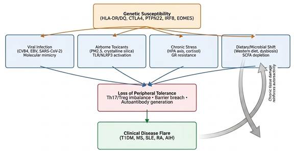
Figure 1. Multi-hit convergence model of autoimmune flare pathogenesis. Genetic susceptibility loci establish baseline risk, while four largely independent environmental exposure categories - viral infection, airborne toxicant inhalation, chronic HPA-axis stress, and dietary/microbial disruption - act on this substrate through distinct but convergent molecular pathways. Each pathway funnels toward a shared intermediate state of Th17/Treg imbalance, epithelial barrier breach, and autoantibody generation, which precipitates clinical flare and, through ongoing tissue damage, reinforces the underlying autoreactive state (cited in Section 2.2).
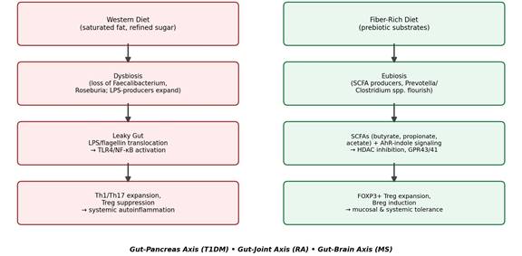
Figure 2. Diet-microbiome-immune signaling along the gut-organ axes. The left pathway traces the consequences of a Western diet through dysbiosis, epithelial barrier failure, and lipopolysaccharide-driven TLR4/NF-κB activation toward Th1/Th17 expansion and systemic autoinflammation. The right pathway traces the alternative trajectory supported by a fiber-rich diet, in which eubiosis and short-chain fatty acid production sustain FOXP3+ regulatory T-cell and regulatory B-cell expansion, preserving mucosal and systemic tolerance across the gut-pancreas, gut-joint, and gut-brain axes (cited in Section 2.4).
2.4 The Gut-Organ Axes and the Microbiome-Metabolite Network
The gut, it turns out, is not a passive bystander in any of this. The host-microbiome interface functions as a genuinely active regulatory hub where diet, microbial ecology, and immune cells negotiate the balance between tolerance and inflammation (Mittal et al., 2026; Rodziewicz & Bryl, 2026). This crosstalk is often conceptualized through specific tissue axes - the gut-pancreas axis in T1DM, the gut-joint axis in RA - each following broadly similar logic even as the downstream organ differs (Mittal et al., 2026; Rodziewicz & Bryl, 2026). Under favorable conditions, gut microbiota ferment dietary fiber into SCFAs (butyrate, propionate, acetate), which reinforce tight-junction stability and mucin synthesis while also acting as potent epigenetic regulators - butyrate, notably, inhibits HDACs, promoting FOXP3+ Treg expansion at the expense of Th1/Th17 lineages (Mittal et al., 2026). SCFAs further signal through FFAR2/GPR43 and FFAR3/GPR41 to suppress NF-κB activity and preserve mucosal tolerance (Mittal et al., 2026; Rodziewicz & Bryl, 2026).
A Western diet reverses this arrangement almost point for point. Saturated fats and refined sugars, often alongside environmental stressors, deplete SCFA-producing taxa such as Faecalibacterium prausnitzii and Roseburia intestinalis, weakening tight junctions and permitting translocation of lipopolysaccharide (LPS) and flagellin into systemic circulation (Mittal et al., 2026; Rodziewicz & Bryl, 2026). These microbial products then engage pattern recognition receptors on antigen-presenting cells in mesenteric and pancreatic lymph nodes, promoting self-antigen cross-presentation; in RA, comparable translocation events appear to seed synovial inflammation directly (Rodziewicz & Bryl, 2026). A separate but related pathway involves microbial tryptophan metabolism: commensal-derived indole compounds activate the aryl hydrocarbon receptor (AHR), supporting IL-22 production, epithelial repair, and Treg stability, while dysbiosis-associated tryptophan deficiency undermines this protective circuit (Mittal et al., 2026). Encouragingly, targeted interventions appear capable of reversing at least part of this damage: oral Clostridium butyricum restores Th17/Treg balance and expands gut-homing α4β7+ Tregs in preclinical T1DM models (Mittal et al., 2026), while Prevotella histicola supplementation raises acetate and butyrate levels and suppresses joint inflammation in arthritis models (Sasidharan et al., 2026; Rodziewicz & Bryl, 2026). Figure 2 summarizes this bidirectional dietary-microbial architecture, contrasting the dysbiotic and eubiotic trajectories side by side (Figure 2).
2.5 Advanced Computational Diagnostics and Precision Immunoengineering
Translating this considerable mechanistic complexity into something clinically actionable has proven difficult using conventional statistical methods alone, which is presumably why artificial intelligence (AI) and machine learning (ML) architectures have gained traction so quickly in this space (Ponce de León-Sánchez et al., 2026; Szili et al., 2026). In MS, roughly 30-50% of patients respond inadequately to interferon-beta (IFN-β) therapy, a variability that traditional clustering methods struggle to capture (Ponce de León-Sánchez et al., 2026). Ponce de León-Sánchez et al. (2026) addressed this by building a Mamdani-type fuzzy expert system, developed in collaboration with neurologists, to translate ambiguous clinical variables - age, sequential Expanded Disability Status Scale (EDSS) scores - into defined response categories; these categories then trained an artificial neural network (ANN) on 13 IFN-β-pathway biomarkers (including IL-2, IFNG, TNF, IL-4, IL-10, and TGFB), with a genetic algorithm optimizing the network's hyperparameters and achieving predictive accuracy between 0.80 and 1.0 (Ponce de León-Sánchez et al., 2026).
Parallel advances have emerged on the therapeutic side. Standard systemic immunosuppression, while effective, often carries a heavy side-effect burden and incomplete response rates (Brozek et al., 2026; Makkar & Morris, 2026). Specialized pro-resolving mediators (SPMs) - resolvins, protectins, and maresins - offer a more targeted alternative, coordinating inflammation resolution through G-protein-coupled receptor signaling on neutrophils and monocytes without the blunt instrument of systemic immunosuppression (Aminu & Wang, 2026). Because SPMs degrade quickly in vivo, nanoparticle-based delivery platforms (PLGA or lipid-based) are being engineered to concentrate them directly at inflamed sites, with encouraging results in experimental MS and cardiovascular models (Aminu & Wang, 2026). In RA, hybrid nanoparticles designed to co-target B and T cells promote antigen-specific tolerance and reduce pathogenic autoantibody production (Brozek et al., 2026), while mesenchymal stem cells (MSCs) and their derived exosomes secrete immunomodulatory factors (PGE2, TGF-β) that suppress Th17 activity, expand Tregs, and reprogram synovial macrophages toward a homeostatic M2 phenotype (Makkar & Morris, 2026).
2.6 Synthesis and Emerging Gaps
Read together, these strands resist reduction to any single explanatory mechanism. Autoimmune disease, at least as this literature currently describes it, looks more like a dynamic, self-reinforcing network - genetic risk, epithelial integrity, neuroendocrine tone, viral exposure history, and gut-microbial ecology all interacting simultaneously - than a linear causal chain (Alonso-Bernáldez et al., 2026; Steri et al., 2026). What remains genuinely uncertain is how these dimensions should be weighted against one another in individual patients, and whether large-scale, longitudinal, multi-omic cohorts will be sufficient to resolve that uncertainty. The computational tools reviewed above represent a promising, if still early, step toward that goal.