Integrative Biomedical Research

Integrative Biomedical Research (Journal of Angiotherapy) | Online ISSN  3068-6326
685
Citations
1.4m
Views
733
Articles
Your new experience awaits. Try the new design now and help us make it even better
Switch to the new experience
Figures and Tables
CASE STUDY   (Open Access)

Serum Uric Acid Reduction Following Use of Canssu5®, a Standardized Orthosiphon stamineus Extract, in Adults with Hyperuricemia: A Retrospective Case Study

Aman Shah Abdul Majid 1*

+ Author Affiliations

Integrative Biomedical Research 10 (1) 1-8 https://doi.org/10.25163/biomedical.10110722

Submitted: 31 January 2026 Revised: 31 March 2026  Published: 06 April 2026 


Abstract

Background: Hyperuricemia remains a central biochemical driver of gout and is increasingly implicated in broader cardiometabolic and renal disorders. Although pharmacologic urate-lowering therapies are effective, their use is sometimes constrained by intolerance, adherence challenges, or patient preference for alternative approaches. In this context, botanically derived compounds with multi-target activity have begun to attract interest, though clinical evidence remains limited.

Methods: This retrospective case series evaluated ten adults with laboratory-confirmed hyperuricemia who consumed Canssu5®, a standardized Orthosiphon stamineus extract, under real-world conditions. Serum uric acid (SUA) values obtained from accredited laboratories before and after supplementation were analyzed using paired-sample statistical methods. Individual-level responses, percentage reductions, and achievement of clinically relevant SUA thresholds were examined descriptively.

Results: All participants demonstrated reductions in SUA following product use (Table 1; Figure 1). Mean SUA declined from 0.592 ± 0.091 mmol/L to 0.364 ± 0.059 mmol/L, corresponding to a mean reduction of 0.228 mmol/L (37.4%). The change was statistically significant (t(9) = 7.78, p < 0.001), with a large effect size (Cohen’s d ≈ 2.46). Seventy percent of participants achieved post-treatment SUA levels below 0.40 mmol/L. No adverse events were reported.

Conclusion: Use of a standardized Orthosiphon stamineus extract was associated with consistent reductions in serum uric acid across all cases. While the findings are encouraging, the retrospective design and small sample size limit causal interpretation. These observations should be considered preliminary and warrant confirmation in controlled clinical trials.

Keywords: Hyperuricemia; Gout; Orthosiphon stamineus; Rosmarinic acid; Botanical therapeutics

1. Introduction

Hyperuricemia, broadly defined as an elevation in serum uric acid beyond physiological solubility thresholds, is increasingly recognized as more than a simple biochemical abnormality. It is, of course, the central pathogenic driver of gout, but its clinical significance extends well beyond acute arthritis alone. Persistently elevated urate levels have also been associated with chronic kidney disease, hypertension, metabolic syndrome, insulin resistance, and broader cardiometabolic dysfunction (Borghi et al., 2014; Dalbeth et al., 2016; Johnson et al., 2013). In many patients, hyperuricemia remains clinically silent for years, yet the underlying biochemical environment continues to favor monosodium urate crystal deposition, low-grade inflammation, and progressive metabolic disturbance. That silent progression is precisely what makes the condition both deceptively common and, in some cases, therapeutically frustrating.

Conventional urate-lowering therapy remains the cornerstone of clinical management. Agents such as allopurinol and febuxostat, primarily through xanthine oxidase inhibition, are effective in reducing urate production and preventing long-term crystal burden (Pacher et al., 2006; Richette et al., 2017). Still, the real-world management of hyperuricemia is often less straightforward than treatment guidelines may suggest. Some patients experience adverse effects, hypersensitivity reactions, gastrointestinal intolerance, or poor long-term adherence, while others remain reluctant to initiate chronic pharmacotherapy for what is initially perceived as an asymptomatic laboratory abnormality (Stamp & Chapman, 2013). In practice, this creates a persistent therapeutic gap—particularly for individuals seeking gentler, adjunctive, or more “natural” approaches that might support urate control before recurrent gout or organ-level complications become established.

It is within this gap that botanical and phytopharmaceutical interventions have drawn growing attention. Not all plant-based products, admittedly, are scientifically persuasive, and many suffer from weak standardization or exaggerated health claims. However, some medicinal plants have accumulated a more credible mechanistic foundation and may warrant serious clinical exploration. Among these, Orthosiphon stamineus Benth., commonly known as cat’s whiskers or Java tea, has emerged as a particularly interesting candidate. Traditionally used in Southeast Asian medicine for urinary, renal, and metabolic complaints, O. stamineus has been investigated for a range of biological activities, including antioxidant, anti-inflammatory, nephroprotective, and metabolic effects (Ameer et al., 2012; Yam et al., 2009).

From a urate-lowering perspective, the pharmacologic rationale is especially compelling. Preclinical studies suggest that O. stamineus and its polyphenolic constituents may influence serum uric acid through several complementary mechanisms rather than a single isolated pathway. These include possible inhibition of xanthine oxidase, enhancement of renal urate excretion, and modulation of inflammatory and oxidative pathways that are increasingly understood to interact with urate metabolism and gout pathophysiology (Cos et al., 1998; Kong et al., 2000; Lin et al., 2002). This “polymolecular” profile may be particularly relevant in metabolic disorders, where disease expression rarely arises from one pathway alone.

A major contributor to this therapeutic interest lies in the phytochemical composition of the plant. Standardized extracts of O. stamineus commonly contain rosmarinic acid, sinensetin, eupatorin, and related methoxylated flavones, compounds with documented antioxidant and anti-inflammatory properties (Akowuah et al., 2004; Damsud et al., 2014; Petersen & Simmonds, 2003). Rosmarinic acid, for instance, has been widely studied for its ability to attenuate oxidative stress and inflammatory mediator activity, both of which are relevant to urate-induced tissue injury and gout flare biology (Al-Dhabi et al., 2020). Likewise, flavonoid compounds such as sinensetin and eupatorin have shown biochemical activities that plausibly intersect with urate metabolism and inflammatory signaling. Taken together, these constituents suggest that O. stamineus may not simply act as a “traditional herb,” but rather as a standardized botanical with mechanistic relevance to hyperuricemia.

Yet despite this promising preclinical and phytochemical foundation, the clinical evidence base remains surprisingly limited. There is still a noticeable gap between laboratory plausibility and documented human outcome data, particularly in real-world settings. That gap matters. Mechanistic promise alone is not enough; what ultimately matters is whether measurable biochemical improvement can be observed in patients under routine use conditions. Even small observational datasets, when carefully reported and appropriately interpreted, can provide useful preliminary insight and help justify more rigorous prospective investigation.

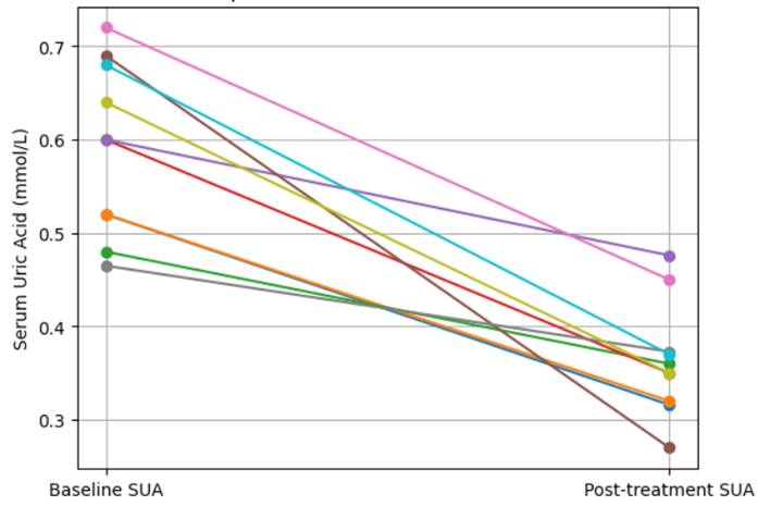
Against this background, the present retrospective case series was undertaken to describe changes in serum uric acid levels among adults with laboratory-confirmed hyperuricemia who consumed Canssu5®, a standardized Orthosiphon stamineus extract formulated to contain approximately 6% rosmarinic acid. Rather than attempting to make definitive therapeutic claims, this report aims to provide early clinical observations on a standardized botanical intervention that may hold translational relevance in the management of hyperuricemia. In doing so, it seeks to contribute a modest but clinically meaningful piece of evidence to an area where patient interest is growing, yet formal human data remain scarce.

2. Methods

2.1 Study Design and Clinical Context

This study was designed as a retrospective observational case series examining changes in serum uric acid (SUA) levels among adults with documented hyperuricemia who had consumed Canssu5®, a standardized Orthosiphon stamineus extract, as part of routine real-world use. The decision to structure the work as a retrospective case series was deliberate. At this early stage, the aim was not to test a formal therapeutic hypothesis under controlled trial conditions, but rather to document whether a clinically meaningful urate-lowering signal could be observed in practice among individuals who had already used the product and undergone laboratory testing.

Case series, admittedly, occupy a modest place in the hierarchy of evidence. Still, when reported transparently and interpreted with appropriate caution, they can provide useful preliminary clinical observations, especially in areas where human data remain limited. In the present context, Orthosiphon stamineus has shown pharmacological relevance in preclinical and phytochemical literature, including anti-inflammatory, antioxidant, and metabolic effects, as well as potential interactions with urate-related pathways (Ameer et al., 2012; Yam et al., 2009). This study was therefore undertaken as an exploratory clinical documentation effort intended to support or refute the plausibility of those earlier findings in a real-world setting.

2.2 Participant Identification and Eligibility

A total of ten adult individuals were included in this case series. Cases were identified retrospectively on the basis of the availability of documented baseline and follow-up serum uric acid measurements obtained from accredited clinical laboratories, together with a confirmed history of consumption of the study product during the interval between those two laboratory assessments.

To be included, individuals had to meet the following practical eligibility criteria:

  1. Adult age at the time of product use
  2. Laboratory-confirmed elevated baseline serum uric acid
  3. Documented use of Canssu5® standardized Orthosiphon stamineus extract
  4. Availability of at least one pre-intervention and one post-intervention SUA value
  5. A minimum product exposure duration sufficient to allow a biologically plausible effect window

The series included 6 males and 4 females, with observed durations of use ranging from 2 to 36 weeks. Because this was a retrospective case series derived from real-world use rather than a protocol-driven interventional trial, participants were not recruited prospectively, randomized, or stratified according to disease severity, comorbidities, or baseline urate burden.

No formal exclusion algorithm had been prospectively predefined at the time the cases arose. However, records were reviewed to ensure that the included cases contained sufficient biochemical documentation to permit before-and-after comparison. Cases with missing laboratory confirmation or insufficient follow-up data would not have been eligible for inclusion.

2.3 Study Product and Exposure Characterization

The intervention evaluated in this case series was Canssu5®, a Malaysian-registered traditional medicinal product formulated from a standardized extract of Orthosiphon stamineus (cat’s whiskers). The extract was standardized to contain approximately 6% rosmarinic acid, with additional phytochemical constituents characteristic of the plant, including sinensetin, eupatorin, and trace-to-low levels of 3′-hydroxy-5,6,7,4′-tetramethoxyflavone (TMF). These compounds have been described in prior phytochemical and pharmacological studies as contributing to antioxidant, anti-inflammatory, and metabolic effects (Akowuah et al., 2004; Damsud et al., 2014; Petersen & Simmonds, 2003).

Because the present study was retrospective and reflected routine consumer or patient use rather than a standardized clinical trial protocol, dosage frequency and duration were not fully uniform across all cases. This variability is important to acknowledge. Rather than imposing artificial standardization after the fact, the study was intentionally reported in a manner that reflects actual real-world exposure conditions. Duration of use was recorded individually and is presented in the results table.

2.4 Outcome Assessment

The primary outcome measure was the change in serum uric acid concentration, expressed in mmol/L, between the baseline laboratory value and the post-intervention laboratory value obtained after use of the study product.

Serum uric acid was selected as the primary endpoint because it is the most clinically relevant biochemical marker of hyperuricemia and the central laboratory parameter used in gout risk assessment and urate-lowering management (Dalbeth et al., 2016; Richette et al., 2017). Unlike symptom-based outcomes, which may fluctuate substantially and be influenced by subjective reporting, SUA provides a more objective and clinically interpretable measure of biochemical response.

For each included case, the following data were extracted and tabulated:

  • Age
  • Sex
  • Duration of product use (weeks)
  • Baseline serum uric acid value
  • Post-intervention serum uric acid value
  • Percentage reduction in serum uric acid

A secondary descriptive outcome of interest was the proportion of participants achieving a post-treatment SUA level below 0.40 mmol/L, a threshold that may be considered clinically meaningful in the context of urate reduction and gout risk modification, particularly in relation to crystal burden reduction (Khanna et al., 2012; Richette et al., 2017).

An additional observational outcome was the presence or absence of reported adverse events during the documented period of use. Because this was not a formal safety trial, adverse event capture was descriptive rather than systematically adjudicated.

2.5 Data Handling and Statistical Analysis

Data were compiled in tabular format and analyzed using a within-subject pre–post comparison approach. Since each participant had both a baseline and follow-up SUA measurement, the principal statistical analysis evaluated whether the mean post-intervention SUA differed significantly from the corresponding baseline value within the same individual.

A paired-sample t-test was used to compare baseline and post-treatment SUA levels. This method was selected because the analysis focused on paired continuous measurements from the same participants before and after exposure. For each participant, the absolute change in SUA was calculated as:

Change in SUA=Post-treatment SUA-Baseline SUA 

In addition to hypothesis testing, the following descriptive and inferential statistics were summarized:

  • Mean ± standard deviation (SD) for baseline and post-treatment SUA
  • Mean absolute reduction in SUA
  • Percentage reduction from baseline
  • 95% confidence interval (CI) for mean change
  • Paired t-statistic and corresponding p value
  • Effect size using Cohen’s d for paired change

A two-sided p-value < 0.05 was considered statistically significant. Given the small sample size, the statistical findings were interpreted cautiously and were regarded as exploratory rather than confirmatory.

2.6 Ethical and Reporting Considerations

This work was conducted as a retrospective analysis of documented laboratory observations rather than as a prospective interventional clinical trial. No experimental treatment allocation, invasive procedure, or protocol-driven therapeutic manipulation was introduced for the purpose of this report. The study therefore represents a descriptive clinical case series based on existing records.

Because the product evaluated in this report is commercially relevant to the author, this relationship has been explicitly disclosed in the Conflict of Interest statement to preserve transparency and allow readers to interpret the findings with appropriate context. In view of this, particular care was taken to present the data conservatively and to avoid overstating causal inference.

The manuscript was prepared in a reporting style intended to maximize transparency, reproducibility, and interpretability for future clinical and translational work. Although limited by its retrospective design and small sample size, the methodological structure was intended to provide a sufficiently clear framework for replication in a larger prospective observational cohort or randomized controlled trial.

3. Results

3.1 Participant Characteristics and Observation Window

A total of ten adults with documented hyperuricemia were included in this retrospective case series. The cohort comprised six males and four females, with ages ranging from 41 to 66 years. The duration of exposure to the standardized Orthosiphon stamineus extract varied across cases, ranging from 2 weeks to 36 weeks, thereby reflecting a heterogeneous but clinically realistic real-world usage pattern rather than a tightly protocolized intervention window (Table 1).

At baseline, all included individuals had serum uric acid (SUA) concentrations above what would generally be considered desirable for long-term urate control. Although the cohort was small, the dataset was notable for one immediately visible feature: every participant demonstrated a downward change in SUA following use of the study product. That uniformity of direction does not, by itself, establish causality, but it does suggest a pattern sufficiently consistent to warrant careful reporting and further scrutiny.

3.2 Baseline and Post-Intervention Serum Uric Acid Profiles

Baseline SUA values ranged from 0.465 mmol/L to 0.720 mmol/L, indicating that the included cases spanned mild to moderately elevated urate burden. Following supplementation with Canssu5®, post-intervention SUA values declined to a range of 0.270 mmol/L to 0.476 mmol/L (Table 1).

When examined at the group level, the mean baseline SUA was 0.592 ± 0.091 mmol/L, whereas the mean post-intervention SUA declined to 0.364 ± 0.059 mmol/L (Table 1). This represented an average absolute reduction of 0.228 mmol/L, corresponding to a 37.4% mean decrease from baseline.

This degree of reduction is clinically notable, particularly in the context of hyperuricemia management, where even modest downward shifts in urate concentration may carry biological relevance over time. More importantly, however, the reduction observed here was not confined to a few extreme responders. Instead, the pattern appeared across the entire dataset, with all ten individuals showing some degree of biochemical improvement.

3.3 Individual-Level Response Pattern

Inspection of the individual case data revealed a relatively broad but consistently favorable response range (Table 1). The smallest observed reduction was 19.8%, recorded in a participant with a shorter exposure duration of 2 weeks, while the largest reduction reached 60.9%, observed in a participant whose SUA declined from 0.690 mmol/L to 0.270 mmol/L after 12 weeks of use (Table 1).

Several cases demonstrated reductions exceeding 40%, including:

  • Case 4: 41.7% reduction
  • Case 6: 60.9% reduction
  • Case 9: 45.3% reduction
  • Case 10: 45.6% reduction (Table 1)

These individual-level reductions suggest that the observed biochemical effect was not merely a statistical artifact of averaging. Rather, the dataset appears to reflect a broadly distributed pattern of urate decline, albeit with expected inter-individual variability. That variability is not surprising. In retrospective real-world observations, response heterogeneity is often influenced by differences in baseline urate burden, adherence, metabolic status, duration of exposure, hydration, dietary purine intake, and potentially unmeasured concomitant therapies.

Interestingly, the data also hint—though only cautiously—at the possibility that longer durations of use may have supported more sustained reductions in some individuals. Still, because this was not a controlled dose-duration study, no formal duration-response inference should be drawn from these observations alone.

3.4 Statistical Analysis of Pre–Post Change

The paired comparison of baseline and post-intervention SUA values demonstrated a statistically significant reduction following product use (Table 1). The mean within-subject reduction was −0.228 mmol/L, with a 95% confidence interval (CI) of −0.296 to −0.160 mmol/L, indicating that the observed change was not only directionally consistent but also statistically robust within the limitations of the dataset.The paired-sample t-test yielded:

Table 1. Clinical outcomes and statistical change in serum uric acid (N=10)

Case

Age/Sex

Duration (weeks)

Baseline (mmol/L)

Post (mmol/L)

% Reduction

1

54/M

12

0.520

0.316

39.2

2

48/F

8

0.520

0.320

38.5

3

61/M

4

0.480

0.360

25.0

4

45/M

16

0.600

0.350

41.7

5

58/F

24

0.600

0.476

20.7

6

52/M

12

0.690

0.270

60.9

7

66/M

36

0.720

0.450

37.5

8

41/F

2

0.465

0.373

19.8

9

55/M

12

0.640

0.350

45.3

10

50/F

20

0.680

0.370

45.6

Figure 1. Individual baseline and post-treatment serum uric acid (SUA) values in the 10 included cases. Each line represents a single participant, illustrating the within-subject change following use of the standardized Orthosiphon stamineus extract (Canssu5®). A consistent downward shift in SUA is observed across all cases.

  • t(9) = 7.78
  • p < 0.001

This suggests that the observed pre–post difference was highly unlikely to have occurred by random variation alone under the assumptions of the applied model.

To further characterize the magnitude of change, an effect size estimate was calculated using Cohen’s d, which was approximately 2.46. From a statistical standpoint, this would be interpreted as a very large within-subject effect. Even so, it is important to interpret this cautiously. In small observational datasets, effect sizes can appear exaggerated relative to what might later be seen in larger, more heterogeneous populations. Nevertheless, within the context of this case series, the signal was undeniably strong.

3.5 Achievement of Clinically Relevant Uric Acid Targets

Beyond mean change alone, the biochemical outcomes were also examined in terms of clinically meaningful post-treatment thresholds. Following use of the standardized Orthosiphon stamineus extract, 7 out of 10 participants (70%) achieved a post-intervention SUA concentration below 0.40 mmol/L (Table 1).

This finding deserves attention because movement toward lower urate thresholds may be more clinically informative than average change alone. In practical terms, reducing serum urate into a lower biochemical range may lessen the likelihood of sustained urate supersaturation and, by extension, reduce the long-term environment that favors crystal formation. Although this case series was not designed to assess flare frequency, tophus burden, or other clinical endpoints, the biochemical shift itself suggests potential translational relevance.

At the same time, three participants remained above the <0.40 mmol/L threshold despite showing measurable reductions. That nuance is important. It suggests that while the intervention may have contributed to meaningful improvement, the degree of response may still vary depending on baseline burden, duration of use, or other unmeasured factors.

3.6 Safety and Tolerability Observations

No adverse events were reported among the ten included participants during the documented period of product use. Although this finding is reassuring at a descriptive level, it should be interpreted conservatively.

Because the present study was retrospective and not designed as a formal safety surveillance trial, adverse event capture was observational rather than systematically monitored. As such, the absence of reported adverse events should not be interpreted as definitive evidence of safety, but rather as an absence of documented tolerability concerns within this limited dataset.

3.7 Overall Interpretation of the Observed Clinical Signal

Taken together, the results reveal a remarkably consistent biochemical pattern: all ten participants demonstrated reductions in serum uric acid, the group-level decline was both statistically significant and quantitatively substantial, and the majority of participants reached a post-treatment urate level below 0.40 mmol/L (Table 1).

What makes these findings particularly interesting is not merely that the mean value decreased, but that the direction of change was universal across cases despite the inherent variability of a retrospective real-world setting. That said, these findings should still be understood as preliminary observational evidence rather than definitive proof of therapeutic efficacy. The dataset is small, uncontrolled, and biologically plausible—but it remains exploratory.

Even so, as an early clinical signal, the findings are difficult to dismiss outright. They provide a coherent basis for more rigorous prospective evaluation and, at the very least, suggest that standardized Orthosiphon stamineus extract may merit further investigation as a candidate urate-lowering botanical intervention.

4. Discussion

4.1 Overview of Principal Findings

The present retrospective case series provides a set of observations that are, at the very least, difficult to ignore. Across all ten included participants, a consistent reduction in serum uric acid (SUA) was observed following the use of a standardized Orthosiphon stamineus extract, with a mean reduction of 0.228 mmol/L and a substantial relative decrease of approximately 37.4% (Table 1). What is perhaps more striking than the magnitude of the reduction itself is the uniformity of its direction—every individual demonstrated a decline in SUA, a pattern that is further illustrated visually in the paired analysis (Figure 1).

Such consistency, particularly in a real-world, non-controlled dataset, invites cautious interpretation. It does not establish causality, nor does it eliminate the possibility of confounding influences. Still, it does suggest that the observed effect is unlikely to be entirely random. In small observational datasets, one might reasonably expect variability in both directions—some individuals improving, others remaining stable, and some even worsening. The absence of such divergence here, while not definitive, strengthens the impression that a biologically relevant signal may be present.

At the same time, it is important to acknowledge that observational consistency alone is not proof of efficacy. Retrospective case series, by their nature, are susceptible to selection bias, regression to the mean, and unmeasured behavioral or metabolic influences. Therefore, the findings should be interpreted as hypothesis-generating rather than confirmatory, serving as an early clinical indication that warrants more rigorous investigation.

4.2 Clinical Interpretation of Uric Acid Reduction

From a clinical standpoint, the magnitude of SUA reduction observed in this study is noteworthy. In many participants, post-intervention urate levels approached or fell below thresholds commonly targeted in gout management, particularly the range of 0.36–0.40 mmol/L, which is often associated with reduced crystal formation and lower risk of recurrent flares (Dalbeth et al., 2016; Richette et al., 2017). In this series, 70% of participants achieved SUA levels below 0.40 mmol/L (Table 1), suggesting that the observed biochemical changes may have potential translational relevance.

However, it is worth pausing here. The temptation to equate biochemical improvement with clinical benefit is understandable, but not always justified. This study did not assess gout flare frequency, tophus resolution, renal outcomes, or patient-reported symptoms. Therefore, while the biochemical shift is encouraging, its direct clinical implications remain uncertain.

Another nuance lies in the variability of individual responses. Although all participants improved, the degree of reduction ranged from approximately 19.8% to 60.9% (Table 1). Such variability likely reflects underlying differences in metabolic state, baseline urate burden, duration of exposure, and possibly adherence or lifestyle factors. This heterogeneity is not a limitation in itself; rather, it reflects the complexity of real-world physiology and reinforces the need for controlled dose-response studies.

4.3 Mechanistic Plausibility and Pharmacological Context

One of the strengths of the present findings is that they are not occurring in a mechanistic vacuum. Orthosiphon stamineus has been studied in preclinical and phytochemical contexts, and several pathways through which it may influence urate metabolism have been proposed.

Hyperuricemia itself is a multifactorial condition, arising from both increased production of uric acid and reduced renal excretion (Becker & Jolly, 2006). Conventional therapies, particularly xanthine oxidase inhibitors, primarily target urate production (Pacher et al., 2006). In contrast, botanical compounds often exhibit multi-target or “polymolecular” effects, which may be particularly relevant in metabolic diseases where single-pathway interventions do not always fully address disease complexity.

Experimental studies have suggested that O. stamineus and its constituent compounds may inhibit xanthine oxidase activity, thereby reducing urate production (Cos et al., 1998; Kong et al., 2000; Lin et al., 2002). At the same time, there is evidence suggesting enhancement of renal urate excretion, though this remains less well characterized in human settings. In addition, anti-inflammatory and antioxidant effects may indirectly influence urate-related pathology by modulating the inflammatory environment associated with monosodium urate crystal deposition.

The phytochemical composition of the extract provides further support for these mechanisms. Rosmarinic acid, one of the principal standardized components (~6%), has been widely reported to exert antioxidant and anti-inflammatory effects, including suppression of reactive oxygen species and inflammatory mediators (Petersen & Simmonds, 2003; Al-Dhabi et al., 2020). These properties are relevant to gout pathophysiology, where oxidative stress contributes to inflammatory amplification.

Similarly, flavonoids such as sinensetin and eupatorin have demonstrated inhibitory effects on xanthine oxidase and pro-inflammatory signaling pathways, including NF-κB (Cos et al., 1998). The presence of TMF, a polymethoxylated flavone, may further enhance bioavailability and cellular activity, although its precise contribution remains to be fully elucidated.

Taken together, these mechanisms suggest that the observed urate reduction may not be attributable to a single pharmacologic action, but rather to a combined effect across multiple biological pathways. This aligns with the concept of botanical therapeutics as systems-level modulators rather than single-target agents.

4.4 Comparison with Existing Therapeutic Approaches

It is tempting, though perhaps premature, to compare the magnitude of urate reduction observed in this case series with that achieved by conventional pharmacologic therapies. Xanthine oxidase inhibitors such as allopurinol and febuxostat are well-established and highly effective, often producing substantial reductions in SUA (Richette et al., 2017). However, they are also associated with limitations, including hypersensitivity reactions, gastrointestinal intolerance, and adherence challenges in some patients (Stamp & Chapman, 2013).

In the present dataset, the observed reductions—particularly in individuals achieving >40% decline—appear, at least numerically, to approach early-phase outcomes reported with low-dose pharmacologic therapy. Yet such comparisons must be made with caution. This study lacks a control group, standardized dosing, and sufficient sample size to support direct equivalence claims. Moreover, pharmacologic trials typically involve more rigorous patient selection and monitoring.

A more appropriate interpretation, therefore, is that Orthosiphon stamineus extract may represent a potential adjunctive or alternative option, particularly for individuals who are unable or unwilling to tolerate standard pharmacotherapy. Whether it could serve as a standalone therapy, or only as part of a combined strategy, remains an open question.

4.5 Interpretation of Consistency and Potential Bias

One of the most intriguing aspects of the dataset is the 100% response rate. While this is encouraging, it also raises important methodological questions. In clinical research, perfect directional consistency is uncommon and often prompts consideration of potential biases.

Several explanations could be considered:

  • Selection bias: Cases included may represent individuals with favorable outcomes
  • Regression to the mean: Elevated baseline values may naturally decline over time
  • Behavioral changes: Participants may have altered diet, hydration, or lifestyle
  • Measurement variability: Differences in laboratory timing or conditions

None of these factors can be definitively ruled out in the present study. That does not invalidate the findings, but it does emphasize the need for cautious interpretation. In this sense, the strength of the signal is also, paradoxically, a reason for careful scrutiny.

4.6 Limitations

The limitations of this study are substantial and should be acknowledged explicitly. The small sample size (n=10) limits generalizability and statistical robustness. The retrospective design precludes control over exposure conditions, introduces potential selection bias, and limits causal inference.

There was also variability in duration of use (2–36 weeks), which complicates interpretation of dose–response relationships. Additionally, dietary intake, hydration status, and concomitant medications were not systematically controlled or recorded, all of which may influence uric acid levels.

Adverse event monitoring was not conducted in a structured manner, and therefore the absence of reported adverse effects should not be interpreted as definitive evidence of safety. Finally, the presence of a declared commercial interest underscores the importance of independent replication.

4.7 Future Directions

The findings of this study point clearly toward the need for prospective, controlled investigation. Randomized controlled trials (RCTs) would be the logical next step to evaluate:

  • Dose-response relationships
  • Comparative efficacy versus standard therapy
  • Long-term safety
  • Effects on clinical endpoints such as gout flares
  • Renal and cardiometabolic outcomes

Additionally, mechanistic studies examining biomarkers of oxidative stress, inflammation, and xanthine oxidase activity could help clarify the pathways through which Orthosiphon stamineus exerts its effects.

5. Conclusion

In summary, this retrospective case series presents a consistent and biologically plausible pattern of serum uric acid reduction following the use of a standardized Orthosiphon stamineus extract. While the findings are encouraging, they remain preliminary and must be interpreted within the constraints of the study design.

Rather than providing definitive evidence of therapeutic efficacy, the results offer a coherent clinical signal—one that aligns with existing mechanistic knowledge and justifies further investigation. In a field where patient interest in botanical and adjunctive therapies continues to grow, such early-stage evidence, when carefully reported and cautiously interpreted, may serve as a meaningful step toward more rigorous clinical evaluation.

Author Contributions

A.S.A.M. conceived the study, curated and analyzed the data, interpreted the findings, drafted the manuscript, critically revised the content, and approved the final version for publication.

Declarations

Ethics Approval and Consent to Participate: This study was conducted as a retrospective case series based on documented laboratory findings and existing observational records. No experimental intervention, treatment assignment, or protocol-driven clinical procedure was performed specifically for research purposes. The manuscript reports retrospective clinical observations and was prepared in accordance with principles of transparency, confidentiality, and responsible scientific reporting.

Conflict of Interest: The author declares a commercial interest in Canssu5® and holds intellectual property rights related to standardized Orthosiphon stamineus extract formulations evaluated in this study. This relationship is disclosed in the interest of full transparency. The findings should therefore be interpreted in light of this declared potential conflict.

Funding: No external funding was received for the preparation, analysis, or reporting of this study.

References


Akowuah, G. A., Zhari, I., Norhayati, I., & Sadikun, A. (2004). Effect of extraction temperature on flavonoid and phenolic contents of Orthosiphon stamineus leaves. Journal of Tropical Medicinal Plants, 5(1), 49–54.

Al-Dhabi, N. A., Arasu, M. V., Park, C. H., & Park, S. U. (2020). An up-to-date review of rutin and its biological and pharmacological activities. Biomedicine & Pharmacotherapy, 131, 110657. https://doi.org/10.1016/j.biopha.2020.110657
Ameer, O. Z., Salman, I. M., Asmawi, M. Z., Ibraheem, Z. O., & Yam, M. F. (2012). Orthosiphon stamineus: Traditional uses, phytochemistry, pharmacology, and toxicology. Journal of Medicinal Food, 15(8), 678–690. https://doi.org/10.1089/jmf.2011.1973

Becker, M. A., & Jolly, M. (2006). Clinical gout and the pathogenesis of hyperuricemia. Rheumatic Disease Clinics of North America, 32(2), 275–293. https://doi.org/10.1016/j.rdc.2006.02.004

Borghi, C., Agabiti-Rosei, E., Johnson, R. J., Kielstein, J. T., Lurbe, E., Mancia, G., Redon, J., Stack, A. G., Tsioufis, C., & Hyperuricaemia and Gout Working Group of the European Society of Hypertension. (2014). Hyperuricaemia and gout in cardiovascular, metabolic and kidney disease. European Journal of Internal Medicine, 25(5), 372–381. https://doi.org/10.1016/j.ejim.2014.01.012
Cos, P., Ying, L., Calomme, M., Hu, J. P., Cimanga, K., Van Poel, B., Pieters, L., Vlietinck, A. J., & Vanden Berghe, D. (1998). Structure–activity relationship and classification of flavonoids as inhibitors of xanthine oxidase and superoxide scavengers. Journal of Natural Products, 61(1), 71–76. https://doi.org/10.1021/np970237h

Dalbeth, N., Merriman, T. R., & Stamp, L. K. (2016). Gout. The Lancet, 388(10055), 2039–2052. https://doi.org/10.1016/S0140-6736(16)00346-9

Damsud, T., Adisakwattana, S., & Phuwapraisirisan, P. (2014). Three new phenylbutanoid glycosides from the leaves of Orthosiphon stamineus. Journal of Natural Medicines, 68(3), 563–567. https://doi.org/10.1007/s11418-014-0823-2

Johnson, R. J., Nakagawa, T., Jalal, D., Sánchez-Lozada, L. G., Kang, D. H., & Ritz, E. (2013). Uric acid and chronic kidney disease: Which is chasing which? Nephrology Dialysis Transplantation, 28(9), 2221–2228. https://doi.org/10.1093/ndt/gft029

Khanna, D., Fitzgerald, J. D., Khanna, P. P., Bae, S., Singh, M. K., Neogi, T., Pillinger, M. H., Merill, J., Lee, S., Prakash, S., Kaldas, M., Gogia, M., Perez-Ruiz, F., Taylor, W., Lioté, F., Choi, H., Singh, J. A., Dalbeth, N., Kaplan, S., … Mandell, B. (2012). 2012 American College of Rheumatology guidelines for management of gout. Part 1: Systematic nonpharmacologic and pharmacologic therapeutic approaches to hyperuricemia. Arthritis Care & Research, 64(10), 1431–1446. https://doi.org/10.1002/acr.21772

Kong, L. D., Cai, Y., Huang, W. W., Cheng, C. H. K., & Tan, R. X. (2000). Inhibition of xanthine oxidase by some Chinese medicinal plants used to treat gout. Journal of Ethnopharmacology, 73(1–2), 199–207. https://doi.org/10.1016/S0378-8741(00)00295-6

Lin, C. M., Chen, C. S., Chen, C. T., Liang, Y. C., & Lin, J. K. (2002). Molecular modeling of flavonoids that inhibits xanthine oxidase. Biochemical and Biophysical Research Communications, 294(1), 167–172. https://doi.org/10.1016/S0006-291X(02)00442-4

Pacher, P., Nivorozhkin, A., & Szabó, C. (2006). Therapeutic effects of xanthine oxidase inhibitors: Renaissance half a century after the discovery of allopurinol. Pharmacological Reviews, 58(1), 87–114. https://doi.org/10.1124/pr.58.1.6

Petersen, M., & Simmonds, M. S. J. (2003). Rosmarinic acid. Phytochemistry, 62(2), 121–125. https://doi.org/10.1016/S0031-9422(02)00513-7

Richette, P., Doherty, M., Pascual, E., Barskova, V., Becce, F., Castañeda-Sanabria, J., Coyfish, M., Guillo, S., Jansen, T. L., Janssens, H., Lioté, F., Mallen, C., Nuki, G., Perez-Ruiz, F., Pimentao, J., Punzi, L., Pywell, A., So, A., Tausche, A. K., … Bardin, T. (2017). 2016 updated EULAR evidence-based recommendations for the management of gout. Annals of the Rheumatic Diseases, 76(1), 29–42. https://doi.org/10.1136/annrheumdis-2016-209707

Sriplang, K., Adisakwattana, S., Rungsipipat, A., & Yibchok-Anun, S. (2007). Effects of Orthosiphon stamineus aqueous extract on plasma glucose concentration and lipid profile in normal and streptozotocin-induced diabetic rats. Journal of Ethnopharmacology, 109(3), 510–514. https://doi.org/10.1016/j.jep.2006.08.027

Stamp, L. K., & Chapman, P. T. (2013). Gout and its comorbidities: Implications for therapy. Rheumatology, 52(1), 34–44. https://doi.org/10.1093/rheumatology/kes211

Yam, M. F., Ang, L. F., Basir, R., Salman, I. M., Ameer, O. Z., Asmawi, M. Z., & Ahmad, M. (2009). Evaluation of the anti-inflammatory and analgesic activities of Orthosiphon stamineus Benth standardized extract. Journal of Ethnopharmacology, 122(2), 305–311. https://doi.org/10.1016/j.jep.2008.12.023


Article metrics
View details
0
Downloads
0
Citations
9
Views

View Dimensions


View Plumx


View Altmetric


0
Save
0
Citation
9
View
0
Share