Integrative Biomedical Research

Integrative Biomedical Research (Journal of Angiotherapy) | Online ISSN  3068-6326
463
Citations
1.8m
Views
770
Articles
Your new experience awaits. Try the new design now and help us make it even better
Switch to the new experience
Figures and Tables
REVIEWS   (Open Access)

Integrative Molecular and Microbial Mechanisms Linking Liver Fibrosis, Cancer Biology, and Host-Microbe Interactions

Anwar Ullah 1* Okti Sri Purwanti 2, Agus Sudaryanto 2, Beti Kristinawati 2, 

+ Author Affiliations

Integrative Biomedical Research 10 (1) 1-8 https://doi.org/10.25163/biomedical.10110621

Submitted: 02 January 2026 Published: 01 March 2026 


Abstract

The liver rarely fails in isolation. Over the past decade, it has become difficult to discuss hepatic fibrosis without also discussing the gut — the two organs are joined by the portal vein so intimately that disturbances on one side of the circuit are, sooner or later, felt on the other. This review draws together that evidence into a single, admittedly ambitious, narrative: how dysbiosis of the intestinal microbiota initiates a chain of events — barrier breakdown, portal translocation of microbial products, innate immune sensing, hepatic stellate cell (HSC) activation — that culminates not only in fibrosis but, in a meaningful subset of patients, in hepatocellular and gastrointestinal cancers. We trace the anatomical and immunological basis of the gut–liver–immune axis, describe how loss of eubiosis shifts the microbiome toward pro-inflammatory, genotoxin-producing pathobionts, and detail the molecular sensing machinery (TLR4/MyD88/NF-κB, NLRP3/NLRP6) through which translocated pathogen-associated molecular patterns activate Kupffer cells and HSCs. A dedicated section examines a newly described structural-immune node — the Nidogen-1–JAK2/STAT3–interleukin-6 axis — and the phytochemical Carpaine as a case study in how a single molecule can simultaneously rebalance hepatic stellate cell activation, macrophage polarization, and gut microecology. We further synthesize how secondary bile acids, short-chain fatty acids, and bacterial genotoxins (colibactin, cytolethal distending toxin, tilimycin) act as molecular switches connecting chronic inflammation to genomic instability, using Fusobacterium nucleatum as a template for pathobiont-driven oncogenesis. Finally, we appraise the current and emerging therapeutic landscape — from receptor-targeted agents such as resmetirom and semaglutide to microbiota-directed and RNA-based strategies — and argue that durable disease modification will likely require interventions that act across, not within, single cellular compartments. Read together, the evidence suggests the gut–liver–immune axis is not a peripheral curiosity but a central organizing principle of chronic liver disease.

Keywords: Gut–liver axis; Intestinal dysbiosis; Hepatic fibrosis; Hepatic stellate cells; Microbial metabolites; Hepatocellular carcinoma.

1.Introduction

For a long time, the relationship between the gut and the liver was described in almost mechanical terms — the intestine absorbed, the portal vein delivered, and the liver filtered whatever came its way. It was, in other words, a plumbing problem more than a physiological one. That framing held up reasonably well for decades, in part because the liver was viewed, somewhat unfairly, as a downstream processing organ rather than an active participant in gut biology (Schnabl, 2014). It has not held up well recently. The arrival of metagenomic sequencing, metabolomics, and — perhaps most tellingly — spatial immunology has forced a fairly substantial revision of that picture, and the field now speaks instead of a gut–liver–immune axis: a continuously bidirectional interface in which immune populations on both the intestinal and hepatic sides act less like sentries and more like translators, converting dietary, microbial, and environmental signals into physiological (or, just as often, pathological) outputs (Yoon et al., 2026).

The anatomy underlying this exchange is, frankly, hard to overstate. Somewhere between 70% and 75% of the blood reaching the liver arrives not from the systemic circulation but from the gastrointestinal tract and spleen, funneled through the hepatic portal vein (Portincasa et al., 2020; Li et al., 2021). Every one of those liters carries with it not just nutrients but live microbes, microbial fragments, and metabolites — a constant, low-grade exposure that shapes hepatic immune tone whether we notice it or not (Yoon et al., 2026; Portincasa et al., 2020). The liver, for its part, does not simply receive; it answers back, secreting primary bile acids, immunoglobulin A, angiogenin, and a suite of antimicrobial peptides into the biliary tract that ultimately reach the gut lumen and help keep the resident microbiota in check — limiting small intestinal bacterial overgrowth and reinforcing the mucosal barrier (Portincasa et al., 2020). Under ordinary circumstances this loop is remarkably stable, supporting immune tolerance and metabolic homeostasis (Yoon et al., 2026). What is less often appreciated is how easily it can be destabilized: metabolic dysfunction-associated steatotic liver disease (MASLD), alcohol-associated liver disease (ALD), chronic viral hepatitis, and inherited susceptibility factors all seem capable of tipping this axis from tolerance into a self-sustaining inflammatory, fibrotic, and — eventually — oncogenic cascade (Yoon et al., 2026; Weiskirchen, 2025).

A healthy gut microbiome is not really a single organism’s worth of description — it is closer to an ecosystem, one that includes bacteria, archaea, viruses, protozoa, and fungi that have, over evolutionary time, settled into something resembling a truce with the host (Canowitz et al., 2026; Yoon et al., 2026). Two phyla, Firmicutes and Bacteroidetes, dominate this community numerically, with smaller but still relevant contributions from Actinobacteria and Verrucomicrobia (Canowitz et al., 2026). The density of colonization is anything but uniform along the gut’s length — roughly 10² cells per gram in the stomach and duodenum, climbing to something on the order of 10¹² cells per gram by the time one reaches the colon (Canowitz et al., 2026). Streptococcus tends to dominate proximally, while Helicobacter carves out its niche specifically within the acidic stomach environment (Canowitz et al., 2026).

Dysbiosis, when it develops, is rarely a subtle shift. It is usually marked by an outright collapse in microbial diversity alongside the expansion of pathobiont populations that would otherwise be kept in check (Cao et al., 2024). In chronic liver disease specifically, this typically looks like a depletion of short-chain fatty acid (SCFA)–producing Clostridia — organisms such as Ruminococcaceae and Faecalibacterium prausnitzii — paired with an overgrowth of pro-inflammatory Gram-negative taxa, particularly Proteobacteria and Enterobacteriaceae (including Escherichia and Shigella species) (Rusman et al., 2025; Weiskirchen, 2025). Certain high-alcohol-producing strains of Klebsiella pneumoniae can also bloom under these conditions, functioning essentially as an endogenous source of hepatotoxic ethanol that promotes steatohepatitis independent of dietary alcohol intake (Yoon et al., 2026). The net effect of losing these SCFA-producing commensals is a thinning of the protective mucus layer and a weakening of tight-junction integrity — the condition colloquially, if imprecisely, termed “leaky gut” (Yoon et al., 2026; Rusman et al., 2025).

Between the gut lumen and the portal circulation sits a two-tier defense: the epithelial barrier itself, and beneath it, the gut vascular barrier (GVB) (Schierwagen et al., 2020). Chronic metabolic stressors — high-fat diets, sustained alcohol exposure — erode this defense directly, damaging enterocytes and loosening tight-junction complexes (Schierwagen et al., 2020; Yoon et al., 2026). Under dysbiotic conditions, pathobionts add insult to injury by secreting cytolytic toxins of their own; Candida albicans releases candidalysin and Enterococcus faecalis releases cytolysin, both of which degrade tight-junction proteins and permeabilize the GVB directly (Yoon et al., 2026).

Once that barrier gives way, what follows might reasonably be called a portal flood — lipopolysaccharide (LPS), flagellin, peptidoglycan fragments, and bacterial DNA all gain relatively unimpeded access to the portal vein and, from there, to the hepatic parenchyma (Yoon et al., 2026; Portincasa et al., 2020). The liver is not defenseless against this; pattern recognition receptors expressed on liver sinusoidal endothelial cells, Kupffer cells, and HSCs themselves are positioned to detect exactly these molecules (Portincasa et al., 2020; Li et al., 2021). LPS in particular binds Toll-like receptor 4 (TLR4), triggering MyD88-dependent activation of nuclear factor-kappa B (NF-κB) (Yoon et al., 2026; Portincasa et al., 2020) — a pathway that drives the production of tumor necrosis factor-alpha (TNF-α), interleukin-6 (IL-6), and interleukin-1 beta (IL-1β) (Yoon et al., 2026; Li et al., 2021). Bacterial DNA and flagellin engage TLR9 and TLR5 respectively, while intracellular danger signals trigger the NLRP3 and NLRP6 inflammasomes — together establishing what amounts to a sterile, self-reinforcing inflammatory state within the hepatic lobule (Yoon et al., 2026; Portincasa et al., 2020) (see Figure 1).

Liver fibrosis is, at its core, a wound-healing response that has overstayed its welcome — the excessive, poorly regulated accumulation of fibrillar extracellular matrix (ECM) that gradually erases normal hepatic architecture (Weiskirchen, 2025; Schierwagen et al., 2020). The cell most responsible for this is the hepatic stellate cell (Akkız et al., 2024). In its resting state, the HSC is a fairly unremarkable, vitamin A–storing cell tucked into the perisinusoidal space of Disse (Akkız et al., 2024; Schierwagen et al., 2020). Chronic injury changes that. Hepatocyte death — whether necrotic or apoptotic — releases damage-associated molecular patterns (DAMPs) and mitochondrial danger signals into this microenvironment, and HSCs respond by transdifferentiating into proliferative, contractile, matrix-producing myofibroblasts (Schierwagen et al., 2020; Akkız et al., 2024). This transformation is driven both directly, through PAMP–TLR4 signaling, and indirectly, through paracrine cytokine release from activated Kupffer cells and recruited monocyte-derived macrophages — chiefly transforming growth factor-beta (TGF-β) and platelet-derived growth factor (PDGF) (Schierwagen et al., 2020; Yoon et al., 2026). TGF-β signaling through Smad and non-Smad cascades upregulates Collagen I and III transcription, while PDGF drives HSC proliferation and migration across the injured parenchyma (Schierwagen et al., 2020; Akkız et al., 2024). In alcohol-associated disease specifically, acetaldehyde generated during ethanol metabolism adds a further, direct fibrogenic push — activating HSCs, upregulating TGF-β, and interacting with acetaldehyde-responsive elements in collagen gene promoters (Schierwagen et al., 2020). Over years, this persistent activation, combined with the accumulation of tissue inhibitors of metalloproteinases (TIMPs), overwhelms the liver’s capacity for matrix degradation and pushes localized scarring toward diffuse cirrhosis (Schierwagen et al., 2020; Akkız et al., 2024; Weiskirchen, 2025).

As fibrosis progresses, mechanical and inflammatory changes within the liver begin to intersect — not always predictably — with dysregulated microbial metabolism, reshaping what might be called the hepatic oncogenic niche (Yoon et al., 2026; Pérez Escriva et al., 2025). The gut microbiota is, among other things, a metabolite factory, and many of its small-molecule products (typically under 1.5 kDa) function as bona fide signaling molecules capable of altering host epigenetics, metabolism, and immune surveillance (Pérez Escriva et al., 2025).

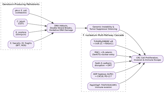
Bile acid metabolism illustrates this well. Bacterial 7α-dehydroxylase, expressed by pathobionts such as Clostridium species, converts primary bile acids into cytotoxic secondary forms — chiefly deoxycholic acid (DCA) and lithocholic acid (LCA) (Yoon et al., 2026). Excess DCA suppresses intestinal farnesoid X receptor (FXR) signaling, further weakening tight junctions and accelerating barrier collapse (Schierwagen et al., 2020; Rusman et al., 2025), and once it reaches the liver, DCA induces DNA damage, genomic instability, and mitochondrial oxidative stress in hepatocytes (Pérez Escriva et al., 2025). Together with lipoteichoic acid, DCA also activates TLR2 on HSCs, inducing cyclooxygenase-2 and a senescence-associated secretory phenotype (SASP) — senescent HSCs then secrete a cocktail of pro-inflammatory factors that plausibly foster a pro-tumorigenic microenvironment conducive to hepatocellular carcinoma (HCC) (Cao et al., 2024; Yoon et al., 2026). Secondary bile acids also appear to suppress natural killer T cell recruitment by downregulating CXCL16 on sinusoidal endothelial cells, effectively blinding hepatic tumor immune surveillance (Cao et al., 2024; Yoon et al., 2026). Other metabolites act through entirely different routes — gut-derived acetate, for instance, can feed the TCA cycle to support de novo lipogenesis and glutamine biosynthesis in HCC cells under high-fructose dietary conditions (Pérez Escriva et al., 2025)The reach of host–microbe interactions extends well beyond the liver, touching genomic stability and oncogenic signaling across the gastrointestinal tract more broadly — a pattern that suggests a shared mechanistic template rather than organ-specific coincidence (Pérez Escriva et al., 2025). Several pathobionts have evolved genotoxins that are directly mutagenic: pks+ Escherichia coli produces colibactin, and Campylobacter jejuni produces cytolethal distending toxin (CDT); both induce covalent DNA adducts and double-strand breaks capable of overwhelming host repair mechanisms (Canowitz et al., 2026; Pérez Escriva et al., 2025). Klebsiella oxytoca contributes tilimycin, an alkylating metabolite implicated in colorectal cancer initiation, while Enterococcus faecalis and Bacteroides fragilis enterotoxin generate high levels of intracellular reactive oxygen species that silence tumor suppressor genes epigenetically (Canowitz et al., 2026; Cao et al., 2024).

Fusobacterium nucleatum is, arguably, the best-characterized example of pathobiont-driven tumor promotion, and it works through several parallel routes rather than a single dominant one (see Figure 3): activation of TLR4/MyD88/NF-κB signaling upregulates miR-21, which suppresses RASA1 and activates RAS-MAPK signaling (Cao et al., 2024); LPS-driven PAK1 phosphorylation promotes β-catenin nuclear translocation and downstream c-Myc/Cyclin D1 expression (Cao et al., 2024); the FadA adhesin disrupts E-cadherin/β-catenin complexes, triggering epithelial–mesenchymal transition and drug-resistance gene expression (Cao et al., 2024); the bacterial metabolite ADP-heptose engages host ALPK1 to drive CXCL8 secretion while simultaneously suppressing mismatch repair genes and upregulating PD-L1 (Cao et al., 2024); and surface proteins Fap2 and CbpF engage inhibitory receptors TIGIT and CEACAM1 on natural killer and T cells, producing an immunosuppressive tumor microenvironment (Cao et al., 2024). Formate, another microbial metabolite, activates aryl hydrocarbon receptor (AhR) signaling in colorectal cancer cells to promote stemness and metastatic dissemination (Pérez Escriva et al., 2025; Yoon et al., 2026) — a striking contrast to tryptophan-derived indole-3-propionic acid from a healthy microbiome, which activates the same AhR pathway to strengthen epithelial barriers instead (Pérez Escriva et al., 2025; Yoon et al., 2026). It is a useful reminder that the same receptor, engaged by different microbial chemistry, can push tissue biology in opposite directions.

Taken individually, the gut–liver axis, hepatic fibrogenesis, and microbial oncology are each reasonably mature fields. What is missing, we think, is a synthesis that treats them as parts of one continuous mechanistic story rather than three adjacent ones. This review attempts that synthesis, with five specific aims: To delineate the structural, cellular, and molecular architecture of the bidirectional gut–liver–immune axis, from homeostatic tolerance to pathological inflammation. To examine the molecular cascades through which translocated PAMPs and microbial metabolites drive HSC activation and progressive ECM deposition — including a focused case study of the newly described Nidogen-1–JAK2/STAT3–IL-6 axis. To decode the dual role of gut-derived metabolites (secondary bile acids, SCFAs, tryptophan derivatives) as regulators of host DNA damage, epigenetic remodeling, and oncogenic signaling. To compare the oncogenic mechanisms of key pathobionts, using Fusobacterium nucleatum and genotoxin-producing Enterobacteriaceae as templates across liver, gastric, and colorectal cancers. To critically evaluate current and next-generation therapeutic strategies targeting this axis — receptor-centric agents, RNA-based therapeutics, phytochemicals, and microbiota-directed interventions — with attention to translational barriers.

2. Gut-Liver-Immune Axis in Chronic Liver Disease: Mechanisms of Fibrosis and Therapeutic Targets

2.1 The Anatomical and Immunological Basis of the Gut–Liver–Immune Axis

It is worth pausing, before wading into pathology, to appreciate how the healthy axis actually works — because most of what goes wrong in disease is best understood as an exaggeration or corruption of normal physiology, not an entirely novel process. The portal venous system is the anatomical backbone of the relationship, delivering the majority of hepatic blood flow directly from the gut and spleen (Portincasa et al., 2020; Li et al., 2021). This is not a passive pipe; hepatic sinusoidal endothelial cells, Kupffer cells, and HSCs are all positioned along this route specifically to sample its contents, and under normal conditions they do so tolerantly rather than reactively (Yoon et al., 2026). Reciprocal traffic in the opposite direction — bile acids, IgA, antimicrobial peptides — helps maintain a microbiota that, in turn, does not overwhelm this tolerance (Portincasa et al., 2020). Figure 1 summarizes this circuit and the point at which chronic disease states interrupt it.

2.2 Dysbiosis as an Initiating Event in Chronic Liver Disease

Several independent lines of evidence converge on the idea that dysbiosis is not merely a bystander phenomenon in chronic liver disease but an active initiating event. Reduced alpha diversity, loss of SCFA-producing Clostridia, and pathobiont overgrowth have each been documented across MASLD, ALD, and viral hepatitis cohorts with a consistency that is somewhat unusual for microbiome research, a field not always known for reproducibility (Rusman et al., 2025; Weiskirchen, 2025). What is less settled — and this seems worth saying plainly rather than glossing over — is the direction of causality in human studies; much of the strongest mechanistic evidence for dysbiosis driving fibrosis, rather than simply accompanying it, still comes from rodent models (Li et al., 2021). That caveat does not undermine the model so much as temper how confidently it should be stated.

2.3 Molecular Sensing of Translocated Microbial Products in the Hepatic Lobule

The TLR4/MyD88/NF-κB axis remains the best-characterized route by which translocated LPS drives hepatic inflammation, but it is not the only one, and treating it as such risks oversimplifying a genuinely multi-receptor system (Yoon et al., 2026; Portincasa et al., 2020). TLR9 (bacterial DNA), TLR5 (flagellin), and the NLRP3/NLRP6 inflammasomes each contribute independently, and there is growing evidence that these pathways cross-talk rather than operate in parallel isolation (Yoon et al., 2026). Table 3 catalogs the principal mechanistic nodes — endotoxemia/LPS, deoxycholic acid and secondary bile acids, short-chain fatty acids, endogenous ethanol, and choline/TMAO metabolism — alongside their receptors, barrier effects, and downstream hepatic consequences.

2.4 Hepatic Stellate Cell Activation: Canonical Pathways and Epigenetic Control

HSC activation has traditionally been described through the lens of TGF-β/Smad and PDGF/PI3K-Akt signaling, and that description remains largely accurate as far as it goes (Akkız et al., 2024; Weiskirchen, 2025). What has changed is an appreciation of how tightly this canonical signaling is intertwined with epigenetic regulation — DNA methylation at the Pten and Pparγ promoters, histone modifications via EZH2 and ASH1, and a dense network of profibrogenic and antifibrotic microRNAs (miR-21, miR-29 family, miR-34) that fine-tune the transcriptional output of activated HSCs (Akkız et al., 2024). Wnt/β-catenin signaling adds yet another layer, cross-talking with hedgehog signaling and directly suppressing PPARγ-driven adipogenic gene expression to lock HSCs into their myofibroblastic phenotype (Weiskirchen, 2025; Elpek, 2014). Table 2 organizes these pathways side by side — canonical TGF-β signaling, PDGF signaling, Wnt/β-catenin, and epigenetic control — to make the redundancy and cross-talk between them easier to see at a glance than the primary literature typically allows.

2.5 The Nidogen-1–JAK2/STAT3–IL-6 Axis: A Structural-Immune Node in Steatohepatitis

Metabolic dysfunction-associated steatotic liver disease (MASLD) — the contemporary name for what was, until relatively recently, called non-alcoholic fatty liver disease — now affects an estimated 25% to 32% of the global population, and a meaningful fraction of those patients progress from simple steatosis to steatohepatitis (MASH) and, eventually, fibrosis (Habibullah et al., 2024; Nian et al., 2026; Rusman et al., 2025). Within that progression, one recently characterized node deserves particular attention, both because it is mechanistically novel and because it illustrates a broader principle: that basement membrane glycoproteins are not inert scaffolding but active signaling participants (Nian et al., 2026; Yoon et al., 2026).

Nidogen-1 (Nid1) is, under normal conditions, a self-assembling, sulfated glycoprotein that physically bridges laminin and type IV collagen networks, stabilizing basement membrane architecture (Nian et al., 2026). During MASH progression, however, integrated proteomic and transcriptomic profiling — in both CDAHFD-fed mouse models and human MASLD/MASH biopsies — has identified a marked upregulation of Nid1, localized specifically to activated, α-SMA-positive HSCs and the surrounding perisinusoidal space (Nian et al., 2026). Exposure to TGF-β1 or PDGF drives HSCs to increase Nid1 transcription substantially, alongside classical fibrotic markers such as α-SMA and Col1a1 (Nian et al., 2026; Seki & Brenner, 2016).

What makes this more than an incidental biomarker is the discovery of a self-reinforcing autocrine loop: exogenous recombinant human Nid1 (rHuNid1), applied to quiescent HSCs, is sufficient on its own to trigger cellular activation — no upstream cytokine stimulus required (Nian et al., 2026). Mechanistically, Nid1 engages cell-surface
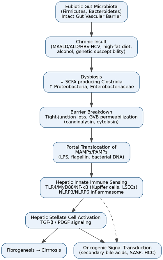
 

Figure 1 traces the sequential breakdown of gut–liver homeostasis under chronic metabolic, alcohol-related, or viral insult. Beginning from an intact gut vascular barrier and eubiotic microbiota, the schematic shows how dysbiosis, tight-junction loss, and portal translocation of MAMPs/PAMPs converge on hepatic innate immune sensing (TLR4/MyD88/NF-κB, NLRP3/NLRP6), driving hepatic stellate cell activation, fibrogenesis, and downstream oncogenic signal transduction. Constructed by the authors from Schnabl (2014), Portincasa et al. (2020), Yoon et al. (2026), and Weiskirchen (2025).
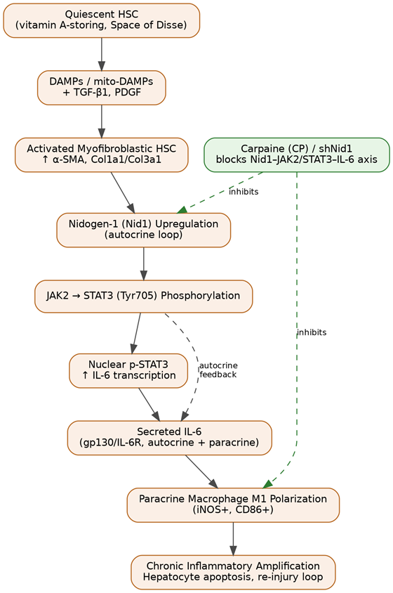
Figure 2 depicts the self-reinforcing feed-forward loop by which basement-membrane-derived Nidogen-1 drives JAK2/STAT3 phosphorylation and autocrine IL-6 secretion in activated hepatic stellate cells, which subsequently polarizes neighboring macrophages toward a pro-inflammatory M1 phenotype through a paracrine, IL-6-dependent mechanism. The points at which Carpaine or genetic Nid1 silencing interrupt this cascade are indicated. Constructed by the authors from Nian et al. (2026) and Solleiro-Villavicencio et al. (2025).

receptors, most plausibly integrins, to initiate JAK2 phosphorylation, which in turn phosphorylates STAT3 at tyrosine 705 (Nian et al., 2026). Phosphorylated STAT3 dimerizes, translocates to the nucleus, and binds directly to the promoters of Acta2, Col1a1, and — critically — the gene encoding IL-6 (Nian et al., 2026; Solleiro-Villavicencio et al., 2025). Secreted IL-6 then binds back to the gp130/IL-6 receptor complex on the same HSCs, reinforcing JAK2/STAT3 phosphorylation in a closed feed-forward loop (Nian et al., 2026). Figure 2 depicts this cascade in full, including the points at which pharmacological or genetic intervention interrupts it.

2.6 HSC–Macrophage Crosstalk: Nid1/IL-6 as a Paracrine Driver of M1 Polarization

Fibrogenesis does not occur in a cellular vacuum, and one of the more interesting findings to emerge from this line of research is how indirect the HSC–macrophage relationship actually is. Hepatic macrophages, whether resident Kupffer cells or recruited monocyte-derived populations, sit along a broad phenotypic spectrum bounded by classically activated M1 and alternatively activated M2 states (Akkız et al., 2024). During MASH, CCL2/CCR2-driven monocyte recruitment expands the M1 population within fibrotic septa, and these cells secrete iNOS, CD86, TNF-α, and IL-1β — mediators that directly worsen hepatocyte injury and further activate HSCs (Akkız et al., 2024; Solleiro-Villavicencio et al., 2025).

The counterintuitive part is what does not drive this polarization directly. When macrophages are exposed to recombinant Nid1 or Carpaine in monoculture, no meaningful shift in M1 markers occurs at all (Nian et al., 2026) — implying that Nid1 lacks a direct receptor-mediated action on macrophages. The polarizing signal instead arrives secondhand, via the HSC secretome: in transwell co-culture, TGF-β1-activated HSCs drive rapid, pronounced M1 polarization in neighboring macrophages, and cytokine profiling identifies IL-6 — not TNF-α, not IL-1β — as the indispensable mediator (Nian et al., 2026). This is a fairly elegant demonstration that HSCs are not simply downstream targets of hepatic inflammation but active upstream orchestrators of it, reprogramming the local immune microenvironment through a single, druggable cytokine node (Nian et al., 2026).

2.7 Carpaine and the Broader Phytotherapeutic Landscape

Carpaine (CP), a dimeric piperidine alkaloid derived from Carica papaya leaves, has emerged as a case study in how a single exogenous compound can intervene at a structural-signaling level rather than a purely metabolic one (Nian et al., 2026). In co-culture systems, Carpaine-treated HSCs show a selective and substantial reduction in IL-6 output, which halts downstream M1 macrophage polarization and dampens the broader inflammatory amplification loop (Nian et al., 2026). Notably — and this detail matters for interpreting its mechanism correctly — Carpaine does not directly resolve hepatocyte steatosis when tested in lipid-laden THLE-2 cells, indicating that its efficacy is not attributable to conventional lipid-clearing pathways but instead to targeted rebalancing of the HSC–ECM–immune axis (Nian et al., 2026).

Carpaine is far from the only phytotherapeutic agent under investigation along the gut–liver–adipose axis. Traditional Chinese Medicine formulations — Si Miao San, Qushi Huayu Decoction, Danshen Yin, and purified Astragalus mongholicus polysaccharides — each engage distinct but overlapping nodes: SREBP-1c/AMPK-mediated lipogenesis suppression, JAK2/STAT3/CPT-1A-driven fatty acid oxidation, Nrf2/HO-1 antioxidant signaling, and TLR4/NF-κB-mediated endotoxin handling, respectively (Tanwar & Sharma, 2026). Table 1 summarizes these agents alongside their molecular targets, gut–liver–adipose mechanisms, and the strength of their preclinical and clinical evidence.

2.8 Systems-Level Integration: Microbiota Remodeling as a Convergent Therapeutic Endpoint

Whatever the entry point — Carpaine, AAV8-mediated Nid1 silencing, or an unrelated TCM formulation — the therapeutic interventions reviewed here tend to converge on a common downstream signature: recovery of gut microbial alpha and beta diversity, enrichment of Bacteroides, Parabacteroides goldsteinii, Blautia, and Lachnospiraceae, and a functional metagenomic shift (measured via PICRUSt2/FAPROTAX) toward SCFA biosynthesis and away from virulence-associated pathways (Nian et al., 2026). This convergence is, we would argue, one of the more persuasive pieces of indirect evidence that the gut–liver axis is genuinely bidirectional rather than liver-centric: correcting hepatic pathology appears sufficient, on its own, to reshape a distant microbial community, presumably by reducing the spillover of inflammatory mediators and abnormal bile-acid profiles into the intestinal lumen (Nian et al., 2026; Portincasa et al., 2020). Figure 4 traces this remodeling process from

Table 1 synthesizes single phytochemical compounds and traditional herbal formulations that engage the gut–liver–adipose axis to resolve steatotic liver disease, listing each agent’s active constituents, primary molecular targets, mechanism of action, and the strength of supporting preclinical or clinical evidence. Carpaine is included as the focal compound of Sections 2.5–2.7 of this review. Adapted by the authors from Nian et al. (2026) and Tanwar and Sharma (2026).

Therapeutic Agent

Key Active Constituents

Primary Molecular/Cellular Targets

Gut–Liver–Adipose Mechanism

Evidence Summary

References

Carpaine (CP)

Dimeric piperidine alkaloid (Carica papaya leaves)

Nid1, JAK2/STAT3, IL-6, gp130

Downregulates Nid1 in activated HSCs; suppresses JAK2/STAT3-driven autocrine IL-6; indirectly blocks paracrine M1 polarization; restores microbial diversity (Bacteroides, Blautia)

In vivo: reduced hepatic lipid vacuoles, α-SMA, ALT/AST in CDAHFD mice; in vitro: arrested LX-2 activation without direct steatosis reversal

Nian et al. (2026)

Si Miao San (SMS)

Atractylodes lancea, Phellodendron chinense, Achyranthes bidentata, Coix lacryma-jobi

SREBP-1c/FAS, AMPK, gut tight junctions, Akkermansia muciniphila

Suppresses de novo lipogenesis; activates AMPK/ACC fatty-acid oxidation; restores mucosal barrier and enriches A. muciniphila

Preclinical: reduced weight gain and hepatic triglycerides in HFHS models; clinical: reduced serum liver enzymes with syndrome-differentiated dosing

Tanwar & Sharma (2026)

Qushi Huayu Decoction (QHD)

Artemisia capillaris, Gardenia jasminoides, Polygonum cuspidatum, Scutellaria baicalensis, Sesamum indicum

CPT-1A, JAK2/STAT3, Cathepsin B/TNF-α, AdipoR2

Upregulates CPT-1A-driven fatty-acid oxidation; restores adipose-liver adiponectin signaling; corrects dysbiosis and gut-vascular barrier

Preclinical: reduced liver TG/ALT in MASH rats; clinical: improved steatosis score and insulin sensitivity

Tanwar & Sharma (2026)

Danshen Yin

Salvia miltiorrhiza, Santalum album, Amomum villosum

Nrf2/HO-1, TLR4/NF-κB, PPARγ

Activates antioxidant Nrf2/HO-1 signaling; blocks TLR4-NF-κB inflammatory signaling in Kupffer cells; preserves HSC PPARγ quiescence

Preclinical: decreased hepatic lipid accumulation and portal LPS; clinical: improved liver histology in MASLD

Tanwar & Sharma (2026)

Astragalus mongholicus Polysaccharides (mAPS)

Purified polysaccharide fraction

TLR4/NF-κB, PI3K/AKT, Firmicutes/Bacteroidetes ratio

Suppresses endotoxin translocation and hepatic TLR4/NF-κB signaling; enhances insulin sensitivity via PI3K/AKT; remodels microbiota ratio

Preclinical: reduced ALT/AST/TG in MASLD and ALD models

Tanwar & Sharma (2026)

Table 2 deconstructs the four principal intracellular signaling and epigenetic systems governing the transdifferentiation of quiescent hepatic stellate cells into extracellular-matrix-producing myofibroblasts, detailing the key mediators, activation mechanisms, downstream effects on collagen and matrix remodeling genes, and cross-talk between pathways. Adapted by the authors from Akkız et al. (2024), Elpek (2014), and Weiskirchen (2025).

Pathway/Regulator

Key Mediators

Activation Mechanism

Downstream ECM Effects

Cross-Talk

References

TGF-β1 Signaling

Smad2/3/7, TGF-βRI/II, JNK/p38 MAPK, BAMBI

Canonical Smad2/3 phosphorylation and nuclear translocation; non-canonical PDGF-driven Smad3 linker phosphorylation

Upregulates Col1a1/Col3a1, α-SMA, CTGF; increases TIMP-1/2, shifting balance toward matrix deposition

Integrated with IL-17 signaling and integrin-αv-mediated latent TGF-β activation

Akkız et al. (2024); Elpek (2014); Weiskirchen (2025); Yoon et al. (2026)

PDGF Signaling

PDGF-A/B/C/D, PDGFR-β, Ras/Raf/MEK/ERK, PI3K/Akt/mTOR

PDGF-BB-induced receptor homodimerization and autophosphorylation

Promotes HSC proliferation/migration; upregulates Col1a1; stimulates LOX/LOXL2-mediated collagen crosslinking

Induces sonic hedgehog ligands; cross-talks with non-canonical TGF-β/JNK pathway

Acharya et al. (2021); Akkız et al. (2024); Weiskirchen (2025)

Wnt/β-Catenin Signaling

Wnt3a/Wnt10b, Frizzled/LRP5/6, β-catenin, CBP/p300

Wnt-Frizzled binding inhibits GSK3β destruction complex, stabilizing β-catenin

Upregulates α-SMA and Col1a1; suppresses adipogenic/lipogenic genes

Cooperates with hedgehog signaling; upregulates MeCP2, repressing PPARγ

Weiskirchen (2025)

DNA/Histone Methylation (Epigenetic Control)

DNMT1/3a/3b, MeCP2, EZH2, ASH1, TET3

Hypermethylation of Pten/Pparγ promoters; H3K27me3/H3K4me histone modification

Silences PPARγ (loss of quiescence); activates profibrogenic genes (Actg2, Loxl1/2, Col4a1)

TET3 loss in NASH causes global 5-hmC loss and permanent gene silencing

Akkız et al. (2024)

the dysbiotic starting state through to the resolution of hepatic inflammation.

2.9 Pharmacological and Investigational Therapeutics Targeting the Axis

Outside the phytotherapeutic space, several receptor-targeted and RNA-based agents have reached clinical or late preclinical stages, with decidedly mixed results (summarized in Table 4). Resmetirom, a selective thyroid hormone receptor-β agonist, achieved FDA approval on the strength of the Phase 3 MAESTRO-NASH trial, showing meaningful reductions in liver fat and at least one-stage fibrosis regression (Tanwar & Sharma, 2026; Liu et al., 2025). Semaglutide, acting through GLP-1 receptor agonism, achieved MASH resolution without fibrosis worsening in the ESSENCE trial, though its direct effect on advanced fibrosis is less consistent (Tanwar & Sharma, 2026). Obeticholic acid, an FXR agonist, met its primary fibrosis endpoint in the REGENERATE trial but is constrained clinically by dose-dependent pruritus and adverse lipid changes (Tanwar & Sharma, 2026; Liu et al., 2025). Lanifibranor, a pan-PPAR agonist, showed encouraging Phase 2b fibrosis regression (Liu et al., 2025). Not every candidate has fared as well: emricasan, a pan-caspase inhibitor, failed to improve liver histology in Phase 2b/3 trials and, if anything, appeared to worsen hepatocyte ballooning — a useful reminder that mechanistic plausibility does not guarantee clinical benefit (Akkız et al., 2024; Elpek, 2014; Schierwagen et al., 2020). RNA-based approaches are earlier in development but conceptually interesting: BMS-986263, an HSP47-targeting siRNA delivered via lipid nanoparticle, showed early dose-dependent histological improvement in Phase 1b/2 testing (Akkız et al., 2024; Schierwagen et al., 2020), while cenicriviroc, a dual CCR2/CCR5 antagonist, demonstrated Phase 2b anti-fibrotic efficacy that did not replicate in the larger Phase 3 AURORA trial (Acharya et al., 2021; Schierwagen et al., 2020).

3. Methods

3.1 Review Design and Reporting Standards

This is a narrative-synthesis review with a systematic search component, structured to satisfy transparency and reproducibility expectations consistent with PubMed/MEDLINE indexing standards and, where applicable, the general reporting logic of the PRISMA framework for identifying and screening literature (though we do not claim formal PRISMA systematic-review status, given the mechanistic and cross-disciplinary scope of the topic). The aim throughout was to make the search strategy specific enough that another investigator could reproduce it and arrive at a materially similar evidence base.

3.2 Information Sources and Search Strategy

We searched PubMed/MEDLINE, Scopus, Web of Science, and the Cochrane Library for records published through early 2026, supplemented by manual screening of reference lists from key included articles (a form of citation-chasing that, admittedly, introduces its own selection biases but remains standard practice for narrative synthesis of this kind). Search terms were combined using Boolean operators and organized around four conceptual clusters: (1) gut–liver axis terms (“gut-liver axis,” “gut-liver-immune axis,” “intestinal permeability,” “dysbiosis,” “portal translocation”); (2) fibrosis and stellate cell terms (“hepatic stellate cell,” “liver fibrosis,” “fibrogenesis,” “Nidogen-1,” “JAK2/STAT3,” “TGF-beta signaling”); (3) oncology terms (“hepatocellular carcinoma,” “colorectal cancer,” “Fusobacterium nucleatum,” “colibactin,” “microbial genotoxins,” “tumor microenvironment”); and (4) therapeutic terms (“resmetirom,” “semaglutide,” “obeticholic acid,” “fecal microbiota transplantation,” “Carpaine,” “Traditional Chinese Medicine,” “siRNA liver fibrosis”). Clusters were combined with AND, and synonyms within each cluster with OR (e.g., (“gut-liver axis” OR “gut-liver-immune axis”) AND (“hepatic stellate cell” OR “liver fibrosis”) AND (“hepatocellular carcinoma” OR “colorectal cancer”)).

3.3 Eligibility Criteria

We included peer-reviewed original research articles, mechanistic and preclinical studies, and review articles published in English between 2014 and 2026, with a deliberate emphasis on the most recent (2024–2026) literature to capture rapidly evolving mechanistic concepts such as the Nid1–JAK2/STAT3–IL-6 axis. Studies were required to address at least one of: gut microbiota composition or function in liver disease; molecular mechanisms of hepatic stellate cell activation; microbial metabolite or genotoxin effects on host DNA damage or oncogenic signaling; or therapeutic interventions (pharmacological, phytochemical, or microbiota-directed) targeting the gut–liver–immune axis. We excluded conference abstracts without full-text availability, non-English-language articles without accessible translation,

Table 3 catalogs the principal microbial metabolites and translocated products implicated in steatohepatitis progression, mapping each to its microbial source, host receptor/pathway, effect on intestinal barrier integrity, and downstream hepatic inflammatory or fibrogenic consequence. Adapted by the authors from Elpek (2014), Yoon et al. (2026), Rusman et al. (2025), Sharpton et al. (2021), and Liu et al. (2025).

Mechanistic Node

Source/Etiology

Receptors/Pathways

Barrier/Translocation Effect

Hepatic Consequence

References

Endotoxemia/LPS

Gram-negative pathobionts (Enterobacteriaceae, Proteobacteria)

TLR4/CD14, NF-κB/MAPK

Degrades claudin-1/ZO-3; disrupts gut-vascular barrier

Kupffer/HSC activation; TNF-α, IL-6, IL-1β secretion; sensitizes HSCs to TGF-β/PDGF

Elpek (2014); Yoon et al. (2026); Rusman et al. (2025); Sharpton et al. (2021)

Deoxycholic Acid & Secondary Bile Acids

7α-dehydroxylating Clostridium spp.

FXR, TGR5, EGFR/Raf-1/ERK

Depletes primary bile acids; suppresses intestinal FXR

Hepatocyte lipotoxicity, mito-DAMP release, NLRP3 activation, HSC SASP

Yoon et al. (2026); Rusman et al. (2025); Liu et al. (2025)

Short-Chain Fatty Acids (SCFAs)

Fermentation by Blautia, Bacteroides, Lachnospiraceae

GPR41/GPR43, GPR109A, HDAC inhibition

Stabilizes tight junctions; reduces macromolecular flux

Promotes M2 macrophage polarization; inhibits NF-κB/NLRP3; activates hepatic AMPK/FAO

Yoon et al. (2026); Rusman et al. (2025); Sharpton et al. (2021)

Endogenous Ethanol

High-alcohol-producing Klebsiella pneumoniae, Escherichia spp.

Alcohol dehydrogenase, CYP2E1, PKC

Impairs tight junctions (“leaky gut”)

Acetaldehyde-driven Col1a1 transcription; ROS-mediated hepatocyte apoptosis

Yoon et al. (2026); Elpek (2014); Rusman et al. (2025)

Choline Depletion/TMA-TMAO

Microbial TMA production by E. coli, Enterobacteriaceae

Hepatic FMO3, FXR suppression

Depletes choline bioavailability

Impairs VLDL packaging (steatosis); TMAO worsens inflammation and vascular stiffness

Sharpton et al. (2021)

Table 4 compares the mechanism of action, cellular target, preclinical findings, and clinical trial outcomes (including reported adverse effects) of receptor-targeted small molecules, RNA-based therapeutics, and approved agents currently under investigation for steatohepatitis and hepatic fibrosis, situating the Carpaine/Nid1 findings of this review (Section 4) within the broader investigational landscape. Adapted by the authors from Tanwar and Sharma (2026), Liu et al. (2025), Akkız et al. (2024), and Schierwagen et al. (2020).

Candidate

Mechanism/Class

Target

Preclinical Findings

Clinical Status/Outcomes

References

Resmetirom (MGL-3196)

Selective THR-β agonist

Hepatocytes

Activated fatty-acid β-oxidation; reduced mitochondrial lipotoxicity

FDA-approved for noncirrhotic MASH with F2–F3 fibrosis; MAESTRO-NASH showed fibrosis regression ≥1 stage; mild diarrhea/nausea

Tanwar & Sharma (2026); Liu et al. (2025); Yoon et al. (2026)

Semaglutide

GLP-1 receptor agonist

CNS, pancreas, adipose, liver

Promoted BAT thermogenesis; reduced TLR-driven inflammation

ESSENCE trial: MASH resolution without fibrosis worsening; GI side effects, weight loss

Tanwar & Sharma (2026); Yoon et al. (2026)

Obeticholic Acid (OCA)

FXR agonist

Intestine, liver

Downregulated de novo lipogenesis; suppressed HSC activation via BAMBI

REGENERATE: ≥1-stage fibrosis improvement; dose-dependent pruritus, elevated LDL

Tanwar & Sharma (2026); Liu et al. (2025)

Lanifibranor

Pan-PPAR agonist

Hepatocytes, Kupffer cells, HSCs, adipose

Combined lipid-clearing and anti-fibrotic effects

Phase 2b: ≥2-point SAF score reduction; weight gain, edema, diarrhea, anemia

Liu et al. (2025)

Emricasan

Pan-caspase inhibitor

Hepatocytes

Blocked apoptosis; reduced inflammation in CCl4 model

Phase 2b/3: failed to improve histology; worsened ballooning

Akkız et al. (2024); Elpek (2014); Schierwagen et al. (2020)

BMS-986263 (ND-L02-s0201)

HSP47 siRNA (lipid nanoparticle)

Hepatic stellate cells

Blocked collagen chaperone activity

Phase 1b/2: good safety; early dose-dependent histological improvement

Akkız et al. (2024); Schierwagen et al. (2020)

Cenicriviroc

Dual CCR2/CCR5 antagonist

Monocytes, macrophages, HSCs

Prevented Ly6C-high monocyte recruitment

Phase 2b (CENTAUR) efficacy signal; Phase 3 AURORA terminated for lack of efficacy; favorable safety

Acharya et al. (2021); Schierwagen et al. (2020)

and case reports lacking mechanistic data.

3.4 Study Selection and Data Extraction

Titles and abstracts were screened for topical relevance against the eligibility criteria above; full texts of potentially eligible records were then reviewed in full. For mechanistic and preclinical studies, we extracted the experimental model (cell line, animal model, or human cohort), the specific molecular pathway or receptor interaction under investigation, key quantitative or qualitative findings, and the original authors’ interpretation, cross-checking claims against the primary data presented in figures and tables where accessible. For pharmacological and clinical studies, we additionally extracted trial phase, primary endpoints, and reported adverse events, to allow the efficacy summaries in Table 4 to reflect both benefit and risk rather than benefit alone.

3.5 Synthesis Approach

Given the heterogeneity of study designs — spanning in vitro cell culture, murine diet-induced disease models, human biopsy cohorts, and randomized clinical trials — a quantitative meta-analytic synthesis was not appropriate, and we instead performed a structured narrative synthesis organized around mechanistic themes (barrier integrity, innate immune sensing, stellate cell signaling, oncogenic transformation, and therapeutic intervention). Where multiple sources converged on the same mechanistic claim, we cited the most direct or mechanistically detailed source in the main text while cross-referencing supporting sources in the accompanying tables. Discrepancies between sources — for instance, differing views on whether hepatic Nid1 suppression causally drives, or merely correlates with, gut microbial remodeling — are flagged explicitly in the relevant discussion subsections rather than resolved by omission.

3.6 Figure and Table Construction

The four schematic figures accompanying this review (Figures 1–4) were constructed de novo by the authors to visually integrate mechanistic findings scattered across multiple primary sources; they are original graphical syntheses rather than reproductions of any single published figure, and each is cited to the primary literature underlying its content in the corresponding legend. The four summary tables (Tables 1–4) were similarly constructed to consolidate comparative data — on phytotherapeutic agents, HSC signaling pathways, gut–liver mechanistic nodes, and investigational pharmacotherapies, respectively — extracted during the data-extraction step described in Section 3.4.

4. Synthesis of the Molecular, Cellular, and Microecological Evidence Linking Nidogen-1 Signaling to Hepatic Fibrogenesis

4.1 Multi-Omics Profiling Identifies Nidogen-1 as a Structural Candidate in Steatohepatitis

Across the studies synthesized in this review, a consistent pattern emerges from multi-omics profiling of steatohepatitis models. In C57BL/6J mice subjected to a nine-week choline-deficient, L-amino acid-defined, high-fat diet — a standard and reasonably well-validated model of MASH — histopathological analysis confirmed the expected features: pronounced lipid vacuolation, elevated α-SMA immunohistochemical scores, and correspondingly elevated serum ALT/AST, all consistent with active hepatocellular injury (Nian et al., 2026) (Table 4). Untargeted fecal metabolomics, visualized by principal coordinates analysis, showed clean separation between healthy and MASH-induced cohorts, and among the metabolites most strikingly depleted during the transition from simple steatosis to fibrotic MASH was Carpaine itself (Nian et al., 2026). When these fecal metabolomic profiles were integrated with hepatic tissue proteomics, Nidogen-1 emerged as the strongest negative correlate of Carpaine depletion — a relationship subsequently confirmed by immunohistochemistry showing elevated Nid1 expression localized to the perisinusoidal space of Disse, closely aligned with collagen type I accumulation and α-SMA-positive HSCs in both murine and human MASLD tissue (Nian et al., 2026) (Figure 1).

4.2 Direct Effects on HSC Activation State Confirm a Nid1-Dependent Autocrine Loop

Working with human LX-2 stellate cells — a reasonably standard, if imperfect, in vitro model for HSC biology — TGF-β1 stimulation reliably induced the expected transdifferentiation phenotype: elevated α-SMA alongside a corresponding rise in endogenous Nid1 at both the transcript and protein level (Nian et al., 2026). Gene-knockdown experiments (shNid1) reversed this activated phenotype, significantly reducing α-SMA transcription, while exogenous recombinant human Nid1 supplementation was, on its own, sufficient to trigger spontaneous activation — direct evidence for the self-reinforcing autocrine loop described in Section 2.5 (Nian et al., 2026) (Figure 2). Concurrent Carpaine treatment suppressed this loop at both the endogenous and rHuNid1-induced level. Importantly, when the same Carpaine treatment was applied to free-fatty-acid-loaded THLE-2 hepatocytes, BODIPY staining showed no meaningful reduction in lipid accumulation — a negative finding that turns out to be mechanistically informative, since it indicates Carpaine’s anti-fibrotic action operates through the structural-immune interface rather than through conventional hepatocyte lipid-clearing pathways (Nian et al., 2026).

4.3 Flow Cytometric and Co-Culture Evidence for IL-6-Mediated Paracrine Macrophage Polarization

Flow cytometric profiling of liver-resident non-parenchymal cells in untreated MASH mice confirmed heavy infiltration by CD86⁺ M1 macrophages, CD11b⁺Ly6G⁺ neutrophils, and CD3⁺ T cells; daily oral Carpaine administration reduced this infiltration significantly and shifted the macrophage compartment toward CD206⁺/Arg-1⁺ M2 polarization (Nian et al., 2026) (Table 4). Mechanistic dissection using monocultured THP-1-derived macrophages showed no direct effect of Carpaine on M1 markers, ruling out a direct macrophage-intrinsic mechanism. In transwell co-culture, however, vehicle-treated activated HSCs drove robust M1 polarization in neighboring macrophages, an effect that was fully blocked by either shNid1 knockdown or Carpaine treatment of the HSC compartment (Nian et al., 2026). Cytokine-panel profiling of the co-culture secretome identified IL-6, rather than TNF-α, IL-1β, CCL2, or CXCL10, as the sole cytokine selectively downregulated by Carpaine — pointing to IL-6 as the indispensable paracrine mediator of this crosstalk (Nian et al., 2026). Pharmacological blockade with an IL-6 neutralizing antibody or a selective STAT3 inhibitor each independently confirmed the pathway, blocking rHuNid1-induced JAK2/STAT3 phosphorylation and preventing downstream M1 polarization in vitro; in vivo, AAV8-mediated, HSC-specific Nid1 silencing reproduced these effects, reducing hepatic JAK2/STAT3 phosphorylation, depleting intrahepatic IL-6, and resolving M1 macrophage-driven tissue inflammation (Nian et al., 2026) (Figure 2).

4.4 Gut Microecological Restructuring Accompanies Resolution of Hepatic Fibrosis

Consistent with the gut–liver axis framework outlined in Section 2.8, the benefits of both Carpaine treatment and hepatic Nid1 silencing extended to the intestinal compartment. 16S rDNA sequencing of fecal samples from CDAHFD-fed MASH mice showed the expected loss of alpha diversity and expansion of opportunistic Gram-negative pathobionts at baseline; both interventions produced a significant recovery of alpha and beta diversity, restructuring the community toward a more homeostatic composition (Nian et al., 2026) (Figure 4). At genus and species level, this recovery was characterized by enrichment of Bacteroides and Parabacteroides goldsteinii (barrier-reinforcing, endotoxemia-suppressing commensals), Blautia and Lachnospiraceae bacterium 28-4 (SCFA-producing fermenters linked to GPR41/GPR43-mediated M2 polarization), and Sporosarcina pasteurii (implicated in nitrogen metabolism and xenobiotic biodegradation) (Nian et al., 2026). Functional metagenomic prediction using PICRUSt2 and FAPROTAX corroborated these taxonomic shifts at the pathway level, showing upregulation of fatty acid biosynthesis, amino acid metabolism, and xenobiotic biodegradation pathways alongside downregulation of pathogenic virulence factors and secondary bile-acid biosynthetic pathways that generate cytotoxic metabolites such as deoxycholic acid (Nian et al., 2026) (Table 3, Figure 4). Read together, these findings support a model in which correcting a single hepatic structural-signaling node — Nid1 — is sufficient to resolve chronic intestinal dysbiosis, most plausibly by reducing the downstream spillover of inflammatory mediators and pathological bile-acid profiles into the enterohepatic circulation.

5. Reframing Liver Fibrosis and Cancer as Convergent Outputs of Gut–Liver–Immune Dysregulation

5.1 A Structural-Immune, Rather than Purely Metabolic, View of Steatohepatitis

Perhaps the central argument this review has tried to build, across its various sections, is that MASH pathogenesis resists explanation as a purely lipid-overload phenomenon. The Nid1–JAK2/STAT3–IL-6 findings synthesized in Section 4 make that argument fairly concretely: Carpaine resolves fibrosis and inflammation without touching hepatocyte steatosis directly (Nian et al., 2026), which is a strange result if one assumes fibrosis is simply a downstream consequence of lipid accumulation. It fits far more comfortably with the structural-immune model advanced by Akkız et al. (2024) and Weiskirchen (2025), in which basement membrane remodeling and stellate cell signaling operate as a semi-independent axis, coupled to but not entirely dictated by metabolic status.

Figure 3 summarizes two convergent routes to genomic instability and tumor promotion: direct genotoxin production by pks+ Escherichia coli* (colibactin), Campylobacter jejuni (cytolethal distending toxin), Klebsiella oxytoca (tilimycin), and Enterococcus faecalis/Bacteroides fragilis (reactive oxygen species), alongside the five parallel signaling cascades activated by Fusobacterium nucleatum — TLR4/MyD88/NF-κB, PAK1/β-catenin, FadA/E-cadherin, ADP-heptose/ALPK1, and Fap2/CbpF-mediated immune evasion. Constructed by the authors from Canowitz et al. (2026), Cao et al. (2024), and Pérez Escriva et al. (2025).*
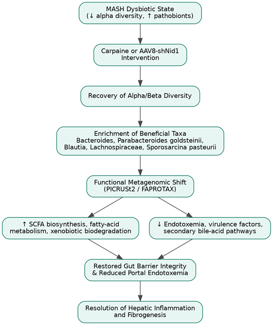
Figure 4 illustrates the downstream restructuring of gut microbial community composition and function following Carpaine treatment or AAV8-mediated hepatic Nid1 silencing, from recovery of alpha/beta diversity and enrichment of beneficial taxa, through functional metagenomic shifts (PICRUSt2/FAPROTAX) in SCFA and virulence-associated pathways, to restored gut barrier integrity and resolution of hepatic inflammation. Constructed by the authors from Nian et al. (2026).

5.2 Convergent Mechanisms Across Fibrosis and Oncogenesis

One pattern that becomes harder to ignore once the evidence is laid out side by side (Tables 2–4) is how often the same signaling nodes recur across fibrosis and cancer biology. IL-6/JAK2/STAT3 signaling drives HSC activation in fibrosis (Nian et al., 2026) and also features prominently in F. nucleatum-driven colorectal oncogenesis via NF-κB and PD-L1 upregulation (Cao et al., 2024); TLR4/NF-κB activation by translocated LPS underlies both fibrogenic priming of HSCs (Yoon et al., 2026) and pro-tumorigenic inflammatory signaling in the gut (Cao et al., 2024). Secondary bile acids, meanwhile, appear at more or less every stage of this story — degrading barrier integrity, inducing hepatocyte DNA damage, and triggering the senescence-associated secretory phenotype that plausibly bridges chronic fibrosis to HCC initiation (Yoon et al., 2026; Cao et al., 2024). We think this convergence is more than coincidental; it suggests that fibrosis and cancer, rather than being sequential stages of one disease, may be better understood as parallel outputs of a shared upstream dysregulation in gut–liver–immune signaling — an interpretation that, if correct, has real implications for how early intervention should be timed.

5.3 Therapeutic Implications and the Case for Multi-Target Intervention

Standard-of-care agents for MASH — resmetirom, semaglutide, obeticholic acid — are, almost by design, receptor-centric, targeting lipid handling or bile-acid signaling within a fairly narrow cellular compartment (Tanwar & Sharma, 2026) (Table 4). Their clinical performance, summarized in Table 4, is genuinely encouraging for steatosis and early fibrosis but noticeably less so for established, advanced scarring — a gap that recurs across several of these agents and is not, we suspect, coincidental. Carpaine’s mechanism, entering the cascade at the structural-signaling level rather than the metabolic one, suggests a complementary rather than competing role: combining a lipid-handling agent with a structural-immune modulator could, at least in principle, address both the upstream metabolic driver and the downstream fibrogenic engine simultaneously (Nian et al., 2026; Tanwar & Sharma, 2026). This is, admittedly, a hypothesis rather than a demonstrated clinical outcome, and it deserves to be tested as such rather than assumed.

The failure of emricasan in Phase 2b/3 trials, despite favorable preclinical data (Akkız et al., 2024; Schierwagen et al., 2020) (Table 4), is worth dwelling on briefly, because it is a useful check against over-optimism elsewhere in this discussion. Mechanistic plausibility in cell culture and rodent models has not, historically, translated reliably into fibrosis reversal in human trials, and there is no obvious reason to assume Nid1-targeted or Carpaine-based strategies will be exempt from that pattern. Humanized organoid systems and primary human co-culture models, as suggested by Nian et al. (2026), would help close this translational gap before resources are committed to large-scale human trials.

5.4 Limitations of the Current Evidence Base

Several limitations recur across the literature synthesized here and deserve to be stated directly rather than buried. First, much of the mechanistic evidence for the Nid1–JAK2/STAT3–IL-6 axis derives from a single research program (Nian et al., 2026); independent replication in separate laboratories and, ideally, separate MASH models would substantially strengthen confidence in its generalizability. Second, murine bile-acid pool composition differs meaningfully from human physiology — mice, unlike humans, synthesize muricholic acids — which complicates direct extrapolation of gut-microbiota findings from CDAHFD mouse models to human MASLD cohorts (Yoon et al., 2026). Third, the pharmacokinetic profile of Carpaine, including its oral bioavailability and first-pass hepatic metabolism, remains incompletely characterized, and translational development will likely require nanoparticle- or lipid-based delivery optimization before human dosing studies are feasible (Nian et al., 2026; Acharya et al., 2021). Finally, causal directionality between hepatic Nid1 suppression and gut microbial remodeling remains, honestly, unresolved; the correlation is robust, but the specific mechanistic chain connecting the two compartments is inferred rather than directly demonstrated (Nian et al., 2026).

5.5 Future Directions

Building on the gaps identified above, we would highlight three priorities for future work: (1) validation of the Nid1–JAK2/STAT3–IL-6 axis in human liver organoid systems and independent patient cohorts; (2) mechanistic dissection of the causal pathway linking hepatic structural remodeling to distal gut microbial community shifts, potentially using germ-free or gnotobiotic mouse models to isolate direction of effect; and (3) combination-therapy trials pairing receptor-centric metabolic agents (resmetirom, semaglutide) with structural-immune modulators (Carpaine or equivalent), designed explicitly to test the complementarity hypothesis raised in Section 5.3 rather than assume it.

6. Conclusion

This review has traced a single thread through what might otherwise look like three separate fields — gut microbiology, hepatic fibrogenesis, and gastrointestinal oncology — and argued that they are, mechanistically, closer to one continuous story than three. Dysbiosis and barrier failure translocate microbial products that hepatic immune cells sense through a now well-characterized receptor repertoire; this sensing activates hepatic stellate cells through both classical (TGF-β/PDGF) and newly described (Nidogen-1–JAK2/STAT3–IL-6) pathways; and the resulting inflammatory, senescent, and metabolically altered microenvironment appears sufficient, in a meaningful subset of cases, to tip fibrosis toward malignancy. Carpaine’s ability to intervene simultaneously at the structural, immune, and microbial level offers a proof of concept for multi-target therapy, though translational confirmation in humans remains outstanding. Whether this framework holds up under independent replication is, ultimately, an empirical question the field is only beginning to answer.

References


Acharya, P., Chouhan, S., Weiskirchen, S., & Weiskirchen, R. (2021). Cellular and molecular mechanisms of liver fibrosis: An update. Frontiers in Pharmacology, 12, Article 671640. https://doi.org/10.3389/fphar.2021.671640         

Akkiz, H., Gieseler, R. K., & Canbay, A. (2024). Liver fibrosis: From basic science towards clinical progress, focusing on the central role of hepatic stellate cells. International Journal of Molecular Sciences, 25(14), Article 7873. https://doi.org/10.3390/ijms25147873      

Canowitz, A., Kamble, N. S., Muck, N., Kaur, K., Garay, N., Bellala, P., ... & Kotagiri, N. (2026). Microbial medicines: unlocking the therapeutic potential of the microbiome in cancer treatment. Journal of Controlled Release, 392, 114720.. https://doi.org/10.1016/j.jconrel.2026.114720         

Cao, C., Yue, S., Lu, A., & Liang, C. (2024). Host-gut microbiota metabolic interactions and their role in precision diagnosis and treatment of gastrointestinal cancers. Pharmacological Research, 207, Article 107321. https://doi.org/10.1016/j.phrs.2024.107321               

Choudhary, N., Kumar, D., Jyoti, T. P., Islam, M. M., Kondaveeti, S. B., Faiyazuddin, M., & Webster, T. J. (2026). Extracellular vesicles mediated intercellular communication in perisinusoidal space, fibrosis and cancer of the liver. Metathesis in Cancer Biology, 16, Article 100176. https://doi.org/10.1016/j.met.2026.100176  

Elpek, G. O. (2014). Pathogenesis of liver fibrosis. World Journal of Gastroenterology, 20(23), 7260–7276. https://doi.org/10.3748/wjg.v20.i23.7260 

Habibullah, M., Jemmieh, K., Ouda, A., Haider, M. Z., Malki, M. I., & Elzouki, A. N. (2024). Metabolic-associated fatty liver disease: A selective review of pathogenesis, diagnostic approaches, and therapeutic strategies. Frontiers in Medicine, 11, Article 1291501. https://doi.org/10.3389/fmed.2024.1291501            

Li, R., Mao, Z., Ye, X., & Zuo, T. (2021). Human gut microbiome and liver diseases: From correlation to causation. Microorganisms, 9(5), Article 1017. https://doi.org/10.3390/microorganisms9051017

Li, Y. G., Yu, Z. J., Li, A., & Ren, Z. G. (2022). Gut microbiota alteration and modulation in hepatitis B virus-related fibrosis and complications: Molecular mechanisms and therapeutic inventions. World Journal of Gastroenterology, 28(28), 3555–3572. https://doi.org/10.3748/wjg.v28.i28.3555 

Liu, A., Huang, M., Xi, Y., Deng, X., & Xu, K. (2025). Orchestration of gut–liver-associated transcription factors in MAFLD: From cross-organ interactions to therapeutic innovation. Biomedicines, 13(6), Article 1422. https://doi.org/10.3390/biomedicines13061422               

Nian, F., Chen, Y., Chen, J., Jiang, Q., Meng, F., Chen, Z., Lu, X., Shen, X., & Li, Y. (2026). Carpaine alleviates NASH-related fibrosis by targeting Nid1 to inhibit IL-6/JAK/STAT3 signaling and macrophage M1 polarization. International Journal of Biological Macromolecules, 337, Article 149451. https://doi.org/10.1016/j.ijbiomac.2025.149451 

Pérez Escriva, P., Tavares Bernardino, C. C., & Letellier, E. (2025). De-coding the complex role of microbial metabolites in cancer. Cell Reports, 44(3), Article 115358. https://doi.org/10.1016/j.celrep.2025.115358 

Portincasa, P., Wang, H. H., Wang, D. Q.-H., Di Ciaula, A., Bajer, J., Garruti, G., & Bazzocco, L. (2020). The gut-liver axis: Aspects involving the gut barrier, intestinal permeability, the microbiome, and the role of local molecules and environmental factors. Journal of Clinical Medicine, 9(8), Article 2648. https://doi.org/10.3390/jcm9082648      

Rusman, R. D. D., Akil, F., Parewangi, M. L., Daud, N. A., Bachtiar, R., Kusuma, S. H., & Rifai, A. (2025). Gut microbiota and metabolic-associated steatosis liver disease: Unveiling mechanisms and opportunities for therapeutic intervention. World Journal of Experimental Medicine, 15(4), Article 107316. https://doi.org/10.5493/wjem.v15.i4.107316        

Schierwagen, R., Ayesh, H., & Editorial Office. (2020). Molecular signaling pathways involved in liver fibrogenesis. Cells, 9(4), Article 875. https://doi.org/10.3390/cells9040875

Schnabl, B. (2014). Linking intestinal homeostasis and liver disease. Current Opinion in Gastroenterology, 30(4), 235–241. https://doi.org/10.1097/MOG.0000000000000058 

Seki, E., & Brenner, D. A. (2016). Activation and regression of hepatic stellate cells. Journal of Hepato-Biliary-Pancreatic Sciences, 23(7), 358–366. https://doi.org/10.1002/jhbp.358   

Sharpton, S. R., Schnabl, B., Knight, R., & Loomba, R. (2021). Current concepts, opportunities, and challenges of gut microbiome-based personalized medicine in nonalcoholic fatty liver disease. Cell metabolism, 33(1), 21-32.

Solleiro-Villavicencio, H., Viurcos-Sanabria, R., Aguayo-Guerrero, J. A., Pineda-Pérez, P. F., & Méndez-García, L. A. (2025). Inflammation: A key mechanism connecting metabolic-associated steatotic liver disease and systemic arterial hypertension. Frontiers in Immunology, 16, Article 1620585. https://doi.org/10.3389/fimmu.2025.1620585      

Tanwar, S. S., & Sharma, S. (2026). Modulatory role of Traditional Chinese Medicine in gut-liver and adipose-liver axis dysfunction in MASLD. Pharmacological Research – Modern Chinese Medicine, 18, Article 100743. https://doi.org/10.1016/j.prmcm.2025.100743               

Tripathi, S., Sharma, Y., & Kumar, D. (2025). Unveiling the link between chronic inflammation and cancer. Metabolism Open, 25, Article 100347. https://doi.org/10.1016/j.metop.2025.100347          

Weiskirchen, R. (2025). Exploring molecular mechanisms of liver fibrosis. International Journal of Molecular Sciences, 26(1), Article 326. https://doi.org/10.3390/ijms26010326             

Yoon, S. J., Eom, J. A., & Suk, K. T. (2026). The gut-liver-immune axis: Structural basis, cellular architecture, molecular mediators, and disease-specific manifestations. Frontiers in Immunology, 17, Article 1864805. https://doi.org/10.3389/fimmu.2026.1864805               


Article metrics
View details
0
Downloads
0
Citations
867
Views

View Dimensions


View Plumx


View Altmetric


0
Save
0
Citation
867
View
0
Share