Biosensors and Nanotheranostics

Bionanotechnology, Drug Delivery, Therapeutics | online ISSN 3064-7789
175
Citations
126k
Views
103
Articles
Your new experience awaits. Try the new design now and help us make it even better
Switch to the new experience
Figures and Tables
REVIEWS   (Open Access)

Biomimetic Cell-Membrane-Coated Nanoparticles for Drug Delivery and Theranostics: Design Principles, Immune Evasion, and Translational Barriers

Erina Parvin Era1, Tusar Ahmed 1

+ Author Affiliations

Biosensors and Nanotheranostics 5 (1) 1-8 https://doi.org/10.25163/biosensors.5111007

Submitted: 23 June 2026 Revised: 13 August 2026  Published: 22 August 2026 


Abstract

Synthetic nanocarriers have reshaped drug delivery, yet most are still flagged as foreign the moment they enter the bloodstream—opsonized within minutes and swept into the liver and spleen long before they reach their target. Biomimetic cell-membrane coating offers a disarmingly simple answer to this old problem: instead of engineering stealth molecule by molecule, it wraps a synthetic core in a plasma membrane harvested from a living cell, transferring that cell's entire surface identity onto the particle. This review traces how the strategy works and why it matters. We first examine the design logic of the core–shell architecture, comparing polymeric, inorganic, and hybrid cores alongside the three dominant membrane–core fusion routes—mechanical extrusion, ultrasonic cavitation, and microfluidic electroporation—against coating completeness and protein fidelity. We then dissect the molecular basis of immune evasion, centering on the CD47–SIRPα “don't-eat-me” axis and the way a biological cloak reshapes, rather than abolishes, the protein corona. A survey of membrane donors—erythrocyte, platelet, leukocyte, cancer, stem-cell, bacterial, and engineered hybrids—maps how each cellular source programs a distinct disease tropism, from homotypic tumor homing to blood–brain-barrier crossing. We next consider theranostic integration across magnetic resonance, photoacoustic, fluorescence, and nuclear imaging coupled to photothermal, photodynamic, chemodynamic, and radioimmunotherapeutic modalities. Finally, and perhaps most consequentially, we confront the translational bottlenecks: right-side-out orientation, batch heterogeneity, oncogenic and endotoxin contamination, storage instability, and the regulatory ambiguity of “combination products.” Throughout, we argue that the field's future hinges less on discovering new cell types than on building standardized, GMP-compliant manufacturing.

Keywords: biomimetic nanoparticles; cell-membrane coating; immune evasion; CD47–SIRPα; protein corona; theranostics; drug delivery; translational nanomedicine

1. Introduction

Over the past several decades, nanomedicine has reorganized how drugs and diagnostic probes are delivered, trading free-drug pharmacokinetics for engineered nanoscale vehicles that can improve solubility, tune release, and blunt off-target toxicity (Chennamsetty et al., 2026; Li et al., 2026b). Conventional synthetic carriers—liposomes, polymeric micelles, dendrimers, and an expanding catalogue of inorganic nanomaterials—have carried this idea into the clinic across oncology and inflammatory disease (Guo et al., 2025; Muhammad et al., 2026). Yet the same platforms that behave so well in a cuvette tend to falter in the body, where a crowded, reactive biological environment erodes much of their designed advantage (Li et al., 2026b; Zhang et al., 2023).

The core difficulty is one of recognition. Within seconds of intravenous injection, a pristine synthetic particle is read as foreign: plasma proteins adsorb onto its surface and assemble a dynamic “protein corona” dominated by opsonins such as immunoglobulins and complement fragments, handing the particle a new biological identity it never asked for (Chennamsetty et al., 2026; Fadeel, 2022). That identity is a clearance signal. Tissue-resident phagocytes of the mononuclear phagocyte system (MPS)—chiefly Kupffer cells in the liver and macrophages in the spleen—sequester the particle before it reaches its target (Fadeel, 2022; Zhu et al., 2022). The classic engineering answer, grafting polyethylene glycol (PEG) to create a steric “stealth” layer, has proven only partially durable: repeated dosing provokes anti-PEG antibodies and the accelerated blood clearance (ABC) phenomenon, and PEGylation does nothing to help a particle cross a pathological barrier (Li et al., 2026b; Yun et al., 2026; Zhou et al., 2025). Active targeting through single synthetic ligand–receptor pairs face its own ceiling—receptor saturation, heterogeneous expression, and immunogenicity—leaving many nanotherapeutics stranded between preclinical promise and clinical reality (Chennamsetty et al., 2026; Zhu et al., 2022).

Biomimetic cell-membrane coating reframes the problem rather than attacking it piecemeal. Instead of synthesizing “self” signals one molecule at a time, this top-down approach cloaks a synthetic core in an intact plasma membrane harvested directly from a donor cell, transferring the cell’s surface identity wholesale onto the particle (Fang et al., 2018; Hu et al., 2011; Zhu et al., 2022). The resulting core–shell hybrid pairs the programmable stability, high loading capacity, and physicochemical flexibility of a synthetic core with the antigenic richness and biocompatibility of a natural membrane (Muhammad et al., 2026; Zhou et al., 2025).

What makes the strategy so flexible is that the membrane carries its donor’s functional repertoire with it. Erythrocyte membranes remain the benchmark for immune evasion: their dense display of cluster of differentiation 47 (CD47) engages signal regulatory protein alpha (SIRPα) on macrophages, delivering a “don’t-eat-me” signal that suppresses phagocytosis and stretches circulation into the range of dozens of hours (Oldenborg et al., 2000; Xia et al., 2019; Zhou et al., 2025). Leukocyte membranes exploit integrins and chemokine receptors to migrate toward inflamed tissue and, in some cases, to cross the blood–brain barrier (BBB) (Muhammad et al., 2026; Yun et al., 2026). Platelet membranes home to damaged endothelium and circulating tumor cells through P-selectin and glycoprotein complexes (Hu et al., 2015; Zhou et al., 2025), while cancer-cell membranes exploit homotypic adhesion for tumor-specific accumulation and antigen presentation (Chen et al., 2016; Sun et al., 2017). Fusing two or more membranes yields hybrids that combine these behaviors in a single shell (Dehaini et al., 2017; Jiang et al., 2019; Zhang et al., 2026).

Beyond targeted therapy, the same envelope has opened a path into theranostics. Encapsulating inorganic nanocores—superparamagnetic iron oxide, gold nanocages, copper sulfide, quantum dots—or radiolabeled chelates lets a single platform support magnetic resonance imaging (MRI), computed tomography (CT), near-infrared fluorescence (NIR-FL), photoacoustic (PA) imaging, and nuclear imaging (SPECT/PET), while the biological cloak shields fragile probes from premature clearance (Chen et al., 2016; Chetty et al., 2020; Liu et al., 2026; Zhu et al., 2022). Responsive cores then enable image-guided photothermal therapy (PTT), photodynamic therapy (PDT), chemodynamic therapy (CDT), and radioimmunotherapy with tight spatiotemporal control (Li et al., 2026b; Zhang et al., 2026; Zhu et al., 2022).

For all this preclinical momentum, translation stays constrained by technical, biological, and regulatory barriers (Chennamsetty et al., 2026; Li et al., 2026b; Shan et al., 2026). Manufacturing spans cell expansion, lysis, gradient purification, and membrane–core assembly, and each step introduces quality-control risk; donor heterogeneity drives batch-to-batch variability; incomplete removal of genomic DNA or oncogenic material raises biosafety concerns; and the correct right-side-out orientation of membrane proteins is difficult to guarantee at scale (Li et al., 2026b; Liu et al., 2021; Muhammad et al., 2026; Zhou et al., 2025). Layered onto storage instability and the regulatory ambiguity of “combination products,” these issues make standardized, good-manufacturing-practice (GMP)-compliant production the field’s central unsolved problem (Chennamsetty et al., 2026; Shan et al., 2026).

Although many reviews treat individual nanocarriers or specific applications of cell-membrane cloaking, few bring interfacial design, quantitative immune-evasion mechanisms, theranostic modalities, and translational barriers under one framework (Chennamsetty et al., 2026; Muhammad et al., 2026; Zhu et al., 2022). This review aims to:  Delineate the design principles and fabrication engineering of biomimetic nanoplatforms, comparing organic, inorganic, and hybrid cores and evaluating extrusion, sonication, and microfluidic electroporation for coating efficiency, structural integrity, and protein fidelity (Muhammad et al., 2026; Zhou et al., 2025; Zhu et al., 2022); Elucidate the molecular and immunological mechanisms of immune evasion, including the CD47–SIRPα axis, complement regulation, and selectin/integrin pathways, and their role in reshaping the protein corona (Fadeel, 2022; Muhammad et al., 2026; Yun et al., 2026);  Examine membrane-donor selection—erythrocytes, platelets, leukocytes, stem cells, cancer cells, bacterial vesicles, and hybrids—and how each cloak dictates disease-homing specificity (Muhammad et al., 2026; Yun et al., 2026; Zhou et al., 2025); Appraise the integration of precision delivery and multimodal theranostics across MRI, CT, optical, photoacoustic, and nuclear imaging coupled to synergistic therapy (Chennamsetty et al., 2026; Zhang et al., 2026; Zhu et al., 2022); Dissect translational bottlenecks—GMP-compliant production, CQA standardization, batch heterogeneity, membrane purity, immunological safety, and shelf life (Chennamsetty et al., 2026; Li et al., 2026b; Shan et al., 2026); and Offer a forward-looking roadmap built on biointerface engineering, genetic membrane modification, AI-guided formulation, and continuous-flow biomanufacturing (Li et al., 2026b; Muhammad et al., 2026; Yun et al., 2026).

2. Cell Membrane-Coated Biomimetic Nanoparticles for Immune Evasion, Targeted Delivery and Theranostic Applications in Disease

2.1 Conceptual framework and biointerfacial engineering

Modern pharmacotherapy has been pulled steadily toward targeted, nanoscale delivery—systems able to carry a potent payload or an imaging probe to diseased tissue while sparing healthy organs (Chennamsetty et al., 2026; Li et al., 2026b). Decades of synthetic chemistry have supplied a broad palette for this purpose, from organic carriers such as polymeric nanoparticles, liposomes, micelles, and nanosponges to inorganic platforms including gold nanoparticles, mesoporous silica, quantum dots, and superparamagnetic iron oxide (Guo et al., 2025; Liu et al., 2026; Yasin et al., 2025). These materials offer tunable size, high payload capacity, and controllable release, but their clinical translation is repeatedly undermined by the same biological clearance cascades described above (Li et al., 2026b; Zhang et al., 2023). Against that backdrop, cell-membrane coating is best understood not as another material but as a way of borrowing an interface the body already trusts (Fang et al., 2018; Hu et al., 2011; Zhou et al., 2025).

The overall logic of the approach—from donor cell, through membrane isolation and synthetic-core fabrication, to the assembled core–shell particle and its inherited functions—is summarized in Figure 1. The remainder of this section unpacks each element: the synthetic cores that define cargo and signal (Table 1), the fusion technologies that assemble the shell (Table 3), the donor membranes that program biological fate (Table 2, Figure 3), the immune-evasion machinery that keeps the particle in circulation (Figure 2), and the theranostic modalities these particles enable.

2.2 Synthetic core composition and functional specialization

The inner core is the cargo reservoir and physical scaffold; it sets encapsulation efficiency, stability, degradation behavior, and stimuli-responsiveness (Chennamsetty et al., 2026; Muhammad et al., 2026). Biodegradable polymers dominate the organic category. Poly(lactic-co-glycolic acid) (PLGA), polydopamine (PDA), and cyclodextrin-based nanosponges are widely used to deliver hydrophobic and hydrophilic drugs, chemotherapeutics such as doxorubicin and paclitaxel, and nucleic acids, with release governed by ester hydrolysis and matrix diffusion over days to weeks (Garg et al., 2024; Hu et al., 2011; Jan et al., 2023). PDA and metal–organic frameworks add photothermal conversion and pH-responsive dissociation (Liu et al., 2026).

Inorganic cores contribute intrinsic theranostic function (Zhu et al., 2022). Superparamagnetic iron oxide nanoparticles (SPIONs) and doped ferrites provide transverse relaxivity for MRI and allow magnetic-field-guided accumulation (Lin et al., 2020; Su et al., 2026). Plasmonic gold nanostructures and transition-metal chalcogenides—copper sulfide, bismuth selenide, molybdenum disulfide—absorb strongly in the near-infrared windows and serve as PA and PTT agents (Chen et al., 2016; Deng et al., 2022; Huang et al., 2025). More

Table 1. Synthetic and inorganic nanoparticle cores: physicochemical properties, cargo encapsulation, and theranostic modalities in biomimetic delivery systems. This matrix compares the principal core materials used beneath biomimetic membrane coatings, spanning biodegradable polymers, mesoporous and plasmonic inorganic scaffolds, coordination metallacycles, and liquid-crystalline cubosomes. For each platform it summarizes material classification and architecture, representative encapsulated drugs and imaging probes, the stimuli that trigger release (pH, glutathione, near-infrared light, or magnetic field), and the diagnostic or therapeutic modality conferred. The final column lists key biological findings and disease targets reported for each core, illustrating how the inner scaffold sets payload capacity, degradation kinetics, and intrinsic contrast while the membrane shell governs biological fate.

Core material & architecture

Encapsulated payloads & release trigger

Diagnostic / theranostic modality

Key findings & disease targets

References

Poly(lactic-co-glycolic acid) (PLGA) — biodegradable aliphatic polyester, solid spherical matrix

Doxorubicin, paclitaxel, docetaxel, senolytic ABT-263, siRNA; bulk ester hydrolysis, slow release over 7–14 days

Carrier combinable with NIR tracers (DiR, Cy5) or embedded nanocrystals

Suppresses lung and gastric tumor progression; clears senescent macrophages and endplate sclerosis

Chi et al. (2019); Hu et al. (2011); Jan et al. (2023); Su et al. (2026)

Iron-functionalized mesoporous polydopamine (MPDAFe) — catechol/amine biopolymer with coordinated Fe

Doxorubicin; acidic pH-triggered coordination dissociation and NIR photothermal expansion

T1/T2-weighted MRI

Negligible normal-cell toxicity; synergistic chemo-photothermal ablation of HepG2 carcinoma

Bigaj-Józefowska et al. (2023); Pu et al. (2021)

Phytantriol / DMG-PEG lipidic cubosomes — bicontinuous cubic liquid-crystalline nanostructure

α-Lipoic acid and boronate-caged NIR probe; high-ROS-triggered boronate cleavage

“Turn-on” NIR fluorescence with low baseline background

Real-time optical dosimetry of renal ischemia-reperfusion injury in acute kidney injury

Wu et al. (2026)

Superparamagnetic iron oxide (SPION / Fe₃O₄) — magnetite/maghemite nanocrystals

Cisplatin, doxorubicin, photosensitizers, siRNA; magnetic-field guidance and acidic endosomal dissolution

T2-weighted MRI, magnetic particle imaging

Deep magnetic homing to glioblastoma; catalytic Fenton-like ROS generation

Jin et al. (2014); Su et al. (2026); Tomitaka et al. (2019); Zhu et al. (2022)

Plasmonic gold nanostructures (nanorods, nanocages, Au–carbon dots) — LSPR-active noble metal

Doxorubicin, siRNA, photosensitizers; NIR-I/II photothermal heating, pH-responsive hydrazone cleavage

Photoacoustic imaging, CT, NIR fluorescence

High-resolution photothermal ablation of breast and cervical tumors

Deng et al. (2022); Sun et al. (2017); Xuan et al. (2016); Zhu et al. (2022)

Aza-BODIPY–platinum supramolecular metallacycles — discrete organoplatinum macrocycles

Intrinsic Pt(II) chemotherapy and aza-BODIPY NIR photosensitizer; GSH reduction and NIR-triggered ¹O₂

NIR optical and photothermal imaging

Synergistic chemo/photodynamic/photothermal therapy with calreticulin exposure and checkpoint blockade in cervical cancer

Li et al. (2026a)

Mesoporous copper/manganese silicate nanospheres (CMSNs) — biodegradable metal-silicate framework

Doxorubicin, indocyanine green; GSH-depletion-triggered framework collapse at tumor acidity

GSH-responsive T2-to-T1 switchable MRI, photoacoustic imaging

Intracellular GSH exhaustion, hypoxia relief, hydroxyl-radical chemodynamic therapy

Liu et al. (2019); Zhu et al. (2022)

Hollow bismuth selenide (Bi₂Se₃) / Prussian blue — high-Z chalcogenide and coordination polymers

Quercetin, doxorubicin, perfluoropentane; catalytic H₂O₂ disproportionation and NIR photothermal release

High-contrast CT, photoacoustic imaging, T1-MRI

Reversal of tumor hypoxia; inhibition of breast-cancer lung metastasis via CCL2-directed macrophage camouflage

Peng et al. (2017); Xiao et al. (2019); Zhao et al. (2018); Zhu et al. (2022)

Cyclodextrin-based nanosponges (CD-NSs) — crosslinked hyper-porous colloidal networks

Curcumin, doxorubicin, paclitaxel, camptothecin; esterase cleavage and pH-triggered guest displacement

Matrix combinable with SPIONs or cyanine dyes for MRI/optical imaging

Enhanced aqueous solubility of hydrophobic drugs; sustained release reducing cardiotoxicity

Garg et al. (2024); Gholibegloo et al. (2019); Kaur & Kumar (2019)

Table 2. Biological membrane donors, biointerfacial molecular signatures, and mechanistic disease-targeting tropisms. This table maps the major cellular sources used to camouflage synthetic cores onto the surface markers they contribute and the disease tropism those markers program. For each donor it records the key surface proteins and glycocalyx components, the molecular signaling or adhesion mechanism that drives targeting, the immune-evasion attributes transferred to the particle, and representative preclinical applications. Donors range from non-nucleated erythrocytes and platelets through myeloid leukocytes, cancer cells, bacterial outer-membrane vesicles, and mesenchymal stem cells, to engineered multi-membrane hybrids. The comparison shows how cellular origin, rather than core chemistry, dictates stealth, circulation longevity, and homing specificity.

Membrane donor source

Key surface markers & components

Targeting / immune-evasion mechanism

Representative preclinical applications

References

Erythrocyte (red blood cell)

CD47, CD55 (DAF), CD59, CR1, glycophorin A, sialic acid glycocalyx

CD47 engages SIRPα to inhibit myosin-IIA; CD55/CD59 deactivate complement; passive retention

Long-circulating drug delivery, atherosclerosis plaque stabilization, toxin neutralization, vascular imaging

Hu et al. (2011); Oldenborg et al. (2000); Xia et al. (2019); Zhou et al. (2025); Zhu et al. (2022)

Platelet

P-selectin (CD62P), αIIbβ3 (CD41/CD61), GPVI, CD42b

Adhesion to CD44, collagen IV, and von Willebrand factor; reduced opsonization via surface CD47/CD55/CD59

Damaged endothelium, circulating-tumor-cell interception, rheumatoid arthritis, atherosclerosis

Chi et al. (2019); Hu et al. (2015); Kunde & Wairkar (2021); Muhammad et al. (2026); Zhou et al. (2025)

Macrophage / monocyte

CCR2, CXCR4, CD14, CD206, α4β1 (VLA-4), TNF decoy receptors

Chemotactic recruitment along CCL2/CXCL12 gradients; cytokine decoy sequestration; transendothelial migration

Sepsis cytokine-storm neutralization, rheumatoid arthritis, orthotopic glioblastoma, endplate sclerosis, Mpox

Deng et al. (2022); Gao et al. (2020); Liao et al. (2024); Su et al. (2026); Zhou et al. (2025)

Neutrophil

β2 integrins (CD11b/CD18, Mac-1/LFA-1), CXCR2, L-selectin, PSGL-1

Rolling and firm adhesion to endothelial ICAM-1; rapid transendothelial migration across blood–brain and alveolar barriers

Ischemic stroke, acute lung injury (ALI/ARDS), inflammatory arthritis, glioblastoma

Kolaczkowska & Kubes (2013); Wang et al. (2021); Zhang et al. (2018); Zhao et al. (2024); Zhou et al. (2025)

Cancer cell (homologous / heterologous)

EpCAM, N-cadherin, galectin-3, CD44, integrin β4, tumor-associated antigens

Homotypic self-recognition adhesion to parent tumor; antigenic camouflage; MHC-I-driven anti-tumor priming

Homologous tumor-targeted chemotherapy, phototherapy, personalized cancer nanovaccines

Bigaj-Józefowska et al. (2023); Chen et al. (2016); Fang et al. (2014); Sun et al. (2017); Zhu et al. (2022)

Bacterial outer-membrane vesicles (OMVs)

OmpA, OmpC, PorB, lipopolysaccharide (LPS), lipoprotein anchors

PAMP engagement of host TLR2/TLR4 on antigen-presenting cells; immune-adjuvant dendritic-cell maturation

Cancer immunotherapy, prophylactic vaccines, antibacterial nanotherapeutics

Chen et al. (2020); Hosseinidoust et al. (2016); Muhammad et al. (2026); Zou et al. (2021)

Mesenchymal stem cell (MSC) & exosomes

CD63, CD9, CD81, TSG101, CXCR4, CD44, SDF-1 receptors

Innate tumor tropism via SDF-1/CXCR4; autologous biocompatibility; low MPS recognition

Ischemic renal imaging, regenerative cytokine delivery, MR tumor imaging, siRNA gene therapy

Chetty et al. (2020); Mu et al. (2018); Wu et al. (2026); Xu et al. (2023)

Engineered hybrid membranes (e.g., RBC–cancer, macrophage–platelet)

Dual signatures (e.g., CD47 + EpCAM; P-selectin + CCR2; CD47 + α4β1)

Cooperative immune avoidance paired with active chemotactic and homotypic guidance; multi-barrier penetration

Multi-antigen radioimmunotherapy, photoacoustic-guided photothermal ablation, combinatorial glioblastoma therapy

Dehaini et al. (2017); Jiang et al. (2019); Wang et al. (2018); Zhang et al. (2026); Zhou et al. (2025)

recent coordination-driven metallacycles, such as organoplatinum acceptors assembled with aza-BODIPY ligands, combine NIR-triggered singlet-oxygen generation with platinum chemotherapy in a single, geometrically defined molecule (Li et al., 2026a). The comparative physicochemical properties, payloads, and theranostic modalities of representative cores are compiled in Table 1.

2.3 Membrane–core fusion technologies

Turning isolated membrane sheets into a uniform coating relies on mechanical, acoustic, or electrical assembly, each with its own trade-offs (Table 3) (Muhammad et al., 2026; Zhou et al., 2025). Mechanical extrusion co-extrudes cores and vesicles through track-etched polycarbonate filters of decreasing pore size; shear forces drive the bilayer to wrap the core, producing monodisperse particles that preserve native protein orientation, though filter fouling and low throughput limit scalability (Van Deun et al., 2020; Zhu et al., 2022). Ultrasonic cavitation uses acoustic energy to drive spontaneous bilayer self-assembly—fast and higher-volume, but prone to local heating that can denature receptors or push polymer cores past their glass transition (Yang et al., 2021). Microfluidic electroporation applies transient electrical pulses to permeabilize vesicles within focused flow streams, achieving coverage above 90% with high reproducibility, while sequential low-shear mixing spreads delicate exosomal membranes without thermal damage (Rao et al., 2017; Zhou et al., 2025).

2.4 Cell-membrane donors and molecular signatures

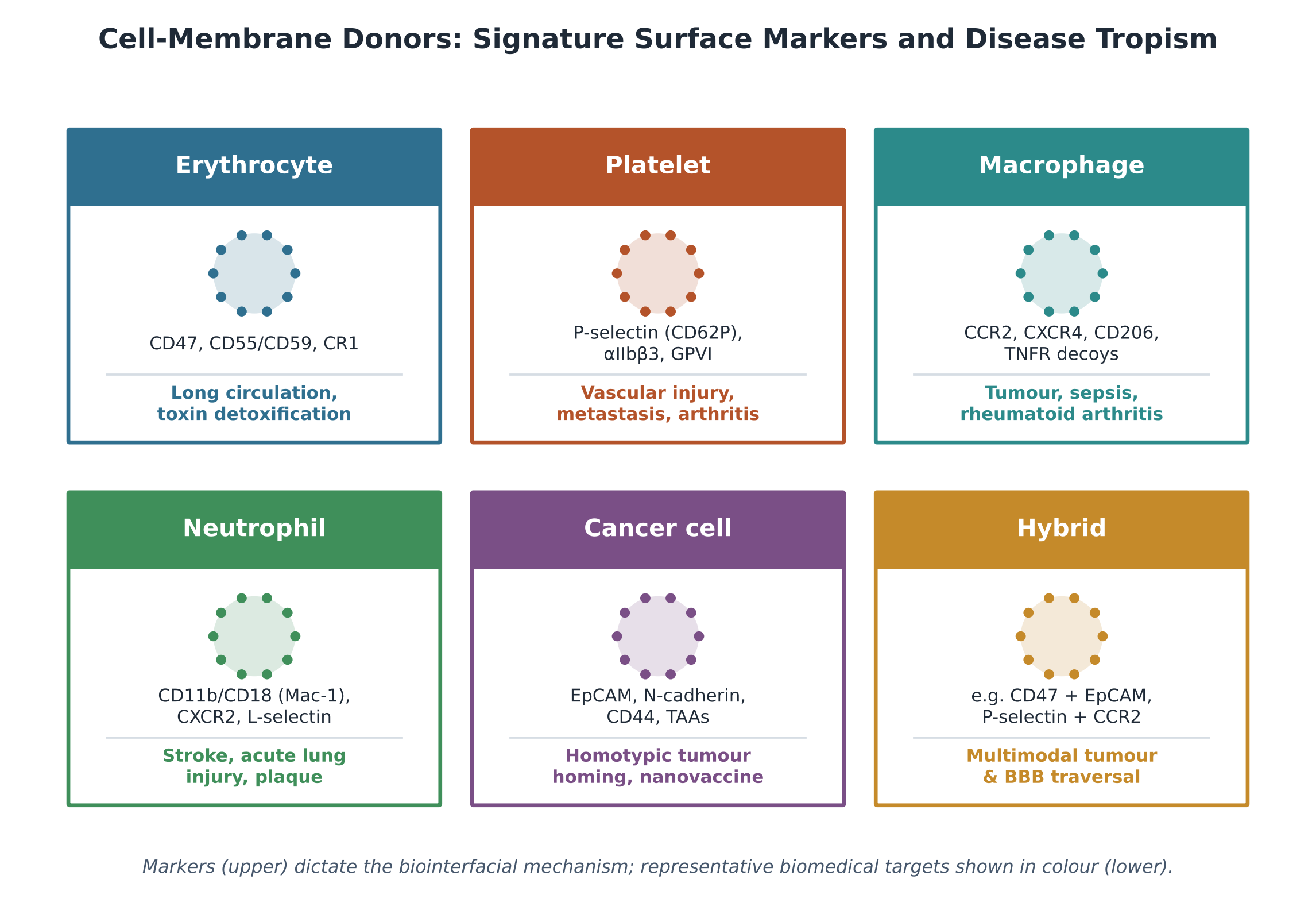
The biological fate, organ tropism, and therapeutic efficacy of a coated particle are governed by the cellular source of its cloak (Table 2, Figure 3) (Muhammad et al., 2026; Zhou et al., 2025). The sections below summarize the dominant donor classes and the surface machinery each contributes.

2.4.1 Erythrocyte membranes

Erythrocytes are the classic stealth donor (Hu et al., 2011; Xia et al., 2019). With a natural lifespan of 100–120 days, they carry a dense CD47 “marker of self” and complement regulators including decay-accelerating factor (CD55), CD59, and complement receptor 1 (CR1), which together dampen alternative complement activation (Oldenborg et al., 2000; Zhou et al., 2025). Wrapping polymeric or metallic cores in these envelopes extends circulation from hours to dozens of hours; the trade-off is a lack of intrinsic homing receptors, so targeting depends on passive extravasation or secondary ligand engineering (Li et al., 2026b; Zhou et al., 2025).

2.4.2 Platelet membranes

Platelet membranes are enriched in P-selectin (CD62P), integrin complexes (αIIbβ3/CD41/CD61), and glycoprotein VI, which bind exposed subendothelial collagen and von Willebrand factor and recognize CD44 on activated endothelium and circulating tumor cells (Hu et al., 2015; Muhammad et al., 2026). Platelet-camouflaged particles have therefore been applied to vascular injury, atherosclerotic plaque, metastatic niches, and anti-inflammatory delivery in rheumatoid arthritis (Li et al., 2021; Su et al., 2026).

2.4.3 Leukocyte, macrophage, and neutrophil membranes

Leukocyte coatings confer inflammatory tropism and barrier-crossing ability (Parodi et al., 2013; Zhou et al., 2025). Macrophage membranes retain CCR2 and CXCR4 alongside scavenger receptors and integrins, letting particles follow chemokine gradients toward inflamed, infected, or hypoxic tumor tissue, and they act as cytokine sponges that neutralize TNF-α, IL-6, and IL-1β to quiet cytokine storms in sepsis, pancreatitis, and fibrosis (Gao et al., 2020; Jiang et al., 2022; Shi et al., 2025). Neutrophil membranes display β2 integrins (CD11b/CD18) and CXCR2 that mediate rolling and firm adhesion to ICAM-1 and VCAM-1 on activated endothelium, enabling transit across the BBB and homing to ischemic or injured lung tissue (Muhammad et al., 2026; Zhang et al., 2018; Zhao et al., 2024).

2.4.4 Cancer-cell membranes

Membranes from cultured cancer cells preserve the parent line’s antigenic spectrum, including EpCAM, N-cadherin, galectins, and CD44 (Chen et al., 2016; Sun et al., 2017). These homologous adhesion molecules drive homotypic self-recognition, concentrating cancer-cell-membrane-coated particles within parent tumors, while retained tumor-associated antigens and MHC molecules allow the particles to act as personalized nanovaccines that prime cytotoxic T-cell responses (Fang et al., 2014; Fontana et al., 2018; Zhou et al., 2025).

2.4.5 Engineered hybrid membranes

To escape the functional trade-offs of any single source, researchers fuse two or more membranes by co-extrusion,

Figure 1. Top-down cell-membrane coating: from donor cell to core–shell nanoplatform. A source cell is lysed and purified—by hypotonic lysis or nitrogen cavitation followed by gradient centrifugation—to yield closed plasma-membrane vesicles that retain the cell's native surface proteins (A). In parallel, a synthetic core is fabricated from polymeric (PLGA, polydopamine, cyclodextrin), inorganic (SPION, gold, copper sulfide, bismuth selenide), metallacycle, or cubosome building blocks to carry drug and imaging cargo (B). Membrane vesicles and cores are then fused by mechanical extrusion, ultrasonic cavitation, or microfluidic electroporation/low-shear mixing to form a core–shell particle that displays surface proteins in their native right-side-out orientation (C). The resulting nanoparticle inherits prolonged circulation, immune evasion, disease-specific homing, and multimodal theranostic capability from its cellular cloak.

Figure 2. The CD47–SIRPα “don't-eat-me” inhibitory signaling axis. A biomimetic nanoparticle that inherits dense CD47 from its donor membrane (typically erythrocyte) presents this “marker of self” to SIRPα receptors on host macrophages. Receptor engagement phosphorylates the immunoreceptor tyrosine-based inhibitory motifs (ITIMs) within the SIRPα cytoplasmic tail, recruiting and activating the phosphatases SHP-1 and SHP-2. The ensuing dephosphorylation cascade blocks assembly of non-muscle myosin-IIA at the phagocytic synapse and halts actin remodeling, so engulfment is suppressed. The net effect is inhibition of macrophage phagocytosis and a corresponding extension of systemic circulation, the central mechanism underlying erythrocyte-based nanoparticle stealth.

sonication, or microfluidic mixing (Dehaini et al., 2017; Muhammad et al., 2026). Erythrocyte–cancer hybrids combine CD47-driven circulation with homotypic tumor homing (Jiang et al., 2019), while macrophage–cancer hybrids merge inflammatory chemotaxis, BBB traversal, and glioblastoma targeting for image-guided radioimmunotherapy (Yun et al., 2026; Zhang et al., 2026).

2.5 Immune evasion and protein-corona modulation

The extended circulation of these particles rests on molecular signaling that overrides immune recognition (Fadeel, 2022; Muhammad et al., 2026). The central mechanism is the CD47–SIRPα axis (Figure 2). When surface CD47 engages SIRPα on a macrophage, it triggers phosphorylation of immunoreceptor tyrosine-based inhibitory motifs (ITIMs) in the SIRPα tail, recruiting SHP-1 and SHP-2 phosphatases; the resulting dephosphorylation cascade prevents non-muscle myosin-IIA from assembling at the phagocytic synapse, so actin remodeling stalls and engulfment is suppressed (Oldenborg et al., 2000; Tsai et al., 2010; Zhu et al., 2022).

A biological cloak does not abolish the protein corona so much as rewrite it (He et al., 2026; Yun et al., 2026). On bare synthetic surfaces, opsonins—IgG, complement C3b, fibrinogen—deposit and engage complement and Fc receptors on hepatic and splenic macrophages, accelerating clearance (Chennamsetty et al., 2026; Papini et al., 2020). Membrane coatings shift this equilibrium toward dysopsonins such as albumin and apolipoproteins, while surface glycans and sialic acids raise a steric hydration barrier against complement (Muhammad et al., 2026; Oldenborg et al., 2000). The corona remains dynamic, however: inflammation and altered metabolic states can enrich particular apolipoproteins, mask targeting ligands, and redirect biodistribution toward clearance organs (Shaw et al., 2025; Tang et al., 2023).

2.6 Multifunctional theranostic integration

By housing a therapeutic agent and an imaging probe in one membrane-cloaked architecture, biomimetic carriers support non-invasive diagnosis alongside spatiotemporally controlled therapy (Chennamsetty et al., 2026; Ma et al., 2025; Zhu et al., 2022). On the imaging side, SPION, iron-functionalized polydopamine, or gadolinium/manganese matrices sharpen MRI contrast, with some copper/manganese silicate systems switching T2-to-T1 contrast on exposure to tumor glutathione (Liu et al., 2019; Su et al., 2026); encapsulated indocyanine green or aggregation-induced-emission luminogens extend fluorescence and PA windows (Li et al., 2020; Zheng et al., 2020); and radiolabeling supports quantitative SPECT/PET tracking (Zhang et al., 2026). On the therapeutic side, NIR-absorbing cores drive PTT and immunogenic cell death, photosensitizers generate reactive oxygen species for PDT, and Fenton-active metals enable CDT; combined with checkpoint blockade, these danger signals can convert a cold tumor into an immunologically active one (Li et al., 2026a; Lin et al., 2025; Pei et al., 2018). Representative preclinical theranostic systems and their in vivo performance are collated in Table 4.

3. Methods

This article is a structured narrative review. Because the literature on biomimetic cell-membrane-coated nanoparticles (CMNPs) is mechanistically heterogeneous and spans materials science, immunology, and pharmaceutics, a narrative synthesis was chosen over a quantitative meta-analysis; nonetheless, the search, screening, and extraction steps were pre-specified and documented so that the review can be reproduced and updated (Muhammad et al., 2026; Zhou et al., 2025). The procedure below is reported at a level of detail consistent with reproducible biomedical reviewing.

3.1 Information sources and search strategy

Three bibliographic databases were queried: PubMed/MEDLINE, Scopus, and Clarivate Web of Science Core Collection. Searches combined controlled vocabulary and free-text terms across three concept blocks joined with the Boolean operator AND, with synonyms within each block joined by OR: (i) carrier—“cell membrane coating” OR “cell-membrane-coated nanoparticle” OR “biomimetic nanoparticle” OR “membrane camouflage” OR “core–shell nanoparticle”; (ii) biological source—“erythrocyte” OR “red blood cell” OR “platelet” OR “leukocyte” OR “macrophage” OR “neutrophil” OR “cancer cell” OR “stem cell” OR “bacterial outer membrane vesicle” OR “hybrid membrane”; and (iii) function—“drug delivery” OR “theranostic” OR “immune evasion” OR “protein corona” OR “photothermal” OR “photodynamic” OR “radioimmunotherapy” OR “blood–brain barrier.” Searches were restricted to peer-reviewed English-language records and were last executed before manuscript preparation; reference lists of key reviews were hand-searched to capture records missed by database indexing (Chennamsetty et al., 2026; Muhammad et al., 2026; Zhu et al., 2022).

3.2 Eligibility criteria

Records were eligible if they (a) reported original fabrication, characterization, or in vitro/in vivo evaluation of a cell-membrane-coated or hybrid-membrane nanoparticle, or (b) provided mechanistic, methodological, or translational analysis directly relevant to such systems. Primary studies were prioritized for data extraction, while authoritative reviews were retained for conceptual framing and cross-checking. Records were excluded if they addressed non-membrane stealth coatings alone (e.g., PEGylation without a biological membrane), reported insufficient methodological detail to interpret the coating step, or were conference abstracts, editorials, or non-English records without an accessible full text.

3.3 Study selection

Retrieved records were de-duplicated and screened in two stages—title/abstract, then full text—against the criteria above. Studies advancing to synthesis were grouped by the dimension they best informed: synthetic-core design, membrane isolation and purification, membrane–core fusion, immune-evasion mechanism, donor-specific tropism, theranostic modality, or translational and regulatory consideration. This mapping allowed each study to contribute to the most relevant comparative table rather than being forced into a single category.

3.4 Data extraction and quality considerations

For each primary study, the following were extracted where reported: synthetic core material and architecture; donor cell type and membrane isolation route; fusion method and critical process parameters (pore-size steps and cycle number for extrusion; amplitude, power, and temperature control for sonication; field strength, flow rate, and core-to-membrane ratio for microfluidic electroporation); coating completeness and topological orientation; particle size, polydispersity, and zeta potential; payload and release trigger; imaging and therapeutic modality; animal model; and reported pharmacokinetic or efficacy endpoints. Methodological rigor was appraised qualitatively, with attention to whether membrane purity, right-side-out orientation, and residual nucleic-acid or endotoxin content were verified, since these attributes determine both reproducibility and biosafety (Liu et al., 2021; Muhammad et al., 2026).

3.5 Synthesis, tables, and figures

Extracted data were synthesized narratively and consolidated into four comparative matrices: synthetic and inorganic cores (Table 1), biological membrane donors and their molecular signatures (Table 2), bioengineering technologies for isolation and assembly (Table 3), and preclinical theranostic applications with their translational bottlenecks (Table 4). Four schematic figures were constructed de novo to integrate the extracted mechanisms: the top-down coating workflow and core–shell architecture (Figure 1), the CD47–SIRPα inhibitory signaling axis (Figure 2), the donor-specific marker-to-tropism map (Figure 3), and the translational-bottleneck framework (Figure 4). All quantitative values reproduced in the text and tables are reported as stated in the primary sources; no new experimental data were generated for this review.

4. Cell Membrane Cloaking Reassigns Biological Fate in Theranostic Nanoparticles but Faces Translational Barriers

Read together, the studies surveyed here point to a fairly specific claim—that cloaking a synthetic core in a natural membrane does not so much improve a single property as reassign responsibility for the particle’s biological behavior. The core still dictates what is carried and what can be imaged; the shell, more often than not, decides how long the particle lasts and where it ends up. The findings below are organized around that split, moving from the core outward to the membrane, then to the assembly step, the integrated theranostic performance, and finally the barriers that keep recurring.

4.1 Physicochemical specialization and payload dynamics of the core

Across delivery models, the inner core behaves as a versatile template that sets encapsulation capacity, degradation timeline, and responsiveness to microenvironmental triggers (Table 1) (Chennamsetty et al., 2026; Li et al., 2026b). Polymeric and supramolecular matrices tend to be the workhorses: PLGA reliably achieves high loading of hydrophobic drugs and senolytics such as ABT-263, with sustained, ester-hydrolysis-controlled release over roughly 7–14 days (Jan et al., 2023; Su et al., 2026), and cyclodextrin nanosponges improve the solubility of labile compounds like curcumin and camptothecin while lowering cardiotoxicity (Garg et al., 2024). Discrete organoplatinum metallacycles are a

Figure 3. Cell-membrane donors, signature surface markers, and disease tropism. Each panel pairs a membrane donor with the characteristic surface markers it contributes (upper) and the representative biomedical target that those markers program (lower). Erythrocyte membranes supply CD47 and complement regulators for long circulation; platelet membranes provide P-selectin and glycoprotein complexes for vascular and metastatic targeting; macrophage and neutrophil membranes contribute chemokine receptors and integrins for inflammatory and barrier-crossing tropism; cancer-cell membranes exploit homotypic adhesion molecules for tumor homing and nanovaccination; and engineered hybrids combine complementary signatures for multimodal targeting and blood–brain-barrier traversal. The figure illustrates that the cellular source, rather than the synthetic core, dictates the biointerfacial mechanism and organ tropism of the particle.

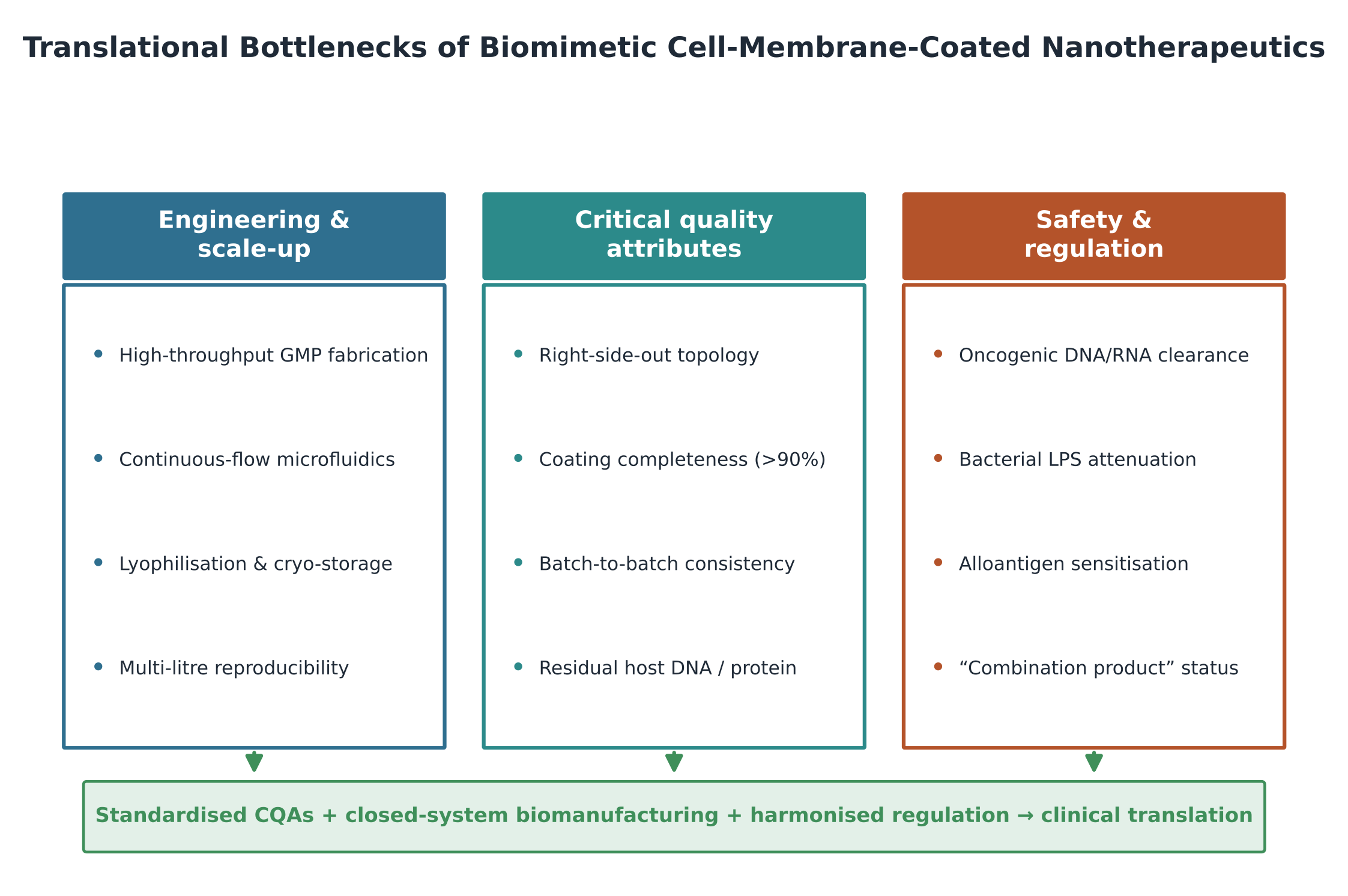
Figure 4. Translational bottlenecks of biomimetic cell-membrane-coated nanotherapeutics. The barriers to clinical translation cluster into three interdependent pillars. Engineering and scale-up challenges include high-throughput GMP fabrication, continuous-flow microfluidics, lyophilization and cryo-storage, and multi-liter reproducibility. Critical quality attributes encompass right-side-out membrane topology, coating completeness above 90%, batch-to-batch consistency, and control of residual host DNA and protein content. Safety and regulatory hurdles span clearance of oncogenic DNA/RNA, attenuation of bacterial lipopolysaccharide, avoidance of alloantigen sensitization, and the ambiguous “combination product” classification. Resolving all three pillars—through standardized CQAs, closed-system biomanufacturing, and harmonized regulatory frameworks—is the prerequisite for clinical translation.

newer entry—they fold chemotherapy and an NIR photosensitizer into one molecule and resist premature burst release (Li et al., 2026a).

Inorganic and catalytic cores add something the polymers cannot: active contrast and on-demand chemistry. Iron-functionalized polydopamine and copper/manganese silicate nanospheres degrade in the glutathione-rich, acidic tumor interior, releasing cargo and, in the silicate case, generating cytotoxic hydroxyl radicals through Fenton-like catalysis (Bigaj-Józefowska et al., 2023; Liu et al., 2019). SPIONs and plasmonic gold do double duty—magnetic homing and photothermal conversion under NIR-I/II illumination (Deng et al., 2022; Su et al., 2026). A smaller but intriguing body of work uses liquid-crystalline cubosomes to co-encapsulate an antioxidant and a caged optical probe for oxidative-stress-triggered reporting in renal injury, with strikingly low background signal (Wu et al., 2026).

4.2 Biointerfacial tropism and immune evasion across donors

If the core is where the chemistry lives, the membrane is where the biology is decided (Table 2, Figure 3) (Muhammad et al., 2026; Zhou et al., 2025). The clearest, most reproducible effect in the whole literature is the erythrocyte-driven extension of circulation: RBC-coated particles move from under two hours for a naked polymeric core to well over 24–40 hours, an effect traceable to CD47 engaging SIRPα and the downstream block on myosin-IIA assembly (Figure 2) (Hu et al., 2011; Oldenborg et al., 2000; Xia et al., 2019). Complement regulators CD55 and CD59 reinforce the effect by quieting the alternative pathway (Muhammad et al., 2026).

Platelet cloaks tell a different story—less about hiding and more about finding. Through P-selectin and glycoprotein complexes they bind CD44 on circulating tumor cells and exposed subendothelial collagen, and preclinical reports describe several-fold increases in accumulation at damaged endothelium, plaque, and metastatic niches relative to bare carriers (Hu et al., 2015; Kunde & Wairkar, 2021). Myeloid coatings go further still: macrophage membranes navigate CCL2/CXCL12 gradients and double as cytokine decoys that strip TNF-α, IL-1β, and IL-6 out of the surrounding fluid (Gao et al., 2020; Jiang et al., 2022), while neutrophil membranes exploit β2 integrins to cross the BBB and the alveolar–capillary interface (Wang et al., 2021; Zhang et al., 2024). Cancer-cell and hybrid membranes, meanwhile, trade on homotypic adhesion and, in the hybrid case, combine prolonged circulation with multivalent, multi-barrier targeting (Dehaini et al., 2017; Jiang et al., 2019; Zhang et al., 2026).

4.3 Biophysical quality attributes and assembly engineering

The fabrication step turns out to matter at least as much as the choice of membrane, and the comparison across methods is not flattering to any single technique (Table 3) (Muhammad et al., 2026; Zhou et al., 2025). Extrusion through track-etched filters gives monodisperse particles (PDI < 0.15) and reliably preserves right-side-out orientation, but it fouls, scales poorly, and throws away most of the membrane input—often retaining only 5–15% (Hu et al., 2011; Van Deun et al., 2020). Sonication is faster and higher-volume, yet uncontrolled heating risks denaturing receptors and pushing PLGA past its glass transition into irreversible aggregation (Yang et al., 2021). Microfluidic electroporation looks, on current evidence, like the most promising route—coating completeness above 90%, high reproducibility, and preserved protein accessibility—while low-shear microfluidic mixing spreads fragile exosomal membranes without degrading sensitive probes (Rao et al., 2017; Wu et al., 2026).

Upstream of assembly, the lysis method shapes safety. Hypotonic lysis suffices for non-nucleated erythrocytes, whereas nucleated cells generally require nitrogen cavitation—pressurization to roughly 350–400 psi followed by rapid decompression—to rupture the plasma membrane while leaving nuclei intact (Li et al., 2026b; Muhammad et al., 2026). Pairing cavitation with a 30–40% sucrose density gradient then removes chromatin, mitochondria, and genomic DNA, which is less a refinement than a prerequisite for translational safety (Muhammad et al., 2026).

4.4 Integrated theranostic performance in preclinical models

When these pieces come together, the preclinical results are, frankly, impressive—though it is worth noting they are almost entirely in rodents (Table 4). In hepatocellular carcinoma and breast-cancer models, cancer-membrane-coated iron-functionalized polydopamine delivered strong T1/T2 MRI contrast alongside efficient photothermal conversion, with 808-nm irradiation driving localized hyperthermia above ~48 °C and, in some reports, complete primary-tumor regression without overt systemic

Table 3. Bioengineering technologies for membrane isolation, core–shell assembly, and quality-control parameters. This matrix contrasts the principal fabrication routes used to isolate plasma membranes and fuse them onto synthetic cores, together with the process parameters that determine product quality. For each technology it specifies the engineering principle and operational setup, the critical process parameters and conditions, the resulting membrane coverage and right-side-out topological fidelity, and the primary advantages and translational drawbacks. Methods span mechanical polycarbonate extrusion, ultrasonic cavitation, microfluidic electroporation and low-shear mixing, nitrogen-cavitation cell lysis, and density-gradient ultracentrifugation. The comparison highlights the recurring trade-off between coating completeness, receptor preservation, and scalable, GMP-compatible throughput.

Fabrication / assembly technology

Working principle & key process parameters

Coverage & topological fidelity

Advantages & limitations

References

Mechanical polycarbonate membrane extrusion

Co-extrusion of cores and vesicles through track-etched filters (800→400→200→100 nm); 10–20 passes at constant temperature

Moderate yield (≈3–15%); preserves native right-side-out topology

Excellent monodispersity and reproducible PDI, broad core compatibility; but filter fouling, material loss, and poor GMP scalability

Hu et al. (2011); Su et al. (2026); Van Deun et al. (2020); Zhou et al. (2025); Zhu et al. (2022)

Ultrasonic cavitation / probe sonication

Acoustic cavitation bubbles drive spontaneous bilayer wrapping; amplitude 20–65%, 100–500 W, pulsed, chilled < 30 °C

Variable completeness; risk of fragmentation and inside-out inversion

Rapid single-step, high-volume; but local heating denatures proteins and aggregates PLGA above its glass transition

Bigaj-Józefowska et al. (2023); Yang et al. (2021); Zhou et al. (2025)

Microfluidic electroporation & hydrodynamic focusing

Electric-field pulses create transient electropores in membranes crossing focused flow; tuned voltage, flow velocity, and core:membrane ratio

High coverage (>90%); preserved extracellular receptor availability

Automated, reproducible, uniform, low operator variability; but high equipment cost and electrode fouling at high ionic strength

Rao et al. (2017); Zhou et al. (2025)

Sequential low-shear microfluidic mixing & thermodynamic spreading

Two-chip assembly: cores formed first, then low-shear mixing with membranes; secondary flow ~200 µL/min, 1:1 lipid:protein, 37 °C

High fusion efficiency (~85%, FRET-verified); preserves tetraspanins (CD63, CD9, TSG101)

Non-destructive, scalable continuous flow; but requires precise multi-channel chips and formulation-specific optimization

Cardellini et al. (2025); Molinaro et al. (2018); Wu et al. (2026)

Nitrogen-cavitation disruption (cell lysis)

Nucleated cells pressurized with N₂ (350–400 psi, 15–30 min on ice), then rapid decompression bursts membranes from within

Harvesting step; yields intact, chemically unperturbed membrane fragments

Rapid, high-yield isolation that leaves nuclei intact, preventing genomic-DNA release; but needs high-pressure vessels and downstream purification

Gao et al. (2017); Li et al. (2026); Muhammad et al. (2026); Xu et al. (2023)

Density-gradient ultracentrifugation (purification)

Centrifugation of lysates through discontinuous sucrose/iodixanol gradients (30–40% interface; 100,000–175,000 × g; 4 °C)

Yields pure plasma-membrane sheets free of chromatin, mitochondria, and cytosolic enzymes

Standard benchmark for removing oncogenic nucleic acids and organelles; but time-consuming, low recovery, limited batch volume

de Weerd et al. (2024); Muhammad et al. (2026); Xu et al. (2023)

Table 4. Preclinical theranostic applications, in vivo delivery performance, and critical translational and CMC bottlenecks. This table consolidates representative biomimetic nanoplatforms evaluated in animal models, linking each formulation to its disease context, theranostic mechanism, and translational obstacles. For every system it records the biomimetic architecture, the in vivo disease model, the synergistic diagnostic-therapeutic mechanism and performance, and the critical translational, chemistry-manufacturing-and-controls (CMC), safety, and regulatory bottlenecks identified by the authors. Examples span homotypic cancer-membrane carriers, macrophage- and erythrocyte-camouflaged systems, bacterial-hybrid vaccines, and neutrophil–macrophage hybrids for brain delivery. The compilation underscores that promising rodent efficacy is consistently tempered by batch heterogeneity, contaminant clearance, storage, and combination-product regulatory classification.

Biomimetic formulation

In vivo disease model

Theranostic mechanism & performance

Critical translational / CMC / regulatory bottleneck

References

MPDAFe@DOX@Mem (cancer-membrane-coated MPDAFe)

BALB/c nude mice bearing HepG2 or MCF-7 tumors

Homotypic targeting; real-time T1/T2 MRI; synergistic chemo-photothermal cytotoxicity; stable >14 days in biofluids

Batch-to-batch variation in cancer-membrane proteins; risk of oncogenic components; lack of standardized release assays

Bigaj-Józefowska et al. (2023); Zhu et al. (2022)

MM@MS@ABT (macrophage-camouflaged Fe₃O₄-PLGA/ABT-263)

C57BL/6J lumbar-spine-instability endplate sclerosis

Dual magnetic + CCR2 inflammatory targeting; senescent-macrophage clearance; sustained release up to 7 days

Coordinating external magnets with systemic carriers; “combination product” regulatory classification

Su et al. (2026)

EXLNPs (exosome–cubosome hybrids; lipoic acid + ROS probe)

C57BL/6 bilateral renal ischemia-reperfusion injury (AKI)

“Turn-on” fluorescence dosimetry of oxidative stress; circulation half-life extended ~5.8-fold; antioxidant protection

Strict liquid cold-chain dependency; need for lyophilization with trehalose/sucrose cryoprotectants

Wu et al. (2026)

CMa / CMb NPs (HeLa-membrane-cloaked Pt-metallacycles)

Subcutaneous HeLa xenograft BALB/c nude mice

Homotypic targeting; NIR-activated ¹O₂ and heat; calreticulin exposure with anti-PD-L1 synergy; stable >7 days

Scalability of supramolecular coordination chemistry; residual platinum removal; allogeneic membrane immunogenicity

Li et al. (2026a)

¹³¹I-LipRM (macrophage–gastric-cancer hybrid-membrane liposomes)

BALB/c nude mice bearing MFC gastric xenografts

Targeted ¹³¹I radioimmunotherapy triggering immunogenic cell death with anti-PD-L1 synergy; superior tumor accumulation

Radiolytic degradation of membrane lipids; radiation safety in manufacturing; hybrid-fusion reproducibility

Zhang et al. (2026)

SPN@RBCM (erythrocyte-coated semiconducting-polymer NPs)

Orthotopic and subcutaneous tumor-bearing mice

Low-bandgap polymer enables stable high-resolution photoacoustic imaging and PTT; reduced RES uptake; extended half-life

Blood-type antigen matching (ABO/Rh); non-biodegradability of conjugated-polymer backbones

Zheng et al. (2020); Zhu et al. (2022)

HPDA@[OMV-CC] (bacterial OMV–melanoma hybrid on polydopamine)

B16-F10 melanoma murine models

Combined photothermal ablation and multi-antigen immune activation via OMV TLR engagement; strong lymph-node localization

Endotoxin (LPS) toxicity requiring genetic attenuation (ΔmsbB); potential cytokine-release syndrome

Chen et al. (2020); Muhammad et al. (2026); Zhu et al. (2022)

NMm-PLGA/RAPA (neutrophil–macrophage hybrid nanocarriers)

Orthotopic glioblastoma and MCAO stroke models

Chemotactic traversal across the blood–brain barrier; anti-inflammatory suppression of microglial activation; superior brain accumulation

Dynamic protein-corona remodeling in brain endothelium; high shear during cerebral microcirculation

Wang et al. (2021); Yin et al. (2022); Yun et al. (2026); Zhao et al. (2024)


toxicity (Bigaj-Józefowska et al., 2023). HeLa-membrane-cloaked platinum metallacycles produced singlet oxygen and heat that exposed calreticulin and released HMGB1; paired with anti-PD-L1 blockade, this biomimetic photoimmunotherapy drove CD8⁺ T-cell infiltration against both primary and distant tumors (Li et al., 2026a). In gastric cancer, ¹³¹I-labeled macrophage–cancer hybrid liposomes enabled SPECT imaging while delivering targeted radioimmunotherapy (Zhang et al., 2026).

The non-oncologic examples are, in some ways, the more surprising ones. Macrophage-camouflaged magnetic nanoparticles carrying ABT-263 combined magnetic guidance with CCR2-mediated homing to clear senescent macrophages and halt aberrant vessel ingrowth in a spinal endplate-sclerosis model (Su et al., 2026), and exosome–cubosome hybrids loaded with lipoic acid provided real-time fluorescent reporting of reactive oxygen species while limiting oxidative damage in renal ischemia–reperfusion injury (Wu et al., 2026). Taken as a set, these studies suggest the platform’s reach extends well beyond oncology, even if the evidence outside cancer is still thin.

4.5 Recurring translational barriers and critical quality attributes

What is striking, reading across otherwise unrelated papers, is how the same four obstacles keep surfacing (Figure 4). First, topological inversion: when vesicles flip inside-out during processing, the extracellular domains of CD47 and targeting receptors end up buried against the core, and cryo-TEM plus functional assays show that even partial core exposure can redirect biodistribution toward clearance organs (Liu et al., 2021). Second, biological heterogeneity—variable surface-protein expression and membrane-to-core ratios across runs—which makes standardized CQAs such as residual host-cell DNA (< 10 ng/dose) and endotoxin limits (< 5 EU/kg) essential rather than optional (Chennamsetty et al., 2026; Shan et al., 2026). Third, colloidal and storage instability, where formulations without optimized cryoprotectants (typically 5–10% trehalose or sucrose) lose integrity on freeze-drying (Li et al., 2026b). And fourth, the regulatory tangle of “combination product” status, which spreads oversight across biological, pharmaceutical, and imaging divisions and, for now, lacks a harmonized international standard (Liu et al., 2026).


5. Moving Cell Membrane-Coated Nanoparticles From Proof of Concept to Manufacturable, GMP-Ready Medicines

5.1 What the evidence consistently supports

It is tempting, given the volume of positive reports, to treat cell-membrane coating as a solved problem. The more careful reading is narrower. What the literature supports with real consistency is a single proposition: a natural membrane can hand a synthetic particle a biological identity that the innate immune system is slow to challenge, and that identity—not the core—largely governs circulation and homing (Fadeel, 2022; Muhammad et al., 2026; Zhou et al., 2025). The erythrocyte-CD47 effect is the strongest pillar here, reproduced across cores and laboratories and mechanistically well explained by the SIRPα cascade (Figure 2) (Oldenborg et al., 2000; Tsai et al., 2010). Much of the rest of the field is built, reasonably, on the inference that if one membrane transfers its function, others should too—and in most cases they appear to.

5.2 The corona is reshaped, not erased

One finding deserves more emphasis than it usually gets: coating does not give a particle a clean, protein-free surface. It changes which proteins arrive (He et al., 2026; Yun et al., 2026). That distinction matters because it reframes the design target. If the goal were simply to prevent adsorption, a better inert polymer might suffice; but if the real mechanism is a shift from opsonins toward dysopsonins like albumin and apolipoproteins, then the membrane’s glycan and sialic-acid landscape becomes a design parameter in its own right (Muhammad et al., 2026; Papini et al., 2020). The uncomfortable corollary is that the corona stays dynamic. Under inflammation or hypercholesterolemia, apolipoprotein enrichment can mask the very ligands the coating was meant to display, quietly redirecting particles toward the liver and spleen (Shaw et al., 2025; Tang et al., 2023). A particle validated in a healthy animal may therefore behave differently in diseased tissue—a caveat that rarely makes it into efficacy summaries.

5.3 The orientation and completeness problem

If there is a single under-appreciated failure mode, it is probably membrane orientation (Figure 4). A coating can look complete by size and zeta potential and still be functionally inert if a meaningful fraction of its vesicles sealed inside-out, burying CD47’s extracellular loops where no macrophage will ever see them (Bu et al., 2021; Liu et al., 2021). The same is true of incomplete coverage: a patch of exposed core is enough to seed opsonin deposition and shift the internalization pathway (Liu et al., 2021; Muhammad et al., 2026). This is where fabrication choice stops being a convenience and becomes a determinant of efficacy—extrusion preserves orientation but will not scale, sonication scales but risks damage, and microfluidic routes currently offer the best, though not yet fully industrialized, compromise (Rao et al., 2017; Van Deun et al., 2020). It is hard to escape the sense that the field’s next real gains will come from the assembly line rather than from new biology.

5.4 Heterogeneity, safety, and the limits of biological sourcing

Biological sourcing is simultaneously the platform’s greatest strength and its most stubborn liability. Donor genetics, passage number, and culture conditions all perturb membrane composition, so batch-to-batch variability is not a manufacturing nuisance to be engineered away but an intrinsic feature of using living material (Chennamsetty et al., 2026; Li et al., 2026b). Safety concerns compound this. Cancer-cell membranes raise the specter of transferring oncogenic DNA or microRNA and demand rigorous validation (Muhammad et al., 2026); bacterial outer-membrane vesicles carry lipopolysaccharide that must be attenuated, for instance by msbB deletion, to avoid endotoxin shock (Chen et al., 2020); and repeated dosing of allogeneic membranes bearing non-self MHC can provoke anti-membrane antibodies and accelerated clearance (Larssen et al., 2019). None of these is necessarily disqualifying, but each converts an elegant idea into a demanding quality-control exercise.

5.5 Toward standardized, GMP-compliant translation

Where does this leave the path to the clinic? Mostly, it argues for a change of emphasis—from discovery to process (Table 4, Figure 4). Continuous-flow, closed-system microfluidics capable of multi-liter throughput under current GMP is the most frequently cited enabling technology, and for good reason: it addresses orientation, reproducibility, and scale at once (Cardellini et al., 2025; Li et al., 2026b). Genetic engineering of donor cells to overexpress targeting ligands while knocking out immunogenic alloantigens offers a way to make membranes more uniform and less reactive by design (Zhou et al., 2025). Standardized, non-destructive assays for corona evolution and receptor accessibility would let manufacturers release product against meaningful specifications rather than surrogate ones (He et al., 2026). And refined lyophilization with trehalose or sucrose cryoprotectants is the unglamorous but necessary step toward an off-the-shelf product (Shan et al., 2026). None of this is conceptually novel; all of it is hard, and most of it is where the field now needs to spend its effort.

5.6 Limitations of the current evidence base

Finally, a note of restraint. The preclinical record is overwhelmingly rodent-based, short-term, and reported by the groups developing the platforms; head-to-head comparisons under standardized conditions are scarce, and negative results are almost certainly under-published. Quantitative claims—circulation half-lives, fold-changes in tumor accumulation—vary with model, dose, and analytical method and should be read as indicative rather than definitive (Zhang et al., 2023; Zhu et al., 2022). This review inherits those limitations: as a structured narrative synthesis rather than a meta-analysis, it integrates heterogeneous studies without pooling them statistically. The pattern it describes is, we think, robust; the individual numbers are not where the confidence lies.

6. Conclusion

Biomimetic cell-membrane coating has matured from a clever laboratory trick into a coherent engineering philosophy—one that borrows the immune fluency of living cells instead of trying to synthesize it from scratch. Across the studies surveyed here, a consistent pattern emerges: the synthetic core sets the payload and the imaging signal, while the biological shell decides how long the particle survives and where it ultimately goes. That division of labor explains both the platform's versatility and its fragility, because the very membranes that confer stealth and homing are also the hardest components to source, orient, and preserve reproducibly. The decisive question, then, is no longer whether these nanoparticles work in animals—they clearly do—but whether they can be made the same way twice at clinical scale. Progress will depend on continuous-flow manufacturing, genetically tailored membranes, standardized critical quality attributes, and regulatory frameworks designed for genuinely hybrid products rather than retrofitted from older categories.

References


Bigaj-Józefowska, M. J., Coy, E., Zaleski, K., Zalewski, T., Grabowska, M., Jaskot, K., Perrigue, P., Mrówczynski, R., & Grzeskowiak, B. F. (2023). Biomimetic theranostic nanoparticles for effective anticancer therapy and MRI imaging. Journal of Photochemistry and Photobiology B: Biology, 249, 112813.

Bu, Y., Zhang, X., Zhu, A., Li, L., Xie, X., & Wang, S. (2021). Inside-out-oriented cell membrane biomimetic magnetic nanoparticles for high-performance drug lead discovery. Analytical Chemistry, 93(22), 7898–7907.

Cardellini, J., Normak, K., Gerlt, M., Makasewicz, K., Seiffert, C., Capasso Palmiero, U., Ye, S., González Gómez, M. A., Piñero, Y., & Rivas, J. (2025). Microfluidics-driven manufacturing and multiscale analytical characterization of nanoparticle–vesicle hybrids. Advanced Healthcare Materials, 14(3), 2403264.

Chen, H.-Y., Deng, J., Wang, Y., Wu, C.-Q., Li, X., & Dai, H.-W. (2020). Hybrid cell membrane-coated nanoparticles: A multifunctional biomimetic platform for cancer diagnosis and therapy. Acta Biomaterialia, 112, 1–13.

Chen, Q., Huang, G., Wu, W., Wang, J., Hu, J., Mao, J., Chu, P. K., Bai, H., & Tang, G. (2020). A hybrid eukaryotic–prokaryotic nanoplatform with photothermal modality for enhanced antitumor vaccination. Advanced Materials, 32(16), e1908185.

Chen, Z., Zhao, P., Luo, Z., Zheng, M., Tian, H., Gong, P., Gao, G., Pan, H., Liu, L., Ma, A., Cui, H., Ma, Y., & Cai, L. (2016). Cancer cell membrane–biomimetic nanoparticles for homologous-targeting dual-modal imaging and photothermal therapy. ACS Nano, 10(11), 10049–10057. https://doi.org/10.1021/acsnano.6b04695

Chennamsetty, S., et al. (2026). Biohybrid nanocarriers for targeted drug delivery by integrating biological interfaces with synthetic platforms for site-specific therapeutics. International Journal of Pharmaceutics, 687, 126400. https://doi.org/10.1016/j.ijpharm.2025.126400

Chetty, S. S., Praneetha, S., Vadivel Murugan, A., Govarthanan, K., & Verma, R. S. (2020). Human umbilical cord Wharton’s jelly-derived mesenchymal stem cells labeled with Mn²? and Gd³? co-doped CuInS2–ZnS nanocrystals for multimodality imaging in a tumor mice model. ACS Applied Materials & Interfaces, 12(3), 3415–3429.

Chi, Y., Song, C., Jia, Q., Zhang, R., Sun, F., Li, Z., Jia, Y., An, X., Wang, Z., & Li, J. (2019). Platelet membrane–coated nanoparticles for targeted drug delivery in lung cancer. Acta Biomaterialia, 92, 290–301.

de Weerd, S., Ruiter, E. A., Calicchia, E., Portale, G., Schuringa, J. J., Roos, W. H., & Salvati, A. (2024). Optimization of cell membrane purification for the preparation and characterization of cell membrane liposomes. Small Methods, 8(5), 2400498. https://doi.org/10.1002/smtd.202400498

Dehaini, D., Wei, X., Fang, R. H., Masson, S., Angsantikul, P., Luk, B. T., Zhang, Y., Ying, M., Jiang, Y., Kroll, A. V., Gao, W., & Zhang, L. (2017). Erythrocyte–platelet hybrid membrane coating for enhanced nanoparticle functionalization. Advanced Materials, 29(16), 1606209. https://doi.org/10.1002/adma.201606209

Deng, T., Wang, J., Li, Y., Han, Z., Peng, Y., Zhang, J., Song, X., Bao, F., Gu, Y., & Zhang, R. (2022). Biomimetic macrophage membrane-coated gold–quantum dots with tumor microenvironment stimuli-responsive capability for tumor theranostic. Materials Today Bio, 16, 100359.

Fadeel, B. (2022). Clearing the way: Nanoparticle interactions with the mononuclear phagocyte system. Biomaterials, 280, 121280.

Fang, R. H., Hu, C.-M. J., Luk, B. T., Gao, W., Copp, J. A., Tai, Y., O’Connor, D. E., & Zhang, L. (2014). Cancer cell membrane-coated nanoparticles for anticancer vaccination and drug delivery. Nano Letters, 14(4), 2181–2188. https://doi.org/10.1021/nl500618u

Fang, R. H., Kroll, A. V., Gao, W., & Zhang, L. (2018). Cell membrane coating nanotechnology. Advanced Materials, 30(23), e1706759. https://doi.org/10.1002/adma.201706759

Fontana, F., Albertini, S., Correia, A., Kemell, M., Lindgren, R., Mäkilä, E., Salonen, J., Hirvonen, J. T., Ferrari, F., & Santos, H. A. (2018). Bioengineered porous silicon nanoparticles@macrophages cell membrane as composite platforms for rheumatoid arthritis. Advanced Functional Materials, 28(32), 1801355.

Gao, C., Huang, Q., Liu, C., Kwong, C. H. T., Yue, L., Wan, J.-B., Lee, S. M. Y., & Wang, R. (2020). Treatment of atherosclerosis by macrophage-biomimetic nanoparticles via targeted pharmacotherapy and sequestration of proinflammatory cytokines. Nature Communications, 11(1), 2622. https://doi.org/10.1038/s41467-020-16439-7

Gao, W., Zhang, L., et al. (2017). Rapid disruption of cellular membranes using nitrogen cavitation for biomimetic nanocarrier assembly. Nanoscale, 9(34), 12540–12548.

Garg, A., Lai, W.-C., Chopra, H., Agrawal, R., Singh, T., Chaudhary, R., & Dubey, B. N. (2024). Multifunctional nanosponges in cancer therapy: Integrating targeted drug delivery and theranostic potential. Heliyon, 10(2), e23303.

Gholibegloo, E., Mortezazadeh, T., Salehian, F., Forootanfar, H., Firoozpour, L., Foroumadi, A., Ramazani, A., & Khoobi, M. (2019). Folic acid decorated magnetic nanosponge: An efficient nanosystem for targeted curcumin delivery and magnetic resonance imaging. Journal of Colloid and Interface Science, 556, 128–139.

Guo, R., et al. (2025). Innovative nanocarriers: Synthetic and biomimetic strategies for enhanced drug delivery. Materials Today Bio, 34, 102180. https://doi.org/10.1016/j.mtbio.2025.102180

He, T., Zhu, L., Ding, J., Fang, X., Gao, Y., Mailänder, V., Crespy, D., Landfester, K., & Jiang, S. (2026). Macrophage phenotype-dependent protein corona formation governs ligand accessibility and immune clearance of biomimetic nanoparticles. Small, 22(4), e2514389. https://doi.org/10.1002/smll.202514389

Hosseinidoust, Z., Mostaghaci, B., Yasa, O., Park, B. W., Singh, A. V., & Sitti, M. (2016). Bioengineered and biohybrid bacteria-based systems for drug delivery. Advanced Drug Delivery Reviews, 106(Pt A), 27–44.

Hu, C.-M. J., Fang, R. H., Wang, K.-C., Luk, B. T., Thamphiwatana, S., Dehaini, D., Nguyen, P., Angsantikul, P., Wen, C. H., Kroll, A. V., & Zhang, L. (2015). Nanoparticle biointerfacing by platelet membrane cloaking. Nature, 526(7571), 118–121. https://doi.org/10.1038/nature15373

Hu, C.-M. J., Zhang, L., Aryal, S., Cheung, C., Fang, R. H., & Zhang, L. (2011). Erythrocyte membrane-camouflaged polymeric nanoparticles as a biomimetic delivery platform. Proceedings of the National Academy of Sciences of the United States of America, 108(27), 10980–10985. https://doi.org/10.1073/pnas.1106634108

Huang, H., Xie, Z., Li, N., Zeng, L., Zeng, Q., Yang, Z., Shen, J., Yang, H., Liu, Y., & Wu, C. (2025). Biomimetic gold nano-modulator for deep-tumor NIR-II photothermal immunotherapy via gaseous microenvironment remodeling strategy. Journal of Nanobiotechnology, 23(1), 220.

Jan, N., Madni, A., Khan, S., Shah, H., Akram, F., Khan, A., Ertas, D., Bostanudin, M. F., Contag, C. H., & Ashammakhi, N. (2023). Biomimetic cell membrane-coated poly(lactic-co-glycolic acid) nanoparticles for biomedical applications. Bioengineering & Translational Medicine, 8(2), e10441. https://doi.org/10.1002/btm2.10441

Jiang, H., Tai, Z., Pan, H., Cui, Z., Chai, R., Zhu, C., Tian, J., Bao, L., Zhu, Q., & Chen, Z. (2022). Macrophage membrane-derived biomimetic nanoparticles for treatment of cytokine release syndrome. Journal of Biomedical Nanotechnology, 18(4), 1064–1074. https://doi.org/10.1166/jbn.2022.3324

Jiang, Q., Liu, Y., Guo, R., Yao, X., Sung, S., Pang, Z., & Yang, W. (2019). Erythrocyte-cancer hybrid membrane-camouflaged melanin nanoparticles for enhancing photothermal therapy efficacy in tumors. Biomaterials, 192, 292–308. https://doi.org/10.1016/j.biomaterials.2018.11.021

Jin, R., Lin, B., Li, D., & Ai, H. (2014). Superparamagnetic iron oxide nanoparticles for MR imaging and therapy: Design considerations and clinical applications. Current Opinion in Pharmacology, 18, 18–27.

Kaur, S., & Kumar, S. (2019). The nanosponges: An innovative drug delivery system. Asian Journal of Pharmaceutical and Clinical Research, 12(7), 60–67.

Kolaczkowska, E., & Kubes, P. (2013). Neutrophil recruitment and function in health and inflammation. Nature Reviews Immunology, 13(3), 159–175.

Kunde, S. S., & Wairkar, S. (2021). Platelet membrane camouflaged nanoparticles: Biomimetic architecture for targeted therapy. International Journal of Pharmaceutics, 598, 120395. https://doi.org/10.1016/j.ijpharm.2021.120395

Larssen, P., Veerman, R. E., Akpinar, G. G., Hiltbrunner, S., Karlsson, M. C., & Gabrielsson, S. (2019). Allogenicity boosts extracellular vesicle–induced antigen-specific immunity and mediates tumor protection and long-term memory in vivo. The Journal of Immunology, 203(3), 825–834. https://doi.org/10.4049/jimmunol.1801628

Li, C., Luo, D., Peng, C., Luo, Q., Xu, W., Zhou, J., Su, W., Wu, W., & Wang, Y.-F. (2026). Nitrogen cavitation enables rapid and high-yield preparation of functional cell-membrane-derived vesicles. Nanoscale Advances, 8(4), 2021–2030.

Li, L., Fu, J., Wang, X., Chen, Q., Zhang, W., Cao, Y., & Ran, H. (2021). Biomimetic “nanoplatelets” as a targeted drug delivery platform for breast cancer theranostics. ACS Applied Materials & Interfaces, 13(3), 3605–3621.

Li, S., Jiang, W., Yuan, Y., Sui, M., Yang, Y., Huang, L., Jiang, L., Liu, M., Chen, S., & Zhou, X. (2020). Delicately designed cancer cell membrane-camouflaged nanoparticles for targeted 19F MR/PA/FL imaging-guided photothermal therapy. ACS Applied Materials & Interfaces, 12(51), 57290–57301.

Li, Y., et al. (2026a). Biomimetic aza-BODIPY-platinum metallacycle nanoparticles for synergistic photoimmunotherapy via chemo-phototherapy and immune checkpoint blockade. Precision Medicine and Engineering, 3, 100061.

Li, Y., et al. (2026b). Nanomaterial-based drug delivery systems: From intelligent delivery to clinical translation and precision nanomedicine. Biomedicine & Pharmacotherapy, 203, 119888. https://doi.org/10.1016/j.biopha.2026.119888

Liao, Y., et al. (2024). Macrophage-derived biomimetic nanoparticles for light-driven theranostics toward Mpox. Cell Reports Physical Science, 5(8), 102120.

Lin, K., Cao, Y., Zheng, D., Li, Q., Liu, H., Yu, P., Li, J., Xue, Y., & Wu, M. (2020). Facile phase transfer of hydrophobic Fe3O4@Cu2??S nanoparticles by red blood cell membrane for MRI and phototherapy in the second near-infrared window. Journal of Materials Chemistry B, 8(6), 1202–1211.

Lin, X., et al. (2025). Synergistic drug homing for glioblastoma: Integrating biomimetic nanocarriers with MR-guided focused ultrasound across the blood-brain barrier. Advanced Functional Materials, 35(12), 2415120.

Liu, C., Wang, D., Zhang, S., Cheng, Y., Yang, F., Xing, Y., Xu, T., Dong, H., & Zhang, X. (2019). Biodegradable biomimic copper/manganese silicate nanospheres for chemodynamic/photodynamic synergistic therapy with simultaneous glutathione depletion and hypoxia relief. ACS Nano, 13(4), 4267–4277.

Liu, L., Bai, X., Martikainen, M. V., Kårlund, A., Roponen, M., Xu, W., Hu, G., Tasciotti, E., & Lehto, V.-P. (2021). Cell membrane coating integrity affects the internalization mechanism of biomimetic nanoparticles. Nature Communications, 12(1), 5726. https://doi.org/10.1038/s41467-021-26052-x

Liu, X., et al. (2026). Magnetic nanorobots in oncology: From targeted drug delivery to smart theranostic systems. Materials Today Bio, 41, 103709.

Ma, J., et al. (2025). Nanoparticle-based drug delivery systems in urologic oncology: From targeted therapy to precision theranostics. Materials Today Bio, 35, 102585.

Molinaro, R., Evangelopoulos, M., Hoffman, J. R., Corbo, C., Taraballi, F., Martinez, J. O., Hartman, K. A., Cosco, D., Costa, G., Romeo, I., Sherman, M., Paolino, D., Alcaro, S., & Tasciotti, E. (2018). Design and development of biomimetic nanovesicles using a microfluidic approach. Advanced Materials, 30(15), 1702749.

Mu, X., Li, J., Yan, S., Zhang, H., Zhang, W., Zhang, F., & Jiang, J. (2018). siRNA delivery with stem cell membrane-coated magnetic nanoparticles for imaging-guided photothermal therapy and gene therapy. ACS Biomaterials Science & Engineering, 4(11), 3895–3905.

Muhammad, W., Savova, G., Tacheva, B., Zheng, W., Paarvanova, B., & Gao, C. (2026). Advances in cell-membrane-coated nanoparticles for inflammation modulation and biomedical applications. Materials Today Advances, 32, 101047.

Oldenborg, P.-A., Zheleznyak, A., Fang, Y.-F., Lagenaur, C. F., Gresham, H. D., & Lindberg, F. P. (2000). Role of CD47 as a marker of self on red blood cells. Science, 288(5473), 2051–2054. https://doi.org/10.1126/science.288.5473.2051

Panico, S., Capolla, S., Bozzer, S., Toffoli, G., Dal Bo, M., & Macor, P. (2022). Biological features of nanoparticles: Protein corona formation and interaction with the immune system. Pharmaceutics, 14(12), 2605.

Papini, E., Tavano, R., & Mancin, F. (2020). Opsonins and dysopsonins of nanoparticles: Facts, concepts, and methodological guidelines. Frontiers in Immunology, 11, 567365. https://doi.org/10.3389/fimmu.2020.567365

Parodi, A., Quattrocchi, N., van de Ven, A. L., Chiappini, C., Evangelopoulos, M., Martinez, J. O., Brown, B. S., Khaled, S. Z., Yazdi, I. K., Enzo, M. V., Isenhart, L., Ferrari, M., & Tasciotti, E. (2013). Synthetic nanoparticles functionalized with biomimetic leukocyte membranes possess cell-like functions. Nature Nanotechnology, 8(1), 61–68. https://doi.org/10.1038/nnano.2012.212

Pei, Q., Hu, X., Zheng, X., Liu, S., Li, Y., Jing, X., & Xie, Z. (2018). Light-activatable red blood cell membrane-camouflaged dimeric prodrug nanoparticles for synergistic photodynamic/chemotherapy. ACS Nano, 12(2), 1630–1641.

Peng, J. R., Yang, Q., Li, W., Tan, L., Xiao, Y., Chen, L., Hao, Y., & Qian, Z. (2017). Erythrocyte-membrane-coated prussian blue/manganese dioxide nanoparticles as H2O2-responsive oxygen generators to enhance cancer chemotherapy/photothermal therapy. ACS Applied Materials & Interfaces, 9(51), 44410–44422.

Pu, Y., Zhu, Y., Qiao, Z., Xin, N., Chen, S., Sun, J., Jin, R., Nie, Y., & Fan, H. (2021). A Gd-doped polydopamine (PDA)-based theranostic nanoplatform as a strong MR/PA dual-modal imaging agent for PTT/PDT synergistic therapy. Journal of Materials Chemistry B, 9(7), 1846–1857.

Rao, L., Cai, B., Bu, L. L., Liao, Q. Q., Guo, S. S., Zhao, X. Z., Dong, W. F., & Liu, W. (2017). Microfluidic electroporation-facilitated synthesis of erythrocyte membrane-coated magnetic nanoparticles for enhanced imaging-guided cancer therapy. ACS Nano, 11(4), 3496–3505. https://doi.org/10.1021/acsnano.7b00133

Shan, Q., et al. (2026). Developing precision nanoparticle-based drug delivery systems for Alzheimer’s disease therapy. Materials Today Bio, 41, 103789.

Shaw, J. R., Caprio, N., Truong, N., Weldemariam, M., Tran, A., Pilli, N., Pandey, S., Jones, J. W., Kane, M. A., & Pearson, R. M. (2025). Inflammatory disease progression shapes nanoparticle biomolecular corona-mediated immune activation profiles. Nature Communications, 16(1), 924. https://doi.org/10.1038/s41467-025-56210-4

Shi, X., et al. (2025). Macrophage-mimicking nanoparticles alleviate acute pancreatitis by suppressing the AKT/mTOR pathway and reducing oxidative stress. Biomaterials, 312, 122718.

Su, W., Shi, C., Pan, Y., et al. (2026). Targeted delivery of senolytic drugs using macrophage membrane-camouflaged magnetic nanoparticles inhibits endplate sclerosis. Journal of Advanced Research, 88, 453–468.

Sun, H., Su, J., Meng, Q., Yin, Q., Chen, L., Gu, W., Zhang, Z., Yu, H., Zhang, P., Wang, S., & Li, Y. (2017). Cancer cell membrane-coated gold nanocages with hyperthermia-triggered drug release and homotypic target inhibit growth and metastasis of breast cancer. Advanced Functional Materials, 27(3), 1604300.

Tang, H., Zhang, Y., Yang, T., Wang, C., Zhu, Y., Qiu, L., Liu, J., Song, Y., Zhou, L., Zhang, X., Wong, Y. K., Liu, Y., Xu, C., Wang, H., & Wang, J. (2023). Cholesterol modulates the physiological response to nanoparticles by changing the composition of protein corona. Nature Nanotechnology, 18(9), 1067–1077.

Tiwari, S., et al. (2026). Biogenic metallic nanoparticles as multifunctional theranostic platforms for targeted drug delivery and imaging. Next Nanotechnology, 10, 100833.

Tomitaka, A., Ota, S., Nishimoto, K., Arami, H., Takemura, Y., & Nair, M. (2019). Dynamic magnetic characterization and magnetic particle imaging enhancement of magnetic-gold core-shell nanoparticles. Nanoscale, 11(14), 6489–6496.

Tsai, R. K., Rodriguez, P. L., & Discher, D. E. (2010). Self inhibition of phagocytosis: The affinity of ‘marker of self’ CD47 for SIRPα dictates potency of inhibition but only at low expression levels. Blood Cells, Molecules, and Diseases, 45(1), 67–74.

Van Deun, J., Roux, Q., Deville, S., Van Acker, T., Rappu, P., Miinalainen, I., Heino, J., Vanhaecke, F., De Geest, B. G., & De Wever, O. (2020). Feasibility of mechanical extrusion to coat nanoparticles with extracellular vesicle membranes. Cells, 9(8), 1797. https://doi.org/10.3390/cells9081797

Wang, D., Dong, H., Li, M., Cao, Y., Yang, F., Zhang, K., Dai, W., Wang, C., & Zhang, X. (2018). Erythrocyte-cancer hybrid membrane camouflaged hollow copper sulfide nanoparticles for prolonged circulation life and homotypic-targeting photothermal/chemotherapy of melanoma. ACS Nano, 12(6), 5241–5252. https://doi.org/10.1021/acsnano.7b08355

Wang, J., Tang, W., Yang, M., Yin, Y., Li, H., Hu, F., Tang, L., Ma, X., Zhang, Y., & Wang, Y. (2021). Inflammatory tumor microenvironment responsive neutrophil exosomes-based drug delivery system for targeted glioma therapy. Biomaterials, 273, 120784. https://doi.org/10.1016/j.biomaterials.2021.120784

Wu, F., et al. (2026). Programming assembly of biomimetic exosomes: An emerging theranostic nanomedicine platform. Materials Today Bio, 38, 103217.

Xia, Q., Zhang, Y., Li, Z., Hou, X., & Feng, N. (2019). Red blood cell membrane-camouflaged nanoparticles: A novel drug delivery system for antitumor application. Acta Pharmaceutica Sinica B, 9(4), 675–689. https://doi.org/10.1016/j.apsb.2019.01.011

Xiao, F., Fan, J., Tong, C., Xiao, C., Wang, Z., Liu, B., Daniyal, M., & Wang, W. (2019). An erythrocyte membrane coated mimetic nano-platform for chemo-phototherapy and multimodal imaging. RSC Advances, 9(48), 27911–27926.

Xu, X., et al. (2023). Programming assembly of biomimetic exosomes: An emerging theranostic nanomedicine platform. Materials Today Bio, 22, 100760.

Xuan, M., Shao, J., Dai, L., Li, J., & He, Q. (2016). Macrophage cell membrane camouflaged Au nanoshells for in vivo prolonged circulation life and enhanced cancer photothermal therapy. ACS Applied Materials & Interfaces, 8(15), 9610–9618.

Yang, F., Cabe, M. H., Ogle, S. D., Sanchez, V., & Langert, K. A. (2021). Optimization of critical parameters for coating of polymeric nanoparticles with plasma membrane vesicles by sonication. Scientific Reports, 11(1), 23996.

Yasin, D., et al. (2025). Biogenic nanoparticles: Understanding their potential role in cancer theranostics. Next Nanotechnology, 8, 100149.

Yin, T., Fan, Q., Hu, F., Ma, X., Yin, Y., Wang, B., Kuang, L., Hu, X., Xu, B., & Wang, Y. (2022). Engineered macrophage-membrane-coated nanoparticles with enhanced PD-1 expression induce immunomodulation for a synergistic and targeted antiglioblastoma activity. Nano Letters, 22(16), 6606–6614.

Yun, D., et al. (2026). Biomimetic membrane-coated metal nanoplatforms for enhanced BBB penetration and targeted glioblastoma therapy. International Journal of Pharmaceutics: X, 12, 100612.

Zhang, C., Chen, H., Yuan, Z., Dong, A., Liu, S., Hu, L., Yang, K., & Zhou, J. (2026). 131I-labeled engineered hybrid cell membrane biomimetic nanocarrier targets gastric cancer and enhances radioimmunotherapy. Materials Today Bio, 40, 103533.

Zhang, P., Xiao, Y., Sun, X., Lin, X., Koo, S., Yaremenko, A. V., Qin, D., Kong, N., Farokhzad, O. C., & Tao, W. (2023). Cancer nanomedicine toward clinical translation: Obstacles, opportunities, and future prospects. Med, 4(3), 147–167.

Zhang, Q., Dehaini, D., Zhang, Y., Zhou, J., Chen, X., Zhang, L., Fang, R. H., Gao, W., & Zhang, L. (2018). Neutrophil membrane-coated nanoparticles inhibit synovial inflammation and alleviate joint damage in inflammatory arthritis. Nature Nanotechnology, 13(12), 1182–1190.

Zhang, Q., Hu, C., Feng, J., Long, H., Wang, Y., Wang, P., Hu, C., Yue, Y., Zhang, C., Liu, Z., & Zhou, X. (2024). Anti-inflammatory mechanisms of neutrophil membrane-coated nanoparticles without drug loading. Journal of Controlled Release, 369, 12–24. https://doi.org/10.1016/j.jconrel.2024.03.030

Zhang, X., et al. (2017). Human cytotoxic T-lymphocyte membrane-camouflaged nanoparticles for targeted delivery in gastric cancer. Advanced Healthcare Materials, 6(18), 1700412.

Zhao, H., Li, L., Zhang, J., Zheng, C., Ding, K., Xiao, H., Wang, L., & Zhang, Z. (2018). C-C chemokine ligand 2 (CCL2) recruits macrophage-membrane-camouflaged hollow bismuth selenide nanoparticles to facilitate photothermal sensitivity and inhibit lung metastasis of breast cancer. ACS Applied Materials & Interfaces, 10(37), 31124–31135.

Zhao, Y., Shen, X., Fan, Y., Wei, N., Ling, Z., Yao, Y., Fan, S., Liu, J., Shao, Y., Zhou, Z., & Jin, H. (2024). Intranasal delivery of macrophage cell membrane cloaked biomimetic drug-nanoparticle system attenuates acute lung injury. Journal of Tissue Engineering, 15, 20417314241287487. https://doi.org/10.1177/20417314241287487

Zheng, D., Yu, P., Wei, Z., Zhong, C., Wu, M., & Liu, X. (2020). RBC membrane camouflaged semiconducting polymer nanoparticles for near-infrared photoacoustic imaging and photothermal therapy. Nano-Micro Letters, 12(1), 94.

Zhou, Y., Wang, X., Zhang, D., Cui, H., Tian, X., Du, W., Yang, Z., Wan, D., Qiu, Z., Liu, C., & Wu, W. (2025). Stealth missiles with precision guidance: A novel multifunctional nano-drug delivery system based on biomimetic cell membrane coating technology. Materials Today Bio, 33, 101922.

Zhu, L., Zhong, Y., Wu, S., Yan, M., Cao, Y., Mou, N., Wang, G., Sun, D., & Wu, W. (2022). Cell membrane camouflaged biomimetic nanoparticles: Focusing on tumor theranostics. Materials Today Bio, 14, 100228.

Zou, D., Wu, Z., Yi, X., Hui, Y., Yang, G., Liu, Y., Tengjisi, Wang, H., Brooks, A., Wang, H., Liu, X., Xu, Z. P., Roberts, M. S., Gao, H., & Zhao, C. X. (2023). Nanoparticle elasticity regulates the formation of cell membrane-coated nanoparticles and their nano-bio interactions. Proceedings of the National Academy of Sciences of the United States of America, 120(18), e2214757120.

Zou, M. Z., Li, Z. H., Bai, X. F., Liu, C. J., & Zhang, X. Z. (2021). Hybrid vesicles based on autologous tumor cell membrane and bacterial outer membrane to enhance innate immune response and personalized tumor immunotherapy. Nano Letters, 21(20), 8609–8618.

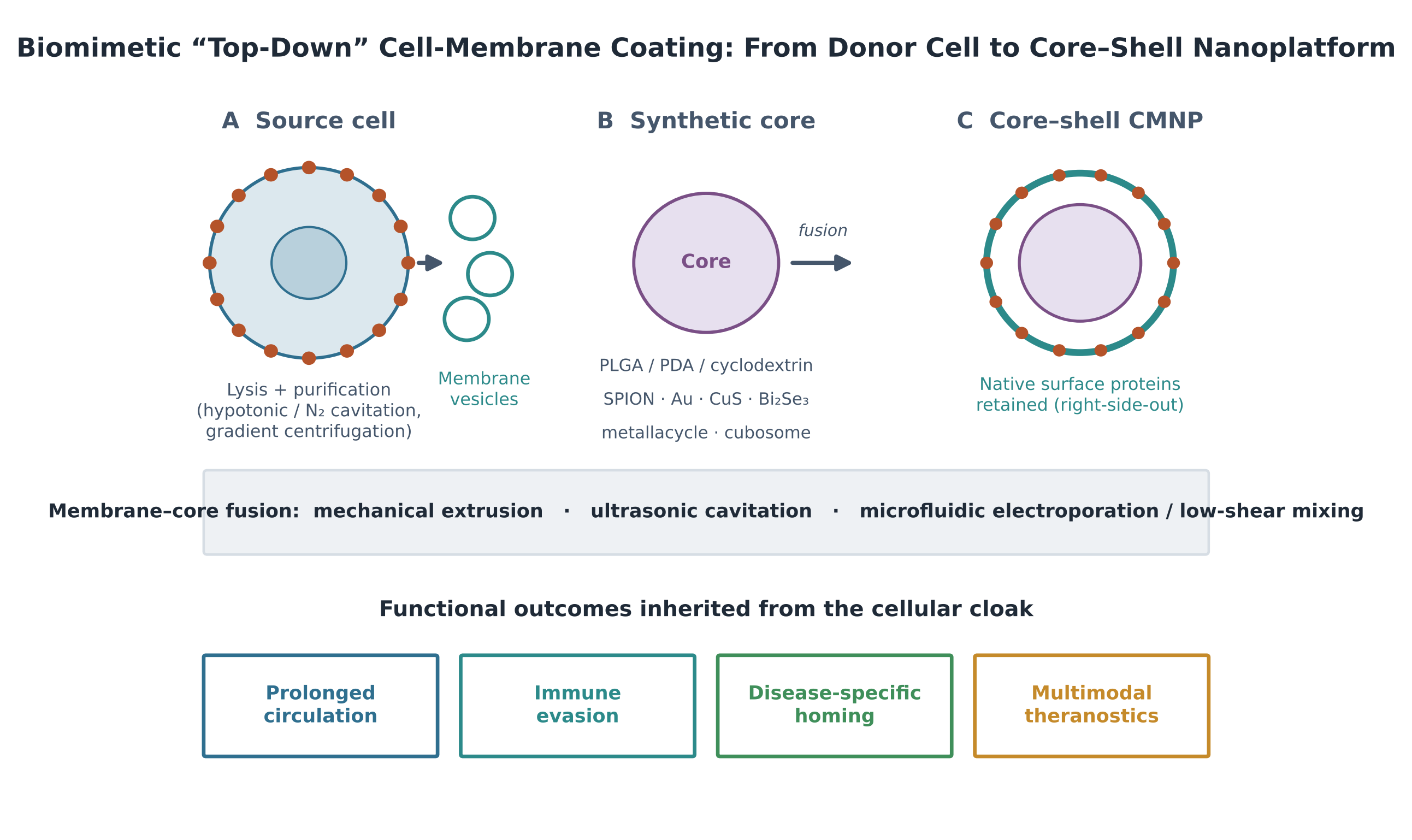
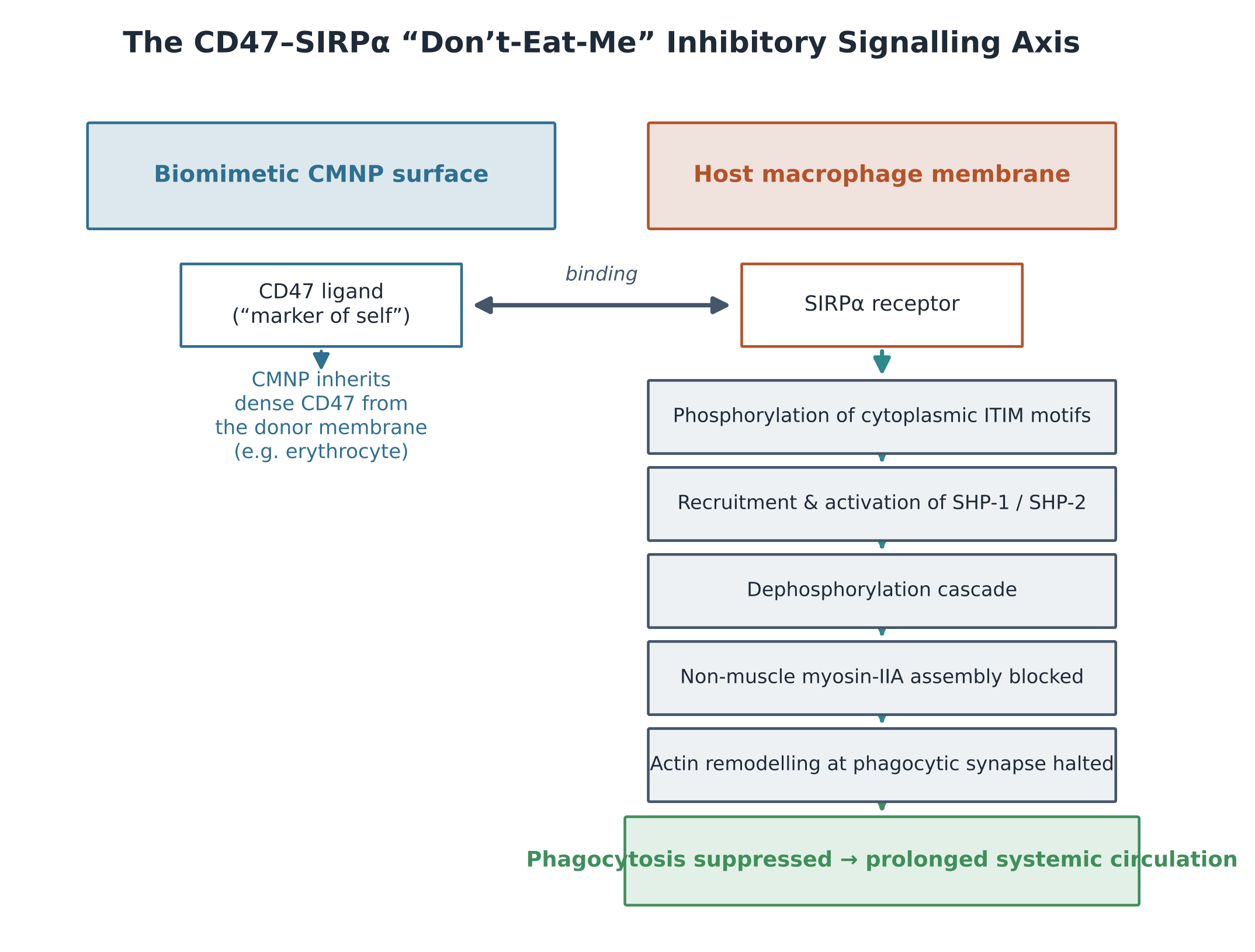
Article metrics
View details
0
Downloads
0
Citations
33
Views

View Dimensions


View Plumx


View Altmetric


0
Save
0
Citation
33
View
0
Share