2.1. Pathophysiological Barriers to Diabetic Foot Ulcer Healing
2.1.1. Clinical Burden and Systemic Drivers
DFUs are among the most debilitating, costly, and recurrent complications of diabetes. The lifetime incidence of foot ulceration ranges from 19% to 34%, about 65% of ulcers recur within three to five years, and the lifetime amputation rate approaches 20% (Armstrong et al., 2017). The persistence of these wounds reflects a cluster of systemic and local abnormalities, notably peripheral neuropathy, peripheral arterial disease, persistent microvascular ischaemia, and metabolic dysregulation sustained by hyperglycaemia (Kushwaha et al., 2022; Renuka et al., 2023). Figure 1 summarises how these factors converge on the ulcer bed.
2.1.2. Arrested Inflammation and Oxidative Stress
Normal cutaneous repair proceeds through four overlapping phases: haemostasis, acute inflammation, proliferation with granulation tissue formation and neovascularisation, and ECM remodelling. In the diabetic wound this sequence becomes uncoupled. Prolonged hyperglycaemia promotes AGE accumulation and intracellular ROS generation, and the resulting toxic microenvironment locks the wound in a chronic pro-inflammatory state (Figure 1) (Renuka et al., 2023). Central to this arrest is the failure of macrophages to shift from the M1 to the M2 phenotype. The wound bed
Table 1. Physicochemical and functional properties of bacterial cellulose (BC) matrices relevant to diabetic foot ulcer dressings. The table compiles the principal structural, hydration, mechanical, and biological properties reported for pristine and modified BC across the included studies. For each parameter, the reported range or representative value is given alongside its functional significance for chronic and diabetic wound management and the analytical method used to obtain it. Values are reproduced as reported and were not pooled; differences reflect variations in strain, culture conditions, hydration state, and post-processing. Abbreviations: AFM, atomic force microscopy; BET, Brunauer–Emmett–Teller; BJH, Barrett–Joyner–Halenda; ECM, extracellular matrix; FE-SEM, field-emission scanning electron microscopy; HDF, human dermal fibroblasts; PXRD, powder X-ray diffraction; WVTR, water vapour transmission rate.
|
Parameter
|
Reported values / observations
|
Functional significance in chronic and diabetic wounds
|
Analytical method
|
Sources
|
|
Nanofibril morphology and diameter
|
Ultrafine 3D network; ribbon-like fibrils 10–100 nm wide (elementary fibrils ~1.5–5 nm aggregating into microfibrils)
|
Mimics fibrillar architecture and nanoscale topography of dermal ECM; supports cell attachment, migration, and proliferation
|
SEM, FE-SEM, AFM
|
Czaja et al. (2007); Gürsoy et al. (2025); Khalid et al. (2026); Sajjad et al. (2025); Yang (2021); Zheng et al. (2020)
|
|
Water-holding capacity
|
98.6%–99.4% water by mass; absorbs 100–200 times its dry weight
|
Maintains wound hydration, aids autolytic debridement, supports epithelial migration, prevents scab formation
|
Gravimetric desiccation; wet/dry swelling ratio
|
Melnikova et al. (2021); Revin et al. (2023); Singla et al. (2017, 2018); Yang (2021); Zheng et al. (2020)
|
|
Tensile strength and elasticity
|
2.02 ± 0.30 MPa (wet hydrogel) to 200–260 MPa (dried/drawn films); Young's modulus 15–138 GPa; elongation at break 22%–63.8%
|
Resists plantar friction and shear; allows painless, atraumatic application and removal
|
Universal tensile testing; stress–strain analysis
|
Gürsoy et al. (2025); Revin et al. (2023); Yang (2021); Zheng et al. (2020)
|
|
Water vapour transmission rate
|
2,500–2,856.60 g/m²/day (2,856.60 g/m²/day for MA-modified nZnO/BC)
|
Balances moisture and gas exchange; prevents maceration and desiccation
|
Cup evaporation / gravimetric permeability (ASTM)
|
Ciecholewska-Juśko et al. (2021); Luo et al. (2020); Melnikova et al. (2021); Zheng et al. (2020)
|
|
Crystallinity index and polymorphism
|
59.77% to >70%; predominantly Iα allomorph; XRD peaks at 2θ = 14.5°, 17.4°, 23.9°
|
Chemical inertness and durability under proteolytic stress; reactive surface for modification
|
PXRD; solid-state ¹³C NMR
|
Melnikova et al. (2021); Revin et al. (2023); Yang (2021); Zheng et al. (2020)
|
|
Pore architecture and surface area
|
Porosity >80%–90%; specific surface area >150 m²/g; pores from nanoscale to 30–90 µm
|
Barrier to exogenous bacteria; high-capacity reservoir for nanoparticle loading and controlled release
|
BET nitrogen adsorption; BJH analysis
|
Gürsoy et al. (2025); Luo et al. (2020); Revin et al. (2023); Yang (2021); Zheng et al. (2020)
|
|
Biocompatibility and endotoxin profile
|
Non-haemolytic; >85%–95% viability in HDF, keratinocytes, and L929 fibroblasts; minimal endotoxin after alkali purification
|
Low immunogenicity; no foreign-body giant cell reaction or necrosis on chronic contact
|
MTT/CCK-8 assays; live/dead staining; ISO 10993-5 direct contact
|
Galateanu et al. (2015); Kart et al. (2024); Luo et al. (2020); Melnikova et al. (2021); Yang (2021)
|
Table 2. Nanoparticle functionalisation strategies used to construct antimicrobial bacterial cellulose (BC) and related cellulose nanocomposites. The table compares the fabrication routes used to incorporate metallic, metal oxide, framework, magnetic, and carbonaceous nanostructures into BC or closely related cellulose matrices. For each formulation it lists the synthetic route, the chemical or physical mechanism holding the particles in place, the resulting particle size and morphology, and the main functional advantage for wound care. Route choice governs particle distribution, leaching risk, and release kinetics, and therefore influences the antimicrobial and regenerative outcomes summarised in Tables 3 and 4. Abbreviations: BDP, betulin diphosphate; CNC, cellulose nanocrystal; FCP, fish collagen peptides; HPβCD, hydroxypropyl-β-cyclodextrin; MA, maleic anhydride; ZIF, zeolitic imidazolate framework.
|
Nanoparticle type / modifying agent
|
Synthetic / fabrication route
|
Interfacial binding mechanism
|
Structure and morphology
|
Key functional advantage
|
Sources
|
|
AgNPs / curcumin–HPβCD
|
Ex situ; AgNO₃ reduced by curcumin–HPβCD complex, then loaded into wet BC hydrogel
|
Hydrogen bonding and physical entrapment of curcumin–AgNP complex in pores
|
Spherical AgNPs (10–30 nm) evenly distributed through hydrogel
|
Overcomes curcumin hydrophobicity; combines silver killing with curcumin-mediated regeneration
|
Gupta et al. (2020); Ibraheem & Shaker (2025); Revin et al. (2023); Zheng et al. (2020)
|
|
Phytofabricated AgNPs on plant/bamboo CNCs
|
In situ green reduction of AgNO₃ with Syzygium cumini or bamboo leaf extracts
|
Adsorption and nucleation on CNC hydroxyl groups without chemical reductants
|
Spherical to quasi-spherical AgNPs (5–20 nm) on rod-shaped CNCs
|
Low silver load (0.05 wt.%); sustained non-toxic Ag⁺ release; no chemical residues
|
Singla et al. (2017, 2018); Ibraheem & Shaker (2025)
|
|
ZnO NPs via MA modification
|
BC esterified with MA, then in situ precipitation / hydrothermal growth of nano-ZnO
|
Carboxyl anchoring groups coordinate Zn²⁺ and prevent aggregation
|
"String-beaded" fibrils; ZnO 30–60 nm; macropores 30–90 µm
|
Flexible membrane; limited leaching; WVTR 2,856.60 g/m²/day
|
Luo et al. (2020); Zheng et al. (2020); Gürsoy et al. (2025)
|
|
ZnO NPs with betulin diphosphate
|
Ultrasonic-assisted impregnation of BDP-coated ZnO NPs (5%–25%; BDP 1.5%) into BC
|
Electrostatic, hydrophobic, and hydrogen-bonding interactions with glucan chains
|
BC crystallites 5–6 nm; ZnO 10–18 nm capped with BDP
|
Anti-inflammatory, antioxidant, and membrane-repair effects; restores microcirculation
|
Melnikova et al. (2021); Revin et al. (2023)
|
|
Graphene oxide nanosheets
|
Ex situ vacuum-assisted immersion and ultrasonic dispersion into purified BC
|
Hydrogen bonding between –OH, –COOH, and epoxide groups of GO and BC
|
Layered interpenetrating network; GO sheets wrapped around BC fibres
|
Greater stiffness; sharp edges disrupt bacterial envelopes
|
Khalid et al. (2026); Zheng et al. (2020); Gürsoy et al. (2025)
|
|
Cu-doped ZIF / fish collagen peptides (Col-Cu@ZIF)
|
One-pot co-precipitation of Cu²⁺/Zn²⁺ with 2-methylimidazole and FCP, embedded in BC hydrogel
|
Metal chelation in framework; physical entanglement and hydrogen bonding in BC
|
Dodecahedral MOF crystals (80–150 nm) in 3D mesh
|
Fenton-like ROS kills biofilms; sustained Cu²⁺ and FCP release aids neovascularisation
|
Sajjad et al. (2025); Gürsoy et al. (2025)
|
|
Magnetic Fe₃O₄ nanoparticles
|
In situ biosynthesis; Fe₃O₄ added to K. xylinus medium during pellicle growth
|
Physical entrapment in self-assembling cellulose ribbons
|
Superparamagnetic cores (10–25 nm) through full thickness
|
Remote magnetic control; supports adipose-derived stem cell adhesion
|
Galateanu et al. (2015); Zheng et al. (2020)
|
|
Multi-nanoparticle hybrids (ZnO–borax / ZnCuO₂)
|
Ultrasonic loading and thermal crosslinking of pre-formed particles into BC films
|
Interfacial coordination and borate-ester crosslinking with hydroxyls
|
Clusters (20–80 nm) anchored in interstitial voids
|
Antibiofilm activity; promotes L929 migration without cytotoxicity
|
Kart et al. (2024); Gürsoy et al. (2025)
|
accumulates IL-1α, interleukin-1β (IL-1β), IL-6, and TNF-α, and this sustained cytokine signalling drives over-secretion of MMPs, which degrade newly synthesised collagen and fibronectin and cleave endogenous growth factors (Khalid et al., 2026; Sajjad et al., 2025).
2.1.3. Impaired Angiogenesis and Matrix Degradation
Diabetic wounds also show marked microvascular rarefaction and endothelial dysfunction. VEGF, basic fibroblast growth factor (bFGF), and PDGF are deficient or rapidly degraded, so the regenerating tissue receives too little oxygen and too few nutrients to support capillary sprouting (Singla et al., 2017, 2018). The combined effect of protease excess and growth factor deficiency is a thin, poorly organised granulation bed that closes slowly, if at all.
2.1.4. Microbial Bioburden and Biofilm Formation
The moist, nutrient-rich, and immunologically compromised ulcer bed is an ideal niche for opportunistic pathogens. More than half of chronic DFUs show overt infection, typically involving S. aureus (including methicillin-resistant S. aureus, MRSA), Streptococcus species, P. aeruginosa, and E. coli (Kushwaha et al., 2022; Zheng et al., 2020). These organisms secrete an EPS matrix that binds colonies into resilient biofilms. Biofilm-embedded bacteria can tolerate antibiotic concentrations up to 1,000-fold higher than their planktonic counterparts and evade phagocytosis by neutrophils and macrophages (Kart et al., 2024). Persistent biofilms fuel necrosis, precipitate osteomyelitis, and raise the risk of amputation.
2.1.5. Limitations of Conventional Dressings
Dry gauze, paraffin tulles, and basic polymeric foams do not address these barriers. They tend to adhere to granulation tissue and strip it on removal, manage exudate poorly, offer limited protection against microbial ingress, and lack bioactive or bactericidal function (Revin et al., 2023; Zheng et al., 2020). These shortcomings explain the interest in multifunctional nanostructured dressings that can suppress infection, resolve inflammation, and support tissue regeneration at the same time (Gürsoy et al., 2025; Renuka et al., 2023).
2.2. Bacterial Cellulose as an ECM-Mimetic Biomaterial Matrix
2.2.1. Biosynthesis and Chemical Purity
Among the biopolymers studied for chronic wound care, BC, also called bacterial nanocellulose, stands out for its structure and physical behaviour (Czaja et al., 2007; Revin et al., 2023). It is produced by oxidative fermentation by non-pathogenic, strictly aerobic Gram-negative bacteria such as Komagataeibacter xylinus (formerly Gluconacetobacter xylinus) and related genera, and accumulates as a gelatinous pellicle at the air–liquid interface of the culture. Chemically, BC consists of linear β-(1→4)-linked D-glucose chains, the same primary structure as plant cellulose, but it is free of lignin, pectin, and hemicellulose. Because no harsh fractionation is required, BC shows excellent biocompatibility, low immunogenicity, and good cytocompatibility with human dermal fibroblasts and keratinocytes (Galateanu et al., 2015; Revin et al., 2023).
2.2.2. Three-Dimensional Nanofibrillar Architecture
The defining feature of BC is its ultrafine 3D reticulated network. Ribbon-like microfibrils 10–100 nm in diameter, themselves assembled from elementary fibrils of about 1.5–5 nm, interweave randomly into a dense, highly porous mesh stabilised by extensive hydrogen bonding (Table 1; Figure 2A) (Czaja et al., 2007; Gürsoy et al., 2025). This architecture closely reproduces the nanoscale topology and fibrous organisation of the dermal ECM, which favours cell attachment, migration, and proliferation (Khalid et al., 2026; Yang, 2021).
2.2.3. Fluid Handling, Mechanical Integrity, and Atraumatic Contact
The large surface area and capillary porosity of BC translate into practical advantages at the wound surface (Table 1). BC retains up to 99 wt.% water and can absorb many times its dry mass in fluid, allowing it to take up heavy exudate while keeping the wound moist, which in turn supports cell migration and re-epithelialisation (Melnikova et al., 2021; Zheng et al., 2020). In the hydrated state it also provides balanced water vapour transmission and gas permeation, protecting against both desiccation and maceration of the surrounding skin (Ciecholewska-Juśko et al., 2021; Luo et al., 2020). Its high crystallinity, typically 60–80%, and high degree of polymerisation give hydrated membranes considerable wet tensile strength and flexibility, so they conform to the contours of the foot, resist shear, and form a physical barrier to secondary contamination (Revin et al., 2023; Yang, 2021). Finally, its hydrophilic, non-adherent surface
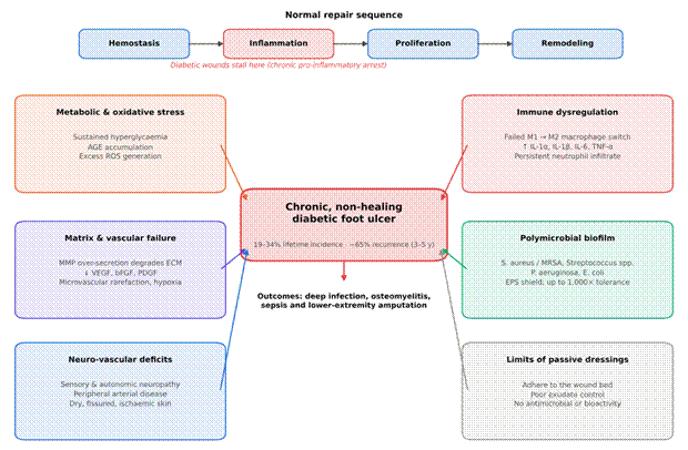
Figure 1. Pathophysiological barriers that converge to produce a chronic, non-healing diabetic foot ulcer. The upper strip shows the normal sequence of cutaneous repair and marks the inflammatory phase at which diabetic wounds typically stall. Six groups of interacting barriers surround the ulcer: metabolic and oxidative stress (hyperglycaemia, advanced glycation end-products, reactive oxygen species), immune dysregulation (failed M1-to-M2 macrophage switching and persistent IL-1α, IL-1β, IL-6, and TNF-α), matrix and vascular failure (MMP excess and deficient VEGF, bFGF, and PDGF), polymicrobial biofilm, neuro-vascular deficits, and the limitations of passive dressings. Together these drive deep infection, osteomyelitis, sepsis, and amputation. Constructed from Armstrong et al. (2017), Kushwaha et al. (2022), Renuka et al. (2023), and Section 2.1.
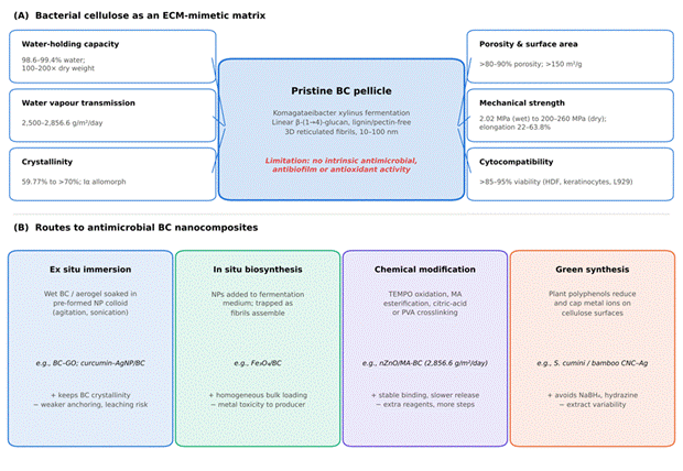
Figure 2. Bacterial cellulose as an ECM-mimetic matrix and the principal routes used to convert it into an antimicrobial nanocomposite. (A) Pristine BC secreted by Komagataeibacter xylinus forms a lignin- and pectin-free 3D network of 10–100 nm fibrils; the surrounding tiles summarise the reported water-holding capacity, vapour transmission, crystallinity, porosity, mechanical strength, and cytocompatibility (values from Table 1). Its key limitation, the absence of intrinsic antimicrobial activity, is highlighted. (B) The four fabrication routes (ex situ immersion, in situ biosynthesis, chemical modification, and green synthesis) are shown with representative examples and their main advantages (+) and drawbacks (−).
allows BC to be applied and removed without damaging the advancing epithelial margin, sparing patients additional pain during dressing changes (Czaja et al., 2007).
2.2.4. The Antimicrobial Gap of Pristine BC
These advantages come with a significant caveat. Pristine BC has no intrinsic antimicrobial, antibiofilm, or antioxidant activity. Placed over a heavily contaminated ulcer, unmodified BC cannot inhibit bacterial growth, and microorganisms may even colonise its porous mesh (Zheng et al., 2020). Functionalising BC with antimicrobial nanoscale agents has therefore become an essential design strategy (Ibraheem & Shaker, 2025; Revin et al., 2023).
2.3. Fabrication and Modification Strategies for BC Nanocomposites
The reactive hydroxyl groups on BC nanofibrils and the open interstitial network make BC an effective template for organic and inorganic nanostructures. Four broad fabrication routes have been used to construct antimicrobial BC nanocomposites (Table 2; Figure 2B).
2.3.1. Ex Situ Incorporation by Physical Absorption and Immersion
The ex situ approach is the most widely used because it is simple, reproducible, and leaves the crystalline BC matrix intact. Purified wet pellicles or freeze-dried aerogels are immersed in colloidal suspensions of pre-synthesised nanoparticles, bioactive molecules, or polymers under agitation or sonication. Particles penetrate the mesh by capillary action and are retained by physical entrapment and non-covalent interactions, including hydrogen bonding, electrostatic attraction, and van der Waals forces (Zheng et al., 2020). Khalid et al. (2026) used this approach to disperse GO nanosheets evenly through the BC network; Fourier-transform infrared spectroscopy (FTIR) and scanning electron microscopy (SEM) indicated close interpenetration mediated by hydrogen bonding between hydroxyl and carboxyl groups. Gupta et al. (2020) similarly loaded purified BC hydrogels with curcumin–cyclodextrin-functionalised AgNPs and achieved high loading without loss of hydration or mechanical stability.
2.3.2. In Situ Biosynthesis
In the in situ route, nanomaterials, polymers, or reinforcing additives are added directly to the fermentation medium. As K. xylinus extrudes cellulose protofibrils, the additives become trapped within the growing network, yielding a homogeneous distribution through the full thickness of the pellicle. Galateanu et al. (2015) produced BC–magnetite (Fe₃O₄) nanocomposites in this way and showed that the resulting scaffolds supported stem cell proliferation. The approach requires careful optimisation, however, because high concentrations of metal precursors or antimicrobial agents can be toxic to the producing bacteria, lowering cellulose yield and altering fibril crystallisation (Revin et al., 2023).
2.3.3. Chemical Surface Modification and Crosslinking
Chemical functionalisation can strengthen interfacial bonding, regulate swelling, and prevent burst release of loaded particles. Options include TEMPO-mediated oxidation to generate carboxylate groups, esterification, etherification, and polymer grafting (Zheng et al., 2020). Luo et al. (2020) modified BC with maleic anhydride (MA) to introduce carboxyl anchoring sites for in situ ZnO deposition; the resulting membrane was flexible and porous, resisted particle agglomeration, and retained a water vapour transmission rate (WVTR) of 2,856.60 g/m²/day. Hybridising cellulose derivatives such as carboxymethyl cellulose (CMC) with poly(vinyl alcohol) (PVA) by citric acid crosslinking has likewise been used to anchor metallic nanoparticles in stable, non-leaching, biocompatible hydrogels (Mansur et al., 2023). Crosslinking can also convert BC into superabsorbent forms suited to highly exudative chronic wounds (Ciecholewska-Juśko et al., 2021).
2.3.4. Green and Bio-Mediated Nanoparticle Synthesis
Concerns about hazardous reducing agents such as sodium borohydride and hydrazine, and about residual solvents, have encouraged green synthesis. Plant extracts rich in polyphenols, flavonoids, and reducing sugars serve as both reducing and capping agents. Singla et al. (2017) used Syzygium cumini leaf extract to synthesise AgNPs directly on cellulose nanocrystals (CNCs), avoiding toxic residues and producing cytocompatible, strongly antibacterial nanobiocomposites; a related bamboo-derived system followed (Singla et al., 2018). Curcumin and Centella asiatica extract have also been incorporated into cellulose- and polymer-based nanocomposites to mediate metal reduction, strengthen antioxidant defences, and stimulate proliferation (Gupta et al., 2020; Wang et al., 2024).
2.4. Mechanistic Antimicrobial and Bioactive Actions of Embedded Nanoparticles
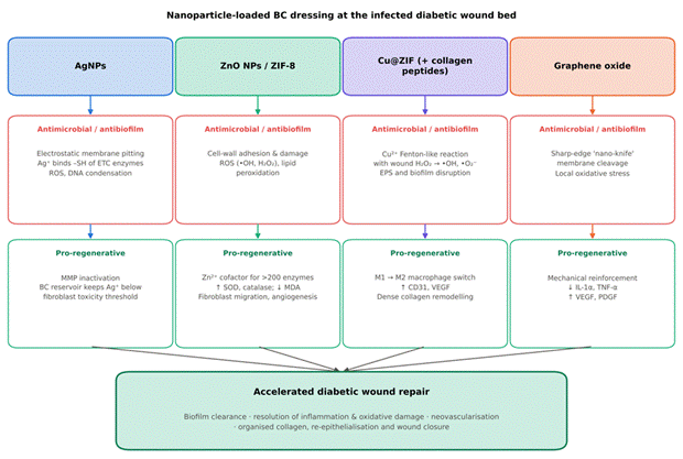
Incorporating nanoscale agents converts BC from a passive cover into an active therapeutic platform able to attack drug-resistant biofilms and alter the pathological wound microenvironment (Figure 3; Table 3).
2.4.1. Silver Nanoparticles
AgNPs are the most extensively studied antimicrobial agents in wound care, owing to their broad-spectrum activity against multidrug-resistant pathogens including S. aureus, MRSA, and P. aeruginosa (Barud et al., 2011; Ibraheem & Shaker, 2025). Within BC, AgNPs act through several overlapping mechanisms. They adhere electrostatically to negatively charged lipopolysaccharide or peptidoglycan, pitting the membrane and causing leakage of intracellular contents. In the aqueous wound environment they continuously release Ag⁺ ions, which bind sulfhydryl groups on respiratory chain enzymes and structural proteins, interrupting electron transport and ATP synthesis. Intracellular silver also catalyses ROS production, leading to DNA condensation, inhibition of replication, and cell lysis. Beyond direct killing, AgNPs can inactivate excess MMPs and so limit inflammatory tissue breakdown (Gupta et al., 2020; Zheng et al., 2020). Immobilisation in the BC network also addresses the longstanding concern about silver toxicity: uncontrolled silver can induce keratinocyte apoptosis and slow re-epithelialisation, whereas the BC matrix acts as a reservoir that keeps local Ag⁺ within the bactericidal window but below the threshold that harms fibroblasts (Gupta et al., 2020; Singla et al., 2017).
2.4.2. Zinc Oxide Nanoparticles and Zeolitic Imidazolate Frameworks
ZnO NPs are an attractive alternative or complement to silver, combining lower cytotoxicity, chemical stability, low cost, and genuine biological functions (Khalid et al., 2017; Luo et al., 2020). Their antibacterial activity arises from electrostatic adhesion to the cell wall, mechanical destabilisation, and ROS generation (hydroxyl radicals and hydrogen peroxide), which drives lipid peroxidation and cell death. ZnO NPs also act as a slow-release source of Zn²⁺, a cofactor for more than 200 enzymes, including alkaline phosphatase, carbonic anhydrases, and DNA and RNA polymerases. Released zinc stimulates fibroblast proliferation and migration, supports keratinocyte re-epithelialisation, and moderates local inflammatory cytokine secretion. Melnikova et al. (2021) showed that BC–ZnO dressings increased superoxide dismutase (SOD) and catalase activity and reduced malondialdehyde (MDA), indicating reversal of oxidative stress. Zeolitic imidazolate framework-8 (ZIF-8) loaded onto BC has been reported to release Zn²⁺ in a controlled, sustained manner, supporting angiogenic sprouting while suppressing E. coli and S. aureus (Gürsoy et al., 2025).
2.4.3. Copper Nanostructures and Metal–Organic Frameworks
Copper nanostructures combine potent antimicrobial activity with a pronounced capacity to stimulate angiogenesis. Sajjad et al. (2025) designed a BC hydrogel membrane containing copper-doped, fish collagen peptide-loaded zeolitic imidazolate frameworks (Col-Cu@ZIF). In infected diabetic wounds, released Cu²⁺ reacted with endogenous hydrogen peroxide in a Fenton-like reaction, generating localised ROS that rapidly disrupted biofilms. The sustained co-delivery of copper ions and collagen peptides also stimulated endothelial migration, increased CD31 and VEGF expression, and promoted dense collagen remodelling.
2.4.4. Graphene Oxide and Carbonaceous Nanostructures
GO nanosheets provide mechanical reinforcement as well as contact-mediated killing. Their sharp edges can cleave bacterial membranes, a so-called "nano-knife" effect, while generating localised oxidative stress (Zheng et al., 2020). In a diabetic mouse model, Khalid et al. (2026) found that BC–GO films produced inhibition zones of 24.6 mm against S. aureus and 23 mm against E. coli and achieved complete wound closure within 21 days, outperforming a commercial ointment.
2.5. In Vitro and In Vivo Biological Outcomes in Diabetic Wound Repair
Preclinical studies suggest that antimicrobial BC dressings accelerate diabetic wound repair through several linked cellular and molecular routes (Table 4; Figure 3; Figure 4).
2.5.1. Suppression of Chronic Inflammation and Oxidative Stress
Real-time reverse-transcription PCR and immunohistochemistry show that functionalised nanocomposites reduce gene and protein expression of IL-1α, IL-1β, IL-6, and TNF-α in diabetic wound tissue. Khalid et al. (2026) reported reduced IL-1α and TNF-α expression
Table 3. Antimicrobial and antibiofilm efficacy of functionalised bacterial cellulose and comparator nanocomposites against common diabetic foot ulcer pathogens. The table summarises quantitative in vitro (and, where stated, in-wound) antimicrobial results for each formulation against Gram-positive, Gram-negative, and fungal pathogens commonly isolated from infected diabetic foot ulcers. The assay method is given for each entry because inhibition zones, viable-count reductions, and biofilm inhibition percentages are not directly interchangeable. The proposed mechanism of action reflects the authors' interpretation in the original studies. Non-BC comparators (CMC/PVA–AgNP hydrogels and antibiotic-loaded coaxial nanofibres) are included as benchmarks. Abbreviations: CFU, colony-forming units; CLSI, Clinical and Laboratory Standards Institute; CV, crystal violet; EPS, extracellular polymeric substance; ETC, electron transport chain.
|
Nanocomposite formulation
|
Target microorganism
|
Assay method
|
Quantitative outcome
|
Proposed mechanism
|
Sources
|
|
BC–GO nanocomposite film
|
S. aureus (Gram-positive); E. coli (Gram-negative)
|
Agar disc diffusion; liquid growth inhibition
|
Inhibition zones: S. aureus 24.6 ± 0.5 mm; E. coli 23.0 ± 1.0 mm (greater than pristine BC and Polyfax)
|
Physical wall disruption by GO edges; oxidative stress; metabolite leakage
|
Khalid et al. (2026); Zheng et al. (2020)
|
|
5 wt.% nZnO/BC membrane
|
S. aureus; E. coli
|
Suspension shaking; viable colony counting
|
>99.9% bactericidal reduction within 24 h
|
Cell-wall adherence, Zn²⁺ release, ROS generation
|
Luo et al. (2020); Zheng et al. (2020)
|
|
Col-Cu@ZIF–BC hydrogel
|
S. aureus; E. coli
|
CFU spread-plate assay (1 × 10⁶ inoculum)
|
>99.5% reduction vs. untreated control and pristine BC; effective in infected wound fluid
|
Cu²⁺-catalysed Fenton-like conversion of H₂O₂ to •OH and •O₂⁻
|
Sajjad et al. (2025)
|
|
Curcumin–AgNP BC hydrogel
|
S. aureus; P. aeruginosa; Candida auris
|
Kirby–Bauer diffusion; broth microdilution
|
Broad-spectrum inhibition including fungal pathogen, without mammalian cell toxicity
|
Ag⁺ binding to thiol groups of respiratory enzymes; curcumin-mediated membrane permeabilisation
|
Gupta et al. (2020); Revin et al. (2023)
|
|
BC–ZnO / BC–borax / BC–ZnCuO₂
|
Preformed biofilms of S. aureus, P. aeruginosa, Listeria innocua
|
CV staining; XTT metabolic assay
|
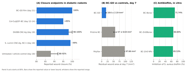
S. aureus biofilm inhibition: BC–borax 71.74%, BC–ZnOBorax 66.60%, BC–ZnO 65.53%; BC–ZnCuO₂ degraded P. aeruginosa biofilm
|
EPS matrix degradation; inhibition of microbial metabolism
|
Kart et al. (2024)
|
|
DH-CNC-Ag and BB-CNC-Ag
|
S. aureus; E. coli; P. aeruginosa
|
Disc diffusion; time-kill turbidity
|
Clear inhibition zones; prevented sepsis and pus formation in vivo within 3 days
|
Sustained low-dose Ag⁺; membrane depolarisation; DNA condensation
|
Singla et al. (2017, 2018); Ibraheem & Shaker (2025)
|
|
CMC/PVA hybrid hydrogel with AgNPs
|
S. aureus ATCC 6538; E. coli ATCC 25922
|
CLSI disc diffusion; broth dilution
|
Concentration-dependent inhibition zones; long-term prevention of colonisation
|
Continuous Ag⁺ elution from carboxylate/hydroxyl complexes
|
Mansur et al. (2023); Ibraheem & Shaker (2025)
|
|
Coaxial moxifloxacin / fusidic acid nanofibres
|
Clinical S. aureus wound isolates
|
Daily wound swab culture; colony enumeration
|
Significant suppression of bioburden vs. plain gauze
|
Topoisomerase IV inhibition plus inhibition of protein synthesis
|
Alzahrani et al. (2024)
|
Table 4. Preclinical in vivo wound healing and tissue regeneration outcomes of antimicrobial nanocellulose-based dressings in diabetic and related animal models. The table collates the in vivo experimental design and outcomes for each dressing, including animal species, method of diabetes induction and glycaemic threshold, wound type and size, group size, treatment regimen, quantitative closure, and histological and molecular endpoints. Closure values are reproduced as reported; studies differ in wound size, follow-up, and comparator, so values should not be compared directly. Most models used streptozotocin (STZ)-induced diabetic rodents with acute excisional wounds, a limitation discussed in Section 5.4. Abbreviations: HEGF, human epidermal growth factor; MDA, malondialdehyde; PLX, poloxamer; SOD, superoxide dismutase.
|
Dressing
|
Model and sample size
|
Regimen and duration
|
Wound closure
|
Histological and molecular outcomes
|
Sources
|
|
BC–GO film
|
Male BALB/c mice; STZ diabetes (>400 mg/dL); 10 mm dorsal excision (n = 5/group)
|
Topical; changed on days 7 and 14; 21 days
|
100% by day 21; day 7 residual area 18.6 ± 2.19 mm² vs. 57 ± 6.63 mm² (BC) and 97.66 mm² (Polyfax)
|
↓ IL-1α, TNF-α mRNA; ↑ VEGF, PDGF; earlier re-epithelialisation; thick collagen
|
Khalid et al. (2026)
|
|
Col-Cu@ZIF–BC hydrogel
|
Male BALB/c mice; STZ diabetes (>15 mM); 15 mm excision with S. aureus biofilm (n = 6/group)
|
Applied every 4 days; 14 days
|
>95% contraction by days 12–14; faster than HEGF gel and pristine BC
|
M1-to-M2 macrophage switch; dense CD31-positive capillaries; mature collagen
|
Sajjad et al. (2025)
|
|
Bamboo CNC–AgNPs (DH/BB-CNC-Ag)
|
Swiss albino mice; STZ diabetes (>200 mg/dL); 8 mm full-thickness excision (n = 9/group)
|
Daily topical hydrogel (50 mg/day); 18 days
|
98%–100% by day 18 vs. 88%–92% in untreated and vehicle controls
|
↓ IL-6, TNF-α; ↑ FGF, PDGF, VEGF; dermal–epidermal junction restored; hair follicle regeneration
|
Singla et al. (2018)
|
|
S. cumini CNC–AgNPs (NC-1)
|
Swiss albino mice; STZ diabetes; 8 mm dorsal excision (n = 9/group)
|
Daily topical ointment; 18 days
|
~99% by day 18; scab loss and epithelial coverage by day 10
|
↑ FGF, PDGF, VEGF; ↓ IL-6, TNF-α; ↑ hydroxyproline; dense parallel collagen
|
Singla et al. (2017)
|
|
BC–ZnO NPs with betulin diphosphate
|
Wistar rats; deep thermal/burn excision wound (n = 12/group)
|
Film changed on days 3, 7, 10, 21
|
Complete dermal reconstitution by day 21; superior to oleogel controls (p < 0.001)
|
↑ SOD, catalase; ↓ MDA; early restoration of microcirculation by day 3
|
Melnikova et al. (2021)
|
|
5 wt.% nZnO/BC membrane
|
BALB/c mice; 6 mm full-thickness excision infected with S. aureus + E. coli (n = 8/group)
|
Changed on days 3, 5, 7, 10, 12, 14
|
Rapid contraction by day 14; smaller residual area than gauze
|
Bacterial clearance; ↓ neutrophil infiltration at day 7; longer epithelial tongue; thicker granulation
|
Luo et al. (2020)
|
|
PLX/ZnO with Centella asiatica (CAE@PLX/ZnO)
|
Male Wistar rats; STZ diabetes; full-thickness excision
|
Daily topical; 14–21 days
|
Faster contraction and closure than standard controls
|
Improved fibroblast survival; suppressed inflammatory mediators; ↑ collagen deposition
|
Wang et al. (2024)
|
|
Coaxial fusidic acid / pirfenidone nanofibres
|
Diabetic Wistar rats; full-thickness dorsal excision (n = 4/group)
|
Daily, secured with Tegaderm; 14 days
|
Near-complete by days 11–13; faster than povidone-iodine dressing
|
Combined antibacterial and anti-inflammatory action; faster re-epithelialisation; lower bioburden
|
Alzahrani et al. (2024)
|
and less inflammatory cell infiltration in BC–GO-treated diabetic mice than in untreated controls. Singla et al. (2017, 2018) found that CNC–AgNP nanobiocomposites lowered IL-6 and TNF-α over 18 days in streptozotocin (STZ)-induced diabetic mice. By damping the inflammatory cascade and scavenging free radicals, these dressings appear to let stalled wounds move on into active proliferation.
2.5.2. Induction of Angiogenesis and Neovascularisation
Restoring a functional capillary bed is essential for reperfusing ischaemic ulcers. Nanoparticle-loaded BC dressings increase VEGF, PDGF, and the endothelial marker CD31 (Sajjad et al., 2025). Sajjad et al. (2025) observed dense CD31-positive capillary networks in diabetic mice treated with Col-Cu@ZIF–BC membranes, and Singla et al. (2017, 2018) documented significant increases in VEGF, bFGF, and PDGF with silver-functionalised nanocellulose, accompanied by greater vessel density and better granulation tissue.
2.5.3. Collagen Organisation, Re-Epithelialisation, and Wound Closure
Macroscopic and histological assessments indicate that functionalised dressings speed closure substantially. In STZ-diabetic mice, wounds treated with pristine controls or commercial ointments typically reached 88–92% closure with persistent purulent exudate by days 18–21, whereas wounds treated with BC–AgNP, BC–GO, or Col-Cu@ZIF–BC dressings reached 98–100% closure over the same period (Figure 4A) (Khalid et al., 2026; Sajjad et al., 2025; Singla et al., 2018). Masson's trichrome staining showed thick, aligned, mature collagen bundles in treated wounds, in contrast to the fragmented, randomly oriented fibrils of untreated diabetic wounds, and sustained antimicrobial release combined with a moist environment encouraged keratinocyte migration and regeneration of hair follicles and sebaceous glands (Luo et al., 2020; Singla et al., 2018).
2.6. Translational Challenges Identified in the Literature
2.6.1. Nanoparticle Toxicity and Long-Term Biocompatibility
The possibility of systemic accumulation and cellular toxicity from inorganic nanoparticles, particularly AgNPs and metal oxides, remains a concern. High intracellular metal ion uptake can cause oxidative DNA damage and mitochondrial apoptosis in healthy dermal cells. Standardised dose–response thresholds, controlled release kinetics, and stable chelation within the BC mesh will be needed to establish long-term safety (Ibraheem & Shaker, 2025; Kushwaha et al., 2022).
2.6.2. Standardisation and Large-Scale Biomanufacturing
Fermentation of BC by K. xylinus varies from batch to batch in membrane thickness, fibre density, pore size, and strength. Industrial-scale production will require tightly controlled bioreactors, inexpensive carbon sources such as agro-industrial by-products, and validated, automated protocols for nanoparticle synthesis and loading (Gürsoy et al., 2025; Revin et al., 2023).
2.6.3. Wound Fluid Complexity and Smart-Dressing Integration
Human DFU exudate is more proteolytic and variable in pH than that of rodent excision wounds, which may alter nanoparticle dissolution, cause premature leaching, or allow a protein corona to mask active sites (Kushwaha et al., 2022). Looking ahead, stimuli-responsive designs that release antimicrobials in response to pH shifts, temperature, or bacterial enzymes, together with chromogenic or bioelectronic sensors embedded in BC, could permit on-demand therapy and real-time monitoring without removing the dressing (Stanescu et al., 2021; Yang, 2021).