Biosensors and Nanotheranostics

Bionanotechnology, Drug Delivery, Therapeutics | online ISSN 3064-7789
175
Citations
119k
Views
88
Articles
Your new experience awaits. Try the new design now and help us make it even better
Switch to the new experience
Figures and Tables
REVIEWS

Salivary exosomal mRNA and biosensors for the non-invasive detection of oral cancer

Yoghinni Manogaran 1* Shazmin Kithur Mohamed 2


+ Author Affiliations

Biosensors and Nanotheranostics 5 (1) 1-8 https://doi.org/10.25163/biosensors.5110996

Submitted: 03 September 2026 Revised: 20 October 2026  Published: 01 November 2026 


Abstract

Oral squamous cell carcinoma (OSCC) accounts for roughly nine in every ten oral cancers, yet survival has barely moved in decades, mainly because most tumors are found late. Patients diagnosed at Stages I–II have five-year survival of 80%–90%, whereas those diagnosed at Stages III–IV fall to about 30%. Scalpel biopsy remains the diagnostic standard, but it is invasive, cannot be repeated freely, and depends on a lesion already being visible. Saliva offers a different starting point. It bathes the tumor directly, and the exosomes shed into it carry messenger RNA (mRNA) inside a lipid bilayer that protects it from salivary nucleases. In this review, we bring together the evidence on salivary exosomal mRNA as a biomarker class for OSCC and on biosensors that might read it at the point of care. A structured search of PubMed/MEDLINE, Scopus, Web of Science, and Google Scholar informed a narrative synthesis covering exosome biology, isolation technologies, biomarker performance, and sensor engineering. In paired-sample data, nine salivary exosomal transcripts were all upregulated, and a TNF-α/OAZ1 panel reached an area under the curve of 0.89 (80% sensitivity, 90% specificity). Salivary MMP-9 and OAZ1 tracked tumor grade, and IL-6 tracked nodal metastasis; serum markers did neither. Electrochemical, SERS, microfluidic, CRISPR-based, and electric field-induced platforms report attomolar-to-femtomolar detection limits in microlitre volumes, although most have not yet targeted mRNA in OSCC saliva. Single-cohort evidence, pre-analytical variability, and vesicle heterogeneity remain the main barriers. We close by outlining the validation and standardization steps needed before such devices can reach routine clinics.

Keywords: oral squamous cell carcinoma; saliva; exosomes; extracellular vesicles; messenger RNA; liquid biopsy; biosensor; point-of-care diagnostics; early detection

1. Introduction

Oral squamous cell carcinoma (OSCC) is the dominant malignancy of the head and neck region, accounting for approximately 90% of all oral cancer diagnoses worldwide (Kumar et al., 2025). It arises from the mucosal epithelium of the tongue, buccal mucosa, floor of the mouth, gingiva, and palate, and it continues to impose a heavy global burden of morbidity and mortality (Kumar et al., 2025; Song & Yang, 2026). The disease is concentrated in people aged 40 to 70 years, and its principal drivers are behavioral: tobacco smoking and chewing, heavy alcohol consumption, and betel nut use together account for much of the malignant transformation seen in clinical practice (Kumar et al., 2025; Song & Yang, 2026). Because these exposures are common, and because the oral cavity is directly accessible, OSCC ought to be one of the more detectable solid cancers. In practice, it is not.

The gap between accessibility and detection is visible in the survival data. Surgery, radiotherapy, chemotherapy, and molecularly targeted agents have all advanced, yet the overall five-year survival of OSCC remains at approximately 50% to 60% (T. Li et al., 2022; Nonaka & Wong, 2022). The decisive variable is stage at diagnosis. Patients whose tumors are detected at localized Stages I and II have five-year survival of 80% to 90%, whereas survival falls to about 30% once the disease has reached Stages III and IV (Kumar et al., 2025) (Figure 1A). Early lesions are frequently painless or clinically non-specific, so they are easy for both patients and clinicians to overlook during the very window in which cure is most attainable (Kumar et al., 2025; Song & Yang, 2026).

Current diagnostic practice compounds this problem. Detection depends on visual and tactile screening, followed by scalpel biopsy, histopathological evaluation, and, where indicated, computed tomography (CT) or magnetic resonance imaging (MRI) (T. Li et al., 2022; Song & Yang, 2026). Histopathology rightly remains the gold standard, but it has inherent drawbacks. It is invasive and uncomfortable, carries risks of bleeding and infection, and samples a single site that may not represent the spatial and temporal heterogeneity of the tumor (Kumar et al., 2025; Nonaka & Wong, 2022). Biopsy also cannot be repeated freely, which makes it poorly suited to longitudinal surveillance, monitoring of treatment response, or screening at population scale (T. Li et al., 2022; Nonaka & Wong, 2022) (Figure 1C). A test that could detect molecular change before morphological change, without cutting tissue, would therefore address a genuine clinical need (Kumar et al., 2025; Tang et al., 2024).

Liquid biopsy, which samples tumor-derived material circulating in body fluids, has emerged as the leading candidate for such a test (Nonaka & Wong, 2022; Tang et al., 2024). Blood and serum have historically been favored for systemic profiling, but saliva has attracted growing interest as the more logical fluid for oral malignancy (Kumar et al., 2025; Lee et al., 2009; Nonaka & Wong, 2022). Its collection is painless, inexpensive, and free of needle-stick and procedural infection risk, which improves compliance in both screening and post-treatment follow-up (Chandrasekaran et al., 2024; Kumar et al., 2025). Saliva also contains a broad repertoire of DNA, cell-free and vesicular RNA, proteins, enzymes, metabolites, and extracellular vesicles that reflect both systemic health and local mucosal pathology (Chandrasekaran et al., 2024; Kumar et al., 2025). Most importantly, saliva is in direct physical contact with the tumor. Molecules shed by a small early lesion are diluted across liters of circulating blood, whereas in saliva they remain concentrated close to their source (Kumar et al., 2025; Nonaka & Wong, 2022) (Figure 1B).

Saliva has its own difficulty, however. It is rich in nucleases and proteases that rapidly degrade naked RNA and protein (Kumar et al., 2025; Nonaka & Wong, 2022). Exosomes offer a biological solution. These endosome-derived vesicles, roughly 30 to 150–200 nm in diameter, are enclosed by a lipid bilayer studded with tetraspanins (CD9, CD63, CD81) and carry endosomal sorting markers such as TSG101 and Alix; the bilayer shields their cargo from enzymatic attack (Kumar et al., 2025; Tang et al., 2024). Tumor cells, including OSCC cells, release exosomes in elevated numbers and use them for intercellular communication, extracellular matrix remodeling, immune suppression, and preparation of pre-metastatic niches (Kumar et al., 2025; T. Li et al., 2022). Among the cargo these vesicles carry, microRNAs have been studied most extensively in oral liquid biopsy (Nonaka & Wong, 2022; Song & Yang, 2026). Exosomal messenger RNA (mRNA), by contrast, has received less attention, even though it reports more directly on active transcriptional programs. In OSCC, exosomal mRNAs reflect chronic inflammatory signaling (TNF-α, IL-1β, IL-6, IL-8), hyperproliferation and polyamine metabolism (OAZ1, SAT, S100P), and matrix remodeling and invasion (MMP-9, Chemerin) (Kumar et al., 2025). In paired-sample analysis, a salivary exosomal TNF-α/OAZ1 panel distinguished OSCC from healthy controls with an area under the receiver operating characteristic curve (AUC) of 0.89, 80% sensitivity, and 90% specificity, and it outperformed the corresponding serum markers. In the same study, salivary MMP-9, OAZ1, and IL-8 separated Grade I from Grade III tumors, and salivary IL-6 was associated with cervical lymph node metastasis (Kumar et al., 2025).

Biomarker validity alone does not make a usable test. Conventional exosome workflows rely on ultracentrifugation, size-exclusion chromatography, or polymer precipitation, followed by RNA extraction and reverse transcription quantitative PCR (RT-qPCR), microarrays, or sequencing. These steps are slow, costly, and labor-intensive, they need relatively large sample volumes, and each one risks losing low-abundance transcripts (Nonaka & Wong, 2022; Tang et al., 2024; Wu et al., 2021). Biosensors are designed to remove these steps. By coupling a biological recognition element to an electrochemical, optical, or electrical transducer, often with nanomaterial interfaces, microfluidic handling, surface-enhanced Raman scattering (SERS), or isothermal amplification, they can convert a binding event into a measurable signal quickly and with small sample volumes (Kashefi-Kheyrabadi et al., 2020; Tang et al., 2024; Wu et al., 2021). Electric field-induced release and measurement (EFIRM), for example, lyses vesicles electrically on the sensor surface and detects their RNA without a separate extraction step (Nonaka & Wong, 2022; Wei et al., 2013). This approach grew out of earlier salivary electrochemical platforms such as the oral fluid nanosensor test (Gau & Wong, 2007). Aptamer-based nanobiosensors, lab-on-a-chip devices, and amplification cascades now report attomolar sensitivity directly from clinical fluids (Kashefi-Kheyrabadi et al., 2020; Nonaka & Wong, 2022; Tang et al., 2024).

The convergence of these two lines of work, an informative salivary exosomal mRNA signature on one side and rapid, portable biosensing on the other, suggests a credible route to decentralized early detection of OSCC (Kumar et al., 2025; Nonaka & Wong, 2022; Tang et al., 2024). The field is young, however, and its evidence is scattered across oncology, extracellular vesicle biology, and bioengineering. This review therefore has five aims. First, it examines the biogenesis, composition, and protective architecture of tumor-derived salivary exosomes. Second, it critically appraises the diagnostic and prognostic performance of salivary exosomal mRNA signatures and compares them with serum (Table 1) and with exosomal RNA biomarkers in other cancers (Table 4). Third, it benchmarks the isolation technologies on which any assay depends (Table 2). Fourth, it evaluates electrochemical, optical, SERS, microfluidic, CRISPR-based, and paper-based biosensors capable of exosome and intra-vesicular RNA detection (Table 3). Fifth, it identifies the pre-analytical, analytical, clinical, and regulatory barriers that must be cleared before a salivary exosomal mRNA biosensor can enter routine practice.

2. Salivary Exosomal mRNA Biosensing for Non-Invasive Early Detection of Oral Squamous Cell Carcinoma

2.1. Clinical Burden of OSCC and the Diagnostic Imperative

OSCC is an aggressive malignancy that accounts for more than 90% of oral cavity cancers (Kumar et al., 2025; Song & Yang, 2026). Radical excision, microvascular reconstruction, intensity-modulated radiotherapy, and systemic therapy have all advanced over the past three decades, yet the gains in survival have been modest (Kumar et al., 2025; Nonaka & Wong, 2022). Five-year survival in general cohorts remains between 50% and 60%. The principal reason is that more than two-thirds of patients present at Stage III or IV, with locoregional invasion or cervical lymph node metastasis already established (Kumar et al., 2025; T. Li et al., 2022). When lesions are found at Stage I or II, five-year survival exceeds 80% to 90% (Kumar et al., 2025). The time of detection, more than any refinement in treatment, therefore determines prognosis in OSCC (Figure 1A).

The diagnostic pathway has changed little. Suspicion is raised by visual inspection and confirmed by scalpel biopsy and histopathology (Kumar et al., 2025; Nonaka & Wong, 2022). Histopathology defines tissue architecture with authority, but it carries clinical costs: pain, a risk of infection and hemorrhage, and sampling bias arising from intra-tumoral heterogeneity (Kumar et al., 2025; Tang et al., 2024). A biopsy captures a single place at a single time. It cannot practically be repeated to follow disease progression, response to therapy, or early recurrence (Nonaka & Wong, 2022). These limitations define what an alternative must offer, namely a test that is non-invasive, repeatable, and capable of sensing molecular change before a lesion becomes histologically obvious (Figure 1C).

2.2. Saliva as a Liquid Biopsy Matrix for Oral Cancer

Liquid biopsy has typically meant blood, with circulating tumor cells (CTCs) and cell-free DNA (cfDNA) as the main analytes. For head and neck malignancy, saliva offers distinct anatomical and biological advantages (Chandrasekaran et al., 2024; Nonaka & Wong, 2022). The idea is not new: saliva was proposed as a diagnostic fluid well before exosomes became a focus, on the grounds that it is easy to collect and carries a molecular record of both oral and systemic disease (Lee et al., 2009). Whole saliva contains metabolites, proteins, electrolytes, and nucleic acids contributed by the major and minor salivary glands, mucosal transudate, and gingival crevicular fluid (Chandrasekaran et al., 2024; Nonaka & Wong, 2022).

The decisive argument is proximity. In the systemic circulation, biomarkers shed by a small early-stage oral lesion are diluted across several liters of blood and mixed with abundant non-tumor material. Saliva, by contrast, is in continuous contact with the neoplastic microenvironment (Kumar et al., 2025). Tumor-derived molecules therefore reach higher local concentrations, which makes saliva an attractive fluid for non-invasive, point-of-care diagnosis (Kumar et al., 2025; Nonaka & Wong, 2022) (Figure 1B). Collection is also patient-friendly and inexpensive, which matters for screening programs where acceptability determines uptake (Chandrasekaran et al., 2024).

2.3. Biogenesis, Structural Integrity, and Cargo Protection of Salivary Exosomes

2.3.1. The Stability Problem in Whole Saliva

Saliva's main weakness as a diagnostic fluid is that it degrades the molecules it carries. Nucleases, proteases, bacterial debris, and mucins rapidly break down naked extracellular RNA and protein (Cancers Editorial Office, 2024; Kumar et al., 2025; Nonaka & Wong, 2022). Extracellular vesicles (EVs), and exosomes in particular, offer a biological solution by enclosing their cargo before it is exposed to this environment (Nonaka & Wong, 2022; Tang et al., 2024).

2.3.2. The Endosomal Origin of Exosomes

Exosomes are endosome-derived vesicles of approximately 30 to 150–200 nm in diameter (Kumar et al., 2025; Tang et al., 2024). Their biogenesis begins with inward budding of the plasma membrane to form early endosomes. Further invagination of the endosomal membrane then produces intraluminal vesicles (ILVs) inside multivesicular bodies (MVBs) (T. Li et al., 2022; Tang et al., 2024). This process is coordinated by the endosomal sorting complex required for transport (ESCRT) together with ESCRT-independent routes involving ceramide and the tetraspanins CD9, CD63, and CD81, and it is during this step that specific mRNAs are selectively loaded (Kumar et al., 2025; T. Li et al., 2022). When mature MVBs fuse with the plasma membrane, the ILVs are released into the extracellular space as exosomes (Tang et al., 2024) (Figure 2A).

2.3.3. Cargo Protection and Tumor-Biased Secretion

The exosomal lipid bilayer shields internal cargo from enzymatic degradation and helps maintain its integrity across pH changes, repeated freeze–thaw cycles, and prolonged storage (Kumar et al., 2025; Nonaka & Wong, 2022) (Figure 2B). Malignant epithelial cells secrete more exosomes than their non-malignant counterparts and use them to remodel the extracellular matrix, induce angiogenesis, establish pre-metastatic niches, and suppress immune surveillance (T. Li et al., 2022; Ten et al., 2024). Exosomes are therefore more than passive carriers. Because they participate in tumor biology, their cargo is likely to reflect it. Isolating tumor-derived exosomes from whole saliva provides an enriched and protected view of the tumor's changing molecular state (Kumar et al., 2025; Tang et al., 2024) (Figure 2C).

2.4. The Molecular Landscape of Salivary Exosomal mRNA in OSCC

2.4.1. Why Messenger RNA?

Non-coding RNAs, particularly microRNAs (miRNAs) and circular RNAs (circRNAs), have dominated research on oral liquid biopsy (Nonaka & Wong, 2022; Song & Yang, 2026). Exosomal mRNAs offer a complementary advantage. Because they are direct templates for protein synthesis, their abundance reflects active transcriptional change, signaling activity, and phenotypic shifts in the tumor microenvironment (Kumar et al., 2025). Recent paired-biofluid profiling has organized candidate OSCC transcripts into three functional modules: inflammation, proliferation and metabolism, and invasion (Kumar et al., 2025) (Figure 3A; Table 1).

2.4.2. Pro-Inflammatory Cytokines: TNF-α, IL-1β, IL-6, and IL-8

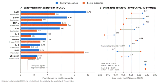
Chronic inflammation is an established driver of head and neck carcinogenesis (Babiuch et al., 2020; Kumar et al., 2025). Transcripts for TNF-α, IL-1β, IL-6, and IL-8 are markedly elevated in the salivary exosomes of OSCC patients, with TNF-α showing the greatest increase among cytokines at 6.0-fold (p = 0.0001) relative to healthy individuals (Kumar et al., 2025) (Table 1). This is clinically important because free salivary cytokines also rise in non-malignant inflammatory conditions such as chronic periodontitis, which can blur their diagnostic signal (Lisa Cheng et al., 2014). Measuring these transcripts within tumor-derived exosomes may improve specificity (Kumar et al., 2025). Salivary exosomal IL-6 also correlates with clinical stage and nodal involvement, and it acts as an indicator of cervical lymph node metastasis (p < 0.05)

Table 1. Diagnostic accuracy and clinicopathologic associations of salivary versus serum exosomal mRNA biomarkers in oral squamous cell carcinoma. Paired saliva and serum samples from 40 patients with OSCC and 40 healthy controls were analyzed by RT-qPCR. Fold change is relative to healthy controls; sensitivity, specificity, AUC, and Youden index are from receiver operating characteristic analysis at the optimal cut-off, and multigene panels were built by logistic regression. Rows are grouped so that each transcript's salivary and serum results can be read together. Salivary transcripts tracked histological grade and nodal metastasis, whereas serum transcripts did not; the bold rows mark the best-performing salivary panels. Abbreviations: AUC, area under the receiver operating characteristic curve; G, histological grade; OSCC, oral squamous cell carcinoma; RT-qPCR, reverse transcription quantitative polymerase chain reaction; —, not reported because the marker was not significant.

Biomarker target (gene/panel)

Functional hallmark and biological role

Biofluid

Fold change vs. control (p value)

Sensitivity (%)

Specificity (%)

AUC

Youden index

Clinicopathologic and staging correlation

Primary reference

TNF-α

Pro-inflammatory cytokine; mediates chronic inflammation, NF-κB signaling, and tumor growth

Saliva

6.02-fold (p = 0.0001)

90.0

80.0

0.88

0.70

Highest individual salivary cytokine predictor; stable across grades

Kumar et al. (2025)

TNF-α

Systemic pro-inflammatory cytokine

Serum

6.88-fold (p = 0.0001)

52.5

92.5

0.75

0.45

Moderate systemic discrimination; lower sensitivity than saliva

Kumar et al. (2025)

OAZ1

Polyamine biosynthesis regulator; modulates cellular hyperproliferation and survival

Saliva

9.76-fold (p = 0.0001)

77.5

87.5

0.87

0.65

Strongly elevated in Grade III vs. Grade I/II (p = 0.006)

Kumar et al. (2025)

OAZ1

Polyamine biosynthesis regulator

Serum

5.09-fold (p = 0.0001)

75.0

82.5

0.80

0.57

Top individual serum proliferation marker; lacks grade stratification

Kumar et al. (2025)

MMP-9

Matrix metalloproteinase; degrades type IV collagen, facilitates basement membrane invasion

Saliva

3.80-fold (p = 0.0001)

72.5

85.0

0.84

0.57

Strong positive correlation with tumor grade (G1/G2 vs. G3, p = 0.001)

Kumar et al. (2025)

MMP-9

Matrix metalloproteinase

Serum

2.94-fold (p = 0.001)

82.5

70.0

0.83

0.52

Elevated in serum; expression does not correlate with histological grade

Kumar et al. (2025)

IL-1β

Pro-inflammatory cytokine; induces invasive tumor microenvironment

Saliva

2.78-fold (p = 0.001)

67.5

71.8

0.71

0.39

Statistically significant elevation; moderate diagnostic power

Kumar et al. (2025)

IL-1β

Pro-inflammatory cytokine

Serum

9.40-fold (p = 0.0001)

87.5

70.0

0.86

0.57

Top individual serum cytokine marker; no correlation with nodal status

Kumar et al. (2025)

IL-6

Pleiotropic inflammatory cytokine; stimulates STAT3-driven cell survival and motility

Saliva

2.92-fold (p = 0.0001)

80.0

67.5

0.79

0.47

Significant positive correlation with lymph node metastasis (p = 0.013)

Kumar et al. (2025)

IL-6

Circulating inflammatory cytokine

Serum

1.40-fold (p = 0.240)

—

—

—

—

Not statistically significant in serum; lacks staging discrimination

Kumar et al. (2025)

IL-8

Pro-angiogenic and chemotactic chemokine; promotes vascularization and motility

Saliva

3.85-fold (p = 0.002)

60.0

77.5

0.69

0.37

Significant progressive elevation across histological grades (G1 vs. G3, p = 0.011)

Kumar et al. (2025)

IL-8

Circulating chemokine

Serum

1.54-fold (p = 0.032)

50.0

77.5

0.63

0.27

Modest upregulation; limited diagnostic utility in serum

Kumar et al. (2025)

S100P

Calcium-binding protein; activates MAPK and NF-κB pathways to drive cell survival

Saliva

6.92-fold (p = 0.0001)

92.5

67.5

0.88

0.55

High diagnostic sensitivity; significantly upregulated in Grade III (p = 0.017)

Kumar et al. (2025)

S100P

Circulating calcium-binding protein

Serum

3.15-fold (p = 0.0001)

72.5

80.0

0.77

0.52

Significantly elevated; lacks grade differentiation in serum

Kumar et al. (2025)

SAT

Spermidine/spermine N1-acetyltransferase; polyamine catabolism enzyme

Saliva

5.80-fold (p = 0.0001)

77.5

77.5

0.82

0.60

Significant stage-related elevation; differentiates G1 from G3 (p = 0.022)

Kumar et al. (2025)

SAT

Polyamine catabolic enzyme

Serum

2.39-fold (p = 0.001)

62.5

87.5

0.73

0.50

Moderate diagnostic accuracy in systemic circulation

Kumar et al. (2025)

Chemerin

Chemoattractant adipokine; regulates leukocyte recruitment and matrix remodeling

Saliva

2.50-fold (p = 0.001)

80.0

71.8

0.78

0.51

Significant baseline elevation in OSCC; stable across histological grades

Kumar et al. (2025)

Chemerin

Circulating adipokine

Serum

2.33-fold (p = 0.001)

65.0

75.0

0.74

0.40

Moderate diagnostic performance; consistent across staging cohorts

Kumar et al. (2025)

TNF-α + OAZ1 (two-gene panel)

Combined inflammation and proliferation panel

Saliva

Multigene logistic model

80.0

90.0

0.89

0.70

Optimal non-invasive diagnostic panel; superior specificity and Youden index

Kumar et al. (2025)

TNF-α + OAZ1 + MMP-9 (three-gene panel)

Combined inflammation, proliferation, and invasion panel

Saliva

Multigene logistic model

85.0

80.0

0.89

0.65

High diagnostic sensitivity; slightly lower specificity due to grade dependency of MMP-9

Kumar et al. (2025)

IL-1β + OAZ1 (two-gene panel)

Combined systemic cytokine and proliferation panel

Serum

Multigene logistic model

75.0

90.0

0.89

0.65

Strong specificity; sensitivity lower than the saliva panel

Kumar et al. (2025)

IL-1β + OAZ1 + MMP-9 (three-gene panel)

Combined systemic multimarker panel

Serum

Multigene logistic model

80.0

85.0

0.89

0.65

Balanced systemic performance; lacks clinicopathologic grade tracking

Kumar et al. (2025

Table 2. Comparison of conventional and emerging technologies for isolating extracellular vesicles (exosomes) from saliva and other biofluids. Each technology is compared on its physical separation principle, typical vesicle recovery, purity and preservation of membrane integrity, processing time, required sample volume, and principal strengths and limitations. Ranges are approximate values compiled from the cited reviews and guidelines; they come from different matrices and laboratories and are not head-to-head measurements. Methods toward the bottom of the table (immunoaffinity capture and centrifugal microfluidics) are the most compatible with integrated point-of-care biosensors. Abbreviations: CSF, cerebrospinal fluid; EV, extracellular vesicle; MWCO, molecular weight cut-off; PEG, polyethylene glycol; RT-qPCR, reverse transcription quantitative polymerase chain reaction.

Isolation technology

Biophysical separation mechanism

Typical yield/recovery (%)

Exosome purity and structural integrity

Processing duration

Sample volume and biofluid scope

Primary advantages

Critical limitations and downstream interferences

Representative references

Differential ultracentrifugation (dUC)

Sequential sedimentation based on particle size and buoyant density across increasing centrifugal fields (up to 100,000–120,000 × g)

30–80

Moderate purity; shear stress can cause vesicle deformation, aggregation, and cargo leakage

6–12 h

> 5–20 mL; cell media, plasma, serum, saliva

Historically established benchmark; reagent-free; handles large starting volumes

Bulky capital equipment; labor-intensive; co-pellets high-molecular-weight protein aggregates and lipoproteins

Tang et al. (2024); Welsh et al. (2024); Xu et al. (2024)

Density gradient centrifugation (DGC)

Isopycnic separation through density gradient cushions (iodixanol, OptiPrep, or sucrose)

20–45

Very high purity; preserves intact spherical morphology and separates vesicles from protein complexes

16–24 h

1–5 mL; plasma, serum, saliva, CSF

Effectively separates EVs from non-vesicular particles and dense lipoproteins

Very long preparation and spin times; labor-intensive; requires extensive gradient fractionation and washing

S. Pan & Zhang (2026); Tang et al. (2024); Xu et al. (2024)

Size-exclusion chromatography (SEC)

Size-based separation through porous gel matrices (Sepharose CL-2B, qEV columns); large vesicles elute first

50–80

High purity; intact membrane structure and functional biological activity preserved

10–30 min

0.1–2 mL; plasma, serum, urine, saliva

Rapid and gentle; highly reproducible; avoids shear-induced rupture; compatible with RNA/protein profiling

Co-elutes non-exosomal particles of similar hydrodynamic radii; dilutes sample, requiring concentration

Nonaka & Wong (2022); Tang et al. (2024); Xu et al. (2024)

Polymer precipitation (PEG/commercial kits)

Polyethylene glycol reduces solubility and water of hydration, precipitating vesicles via low-speed centrifugation (10,000 × g)

60–90

Low purity; high yield but frequent vesicle aggregation and non-vesicular contamination

1–12 h (overnight incubation)

0.1–1 mL; serum, plasma, cell culture

Simple protocol; no ultracentrifuge needed; scalable for high-throughput screening

Polymer residues and co-precipitated lipoproteins/mucins can inhibit downstream RT-qPCR enzymes

Kumar et al. (2025); Tang et al. (2024); Welsh et al. (2024)

Ultrafiltration and tangential flow filtration (TFF)

Size-selective membrane retention using defined MWCO (10–100 kDa)

40–70

Moderate purity; membrane shear can induce mechanical vesicle rupture and clogging

1–2 h

0.5–50 mL; urine, saliva, conditioned media

Rapid volume concentration; low operational cost; suitable for large biofluid volumes

Membrane fouling and vesicle adhesion to filter surfaces lower total particle recovery

Tang et al. (2024); Xu et al. (2024)

Immunoaffinity capture (magnetic beads)

Specific molecular recognition targeting surface tetraspanins (CD9, CD63, CD81) or tumor antigens (EpCAM, EGFR)

40–70

Extremely high purity; specifically isolates distinct tumor-derived exosome subpopulations

1–4 h

0.05–1 mL; raw saliva, plasma, serum

High specificity; isolates cancer-derived vesicles from complex background; directly interfaces with biosensors

High antibody reagent costs; low total yield; harsh elution buffers can compromise membrane integrity

Kashefi-Kheyrabadi et al. (2020); Tang et al. (2024); Zhang et al. (2025)

Centrifugal microfluidic lab-on-a-chip (e.g., ExoTIC)

Integrated microfluidic filtration using nanoporous membrane arrays and hydrodynamic fluid forces

70–85

Very high purity; intact vesicle morphology without high-shear-induced damage

30–60 min

0.02–0.5 mL; whole saliva, serum, plasma

Automated sample-to-answer processing; high throughput; minimal biofluid consumption

Requires specialized microfabrication and chip-packaging instrumentation; potential microchannel clogging

Tang et al. (2024); Xu et al. (2024)


(Kumar et al., 2025).

2.4.3. Proliferation and Metabolic Regulators: OAZ1, S100P, and SAT

Transcripts that govern hyperproliferation, polyamine metabolism, and metabolic remodeling are packaged into salivary exosomes during malignant transformation (Kumar et al., 2025; Senevirathna et al., 2024). Ornithine decarboxylase antizyme 1 (OAZ1), a key regulator of polyamine biosynthesis, shows the largest change of any transcript studied, with a mean 9.7-fold upregulation (p = 0.0001) in OSCC salivary exosomes. S100P, a calcium-binding protein that activates MAPK and NF-κB signaling to support tumor cell survival, rises 6.9-fold, and spermidine/spermine N1-acetyltransferase (SAT) rises 5.8-fold (Kumar et al., 2025) (Figure 3A).

2.4.4. Extracellular Matrix Remodeling and Metastatic Mediators: MMP-9 and Chemerin

Degradation of the extracellular matrix (ECM) is an obligatory step in basement membrane invasion and metastatic spread (Kumar et al., 2025; Song & Yang, 2026). Matrix metalloproteinase-9 (MMP-9) degrades type IV collagen and other basement membrane components, and its transcript is elevated 3.8-fold (p = 0.0001) in OSCC salivary exosomes. Chemerin, a chemoattractant adipokine involved in leukocyte recruitment and angiogenesis, is elevated 2.5-fold (Kumar et al., 2025) (Table 1).

2.4.5. Comparative Diagnostic Performance: Saliva Versus Serum

Paired-biofluid analysis has produced one of the most informative findings in this field: salivary exosomal mRNAs discriminate disease better than matched serum exosomal mRNAs (Kumar et al., 2025). In a cohort of 40 OSCC patients and 40 healthy controls, individual salivary markers achieved AUCs between 0.69 and 0.88. A two-gene logistic regression panel combining TNF-α and OAZ1 reached an AUC of 0.89 with 80% sensitivity, 90% specificity, and a Youden index of 0.70. Adding MMP-9 preserved the AUC at 0.89 and shifted the balance to 85% sensitivity and 80% specificity (Kumar et al., 2025) (Figure 3B; Table 1). Serum exosomal IL-6, by contrast, did not differ significantly between patients and controls (p = 0.240), and serum panels showed lower sensitivity (75%) and no ability to stratify histological grade. In saliva, OAZ1, MMP-9, IL-8, S100P, and SAT separated Grade I from Grade III OSCC (Kumar et al., 2025). Taken together, these data identify saliva as the more responsive and clinically informative matrix for exosomal mRNA detection in OSCC (Kumar et al., 2025).

2.5. Exosomal RNA Biomarkers Across Human Cancers

Placing the OSCC data alongside other cancers helps calibrate expectations. Exosomal and circulating non-coding RNAs have reached high diagnostic accuracy in several malignancies (Table 4). Vesicle-associated miR-10b discriminated lung adenocarcinoma with an AUC of 0.998, outperforming conventional serum markers (Tang et al., 2024; Yuan et al., 2021). A serum miRNA model built on miR-1268b and miR-6075 achieved 99% sensitivity and specificity in a large-scale lung cancer cohort (Asakura et al., 2020), and circulating miRNA profiles were similarly evaluated in symptomatic patients (Fehlmann et al., 2020). Serum exosomal circRNA panels distinguished non-small cell lung cancer from chronic obstructive pulmonary disease and tuberculosis (Kang et al., 2022; Tang et al., 2024). Single circRNAs have shown promise in colorectal cancer (B. Pan et al., 2019), gastric cancer (Roy et al., 2022), hepatocellular carcinoma, and epithelial ovarian cancer (Tang et al., 2024). Long non-coding RNAs, including H19 in breast cancer and lnc-GNAQ-6:1 in gastric cancer, add further examples (S. Li et al., 2020; Zhong et al., 2020). In OSCC, a salivary and plasma miR-21/miR-184 panel has reached an AUC of 0.867 (Nonaka & Wong, 2022; Song & Yang, 2026). Against this background, the salivary mRNA panel performs competitively. It also offers something most of these markers do not, namely a readout of functional pathways that could inform prognosis as well as detection (Kumar et al., 2025).

2.6. Analytical Challenges of Conventional Detection Workflows

2.6.1. Exosome Isolation

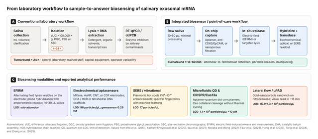
Strong biomarker performance has not yet translated into decentralized testing, largely because conventional laboratory protocols are unsuited to it (Kashefi-Kheyrabadi et al., 2020; Tang et al., 2024) (Figure 4A). Differential ultracentrifugation (dUC), the historical reference method, separates vesicles by sedimentation through successive runs exceeding 100,000 × g (Kashefi-Kheyrabadi et al., 2020; Tang et al., 2024). It isolates vesicles cleanly, but it needs capital-intensive equipment and 4 to 12 hours of processing, and the shear forces involved can rupture
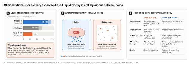
Figure 1. Clinical rationale for salivary exosome-based liquid biopsy in oral squamous cell carcinoma. (A) Approximate five-year overall survival by stage at diagnosis: about 80%–90% for localized Stage I–II disease, 50%–60% across all stages, and about 30% for advanced Stage III–IV disease. Lighter segments show the upper end of each reported range. (B) Anatomical proximity: saliva is in direct contact with the oral lesion and contains a high local concentration of tumor-derived exosomes, whereas in blood the same signal is diluted systemically among abundant non-tumor vesicles. (C) Comparison of scalpel tissue biopsy and salivary exosome liquid biopsy for invasiveness, repeatability, sampling of tumor heterogeneity, timing of molecular detection, and suitability for screening at scale. Data summarized from Kumar et al. (2025), T. Li et al. (2022), Nonaka and Wong (2022), Chandrasekaran et al. (2024), and Song and Yang (2026).
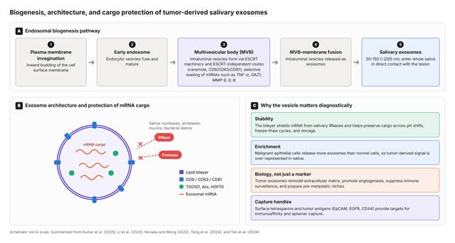
Figure 2. Biogenesis, architecture, and cargo protection of tumor-derived salivary exosomes. (A) Endosomal biogenesis: inward budding of the plasma membrane forms early endosomes, which mature into multivesicular bodies in which ESCRT-dependent and ESCRT-independent (ceramide and tetraspanin) machinery forms intraluminal vesicles and loads selected mRNAs. Fusion of the multivesicular body with the plasma membrane releases 30–150 (–200) nm exosomes into saliva. (B) Exosome architecture, showing the lipid bilayer, surface tetraspanins (CD9, CD63, CD81), luminal markers (TSG101, Alix, HSP70), and mRNA cargo shielded from salivary RNases and proteases. (C) The four properties that make exosomes diagnostically useful: cargo stability, tumor-biased secretion, participation in tumor biology, and surface proteins that can be targeted for capture. The schematic is not to scale. Based on Kumar et al. (2025), T. Li et al. (2022), Nonaka and Wong (2022), Tang et al. (2024), and Ten et al. (2024).

membranes and compromise cargo (Kashefi-Kheyrabadi et al., 2020; Tang et al., 2024). Polymer precipitation kits and size-exclusion chromatography (SEC) columns improve throughput but often co-isolate salivary mucins, amylase, and non-vesicular protein aggregates, which reduces purity (Cancers Editorial Office, 2024; Kumar et al., 2025; Welsh et al., 2024). Density gradient centrifugation, ultrafiltration, immunoaffinity capture, and centrifugal microfluidic systems each occupy a different position in the trade-off between yield, purity, time, and sample volume (Kashefi-Kheyrabadi et al., 2020; S. Pan & Zhang, 2026; Tang et al., 2024; Xu et al., 2024; Zhang et al., 2025) (Table 2).

2.6.2. Downstream mRNA Quantification

Quantification usually follows detergent lysis, organic solvent RNA purification, reverse transcription, and RT-qPCR or droplet digital PCR (ddPCR) (Kumar et al., 2025; Tang et al., 2024). Each manual transfer risks losing trace transcripts, and residual salivary inhibitors can suppress reverse transcriptase and polymerase activity (Song & Yang, 2026; Tang et al., 2024). The cumulative effect is a workflow that takes more than a day and requires a central laboratory. That rules out rapid, near-patient screening (Nonaka & Wong, 2022; Tang et al., 2024) (Figure 4A).

2.7. Biosensor Architectures for Exosome and Intra-Vesicular mRNA Sensing

Biosensors aim to collapse extraction and amplification into a single device (Kashefi-Kheyrabadi et al., 2020; Tang et al., 2024; Wu et al., 2021). They pair a recognition element (an antibody, DNA or RNA aptamer, or antisense oligonucleotide) with a transducer that produces an optical, electrochemical, or mechanical signal (Kashefi-Kheyrabadi et al., 2020; Tang et al., 2024) (Figure 4B; Table 3).

2.7.1. Electric Field-Induced Release and Measurement (EFIRM)

EFIRM is a significant development in salivary diagnostics (Nonaka & Wong, 2022; Wei et al., 2013, 2014). It builds on earlier work with integrated electrochemical saliva platforms such as the oral fluid nanosensor test (OFNASET) (Gau & Wong, 2007). In EFIRM, a low-voltage alternating electric field lyses exosomal membranes and releases intra-vesicular nucleic acids directly onto a functionalized electrode, without chemical detergents (Nonaka & Wong, 2022; Tu et al., 2020). The released sequences hybridize with immobilized capture probes, and an enzymatic reporter provides amperometric quantification within 15 to 30 minutes (Nonaka & Wong, 2022; Wei et al., 2013). EFIRM has detected EGFR mutations in the saliva of patients with lung cancer (Wei et al., 2014) and has been technically validated in plasma and saliva (Tu et al., 2020). Because it works with 10 to 30 µL of unprocessed fluid, it is a functional blueprint for sample-to-answer exosomal mRNA sensing (Nonaka & Wong, 2022; Tu et al., 2020).

2.7.2. Electrochemical Aptasensors and Nanomaterial Interfaces

Electrochemical sensors combine high sensitivity, low fabrication cost, and straightforward compatibility with portable potentiostats (Kashefi-Kheyrabadi et al., 2020; Tang et al., 2024). Low limits of detection (LODs) for vesicles are reached by coating electrodes with conductive nanomaterials, including gold nanoparticles (AuNPs), titanium carbide MXenes (Ti₃C₂Tₓ), carbon nanotubes, and covalent organic frameworks (COFs), which enlarge the electroactive surface and accelerate electron transfer (Kashefi-Kheyrabadi et al., 2020; Tang et al., 2024; Wang et al., 2020). Three design strategies are particularly relevant to intra-vesicular RNA. First, tetrahedral DNA nanostructures on gold electrodes present capture probes in a uniform orientation, reducing steric hindrance (Zeng et al., 2017). Target binding can then initiate catalytic hairpin assembly (CHA) or hybridization chain reaction (HCR), building long duplexes that recruit redox reporters such as methylene blue or ferrocene and amplify the signal to attomolar levels (Tang et al., 2024; Zeng et al., 2017). Second, CRISPR/Cas systems, notably Cas12a and Cas13a, add a further layer of amplification: once the guide RNA binds its target, the enzyme's collateral cleavage destroys nearby reporters and produces a sharp fluorescent or amperometric signal (Hong et al., 2023; Tang et al., 2024). Third, magnetic beads coated with anti-CD63 or anti-EpCAM antibodies capture intact exosomes from raw fluid before lysis and electrochemical hybridization, combining purification with sensitive readout (Tang et al., 2024; Wu et al., 2021).

2.7.3. Optical, SERS, Microfluidic, and Paper-Based Platforms

SERS and microfluidic lab-on-a-chip (LOC) technologies extend the available options (Faur et al., 2023; Shin et al., 2023; Tang et al., 2024). SERS amplifies Raman scattering by several orders of magnitude at plasmonic "hot spots" on anisotropic metal nanostructures, producing narrow spectral fingerprints suited to multiplexed vesicle profiling (Faur et al., 2023; Shin et al., 2023). Combined with machine learning, vibrational spectroscopy of salivary exosomes, by SERS or by Fourier-transform infrared spectroscopy (FTIR), has separated oral cancer patients from controls with sensitivities above 90% (Faur et al., 2023; Zlotogorski-Hurvitz et al., 2019). Microfluidic devices automate sample handling by routing fluid through herringbone channels or acoustic sorting fields (Sackmann et al., 2014; Tang et al., 2024). Such chips can capture exosomes on immobilized aptamers and read them out through quantum dot (QD) probes or integrated organic photodetectors, giving sample-to-answer saliva analysis within an hour (Dong & Pires, 2017; Wu et al., 2021). Lateral flow and paper-based microfluidic devices go further toward instrument-free testing, trading some sensitivity for simplicity and cost (Carrell et al., 2019; Tang et al., 2024) (Figure 4C; Table 3).

2.8. Translational Obstacles and Standardization

2.8.1. Pre-Analytical Variability

Several obstacles separate the current evidence from routine dental and oncological use (Kumar et al., 2025; Welsh et al., 2024). The first is pre-analytical variability (Cancers Editorial Office, 2024; Welsh et al., 2024). Salivary flow, pH, viscosity, and composition change with circadian rhythm, hydration, medication, and diet (Cancers Editorial Office, 2024; Nonaka & Wong, 2022). Consistent results will require defined collection rules, for example morning collection after an overnight fast, an explicit choice between stimulated and unstimulated saliva, and prompt low-speed clarification to remove cells, debris, and bacteria (Cancers Editorial Office, 2024; Nonaka & Wong, 2022; Welsh et al., 2024).

2.8.2. Vesicle Heterogeneity

The second obstacle is heterogeneity. Saliva contains vesicles from keratinocytes, acinar cells, immune cells, and oral microbes, not only from the tumor (Kumar et al., 2025; Song & Yang, 2026). Sensors must therefore isolate the tumor-derived fraction, typically through dual recognition of a general tetraspanin such as CD63 and a tumor-associated antigen such as EpCAM, EGFR, or CD44, before interrogating internal mRNA (Kashefi-Kheyrabadi et al., 2020; Tang et al., 2024).

2.8.3. Analytical and Clinical Validation

The third obstacle is validation. Under the MISEV2023 framework, diagnostic platforms must demonstrate analytical repeatability (coefficients of variation below 15%), reagent stability, and reproducible recovery across sites (Welsh et al., 2024). Larger prospective studies are needed to test the TNF-α/OAZ1/MMP-9 panel in ethnically diverse cohorts and to confirm that it can distinguish early invasive carcinoma from oral potentially malignant disorders (OPMDs) such as oral leukoplakia and oral lichen planus (Kumar et al., 2025; Senevirathna et al., 2024). If these biological, analytical, and manufacturing problems are solved, fully integrated sample-to-answer cartridges could bring salivary exosomal mRNA profiling into community clinics.
3. Methods

3.1. Review Design

This study is a structured narrative review with a systematic, reproducible search strategy. A narrative design was chosen because the evidence spans three disciplines (clinical oncology, extracellular vesicle biology, and biosensor engineering) whose study designs, outcomes, and units of measurement cannot be pooled meaningfully. The search, selection, extraction, and synthesis steps were nonetheless specified in advance and are reported below in sufficient detail to allow replication. The review questions were: (a) what diagnostic and prognostic value do salivary exosome-derived mRNAs have in OSCC, and how does it compare with serum (Table 1) and with exosomal RNA biomarkers in other cancers (Table 4); (b) how do exosome isolation methods perform in terms of yield, purity, time, and sample volume (Table 2); and (c) which biosensing architectures can detect exosomes or intra-vesicular RNA, and with what analytical performance (Table 3)? Because only previously published data were analyzed, ethical approval and informed consent were not required.

3.2. Information Sources

Four electronic sources were searched: PubMed/MEDLINE (National Library of Medicine), Scopus (Elsevier), Web of Science Core Collection (Clarivate), and Google Scholar. For Google Scholar, the first 200 records ranked by relevance were screened, because its ranking algorithm makes complete retrieval impractical. The search covered January 2007 to March, 2026; the

Table 3. Biosensing platforms, nanomaterial interfaces, and signal amplification strategies for detecting exosomes and intra-vesicular RNA. Platforms are organized by transduction principle and described by their nanomaterial interface, biorecognition element, target analyte, amplification strategy, analytical limit of detection, linear dynamic range, and the biofluids in which they were tested. Values are reported in their original units (particles/µL, particles/mL, or molar concentration) and are therefore not directly comparable across rows. Only EFIRM and the magneto-electrochemical genosensor target intra-vesicular nucleic acid; the remaining platforms detect whole vesicles or miRNA. Abbreviations: aM, attomolar; AuNP, gold nanoparticle; CHA, catalytic hairpin assembly; EFIRM, electric field-induced release and measurement; fM, femtomolar; HCR, hybridization chain reaction; HRP, horseradish peroxidase; LOD, limit of detection; MoS₂-AuNSs, molybdenum disulfide–gold nanostars; QD, quantum dot; SERS, surface-enhanced Raman spectroscopy; TMB, 3,3′,5,5′-tetramethylbenzidine.

Biosensing modality and transduction principle

Functional nanomaterials/surface interface

Biorecognition element and architecture

Target analyte (exosome/cargo)

Signal amplification strategy

Analytical LOD

Linear dynamic range

Biofluid matrix and clinical utility

Primary references

Electric field-induced release and measurement (EFIRM)

Conducting polymer-coated electrochemical electrode microarrays

Sequence-specific antisense oligonucleotide capture probes

Intra-vesicular exosomal RNA/mRNA (EGFR, TNF-α, OAZ1)

Non-thermal alternating electric field lyses vesicles directly on-chip; enzymatic amperometric readout

Sub-attomolar (down to single-copy RNA detection)

10¹ to 10⁶ copies/µL

Whole unstimulated saliva (10–30 µL); rapid non-invasive liquid biopsy

Nonaka & Wong (2022); Wei et al. (2013)

Electrochemical aptasensor

Multi-walled carbon nanotubes/titanium carbide (Ti₃C₂ MXene)/gold nanoparticles

Thiolated CD63/EpCAM-specific DNA aptamers

Tumor-derived exosomes and surface tetraspanins

Target-triggered CHA combined with HCR

96 particles/µL (or 5 × 10² exosomes/mL)

1.12 × 10² to 1.12 × 10⁸ particles/µL

Serum and saliva; lung and breast cancer exosome screening

Kashefi-Kheyrabadi et al. (2020); Zhang et al. (2025)

Magneto-electrochemical genosensor

Iron oxide magnetic nanoparticles (Fe₃O₄) and glassy carbon electrode modified with AuNPs

Dual-probe sandwich: anti-CD63 magnetic capture + biotinylated RNA-targeting probes

Exosomal mRNA/miRNA (miR-21, MMP-9, OAZ1)

Catalytic HRP enzymatic reaction with TMB/H₂O₂ redox chemistry

0.29 fM (or 3.4 × 10³ particles/mL)

1 fM to 1 nM

Serum and raw saliva; early diagnosis of gastrointestinal and oral cancers

Tang et al. (2024); Zhang et al. (2025)

Surface-enhanced Raman spectroscopy (SERS)

Plasmonic gold nanostars decorated on molybdenum disulfide nanosheets (MoS₂-AuNSs)

ROX-labeled DNA aptamers targeting exosomal CD63 and EpCAM

Intact exosomes and membrane surface proteins

Nanoplasmonic electromagnetic hot-spot enhancement (10⁶ to 10¹⁰ signal amplification)

17 particles/µL

10¹ to 10⁶ particles/µL

Serum and saliva; non-destructive cancer exosome fingerprinting

Faur et al. (2023); Tang et al. (2024)

Aptamer–quantum dot microfluidic sensor

Core–shell semiconductor quantum dots (CdSe/ZnS QDs) and magnetic microbeads

Aptamer-functionalized DNA concatamers tethered to magnetic particles

Salivary exosomes and oral cancer vesicles

"One exosome–numerous QDs" self-assembled concatamer release; fluorescence amplification

1.1 × 10³ particles/µL

10³ to 10⁷ particles/µL

Clinical saliva; one-step point-of-care oral cancer diagnostic chip

Tang et al. (2024); Wu et al. (2021)

CRISPR/Cas13a-powered optical biosensor

Magnetic capture beads coupled with fluorescent reporter ssRNA molecules

CRISPR-Cas13a crRNA ribonucleoprotein complex

Intra-vesicular microRNA and exosomal mRNA (miR-10b, TNF-α)

Target-activated non-specific collateral cleavage of fluorescent RNA quenchers

Attomolar (~10 aM)

10 aM to 100 pM

Plasma and saliva; ultrasensitive amplification-free vesicular RNA sensing

Hong et al. (2023); Tang et al. (2024)

Lateral flow immunoassay strip (LFIA/µPAD)

Colloidal gold nanoparticles (AuNPs)/upconverting nanoparticles on nitrocellulose

Capture antibodies (anti-CD81/CD9) with detector antibody (anti-CD63-AuNPs)

Whole tumor-derived exosomes

Visual colorimetric accumulation at test line; dual AuNP signal amplification

1.3 × 10³ particles/µL (or 117.9 particles/µL with multi-QDs)

7.4 × 10³ to 5.5 × 10⁴ particles/mL

Urine, saliva, and plasma; rapid disposable strip for decentralized clinics

Carrell et al. (2019); Tang et al. (2024)

Table 4. Exosomal and circulating coding and non-coding RNA biomarkers for liquid biopsy across human cancers. The table places the salivary exosomal mRNA findings in OSCC (first two rows) alongside representative miRNA, circRNA, and lncRNA biomarkers reported for lung, colorectal, gastric, hepatocellular, ovarian, and breast cancer. For each marker, the biofluid, cohort composition, quantitative diagnostic performance, and main clinical association are given. Cohort sizes, control groups, and threshold selection differ widely between studies, so the AUC values indicate the range of reported performance rather than a ranking. Abbreviations: AFP, alpha-fetoprotein; AUC, area under the receiver operating characteristic curve; BC, breast cancer; circRNA, circular RNA; COPD, chronic obstructive pulmonary disease; CRC, colorectal cancer; EOC, epithelial ovarian cancer; GC, gastric cancer; HBV, hepatitis B virus; HCC, hepatocellular carcinoma; LN, lymph node; lncRNA, long non-coding RNA; LUAD, lung adenocarcinoma; NSCLC, non-small cell lung cancer; Sens, sensitivity; Spec, specificity.

Biomarker identity and structural class

Malignancy and cancer subtype

Biofluid matrix

Cohort/sample size (n)

Quantitative diagnostic performance (AUC, sensitivity, specificity)

Clinical significance and biological association

Primary references

TNF-α and OAZ1 (mRNAs)

Oral squamous cell carcinoma (OSCC)

Saliva

40 OSCC vs. 40 healthy controls

AUC 0.89; Sens 80.0%; Spec 90.0% (Youden 0.70)

Non-invasive detection of primary OSCC; outperforms paired serum panel

Kumar et al. (2025)

MMP-9 and IL-6 (mRNAs)

Oral squamous cell carcinoma (OSCC)

Saliva

40 OSCC vs. 40 healthy controls

Grade: G1 vs. G3 (p = 0.001); nodal: LN+ vs. LN− (p = 0.013)

Tracks histological tumor grade (MMP-9) and cervical lymph node metastasis (IL-6)

Kumar et al. (2025)

miR-10b (miRNA)

Lung adenocarcinoma (LUAD)

Serum/plasma

120 LUAD vs. 90 controls

AUC 0.998; Sens 95.0%; Spec 98.0%

Outperforms conventional serum markers (AFP, CEA, CYFRA21-1, NSE)

Tang et al. (2024); Yuan et al. (2021)

miR-1268b and miR-6075 (miRNA panel)

Non-small cell lung cancer (NSCLC)

Serum

1,358 NSCLC vs. 1,970 controls

Sens 99.0%; Spec 99.0%

Robust multi-miRNA model in large-scale validation cohorts

Asakura et al. (2020); Fehlmann et al. (2020)

circRNA_0047921, 0056285, 0007761 (circRNA panel)

Non-small cell lung cancer (NSCLC)

Serum

30 NSCLC vs. 45 non-cancer controls

AUC 0.919

Differentiates NSCLC from COPD and tuberculosis; correlates with TNM stage

Kang et al. (2022); Tang et al. (2024)

circ_0004771 (circRNA)

Colorectal cancer (CRC)

Serum/plasma

135 CRC vs. 35 benign vs. 45 controls

AUC 0.880

Elevated in CRC; independent circulating diagnostic marker

B. Pan et al. (2019); Tang et al. (2024)

circ_0015286 (circRNA)

Gastric cancer (GC)

Plasma

60 GC vs. 30 gastritis vs. 30 controls

AUC 0.778 (0.843 combined with CEA and CA19-9)

Associated with tumor size, TNM stage, lymph node invasion, and overall survival

Roy et al. (2022); Tang et al. (2024)

circ_0028861 (circRNA)

Hepatocellular carcinoma (HCC)

Serum

56 HCC vs. 47 cirrhosis vs. 57 HBV

AUC 0.790

Detects small, early-stage, and AFP-negative HCC tumors

Tang et al. (2024)

circ_0001068 (circRNA)

Epithelial ovarian cancer (EOC)

Serum

95 EOC vs. 53 healthy controls

AUC 0.9697

High diagnostic accuracy for early and advanced ovarian cancer

Tang et al. (2024)

lncRNA H19 (lncRNA)

Breast cancer (BC)

Serum

50 BC vs. 50 benign vs. 50 controls

Sens 87.0%; Spec 70.6% (superior to CA15-3 and CEA)

Correlates with lymph node metastasis (p = 0.039) and TNM stage (p = 0.022)

T. Li et al. (2022); Zhong et al. (2020)

lnc-GNAQ-6:1 (lncRNA)

Gastric cancer (GC)

Plasma/serum

80 GC vs. 80 controls

Downregulated in GC; diagnostic discriminator

Potential non-invasive screening biomarker for disease staging

S. Li et al. (2020); Tang et al. (2024)

miR-21 and miR-184 (miRNA panel)

Oral squamous cell carcinoma (OSCC)

Saliva and plasma

50 OSCC vs. 50 controls

AUC 0.867

Regulates PTEN/PI3K/AKT oncogenic signaling; salivary diagnostic marker

Nonaka & Wong (2022); Song & Yang (2026)

2007 start date captures the first integrated electrochemical salivary nanosensor platforms (Gau & Wong, 2007) and the subsequent development of salivary diagnostics (Lee et al., 2009). Reference lists of all included articles and of key reviews (Nonaka & Wong, 2022; Song & Yang, 2026; Tang et al., 2024) were hand-searched by backward citation chasing, and forward citation searching of the index clinical study (Kumar et al., 2025) was performed in Google Scholar.

3.3. Search Strategy

Search strings combined four concept blocks (disease, biofluid, vesicle and RNA cargo, and detection technology) using Boolean operators, Medical Subject Headings (MeSH), and free-text terms in titles and abstracts. The PubMed string was as follows:

("Carcinoma, Squamous Cell"[MeSH] OR "Mouth Neoplasms"[MeSH] OR "Squamous Cell Carcinoma of Head and Neck"[MeSH] OR "oral squamous cell carcinoma"[tiab] OR OSCC[tiab] OR "oral cancer"[tiab] OR "head and neck cancer"[tiab]) AND ("Saliva"[MeSH] OR saliva*[tiab] OR "oral fluid"[tiab] OR "Liquid Biopsy"[MeSH] OR "liquid biopsy"[tiab]) AND ("Exosomes"[MeSH] OR "Extracellular Vesicles"[MeSH] OR exosom*[tiab] OR "extracellular vesicle*"[tiab] OR "RNA, Messenger"[MeSH] OR mRNA[tiab] OR transcript*[tiab]) AND ("Biosensing Techniques"[MeSH] OR biosensor*[tiab] OR electrochemical[tiab] OR aptamer*[tiab] OR "surface-enhanced Raman"[tiab] OR SERS[tiab] OR microfluidic*[tiab] OR "lab-on-a-chip"[tiab] OR CRISPR[tiab] OR EFIRM[tiab] OR diagnos*[tiab] OR "ROC Curve"[MeSH])

Equivalent strings were adapted to the syntax of Scopus (TITLE-ABS-KEY) and Web of Science (TS=). Because many biosensor platforms have been developed in non-oral cancers before being applied to saliva, a supplementary search removed the disease block and retained the vesicle and technology blocks, to capture transferable sensing architectures (for example, Kashefi-Kheyrabadi et al., 2020; Wang et al., 2020; Zeng et al., 2017). A second supplementary search replaced the oral disease block with lung, colorectal, gastric, hepatocellular, ovarian, and breast cancer terms, combined with exosomal miRNA, circRNA, and lncRNA terms, to populate the cross-cancer comparison (Table 4). Filters restricted results to English-language publications on humans or human-derived samples. Full strings for each database are available from the corresponding author.

3.4. Eligibility Criteria

Studies were eligible if they met at least one of the following criteria: (a) original clinical studies that quantified exosomal or EV-associated RNA, or salivary inflammatory, proliferative, or invasion markers, in patients with OSCC, OPMDs, or other cancers, and reported a measure of diagnostic accuracy (AUC, sensitivity, specificity, or Youden index) or a clinicopathologic association; (b) technical studies describing exosome isolation methods with reported recovery, purity, processing time, or sample volume; (c) biosensor development or validation studies that detected exosomes or intra-vesicular nucleic acids and reported an LOD, linear dynamic range, or validation in a biological fluid; or (d) peer-reviewed reviews, consensus statements, or guidelines that defined terminology, biological mechanisms, or reporting standards, such as MISEV2023 (Welsh et al., 2024). Exclusion criteria were: conference abstracts without full data; non-English publications; studies limited to cell lines or animals without any human-sample validation, unless they described a sensing architecture discussed for transfer to saliva; and duplicate reports of the same cohort, in which case the most complete report was retained.

3.5. Study Selection

Records were exported to a reference manager and deduplicated. Two reviewers independently screened titles and abstracts against the eligibility criteria and then assessed full texts. Disagreements were resolved by discussion or, when necessary, by a third reviewer. The final evidence base comprised 40 sources, all of which are cited in this manuscript.

3.6. Data Extraction

Data were extracted into a piloted spreadsheet by one reviewer and verified independently by a second. Four extraction templates were used, each corresponding to one of the results tables. For clinical biomarker studies (Tables 1 and 4), the extracted fields were first author and year, cancer type, biofluid, cohort composition and size, biomarker identity and RNA class, fold change and p value, AUC, sensitivity, specificity, Youden index, and any association with grade, stage, or nodal status. For isolation methods (Table 2), the fields were separation principle, typical recovery, purity and preservation of vesicle integrity, processing time, sample volume and matrix, advantages, and limitations. For biosensors (Table 3), the fields were transduction principle, nanomaterial interface, recognition element, target analyte, amplification strategy, LOD, linear dynamic range, and biofluid tested. For EV characterization, the methods reported for size and morphology (transmission electron microscopy [TEM] and nanoparticle tracking analysis [NTA]), protein and lipid content, and marker proteins (CD9, CD81, TSG101, HSP70) were extracted to assess conformity with MISEV guidance (Osteikoetxea et al., 2015; Welsh et al., 2024). Values were recorded exactly as reported, in their original units, and were not recalculated. Where a study reported a Youden index, it was checked against the identity J = sensitivity + specificity − 1.

4. Diagnostic Performance of Salivary Exosomal mRNA and the Analytical Capability of Biosensing Platforms

4.1. Characteristics of the Evidence Base

The 40 sources fall, by our classification, into three broad groups. Sixteen are clinical studies that measured salivary or circulating biomarkers in patients, 10 are technical or bioengineering studies concerned with vesicle characterization or sensing, and 14 are reviews, consensus documents, or methodological guidelines. The clinical evidence for salivary exosomal mRNA specifically in OSCC is narrower than this overall count might suggest. Most of the quantitative findings in Table 1 come from a single paired-sample study of 40 patients and 40 controls (Kumar et al., 2025). That should be kept in mind throughout what follows. The other clinical studies add context on salivary cytokines (Babiuch et al., 2020; Lisa Cheng et al., 2014; Senevirathna et al., 2024), on spectroscopic profiling of salivary exosomes (Faur et al., 2023; Zlotogorski-Hurvitz et al., 2019), and on exosomal RNA in other cancers (Table 4).

4.2. Integrity of Isolated Salivary and Serum Exosomes

Before any transcript can be trusted, the vesicles carrying it need to be what they are claimed to be. In the index study, vesicles from paired saliva and serum appeared to meet MISEV criteria (Kumar et al., 2025; Welsh et al., 2024). TEM showed the familiar cup-shaped or spherical bilayer morphology, mostly below 100 nm, while NTA gave a monodisperse distribution centered between 150 and 200 nm, together with negative zeta potentials (Kumar et al., 2025). These two size estimates may seem to disagree. They probably do not: NTA measures hydrodynamic diameter, which includes the hydration shell and any surface corona, and TEM sample preparation tends to shrink vesicles, so a gap of this order is expected.

Total protein content was higher in vesicles from OSCC patients in both fluids. Lipid content behaved differently: it was significantly raised in salivary vesicles from patients but showed no difference in serum (Kumar et al., 2025). The protein-to-lipid ratio stayed between 1 and 2, which suggests intact bilayers rather than protein-rich contaminants (Kumar et al., 2025; Osteikoetxea et al., 2015). Western blotting detected CD81, CD9, TSG101, and HSP70 in both fluids, consistent with an endosomal origin (Kumar et al., 2025). These checks do not prove purity, since co-isolated material can never be fully excluded (Table 2). They do make it reasonably likely that the transcript differences described below come from vesicles rather than from free RNA.

4.3. Differential Expression of Exosomal mRNA in Saliva and Serum

Nine cancer-associated transcripts were measured by RT-qPCR across three functional modules (Kumar et al., 2025). In saliva, every one was significantly raised in OSCC (Figure 3A; Table 1). The largest change was in OAZ1, at 9.76-fold (p = 0.0001), followed by S100P (6.92-fold) and TNF-α (6.02-fold). SAT rose 5.80-fold, while IL-8 (3.85-fold; p = 0.002), MMP-9 (3.80-fold), IL-6 (2.92-fold), IL-1β (2.78-fold; p = 0.001), and Chemerin (2.50-fold; p = 0.001) made up the more modest tier (Kumar et al., 2025).

Serum vesicles showed a broadly similar direction of change but a different pattern. Eight of the nine transcripts were raised, yet the ordering shifted. IL-1β, which ranked low in saliva, was the most elevated serum transcript at 9.40-fold, and TNF-α reached 6.88-fold. IL-6 rose only 1.40-fold and did not reach significance (p = 0.240) (Kumar et al., 2025) (Figure 3A). One plausible reading is that the serum signal partly reflects systemic inflammation, to which IL-1β is especially sensitive, whereas the salivary signal is more closely tied to the tumor itself. That interpretation fits the proximity argument (Figure 1B), but it has not been tested directly. Overall, salivary vesicles showed greater divergence between patients and controls across most of the panel (Kumar et al., 2025).

4.4. Diagnostic Accuracy: Single Markers and Multigene Panels

Taken individually, salivary transcripts discriminated OSCC from controls with AUCs ranging from 0.69 (IL-8) to 0.88 (Figure 3B; Table 1). TNF-α performed best among the cytokines, with an AUC of 0.88, 90.0% sensitivity, 80.0% specificity, and a Youden index of 0.70. OAZ1 performed similarly, with an AUC of 0.87, 77.5% sensitivity, and 87.5% specificity. S100P matched TNF-α on AUC (0.88) but did so mainly through sensitivity (92.5%), at the cost of specificity (67.5%). MMP-9 gave an AUC of 0.84 (Kumar et al., 2025).

Combining markers improved specificity more than AUC. A TNF-α/OAZ1 logistic model reached an AUC of 0.89 with 80.0% sensitivity, 90.0% specificity, and a Youden index of 0.70, and adding MMP-9 left the AUC unchanged while shifting the balance toward sensitivity (85.0% vs. 80.0% specificity) (Kumar et al., 2025). Which of these is preferable depends on the intended use. A screening test in a high-risk population might favor the three-gene panel, because a missed cancer costs more than an extra referral. A triage test before biopsy might favor the two-gene panel. Both panels were derived and evaluated in the same 80 participants. Without an independent validation set, some optimism in these estimates is likely.

Serum told a slightly different story. The best single serum markers were IL-1β (AUC 0.86), MMP-9 (0.83), and OAZ1 (0.80), and the corresponding serum panels also reached an AUC of 0.89 (Kumar et al., 2025) (Table 1). On AUC alone, then, the two fluids look equivalent. The difference is in how that accuracy was achieved: the best salivary panel had higher sensitivity (80.0% vs. 75.0%) and a higher Youden index (0.70 vs. 0.65) (Figure 3B). The margin is not large, and with 40 cases it may not be statistically robust. Its direction is nevertheless consistent with the clinicopathologic findings below.

4.5. Clinicopathologic Correlation With Tumor Grade and Nodal Metastasis

The clearest separation between the two fluids was in prognostic information. Salivary MMP-9 was higher in poorly differentiated Grade III tumors than in Grade II (p = 0.001) or Grade I tumors (p = 0.001), and OAZ1 was higher in Grade III than in Grades I and II (p = 0.006) (Kumar et al., 2025). IL-8 (p = 0.011), S100P (p = 0.017), and SAT (p = 0.022) also increased from Grade I to Grade III (Table 1). None of the serum transcripts showed a significant association with grade (Kumar et al., 2025).

Salivary IL-6 behaved as a marker of regional spread. Patients with histologically confirmed cervical node metastasis had a median relative expression of 4.08 (interquartile range [IQR] 2.11–9.26), compared with 1.83 (IQR 1.24–3.95) in node-negative patients (p = 0.013) (Kumar et al., 2025) (Table 4). No serum marker was associated with nodal status. The overlap between these interquartile ranges is considerable, so IL-6 alone could not classify an individual patient's nodal status. It may still add useful information to imaging. Grade and nodal subgroups within an 80-person study are also small, and these associations should be regarded as hypothesis-generating until they are replicated.

4.6. Salivary Exosomal mRNA in the Wider Landscape of Exosomal RNA Biomarkers

Across cancers, exosomal and circulating RNA markers report AUCs from about 0.78 to above 0.99 (Table 4). The highest values, such as 0.998 for miR-10b in lung adenocarcinoma (Yuan et al., 2021) and 99% sensitivity and specificity for a two-miRNA lung cancer model (Asakura et al., 2020), come from larger cohorts or from markers measured against conventional tumor antigens. CircRNAs gave AUCs of 0.778 to 0.970 in gastric, colorectal, hepatocellular, lung, and ovarian cancer (Kang et al., 2022; B. Pan et al., 2019; Roy et al., 2022; Tang et al., 2024). The lncRNA H19 reached 87.0% sensitivity in breast cancer and was associated with nodal metastasis (Zhong et al., 2020), and lnc-GNAQ-6:1 was proposed as a gastric cancer marker (S. Li et al., 2020). In OSCC, the salivary miR-21/miR-184 panel reached an AUC of 0.867 (Nonaka & Wong, 2022; Song & Yang, 2026), which is close to the salivary mRNA panel (0.89).

These comparisons need caution. Cohort sizes range from 75 to more than 3,300, control groups vary from healthy volunteers to benign disease, and thresholds were often optimized in the same data in which they were tested. Even so, a general pattern emerges. The OSCC mRNA panel is competitive with miRNA alternatives in the same disease, while also providing grade and nodal information that the non-coding markers in Table 4 report less consistently.

4.7. Performance Benchmarks of Exosome Isolation Technologies

No isolation method performs best on every criterion (Table 2). Differential ultracentrifugation remains the most widely used, with recoveries of 30%–80%, but it takes 6 to 12 hours, needs large volumes, and subjects vesicles to more than 100,000 × g, which can deform or rupture them (Tang et al., 2024; Welsh et al., 2024; Xu et al., 2024). Density gradient centrifugation gives very high purity but recovers only 20%–45% and takes up to a day (S. Pan & Zhang, 2026; Tang et al., 2024). Polymer precipitation recovers the most material (60%–90%) with the simplest protocol, but it co-precipitates mucins and lipoproteins that can inhibit RT-qPCR enzymes and interfere with sensor hybridization (Kumar et al., 2025; Tang et al., 2024). This matters directly here, because precipitation-based isolation is common in clinical studies. SEC is gentle, rapid (10–30 minutes), and reproducible, with recoveries of 50%–80%, but it dilutes the sample (Nonaka & Wong, 2022; Tang et al., 2024; Xu et al., 2024).

For point-of-care use, two approaches seem most relevant. Immunoaffinity capture on magnetic beads gives extremely high purity and couples naturally to electrochemical readout, although yield is modest and reagents are costly (Kashefi-Kheyrabadi et al., 2020; Tang et al., 2024; Zhang et al., 2025). Centrifugal microfluidic systems such as ExoTIC report 70%–85% recovery in 30 to 60 minutes from less than 0.5 mL, without high-shear pelleting (Tang et al., 2024; Xu et al., 2024). These figures come from varied matrices and are not directly comparable, but they indicate that capture and enrichment need no longer be the slowest step (Figure 4B).

4.8. Analytical Performance of Biosensing Modalities

The sensing literature reports sensitivities that far exceed what conventional workflows need (Table 3; Figure 4C). EFIRM reports sub-attomolar detection from 10 to 30 µL of raw saliva in 15 to 30 minutes, using non-thermal field-induced permeabilization to release vesicular RNA for immediate on-chip hybridization (Nonaka & Wong, 2022; Wei et al., 2013). Electrochemical aptasensors built on MXene, AuNP, or carbon nanotube interfaces with CHA or HCR amplification report LODs down to 96 particles/µL, with linear ranges spanning about six orders of magnitude (Kashefi-Kheyrabadi et al., 2020; Zhang et al., 2025). Magneto-electrochemical genosensors for vesicular RNA reach 0.29 fM (Tang et al., 2024). SERS on plasmonic gold nanostars reports enhancement factors of 10⁶ to 10¹⁰ and an LOD of 17 particles/µL (Faur et al., 2023; Tang et al., 2024). Aptamer–QD concatamers detect salivary exosomes at 1.1 × 10³ particles/µL in a single step (Wu et al., 2021), and CRISPR/Cas13a platforms reach roughly 10 aM without thermal cycling (Hong et al., 2023; Tang et al., 2024). Lateral flow and paper-based formats give a visual result in about 15 minutes, at LODs between 117.9 and 1.3 × 10³ particles/µL (Carrell et al., 2019; Tang et al., 2024).

These numbers are encouraging, but they are not as comparable as Table 3 might make them appear. LODs are reported in particles per microliter for some platforms and in molar units for others, and many were established in buffer or spiked samples rather than in clinical saliva. More importantly, only EFIRM and the magnetic genosensor in Table 3 target intra-vesicular nucleic acid. Although TNF-α, OAZ1, and MMP-9 appear among the targets these platforms are suited to, we found no study that reported measuring them with a biosensor in a clinical OSCC cohort. The analytical capability exists, but it has yet to be applied to the clinically validated mRNA panel.

5. Toward Point-of-Care Salivary Exosomal mRNA Biosensors for OSCC

5.1. Principal Findings

This review brought together two bodies of work that have mostly developed separately. The clinical evidence suggests that salivary exosomal mRNA carries a disease signal in OSCC that is detectable, functionally coherent, and in several respects more informative than the matching serum signal (Kumar et al., 2025) (Figure 3; Table 1). The engineering evidence shows that biosensors can now detect exosomes and intra-vesicular RNA at sensitivities that exceed the clinical need, from microlitre volumes and in under an hour (Tang et al., 2024; Wei et al., 2013; Wu et al., 2021) (Figure 4; Table 3). The main gap lies between these two bodies of evidence. The validated transcripts have not yet been read by a validated sensor in patients with OSCC.

5.2. Why Saliva Seems to Outperform Serum

The advantage of saliva over serum was modest for detection and clearer for prognosis (Table 1). Why should this be? The simplest explanation is anatomical. Vesicles shed from an oral lesion enter saliva at high local concentration, whereas in blood they are diluted and mixed with vesicles from every other tissue (Kumar et al., 2025; Nonaka & Wong, 2022) (Figure 1B). A second possibility is that the two fluids sample different biology.

Figure 3. Expression and diagnostic accuracy of salivary versus serum exosomal mRNA biomarkers in oral squamous cell carcinoma. (A) Fold change in exosomal mRNA expression in patients with OSCC relative to healthy controls for nine transcripts, grouped as inflammatory, proliferation/metabolic, and invasion/extracellular matrix (ECM) markers. Blue bars show salivary exosomes and orange bars show serum exosomes; the dashed line marks no change, and ns indicates a non-significant difference. (B) Area under the receiver operating characteristic curve (AUC) for each transcript in saliva (blue) and serum (orange), with the best two-gene logistic regression panel for each fluid shown below the divider. Salivary TNF-α + OAZ1 and serum IL-1β + OAZ1 both reached an AUC of 0.89, but the salivary panel had higher sensitivity and a higher Youden index. Values from Kumar et al. (2025) (n = 40 OSCC, 40 controls); see Table 1.

Figure 4. From laboratory workflow to sample-to-answer biosensing of salivary exosomal mRNA. (A) Conventional workflow: millilitre-scale saliva collection, exosome isolation by ultracentrifugation, density gradient, precipitation, or size-exclusion chromatography (4–24 h), chemical lysis and RNA extraction, and RT-qPCR or ddPCR. Total turnaround exceeds 24 h. (B) Integrated biosensor workflow: 10–50 µL of raw saliva undergoes on-chip vesicle capture, in-situ release of RNA by an electric field or targeted lysis, and hybridization with electrochemical, optical, or SERS transduction in about 15–60 min. (C) Principal biosensing modalities and their reported limits of detection (see Table 3). Values are from Wei et al. (2013), Kashefi-Kheyrabadi et al. (2020), Wu et al. (2021), Nonaka and Wong (2022), Faur et al. (2023), Hong et al. (2023), Tang et al. (2024), and Zhang et al. (2025).

The pattern in serum, dominated by IL-1β and without a significant IL-6 change, looks more like systemic inflammation, which could explain why serum markers detected disease without tracking grade or nodal status. Neither explanation has been tested directly, and the two are not mutually exclusive. It is also worth remembering that the serum and saliva panels reached the same AUC. Equivalent overall accuracy combined with different prognostic behavior suggests that the two fluids may complement each other rather than compete.

5.3. Biological Coherence of the mRNA Signature

The salivary signature makes biological sense, which adds to its credibility. The three modules, inflammation, polyamine-driven proliferation, and matrix invasion, correspond to recognized hallmarks of oral carcinogenesis (Babiuch et al., 2020; Kumar et al., 2025; Senevirathna et al., 2024). The grade-associated transcripts (MMP-9, OAZ1, S100P, SAT, and IL-8) are those most closely linked to proliferation and invasion. The node-associated transcript, IL-6, drives STAT3 signaling, which supports cell survival and motility (Kumar et al., 2025). Biological coherence does not prove diagnostic utility, but it makes the result less likely to be a statistical artifact.

The inflammatory markers also raise a specific concern. Free salivary IL-6 and IL-8 rise in chronic periodontitis and other inflammatory oral conditions (Lisa Cheng et al., 2014). Salivary IL-1β, IL-8, SAT, S100P, and OAZ1 also change in oral submucous fibrosis and oral lichen planus (Senevirathna et al., 2024). Packaging within exosomes may reduce some of this cross-reactivity (Kumar et al., 2025), but the published comparison was against healthy controls, not against patients with periodontitis or OPMDs. Until such a comparison is made, specificity in a real screening population, where gingival inflammation is common, remains uncertain and could be lower than reported.

5.4. Matching Sensor Architecture to an mRNA Target

Most biosensors in Table 3 detect whole vesicles through surface proteins such as CD63, EpCAM, or CD81, or they detect miRNA. That is useful for counting tumor-derived vesicles but is not the same as measuring the mRNA panel described above. Reading mRNA requires a sensor that opens the vesicle, hybridizes a sequence of several hundred nucleotides or more, and does so in the presence of salivary RNases released at the same moment. EFIRM is the closest existing solution. It lyses and hybridizes in a single step on the electrode, works in raw saliva, and has already detected a nucleic acid target (EGFR) in clinical saliva (Tu et al., 2020; Wei et al., 2013, 2014). Its development from earlier integrated salivary sensors (Gau & Wong, 2007) shows that the platform has been refined over many years.

A practical design might combine elements from several platforms: dual-recognition capture (CD63 plus EpCAM or EGFR) to enrich tumor vesicles (Kashefi-Kheyrabadi et al., 2020; Tang et al., 2024), on-chip electrical or targeted lysis, and multiplexed electrochemical readout of TNF-α, OAZ1, and MMP-9 on tetrahedral DNA probes (Zeng et al., 2017) or with CRISPR/Cas13a amplification (Hong et al., 2023). Electrochemical readout is probably preferable to SERS for a first device, because potentiostats are cheap and portable, whereas Raman instruments are not. SERS, especially combined with machine learning (Faur et al., 2023; Shin et al., 2023), may still prove valuable as a complementary vesicle fingerprinting method. Microfluidic integration of these steps is technically feasible (Dong & Pires, 2017; Sackmann et al., 2014). The difficulty lies more in manufacturing such devices reproducibly than in proving that the concept works.

5.5. Standardization as the Rate-Limiting Step

The barriers that remain are mostly unglamorous. Saliva composition varies with time of day, hydration, medication, diet, and stimulation method (Cancers Editorial Office, 2024; Nonaka & Wong, 2022). A biosensor with attomolar sensitivity will faithfully detect that variation along with the disease signal. Standard operating procedures for collection, clarification, storage, and freeze–thaw handling are therefore prerequisites for interpreting results, not optional additions (Welsh et al., 2024). The choice of isolation method affects the result directly: precipitation-derived contaminants inhibit enzymes and hybridization, and high-shear methods may damage the vesicles whose contents are being measured (Kumar et al., 2025; Tang et al., 2024) (Table 2). Reference materials, such as characterized vesicle standards spiked into pooled saliva, would allow LODs from different laboratories to be compared, which they currently cannot be. MISEV2023 provides the reporting framework (Welsh et al., 2024). Diagnostic studies in this field will need to follow it more consistently than they have so far.

5.6. Limitations of the Evidence and of This study

Several limitations should temper any conclusions. The mRNA findings for OSCC rest largely on one cohort of 80 participants, with panels derived and assessed in the same data and without external validation (Kumar et al., 2025). Grade and nodal subgroups are small. Disease controls (OPMDs and periodontitis) were not included. The cross-cancer comparison (Table 4) combines different RNA classes, platforms, and control groups. Biosensor LODs (Table 3) were often established outside clinical saliva and are reported in non-equivalent units. This review also has limitations of its own. It was restricted to English-language publications, and the narrative design allows judgment to influence which findings are emphasized. Publication bias is likely in a field where positive diagnostic results are more readily published, and we could not assess it formally.

5.7. Future Directions

The next steps follow from these gaps. First, the TNF-α/OAZ1/MMP-9 panel needs prospective, multicenter validation in diverse populations, with pre-specified thresholds and with OPMD and periodontitis comparison groups (Kumar et al., 2025; Senevirathna et al., 2024). Second, longitudinal sampling would show whether these transcripts rise before malignant transformation in patients with leukoplakia or other OPMDs. This is the scenario in which a non-invasive test would add the most value (Song & Yang, 2026). Third, a prototype sensor targeting these specific transcripts should be benchmarked against RT-qPCR in the same clinical samples. Fourth, combining mRNA with salivary miRNA (Nonaka & Wong, 2022) or vesicle surface signatures (Shin et al., 2023) might improve specificity, although each added marker also increases cost and complexity. If these steps succeed, a salivary exosomal mRNA cartridge could serve both as a screening aid in primary dental care and as a surveillance tool after treatment.

6. Conclusion

OSCC is mainly lethal because it is diagnosed late, and scalpel biopsy cannot be used for repeated screening. Salivary exosomes provide a promising alternative. They carry tumor-derived mRNA, protected from salivary nucleases, in a fluid that is in direct contact with the lesion. Current evidence shows that a TNF-α/OAZ1 panel distinguishes OSCC from healthy controls with an AUC of 0.89. Salivary MMP-9, OAZ1, and IL-6 also track tumor grade and nodal spread, which matched serum markers do not. In parallel, EFIRM, electrochemical, CRISPR-based, SERS, and microfluidic biosensors can detect vesicles and their RNA in microlitre samples, often within an hour. These two lines of evidence, however, have not yet been brought together in actual patients. Multicenter validation that includes premalignant and inflammatory controls, standardized saliva handling, and sensors designed for intra-vesicular mRNA are needed before salivary exosomal mRNA testing can reliably support routine early detection in dental and oncology clinics.

References


Asakura, K., Kadota, T., Matsuzaki, J., Yoshida, Y., Yamamoto, Y., Nakagawa, K., Takizawa, S., Aoki, Y., Nakamura, E., Miura, J., Sakamoto, H., Ochiya, T., & Sanada, M. (2020). A large-scale validation study of a diagnostic model based on serum microRNA expression profiles for lung cancer. Cancer Science, 111(9), 3326-3336.

Babiuch, K., Bednarczyk, A., Gawron, K., Krzysciak, P., Budzynska, A., & Chomyszyn-Gajewska, M. (2020). Evaluation of proinflammatory, NF-kappaB dependent cytokines: IL-1α, IL-6, IL-8, and TNF-α in tissue specimens and saliva of patients with oral squamous cell carcinoma and oral potentially malignant disorders. Journal of Clinical Medicine, 9(3), Article 856. https://doi.org/10.3390/jcm9030867

Cancers Editorial Office. (2024). Advances in saliva diagnostics and exosome analysis for non-invasive cancer detection. Cancers, 16(18), Article 3298.

Carrell, C., Kava, A., Nguyen, M., Menger, R., Munshi, Z., Call, Z., & Henry, C. S. (2019). Beyond the lateral flow assay: A review of paper-based microfluidics. Microelectronic Engineering, 206, 45-54.
https://doi.org/10.1016/j.mee.2018.12.002

Chandrasekaran, Y., Kayal, L., Babu, N. A., Jothinathan, M. K., & Behera, A. (2024). Salivary biomarkers: A promising avenue for advancing oral cancer detection. Oral Oncology Reports, 1, Article 100315.
https://doi.org/10.1016/j.oor.2024.100315

Dong, T., & Pires, N. M. M. (2017). Immunodetection of salivary biomarkers by an optical microfluidic biosensor with polyethylenimine-modified polythiophene-C70 organic photodetectors. Biosensors and Bioelectronics, 94, 321-327.
https://doi.org/10.1016/j.bios.2017.03.005

Faur, C. I., Dinu, C., Toma, V., Jurj, A., Marginean, R., Onaciu, A., Moldovan, C. S., Cri?an, B., Baciut, M., Baciut, G., & Berindan-Neagoe, I. (2023). A new detection method of oral and oropharyngeal squamous cell carcinoma based on multivariate analysis of surface enhanced Raman spectra of salivary exosomes. Journal of Personalized Medicine, 13(5), Article 762.
https://doi.org/10.3390/jpm13050762

Fehlmann, T., Kahraman, M., Ludwig, N., Backes, C., Galata, V., Keller, V., Geffers, L., Mergenthaler, P., Jost, N. J., & Keller, A. (2020). Evaluating the use of circulating microRNA profiles for lung cancer detection in symptomatic patients. JAMA Oncology, 6(5), 714-723. https://doi.org/10.1001/jamaoncol.2020.0001

Gau, V., & Wong, D. (2007). Oral fluid nanosensor test (OFNASET) with advanced electrochemical-based molecular analysis platform. Annals of the New York Academy of Sciences, 1098, 401-410.
https://doi.org/10.1196/annals.1384.005

Hong, J. S., Son, T., Castro, C. M., & Im, H. (2023). CRISPR/Cas13a-based microRNA detection in tumor-derived extracellular vesicles. Advanced Science, 10(18), Article e2301766.
https://doi.org/10.1002/advs.202301766

Kang, Y., Ding, Y., Chen, Z., & Wang, Y. (2022). Evaluation of serum exosomal circRNAs as novel diagnostic biomarkers for lung adenocarcinoma. Respiratory Research, 23(1), Article 183.

Kashefi-Kheyrabadi, L., Nguyen, H. V., Go, A., Baek, C., Jang, N., Ghahremanloo, M., & Kim, M. I. (2020). Ultrasensitive electrochemical detection of cancer-derived exosomes using an aptamer-based biosensor. Biosensors and Bioelectronics, 150, Article 111863.

Kumar, S. K., Kanaparthi, N., Annes, S. B., Bendre, R. V., Balakrishna, N., Kumar, S., Hakeem, A. H., & Sasidhar, M. V. (2025). Evaluation of salivary and serum exosomal mRNAs as biomarkers for the diagnosis and prognosis of oral squamous cell carcinoma. Scientific Reports, 15(1), Article 22246.
https://doi.org/10.1038/s41598-025-22246-1

Lee, J. M., Garon, E., & Wong, D. T. (2009). Salivary diagnostics. Orthodontics & Craniofacial Research, 12(3), 206-211.
https://doi.org/10.1111/j.1601-6343.2009.01454.x

Li, S., Zhang, M., Zhang, H., & Wang, X. (2020). Exosomal long noncoding RNA lnc-GNAQ-6:1 may serve as a diagnostic marker for gastric cancer. Clinica Chimica Acta, 501, 252-257.
https://doi.org/10.1016/j.cca.2019.10.047

Li, T., Li, J., Wang, H., Zhao, J., Yan, M., He, H., & Yu, S. (2022). Exosomes: Potential biomarkers and functions in head and neck squamous cell carcinoma. Frontiers in Molecular Biosciences, 9, Article 881794.
https://doi.org/10.3389/fmolb.2022.1056179

Lisa Cheng, Y. S., Jordan, L., Rees, T., Chen, H. S., & Oxford, L. (2014). Salivary interleukin-6 and -8 in patients with oral cancer and patients with chronic oral inflammatory diseases. Journal of Periodontology, 85(7), 956-965.
https://doi.org/10.1902/jop.2013.130320

Nonaka, T., & Wong, D. T. W. (2022). Liquid biopsy in head and neck cancer: Promises and challenges. Non-Coding RNA, 8(5), Article 60.

Osteikoetxea, X., Balogh, A., Szabó-Taylor, K., Németh, A., Szabó, T. G., Pálóczi, K., Sódar, B., Kittel, Á., György, B., Pállinger, É., & Buzás, E. I. (2015). Improved characterization of EV preparations based on protein to lipid ratio and lipid properties. PLoS ONE, 10(3), Article e0121184. https://doi.org/10.1371/journal.pone.0121184

Pan, B., Qin, J., Liu, X. X., & Xu, T. (2019). Identification of serum exosomal hsa-circ-0004771 as a novel diagnostic biomarker of colorectal cancer. Frontiers in Genetics, 10, Article 10.
https://doi.org/10.3389/fgene.2019.01096

Pan, S., & Zhang, C. (2026). Extracellular vesicles as regulators of chemotherapy resistance in oral squamous cell carcinoma. Discover Oncology, 17(1), Article 5096. https://doi.org/10.1007/s12672-026-05096-9
https://doi.org/10.1007/s12672-026-05096-9

Roy, S., Kanda, M., Nomura, S., Zhu, Z., Toiyama, Y., Taketomi, A., & Kodera, Y. (2022). Diagnostic efficacy of circular RNAs as noninvasive, liquid biopsy biomarkers for early detection of gastric cancer. Molecular Cancer, 21(1), Article 42.
https://doi.org/10.1186/s12943-022-01527-7

Sackmann, E. K., Fulton, A. L., & Beebe, D. J. (2014). The present and future role of microfluidics in biomedical research. Nature, 507(7491), 181-189. https://doi.org/10.1038/nature13118

Senevirathna, K., Jayasinghe, R. D., & Amarasinghe, H. (2024). Diagnostic potential of salivary IL-1β, IL-8, SAT, S100P, and OAZ1 in oral squamous cell carcinoma, oral submucous fibrosis, and oral lichen planus based on findings from a Sri Lankan cohort. Scientific Reports, 14(1), Article 27226.
https://doi.org/10.1038/s41598-024-75735-0

Shin, H., Choi, B. H., Shim, O., Kim, J., Park, Y., Cho, S. K., Kim, H. K., & Choi, Y. (2023). Single test-based diagnosis of multiple cancer types using exosome-SERS-AI for early stage cancers. Nature Communications, 14(1), Article 1644.
https://doi.org/10.1038/s41467-023-37403-1

Song, E., & Yang, D. (2026). Emerging non-invasive diagnostic approaches for early detection of oral squamous cell carcinoma. Cancer Management and Research, 18, 1-18. https://doi.org/10.2147/CMAR.S569352
https://doi.org/10.2147/CMAR.S569352

Tang, H., Yu, D., Zhang, J., Wang, M., Fu, M., Qian, Y., Zhang, X., Ji, R., Gu, J., & Zhang, X. (2024). The new advance of exosome-based liquid biopsy for cancer diagnosis. Journal of Nanobiotechnology, 22(1), Article 610. https://doi.org/10.1186/s12951-024-02863-0

Ten, A., Kumeiko, V., Farniev, V., Gao, H., & Shevtsov, M. (2024). Tumor microenvironment modulation by cancer-derived extracellular vesicles. Cells, 13(8), Article 682. https://doi.org/10.3390/cells13080682

Tu, M., Cheng, J., Chen, Y. L., Jea, W. C., Chen, W. L., Chen, C. J., & Wong, D. T. (2020). Electric field-induced release and measurement (EFIRM): Characterization and technical validation of a novel liquid biopsy platform in plasma and saliva. The Journal of Molecular Diagnostics, 22(8), 1050-1062. https://doi.org/10.1016/j.jmoldx.2020.05.005

Wang, M., Pan, Y., Wu, S., Sun, Z., Wang, L., Yang, J., Yin, Y., & Li, G. (2020). Detection of colorectal cancer-derived exosomes based on covalent organic frameworks. Biosensors and Bioelectronics, 169, Article 112638.
https://doi.org/10.1016/j.bios.2020.112638

Wei, F., Lin, C. C., Joon, A., Feng, Z., Troche, G., Lira, M. E., Chia, D., Mao, M., Tu, M., & Wong, D. T. (2014). Noninvasive saliva-based EGFR gene mutation detection in patients with lung cancer. American Journal of Respiratory and Critical Care Medicine, 190(10), 1117-1126. https://doi.org/10.1164/rccm.201406-1003OC

Wei, F., Yang, J., & Wong, D. T. (2013). Detection of exosomal biomarker by electric field-induced release and measurement (EFIRM). Biosensors and Bioelectronics, 44, 115-121.
https://doi.org/10.1016/j.bios.2012.12.046

Welsh, J. A., Goberdhan, D. C. I., O'Driscoll, L., Buzas, E. I., Blenkiron, C., Bussolati, B., Cai, H., Di Vizio, D., Driedonks, T. A. P., Erdbrügger, U., Falcon-Perez, J. M., Fu, Q. L., Hill, A. F., Lenassi, M., Lötvall, J., Nieuwland, R., Ochiya, T., Romanov, S., Sahoo, S., . . . Witwer, K. W. (2024). Minimal information for studies of extracellular vesicles (MISEV2023): From basic to advanced approaches. Journal of Extracellular Vesicles, 13(2), Article e12404.
https://doi.org/10.1002/jev2.12404

Wu, M., Chen, Z., Xie, Q., Xiao, B., Zhou, G., Chen, G., & Bian, Z. (2021). One-step quantification of salivary exosomes based on combined aptamer recognition and quantum dot signal amplification. Biosensors and Bioelectronics, 171, Article 112733. https://doi.org/10.1016/j.bios.2020.112733

Xu, J., Zhang, L., & Wang, Y. (2024). Saliva-derived exosomes: Biological roles and clinical applications in salivary gland tumors and oral cancers. Cancers, 16(19), Article 3298. https://doi.org/10.3390/cancers16193298
https://doi.org/10.3390/cancers16193298

Yuan, G., Xie, H., Wei, T., Zhu, D., Zhang, C., & Yang, Y. (2021). Diagnostic potential of extracellular vesicle-associated microRNA-10b and tumor markers for lung adenocarcinoma. Oncology Letters, 22(2), Article 614.
https://doi.org/10.3892/ol.2021.12875

Zeng, D., Wang, Z., Meng, Z., Muranko, J. E., & Xia, J. (2017). DNA tetrahedral nanostructure-based electrochemical miRNA biosensor for simultaneous detection of multiple miRNAs in pancreatic carcinoma. ACS Applied Materials & Interfaces, 9(28), 24118-24125. https://doi.org/10.1021/acsami.7b05981

Zhang, H., Wang, Z., & Liu, Y. (2025). Emerging electrochemical biosensors and nanomaterials for extracellular vesicle detection. Molecules, 30(5), Article 2708. https://doi.org/10.3390/molecules30132708

Zhong, G. B., Wang, K. Q., Li, J. W., & Zhang, J. (2020). Determination of serum exosomal H19 as a noninvasive biomarker for breast cancer diagnosis. OncoTargets and Therapy, 13, 2563-2571. https://doi.org/10.2147/OTT.S243601

Zlotogorski-Hurvitz, A., Dekel, B. Z., Malonek, D., Yahalom, R., & Vered, M. (2019). FTIR-based spectrum of salivary exosomes coupled with computational-aided discriminating analysis in the diagnosis of oral cancer. Journal of Cancer Research and Clinical Oncology, 145(3), 685-694. https://doi.org/10.1007/s00432-018-02827-6


Article metrics
View details
0
Downloads
Citations
12
Views

View Dimensions


View Plumx


View Altmetric


0
Save

Citation
12
View
0
Share