2.1 The Epidemiological Imperative and the Therapeutic Shift
Epidemiological assessments converge on a sobering picture: roughly 10.7–10.8 million people developed active TB in 2024, with 1.23–1.25 million deaths, the burden concentrated across South-East Asia, the Western Pacific, and sub-Saharan Africa (Afreen et al., 2026; Carnero et al., 2026; Gautam & Venugopal, 2026). Resistance compounds the problem. MDR-TB contributes more than 400,000 new cases annually and is a major driver of antimicrobial-resistance mortality (Carnero et al., 2026; Tiwari et al., 2026). Against drug-susceptible disease, success rates sit near 60% for MDR-TB and fall to roughly 39% for XDR-TB (Baranyai et al., 2021). The conceptual response, traced in Figure 1, is a shift away from viewing nanoparticles as passive drug vectors and toward treating them as active platforms that overcome anatomical, microenvironmental, and mycobacterial barriers at once (Baranyai et al., 2021; Onnainty & Granero, 2025).
2.2 Pathophysiological, Anatomical, and Cellular Barriers
The failure of systemic chemotherapy is bound up with the niches M. tb establishes in the host (Baranyai et al., 2021; Ranjith Singh et al., 2025). Within alveolar macrophages the bacillus arrests phagosome–lysosome fusion, blocks vacuolar acidification, and suppresses apoptosis and autophagy, converting the phagosome into a permissive compartment and drawing on host lipids to enter a slow-growing, dormant state (Afreen et al., 2026; Omoteso et al., 2025). As infection progresses, the granuloma takes on its characteristic layered architecture, shown in Figure 2: a caseous necrotic core, a foamy-macrophage layer, a cellular rim of epithelioid and giant cells, and a peripheral fibrotic cuff (Lyu et al., 2024; Tiwari et al., 2026). Each layer raises the barrier to drug penetration. The core is hypoxic, acidic, lipid-rich, and avascular (Prideaux et al., 2015; Ranjith Singh et al., 2025). Inhaled agents must additionally survive mucociliary clearance and sequestration by non-infected phagocytes (Gao et al., 2025; Onnainty & Granero, 2025), and at the alveolar air–liquid interface they acquire a surfactant corona of lipids and proteins (SP-A, SP-D) that reshapes aggregation, uptake, and residence time (Gao et al., 2025; Uskoković, 2023). The physicochemical and formulation strategies that answer these barriers are catalogued in Table 1.
2.3 Formulation Engineering of Inhalable Nanocarriers
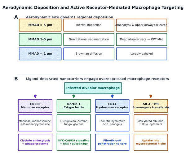
Pulmonary administration concentrates drug in the alveoli while sparing systemic tissues (Makled et al., 2021; Ramachandran et al., 2023). Where a particle lands is governed by its mass median aerodynamic diameter (MMAD), as summarized in the upper panel of Figure 3: particles above 5 µm impact in the upper airways and are cleared, those below 1 µm are largely exhaled, and the 1–5 µm window, achievable directly or through nano-in-micro aggregates, deposits efficiently in the deep alveoli (Ramachandran et al., 2023; Uskoković, 2023). The principal carrier classes and their aerodynamic performance are compared in Table 1.
Lipid-based nanocarriers
Liposomes, solid lipid nanoparticles (SLNs), and nanostructured lipid carriers (NLCs) encapsulate both
Table 1. Physicochemical properties, formulation engineering, and aerodynamic performance of inhalable nanocarrier platforms for anti-TB delivery. Comparison of the major inhalable nanocarrier classes evaluated for anti-tuberculosis delivery, aligning each platform's composition and particle size with its aerodynamic performance (mass median aerodynamic diameter, MMAD; fine particle fraction, FPF), drug payload and encapsulation efficiency (EE), and principal advantages and limitations. Across classes, formulation into spray-dried nano-in-micro aggregates brings the MMAD into the 1–5 µm window required for deep-alveolar deposition. Lipid and polymeric carriers favor sustained release and biocompatibility, whereas inorganic frameworks offer the highest loading and stimulus-responsive gating at the cost of slower biodegradation. Abbreviations: DPI, dry-powder inhaler; RIF, rifampicin; INH, isoniazid; P-gp, P-glycoprotein; ζ, zeta potential.
|
Nanocarrier class & material
|
Physicochemical properties & size
|
Aerodynamic behavior (MMAD / FPF)
|
Encapsulated payload & loading
|
Key therapeutic advantages
|
Formulation drawbacks
|
APA citation
|
|
Solid lipid NPs (SLNs) (Precirol ATO 5, glyceryl monostearate, palmitic acid)
|
Solid lipid core; 80–300 nm; surface charge −15 to −40 mV
|
Spray-dried with mannitol (DPI); MMAD 1–5 µm; FPF > 65%
|
RIF, INH, pyrazinamide, ethambutol; EE 60–90%
|
Sustained matrix release; bypasses P-gp efflux; lowers systemic toxicity
|
Limited capacity for hydrophilic drugs; drug expulsion on storage
|
Bera et al., 2024; Makled et al., 2021; Ramachandran et al., 2023
|
|
Nanostructured lipid carriers (NLCs) (solid + liquid lipids, e.g. Miglyol 812)
|
Fluidic solid–liquid matrix; 30–100 nm; ζ −37 to −59 mV
|
Nano-embedded spray-dried microspheres; MMAD ~2.0 µm; FPF ~93%
|
Linezolid, rifabutin, clofazimine; loading ~19–25%
|
Prevents crystallization; high payload; rapid mucus penetration (73% in 1 h)
|
Complex multi-lipid prep; careful surfactant choice to avoid irritation
|
Makled et al., 2021; Omoteso et al., 2025
|
|
Polymeric NPs (PNPs) (PLGA, chitosan, poly-caprolactone)
|
Biodegradable matrix; 40–200 nm; +15 to +35 mV (chitosan)
|
Nebulized or spray-dried nanocomposites; MMAD 2.1–3.4 µm; FPF > 70%
|
Linezolid, bedaquiline, moxifloxacin, levofloxacin; EE 57–85%
|
Sustained release (to 120 h); mucoadhesion; endosomal escape
|
Higher cost; initial burst release; acidic PLGA degradation products
|
Bourguignon et al., 2023; Garg et al., 2016; Ramachandran et al., 2023
|
|
Nanoliposomes & deformable vesicles (DPC, cholesterol, phosphatidylcholine)
|
Phospholipid bilayer vesicles; 80–350 nm; neutral/negative heads
|
Nebulized aerosols or spray-dried lipo-microspheres; MMAD 1.8–3.8 µm
|
Amikacin, INH, RIF, pyrazinamide; EE 37–90%
|
Biocompatible; co-encapsulates hydrophilic and lipophilic drugs
|
Instability on nebulization; leakage, oxidation; clearance by non-infected cells
|
Omoteso et al., 2025; Ramachandran et al., 2023
|
|
Inorganic NPs (MSNs, MOFs) (mesoporous silica, Fe-MIL-101-NH2, ZnO)
|
Rigid porous frameworks; 100–300 nm; surface area > 800 m²/g
|
Spray-dried micro-porous aggregates; MMAD 2.5–4.2 µm
|
INH, RIF, NZX antimicrobial peptide; payload > 30 wt%
|
Very high loading; stimulus-responsive gating (pH/enzyme); intrinsic antibacterial (ZnO/Ag)
|
Slow biodegradation; bio-persistence; local oxidative stress
|
Clemens et al., 2012; Wyszogrodzka et al., 2018
|
Table 2. Active receptor-mediated macrophage and granuloma targeting ligands in nanomedicine-based TB management. Receptor-targeting strategies that convert non-specific phagocytic capture into active, ligand-directed internalization of nanocarriers by infected macrophages. For each overexpressed receptor the table lists the matched surface ligand, the target cell or microenvironmental niche, the endocytic pathway engaged, a representative carrier–payload pairing, and the reported biological outcome. Mannose (CD206) and β-glucan (Dectin-1) systems dominate the evidence base, the latter coupling delivery to innate immune activation, while hyaluronic acid (CD44) and biomimetic membrane coatings extend reach into the dense fibrotic cuff and hypoxic lesion core. Abbreviations: RIF, rifampicin; INH, isoniazid; ROS/RNS, reactive oxygen/nitrogen species; TNF-α, tumor necrosis factor-α.
|
Target receptor & marker
|
Functional targeting ligand / coating
|
Primary target cell / niche
|
Internalization & trafficking pathway
|
Representative carrier & payload
|
Key biological & pharmacological outcomes
|
APA citation
|
|
Mannose receptor (CD206)
|
Mannose, mannosamine, methyl α-D-mannopyranoside
|
Alveolar & monocyte-derived macrophages
|
Clathrin-mediated, receptor-mediated endocytosis to early phagosomes
|
Mannosylated PLGA/chitosan NPs & SLNs loading RIF, INH, or rifapentine
|
8- to 15-fold higher macrophage uptake; phagolysosomal colocalization; lower systemic toxicity
|
Bera et al., 2024; Baranyai et al., 2021; Luan et al., 2025
|
|
Dectin-1 receptor (C-type lectin)
|
1,3-β-glucan, curdlan, fungal glucan polymers
|
Activated macrophages, dendritic cells
|
Dectin-1-mediated phagocytosis and endosomal routing
|
Curdlan-functionalized PLGA & chitosan-sulfate NPs co-loading RIF & d-pinitol
|
SYK–CARD9 signaling; TNF-α and ROS/RNS; drives autophagy
|
Bekale et al., 2025; Kutscher et al., 2026; Omoteso et al., 2025
|
|
Hyaluronic acid receptor (CD44)
|
Low-molecular-weight hyaluronic acid (HA)
|
Inflamed alveolar macrophages, epithelioid cells in granulomas
|
CD44 receptor-mediated endocytosis; hyaluronidase degradation
|
HA-grafted chitosan/PLGA nanogels delivering INH, RIF, or LLKKK18 peptide
|
Penetration across fibrotic capsule; selective uptake by CD44-overexpressing cells
|
Baranyai et al., 2021; Carnero et al., 2026
|
|
Tuftsin & formyl peptide receptors (FPRs)
|
Tuftsin tetrapeptide (Thr-Lys-Pro-Arg), fMLF peptides
|
Infected macrophages, granulomatous lesions
|
Receptor-mediated pinocytosis; phagocytic acceleration
|
Tuftsin-modified Pluronic F127-PLGA NPs & NLCs loading RIF or TB515
|
~2-fold higher bactericidal activity; immunostimulatory macrophage activation
|
Baranyai et al., 2021; Ramachandran et al., 2023
|
|
Scavenger receptors (SR-A, MARCO)
|
Maleylated bovine serum albumin (mBSA), anionic lipids
|
Resident pulmonary phagocytes, foamy macrophages
|
Scavenger receptor-mediated endocytosis and lysosomal delivery
|
mBSA-coated polymeric/lipid NPs loading ethionamide or clofazimine
|
Exploits polyionic binding on infected macrophages; avoids off-target organ distribution
|
Baranyai et al., 2021; Omoteso et al., 2025
|
|
Biomimetic membrane homotypic receptors
|
Pre-activated macrophage / erythrocyte plasma membranes
|
Granuloma necrotic cores, hypoxic infected tissue
|
Homotypic recognition, cell-fusion, chemokine-guided extravasation
|
BBTD@PM biomimetic NPs & RBC-O2/TQ@PB nanoplatforms
|
Immune evasion (half-life ~18.8 h); dual granuloma homing; hypoxia-targeted delivery
|
Li et al., 2024; Wang et al., 2024
|
hydrophilic and lipophilic antibiotics (Bourguignon et al., 2023; Ramachandran et al., 2023). Liposomes, while versatile, are prone to instability during nebulization and to lipid oxidation and leakage, whereas SLNs and NLCs offer better physical stability, high encapsulation efficiency, and sustained release of lipophilic second-line drugs such as linezolid or clofazimine (Makled et al., 2021; Ramachandran et al., 2023).
Polymeric nanoparticles
Carriers built from poly(lactic-co-glycolic acid) (PLGA) or from natural biopolymers such as chitosan and alginate combine high loading capacity with sustained intracellular release (Bourguignon et al., 2023; Zhu et al., 2026). Chitosan adds mucoadhesion and transient tight-junction opening to prolong retention (Garg et al., 2016), and cationic polymers promote endosomal escape through the proton-sponge effect (Zhu et al., 2026).
Inorganic and hybrid constructs
Mesoporous silica nanoparticles, SPIONs, gold nanoparticles, quantum dots, and metal–organic frameworks provide rigid, high-surface-area scaffolds that can carry drug and diagnostic function simultaneously (Onnainty & Granero, 2025; Uskoković, 2023).
2.4 Active Receptor-Mediated Macrophage and Granuloma Targeting
Passive accumulation in inflamed tissue yields only modest benefit; active surface targeting markedly increases uptake into infected macrophages (Baranyai et al., 2021; Cung et al., 2025). Infection upregulates specific pattern-recognition and endocytic receptors, each of which can be addressed with a matched ligand, as laid out in the lower panel of Figure 3 and catalogued in Table 2 (Baranyai et al., 2021; Cung et al., 2025).
Mannose receptor (CD206)
CD206 internalizes terminal mannose, fucose, and N-acetylglucosamine residues (Baranyai et al., 2021; Bera et al., 2024). Functionalizing PLGA, chitosan, or SLNs with mannose, mannosamine, or methyl α-D-mannopyranoside raises intracellular accumulation by up to 15-fold and delivers payloads straight to the phagolysosome (Bera et al., 2024; Luan et al., 2025).
Dectin-1 receptor
Dectin-1 recognizes 1,3-β-glucan, and coating nanoparticles with curdlan triggers Dectin-1-mediated phagocytosis while activating SYK–CARD9 signaling, pro-inflammatory cytokine release (TNF-α, IL-12), and ROS production, uniting delivery with immune stimulation (Bekale et al., 2025; Omoteso et al., 2025).
Hyaluronic acid receptor (CD44)
CD44, upregulated on inflamed macrophages and epithelioid cells, binds low-molecular-weight hyaluronic acid with high affinity, improving penetration across the dense fibrotic cuff of mature granulomas (Baranyai et al., 2021; Carnero et al., 2026).
Scavenger, transferrin, and peptide receptors
Scavenger receptors (SR-A, MARCO) take up maleylated albumin and anionic lipids, while transferrin-binding peptides, camelid antibodies, and tuftsin enhance internalization and deep accumulation in avascular caseous centers (Baranyai et al., 2021; Tiwari et al., 2026).
2.5 Integrated Diagnostic Imaging and Biomimetic Phototherapeutics
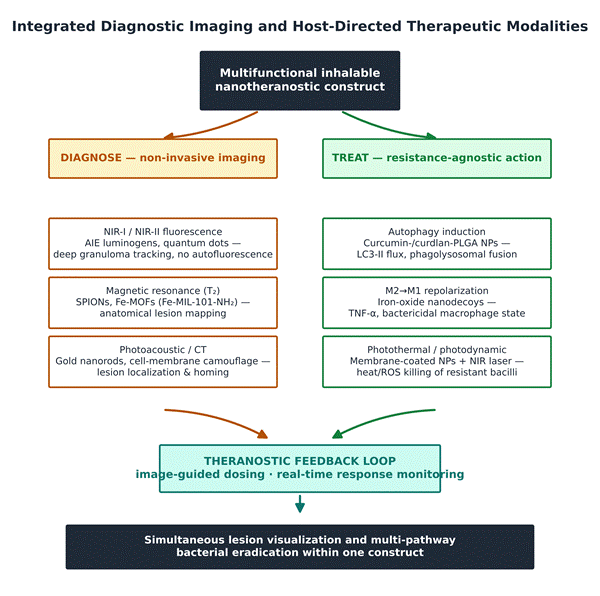
The defining move of nanotheranostics is to make therapy visible, pairing drug delivery with non-invasive imaging and, increasingly, light-activated killing within one construct, as mapped in Figure 4 and detailed in Table 3 (Bueno, 2025; Onnainty & Granero, 2025).
NIR imaging and AIE luminogens
Conventional dyes photobleach and quench in aqueous media; aggregation-induced emission (AIE) luminogens instead brighten upon aggregation (Liao et al., 2020). RIF-loaded AIE nanocarriers emit strong NIR-II fluorescence inside granulomas, enabling high-resolution, background-free tracking in live animals (Liao et al., 2020; Onnainty & Granero, 2025).
MRI contrast
SPIONs and iron-based frameworks such as Fe-MIL-101-NH2 act as T2 contrast agents for pulmonary lesions and, when ligand-decorated, combine anatomical imaging with sustained antibiotic release (Onnainty & Granero, 2025; Wyszogrodzka et al., 2018).
Cell-membrane camouflage and resistance-agnostic phototherapy
Nanoparticles cloaked in membranes from pre-activated,
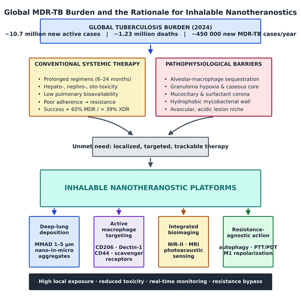
Figure 1. Global MDR-TB burden and the rationale for inhalable nanotheranostics. Schematic overview linking the epidemiological burden of tuberculosis to the design logic of inhalable nanotheranostic platforms. The persistent global case and death toll, together with the high annual incidence of multidrug-resistant disease, exposes two convergent problems: the pharmacological failures of conventional systemic therapy (prolonged, toxic regimens with low pulmonary bioavailability) and the pathophysiological barriers that shelter the pathogen (macrophage sequestration, granuloma hypoxia, mucociliary clearance, and the surfactant corona). These define an unmet need that inhalable nanotheranostics address through four coordinated capabilities, deep-lung deposition, active macrophage targeting, integrated bioimaging, and resistance-agnostic action, yielding high local exposure, reduced toxicity, real-time monitoring, and resistance bypass.
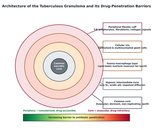
Figure 2. Architecture of the tuberculous granuloma and its drug-penetration barriers. Concentric structure of the mature tuberculous granuloma and the gradient of drug accessibility it imposes. Moving inward from the vascularized periphery, the peripheral fibrotic cuff (T/B lymphocytes, fibroblasts, collagen), the cellular rim (epithelioid and multinucleated giant cells), the lipid-laden foamy-macrophage layer, and the hypoxic intermediate zone surround a central caseous necrotic core harboring dormant, non-replicating bacilli. Progressive hypoxia, acidification, and loss of vascularization toward the core, depicted by the lower gradient bar, steadily raise the barrier to antibiotic penetration, explaining why systemically delivered drugs reach the caseum at sub-therapeutic concentrations and why lesion-penetrating nanocarriers are needed.
M. tb-stimulated macrophages home to infected tissue while evading clearance (Li et al., 2024; Ranjith Singh et al., 2025). Under 1064 nm irradiation they generate localized heat and ROS, destroying drug-resistant bacilli independently of antibiotic susceptibility while permitting simultaneous NIR-II imaging (Li et al., 2024; Onnainty & Granero, 2025).
2.6 Nanocarrier-Enabled Host-Directed Therapies
Because M. tb so readily evolves resistance to direct-acting drugs, host-directed therapy (HDT) offers a resistance-agnostic paradigm, modulating host pathways rather than bacterial targets and thereby removing the selective pressure that breeds resistance (Bekale et al., 2025; Omoteso et al., 2025; Tiwari et al., 2026). The principal mechanisms, with representative nanomaterials and outcomes, are compiled in Table 4 and summarized in Figure 4.
-
Autophagy stimulation. Curcumin-loaded and curdlan-coated PLGA nanoparticles engage Dectin-1 and drive the LC3-II autophagic cascade, overriding M. tb-induced phagolysosomal arrest and cutting intracellular burden (Bekale et al., 2025; Omoteso et al., 2025).
-
Macrophage repolarization. Iron-tropic iron oxide nanoparticles deliver intracellular iron that repolarizes permissive M2 macrophages toward the bactericidal M1 phenotype, with release of TNF-α and IL-12 (Onnainty & Granero, 2025; Tiwari et al., 2026).
-
Vascular normalization and matrix remodeling. Carriers delivering anti-VEGF or matrix-metalloproteinase inhibitors normalize aberrant granuloma vasculature and loosen the surrounding matrix, improving penetration of co-administered antibiotics (Datta et al., 2024; Onnainty & Granero, 2025).
2.7 Translational Challenges, Safety, and a Clinical Roadmap
The preclinical case is strong, but translation is constrained on several fronts (Erothi et al., 2026; Tiwari et al., 2026). Non-biodegradable carriers can provoke pulmonary inflammation, oxidative damage, and fibrosis on repeated dosing (Poojary & Dash, 2025; Uskoković, 2023). Interaction with native surfactant can disturb alveolar surface tension and mask targeting ligands through corona formation (Poojary & Dash, 2025; Tiwari et al., 2026). Nanoparticles aggregate during nebulization or spray-drying, degrading aerosol performance (Erothi et al., 2026; Ramachandran et al., 2023). And GMP-scale synthesis with batch-to-batch consistency in size, loading, ligand density, and sterility remains a formidable industrial hurdle (Poojary & Dash, 2025; Tiwari et al., 2026). Addressing these will require standardized nanotoxicology panels, microfluidic manufacturing, and long-term, multi-dose toxicity studies in non-human primates (Erothi et al., 2026; Poojary & Dash, 2025).