Biosensors and Nanotheranostics

Bionanotechnology, Drug Delivery, Therapeutics | online ISSN 3064-7789
175
Citations
116.7k
Views
81
Articles
Your new experience awaits. Try the new design now and help us make it even better
Switch to the new experience
Figures and Tables
REVIEWS   (Open Access)

Scanning Electrochemical Microscopy in Biosensing and Cell Imaging: Principles, Hybrid Probe Architectures, and Emerging Bio-Interface Applications

Tina Thai 1*, Annachiara Bazzoni 2


+ Author Affiliations

Biosensors and Nanotheranostics 5 (1) 1-8 https://doi.org/10.25163/biosensors.5110987

Submitted: 13 May 2026 Revised: 01 July 2026  Published: 11 July 2026 


Abstract

Living systems do their chemistry in small places. A neuron releases dopamine from a patch of membrane a few hundred nanometres across, a biofilm acidifies one pocket while its neighbour stays near neutral, and a single phagolysosome may carry a reactive oxygen burst that the next one lacks. Bulk electrochemistry averages all of this away. Scanning electrochemical microscopy (SECM), together with the wider family of scanning electrochemical probe microscopies, was developed to recover that lost spatial information, yet its classical form struggles on soft, uneven biological specimens, where changes in topography and changes in reactivity become hard to separate. This review traces how the field has responded. We first revisit the operating principles of direct-current, alternating-current and intermittent-contact SECM, and examine the micro/nanohybrid probes (coaxial, theta-type and multi-barrel) and multimodal couplings with scanning ion conductance microscopy, atomic force microscopy and scanning electrochemical cell microscopy that now separate topography from faradaic signal. Drawing on a structured narrative synthesis, we compare reported spatial resolutions, collection efficiencies and biological outcomes, from single-organelle reactive species profiling to three-parameter mapping of live oral biofilms. We follow the same interfacial logic outward, into nanostructured point-of-care biosensors, bacterial nanocellulose and hydrogel bio-interfaces, and the antifouling and signal-amplification chemistries that decide whether a sensor survives real biofluids. Across these areas, analytical performance is often impressive, but most platforms remain at technology readiness levels 3–5. We close by discussing fabrication reproducibility, temporal resolution, fouling and validation as the main obstacles still separating demonstrations from routine biological and clinical use.

Keywords: scanning electrochemical microscopy; hybrid nanoprobes; single-cell imaging; biofilm mapping; electrochemical biosensors; bacterial nanocellulose; antifouling interfaces; point-of-care diagnostics

1. Introduction

Interfacial events in biological and electrochemical systems, from transmembrane transport and cellular signalling to localized enzymatic catalysis and electron transfer across biological membranes, are heterogeneous in both space and time (Liu & Zhang, 2026; Yan & Zhang, 2025). This heterogeneity is not a nuisance to be averaged out; it is frequently where the biology lies. A cell population that appears quiescent in a bulk measurement may contain a minority of highly active cells, and a catalytic surface that seems uniformly mediocre may in fact host a few very active sites surrounded by inert regions. Conventional macroscale techniques, including cyclic voltammetry (CV), electrochemical impedance spectroscopy (EIS) and open-circuit potentiometry, record signals that are integrated over the entire electrode. They remain indispensable for macroscopic kinetic characterization, but by construction they conceal local variations in charge-transfer kinetics, concentration gradients and interfacial dynamics at the micro- and nanoscale (Garcia et al., 2024; Polcari et al., 2016).

Recovering this hidden information requires a measurement that is itself local. Scanning electrochemical probe microscopy (SEPM) meets that requirement by moving a miniaturized electrochemical sensor across, or into, the specimen of interest and recording how its response changes from point to point, in situ and in real time (Bard et al., 1991; Liu & Zhang, 2026). The most established member of this family, scanning electrochemical microscopy (SECM), emerged in the late 1980s. In SECM, a movable ultramicroelectrode (UME), a micro- or nanoscale conductive disk sealed in an insulating sheath, is scanned close to a substrate immersed in an electrolyte, and the faradaic current produced by the oxidation or reduction of a dissolved redox mediator at the tip is used as the imaging signal (Bard et al., 1991; Polcari et al., 2016). Because this current depends on how freely the mediator can diffuse into the narrow gap between tip and substrate, and on whether the substrate regenerates or consumes it, the tip current encodes information about both the distance to the surface and the surface's local chemical activity.

That dual sensitivity is exploited through a set of complementary operating modes. In feedback mode, approaching a conductive or electrochemically active surface regenerates the mediator and enhances the tip current (positive feedback), whereas approaching an insulating or inactive surface hinders diffusion and suppresses it (negative feedback) (Liu & Zhang, 2026; Polcari et al., 2016). In the substrate-generation/tip-collection (SG/TC) and tip-generation/substrate-collection (TG/SC) modes, one interface produces an electroactive species, such as hydrogen peroxide, another reactive oxygen species or dopamine, and the other collects it quantitatively (Liu & Zhang, 2026). Redox competition mode instead monitors how tip and substrate compete for a shared analyte (Liu & Zhang, 2026; Polcari et al., 2016). Where an added mediator could perturb cell viability or interfere with native chemistry, alternating-current SECM (AC-SECM) and intermittent-contact SECM (IC-SECM) allow mediator-free mapping of local impedance and activity (Liu & Zhang, 2026; X. Shi et al., 2020).

The difficulty is that the very property that makes SECM informative also makes it ambiguous. In classical constant-height imaging with a single-channel disk UME, any change in tip-to-substrate distance alters the current just as a change in reactivity would. On flat, rigid electrodes this is manageable. On soft, deformable or non-planar specimens, such as living mammalian cells, bacterial biofilms and tissue matrices, topographic height fluctuations generate current artefacts that are convolved with the electrochemical signal of interest (Liu & Zhang, 2026; Takahashi et al., 2010). A second limitation is informational: a single-channel UME returns one data stream per scan, so mapping several parameters (for instance local pH, ion fluxes and a redox-active metabolite) requires repeated scans that introduce spatial and temporal misregistration, which is especially problematic for systems that change while they are being imaged (Liu & Zhang, 2026; S. Park et al., 2023).

Two broad lines of technological development have addressed these constraints. The first decouples topography from reactivity by adding an independent, non-faradaic channel for distance control. Scanning ion conductance microscopy (SICM) uses the ionic current through an electrolyte-filled nanopipette as feedback and can track the surface of a living cell without touching it; when combined with an SECM channel (SECM–SICM), it maps faradaic activity at constant separation (Takahashi et al., 2010, 2011). Atomic force microscopy combined with SECM (AFM–SECM), implemented with conductive cantilevers or colloidal probes, couples nanomechanical force mapping with localized electrochemical imaging (Knittel et al., 2016; X. Shi et al., 2020). Scanning electrochemical cell microscopy (SECCM) takes a different route altogether, confining the electrochemical reaction to a femtolitre-to-picolitre meniscus droplet at a pipette tip so that the sample need not be immersed (Ebejer et al., 2010; Liu & Zhang, 2026).

The second line concerns the probe itself. Coaxial (disk@ring, ring@pipette), theta-type (dual-amperometric, amperometric–potentiometric, dual-potentiometric) and multi-barrel micro/nanohybrid probes place several sensing or feedback channels within one tip, so that different chemical quantities can be recorded at the same pixel and at the same moment (Liu & Zhang, 2026). Coaxial Ca²⁺-selective disk@IrOx-modified pH ring probes, for example, have visualized biomineralization and local acidification during biofilm-induced corrosion (Liu & Zhang, 2026). Theta-type Pt-decorated multiwalled carbon nanotube (MWCNT) nanoprobes and enzyme-functionalized microsensors have mapped glucose uptake and H₂O₂ dynamics across microbial biofilms (Jayathilake & Koley, 2020; Liu & Zhang, 2026), and triple-barrel probes combining pH, H₂O₂ and topographic feedback have exposed pronounced microenvironmental heterogeneity in living oral biofilms (S. Park et al., 2023).

In parallel, a materials-oriented literature has developed functional bio-interfaces that make local and continuous electrochemical sensing practical in real biological media. Bacterial nanocellulose (BNC) hydrogels, conductive polymers such as polyaniline (PANI) and poly(3,4-ethylenedioxythiophene):polystyrene sulfonate (PEDOT:PSS), carbon nanomaterials (carbon nanotubes, graphene and MXenes) and biomimetic cell-membrane coatings now provide stable, low-fouling electrode surfaces for cell capture, neurotransmitter monitoring and wearable or implantable biomarker detection (Lertanantawong et al., 2026; Priya et al., 2026). Together, these developments have extended SECM and related SEPM platforms towards single-entity bio-events, cellular secretion and dynamic communication at the cell surface (Garcia et al., 2024; Yan & Zhang, 2025).

These two literatures, one concerned with how to resolve local chemistry and the other with how to sense it robustly in complex fluids, are usually reviewed separately, even though they face many of the same interfacial problems. The present review brings them together. Its specific objectives are (i) to set out the working principles and operating modes of SECM, including DC (feedback, SG/TC, TG/SC and redox competition), AC and IC variants, with attention to mediator choice, mass transport and the limits of spatial resolution; (ii) to classify and critically analyse micro/nanohybrid and multimodal probe architectures (coaxial, theta-type, multi-barrel, SECM–SICM, SECM–SECCM and AFM–SECM) as strategies for decoupling topography from reactivity; (iii) to evaluate emerging biological applications, including cellular secretion, intracellular and pericellular reactive oxygen and nitrogen species (ROS/RNS) profiling, membrane transport, biofilm metabolic mapping and label-free cell capture on nanostructured surfaces, alongside the biosensing and bio-interface materials that share the same design logic; and (iv) to identify the technical bottlenecks that still limit translation, namely probe fabrication yield, signal crosstalk, temporal resolution and fouling, and to outline future directions in adaptive probes, operando multi-technique coupling and high-throughput point-of-care diagnostics.

2. Hybrid Scanning Probes and Smart Bio-Interfaces Bring Electrochemical Mapping Toward Clinical Biosensing

2.1 Theoretical Framework and Core Mechanics of SECM

2.1.1 The probe and the tip–substrate gap

Interfacial electrochemical processes, from transmembrane ion fluxes and single-cell metabolic bursts to localized enzymatic catalysis, vary sharply across space and time (Liu & Zhang, 2026). Bulk measurements average the contributions of millions of chemical entities and so obscure microscale reactive hot spots and transient events (Priya et al., 2026; Saweres-Argüelles et al., 2027). SECM was conceived to close this gap, providing maps of local electrochemical reactivity, and indirectly of topography, with high spatiotemporal resolution (Liu & Zhang, 2026; X. Shi et al., 2020). Over more than three decades the technique has matured from a qualitative imaging tool into a quantitative toolbox for interrogating the chemistry inside electrochemical processes (Polcari et al., 2016; Putnam et al., 2025).

At the heart of classical SECM is a UME or nanoelectrode, typically a platinum, gold or carbon core sealed in a glass or quartz sheath, immersed in an electrolyte containing a redox mediator (Liu & Zhang, 2026). The spatial sensitivity of the measurement arises from distance-dependent mass transfer of the mediator within the thin layer of solution trapped between the tip and the substrate (Kranz et al., 2001; Liu & Zhang, 2026). Far from any surface the tip reaches a steady-state, diffusion-limited current; as the tip approaches a surface this current is perturbed in a way that depends on both the separation and the nature of the surface. The relative size of the electroactive disk and its insulating sheath, the mediator concentration and the scan speed all shape the achievable resolution and the time needed to acquire an image, which is why careful reporting of these experimental parameters has long been emphasized (Polcari et al., 2016).

2.1.2 Direct-current operating modes

In feedback mode, an indirect redox mediator is added to the solution and the tip is held at a potential that drives its diffusion-limited oxidation or reduction (Liu & Zhang, 2026). Over a conductive or electrochemically active region, the species generated at the tip diffuses to the substrate and is converted back to its original form, establishing a cyclic flux that raises the tip current above its bulk value (positive feedback; Figure 1a). Over an insulating or passive region, the substrate simply blocks diffusion into the gap and the current falls (negative feedback; Figure 1b) (Liu & Zhang, 2026). The approach curve, the tip current plotted against distance, therefore serves both as a positioning tool and as a measure of local heterogeneous kinetics.

In generator–collector (GC) modes, one electrode produces an electroactive species and the other detects it. In SG/TC mode, cellular processes or enzymatic turnover release electroactive species into solution, which are collected and quantified at the polarized tip (Figure 1c) (Liu & Zhang, 2026). In TG/SC mode, the tip locally delivers a mediator or reactant so that its consumption or catalytic conversion at the substrate can be interrogated (Figure 1d) (Liu & Zhang, 2026). Generator–collector configurations have proved particularly useful for corrosion studies, where they permit direct mapping of corrosion products and gas evolution without external mediators (Oyeleye et al., 2026).

In redox competition mode, tip and substrate compete for the same dissolved species, commonly dissolved oxygen, so that regions of high substrate consumption appear as dips in the tip current (Figure 1e) (Liu & Zhang, 2026). The approach is attractive for electrocatalyst screening because it reports on the substrate reaction directly rather than through a mediator.

2.1.3 Alternating-current and intermittent-contact SECM

AC-SECM superimposes a small sinusoidal voltage on the tip and measures the resulting local impedance, so that neither an exogenous mediator nor polarization of the sample is required (Liu & Zhang, 2026). Typical excitation frequencies lie in the low-kilohertz range, and the technique performs well in low-conductivity media, which has made it a mainstay for mapping weld joints, aluminium alloys and steel reinforcement in concrete (Oyeleye et al., 2026). Intermittent-contact operation adds a vibrating tip whose damping provides an independent measure of distance, partially decoupling topography from electrochemical activity (Oyeleye et al., 2026; X. Shi et al., 2020). For soft biological samples, the mediator-free character of AC-SECM is its principal advantage: the measurement need not introduce redox-active chemicals that might alter cell physiology.

2.1.4 The signal convolution problem

Despite its successes, conventional single-channel SECM has a basic limitation: signal convolution (Liu & Zhang, 2026). In constant-height scanning, a change in tip current may reflect a change in local reactivity, a change in tip-to-sample distance caused by surface roughness, or both (Figure 2e). On a living cell several micrometres tall, the distance contribution can dominate. This limitation has driven the development of hybrid probe architectures that separate physical distance regulation from chemical sensing (Kranz et al., 2001; Liu & Zhang, 2026; X. Shi et al., 2020; Takahashi et al., 2010), which are the subject of the next section.

2.2 Micro/Nanohybrid Probe Architectures and Multimodal Integration

2.2.1 Coaxial, theta-type and multi-barrel geometries

To overcome signal convolution and the single-output constraint, recent SEPM developments have introduced hybrid probes that integrate several sensing, feedback and microfluidic channels within one micro- or nanoscale tip (Liu & Zhang, 2026). These designs fall into three geometric families (Figure 2a–d). Coaxial probes place an axisymmetric outer ring of gold, platinum or carbon around an inner UME or nanopipette. Their natural mode is disk-generation/ring-collection (DG/RC), in which species produced at the disk are intercepted by the surrounding ring, enabling localized species partitioning and dual-parameter mapping (Liljeroth et al., 2002; Liu & Zhang, 2026). Theta-type probes are drawn from dual-barrel capillaries whose two barrels can be modified independently, for example as two amperometric electrodes, as an amperometric electrode paired with a potentiometric pH or ion sensor, or as an electrode paired with an open electrolyte channel for SICM feedback (Liu & Zhang, 2026). A double-barrel assembly has also been used to suppress electrical-field artefacts in potentiometric SECM imaging of galvanic corrosion (Filotás et al., 2018). Multi-barrel probes extend the principle to three or four barrels, accommodating simultaneous measurement of three or more parameters, such as pH, H₂O₂ and
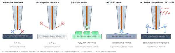
Figure 1. Principal operating modes of scanning electrochemical microscopy (SECM). (a) Positive feedback: over a conductive or active substrate, the oxidized mediator (O) generated at the ultramicroelectrode tip is reduced back to R at the surface, raising the tip current above its bulk value. (b) Negative feedback: an insulating substrate hinders diffusion of R into the tip–substrate gap and the current falls. (c) Substrate-generation/tip-collection (SG/TC): cells or enzymes release an electroactive product (P), such as H₂O₂, reactive oxygen species or dopamine, that is collected at the tip. (d) Tip-generation/substrate-collection (TG/SC): a tip-generated species (G) is delivered locally and consumed at the substrate. (e) Redox competition and AC-SECM: tip and substrate compete for dissolved O₂, or a small AC perturbation (1–8 kHz) measures local impedance without a mediator. Schematic drawn by the authors on the basis of Liu and Zhang (2026), Polcari et al. (2016) and Oyeleye et al. (2026); not to scale.
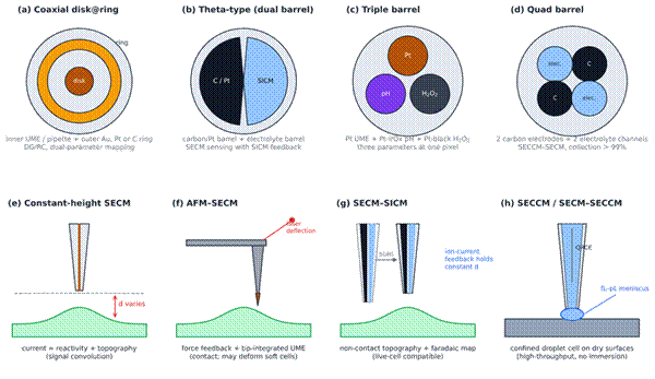
 

Figure 2. Micro/nanohybrid probe architectures and multimodal distance-regulation strategies. Top row, tip-face cross-sections: (a) coaxial disk@ring probe for disk-generation/ring-collection; (b) theta-type dual-barrel probe pairing a carbon or Pt electrode with an electrolyte barrel for SICM feedback; (c) triple-barrel probe combining a Pt UME, a Pt–IrOx pH sensor and a Pt-black H₂O₂ sensor; and (d) quad-barrel probe with two carbon electrodes and two electrolyte channels. Bottom row, imaging over a soft, non-planar specimen: (e) constant-height SECM, in which the changing tip–sample distance (d) convolves topography with reactivity; (f) AFM–SECM with a cantilever-integrated electrode and optical force feedback; (g) SECM–SICM, in which ion-current feedback holds d constant without contact; and (h) SECCM, in which a femtolitre-to-picolitre meniscus confines the reaction on a dry surface. Drawn by the authors from Liu and Zhang (2026), Takahashi et al. (2010), Kranz et al. (2001) and Ebejer et al. (2010); not to scale.

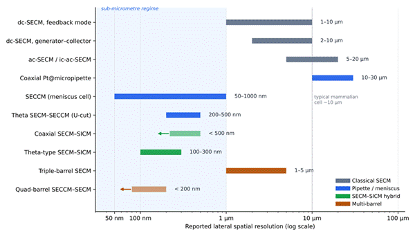
topography, together with built-in working and reference cells (Liu & Zhang, 2026; S. Park et al., 2023). Quad-barrel nanopipettes combining two carbon electrodes with two electrolyte channels have been used for both voltammetric analysis and ion conductance imaging (Paulose Nadappuram et al., 2015). The characteristic designs, feedback mechanisms and reported resolutions of these architectures are compared in Table 1, and the reported spatial resolutions are summarized graphically in Figure 3.

2.2.2 Decoupling topography and reactivity: AFM–SECM and SECM–SICM

Integrating atomic force microscopy with SECM was one of the earliest attempts to separate height artefacts from faradaic measurements (Kranz et al., 2001; X. Shi et al., 2020). By building a UME or nanoelectrode directly into a flexible AFM cantilever or tip, AFM–SECM records nanoscale mechanical topography and localized electrochemistry at the same time (Figure 2f) (Kranz et al., 2001; X. Shi et al., 2020). Conductive colloidal probes have extended this approach to soft interfaces, for instance in studies of the PEDOT:PSS–cell interface (Knittel et al., 2016). The drawback is that physical contact, even when gentle, can deform soft biological specimens or damage fragile membranes.

SICM offers a non-contact alternative that is well suited to living systems (Takahashi et al., 2010). An electrolyte-filled nanopipette containing an internal reference electrode passes an ionic current through its nanoscale aperture; as the pipette approaches a surface, partial obstruction reduces that current, providing a sensitive distance signal without mechanical contact (Liu & Zhang, 2026; Takahashi et al., 2010). In SECM–SICM hybrid probes, such as coaxial nanopipette@Au ring or theta-type carbon/nanopipette designs, one channel maintains SICM distance feedback while an adjacent conductive channel performs amperometric or potentiometric SECM sensing (Figure 2g) (Liu & Zhang, 2026; Takahashi et al., 2010). The result is simultaneous, nanoscale mapping of living-cell morphology and local metabolic flux (Liu & Zhang, 2026; Takahashi et al., 2010). Multifunctional nanoprobes of this kind can also deliver chemicals locally, adding a "write" capability to the "read" function of imaging (Takahashi et al., 2011), and their biological applications have been reviewed in depth (Takahashi et al., 2021).

2.2.3 Confinement and localized delivery: SECM–SECCM and microfluidic probes

SECCM addresses a different drawback of conventional SECM, namely the requirement to submerge the entire sample in electrolyte (Liu & Zhang, 2026). A micro- or nanopipette filled with electrolyte and fitted with a quasi-reference counter electrode (QRCE) forms a confined femtolitre-to-picolitre meniscus on contact with a substrate, and only the wetted footprint participates in the measurement (Figure 2h) (Ebejer et al., 2010; Liu & Zhang, 2026). When combined with SECM working electrodes in SECM–SECCM hybrid probes, the meniscus becomes a mobile micro-reactor that supports electrocatalytic, enzymatic or surface-reactivity measurements on dry, vertical or highly localized surfaces (Liu & Zhang, 2026). Hybrid SECCM probes have been used to resolve microscale activity and product selectivity during CO₂ reduction (M. Kim et al., 2026; C. H. Ryu & Ren, 2024).

Push–pull microfluidic SECM probes, finally, carry two microchannels for simultaneous electrolyte delivery and aspiration. They regulate the microenvironment around a single cell while sampling secreted biomolecules for downstream mass spectrometric or fluorescence analysis (Liu & Zhang, 2026).

2.3 SEPM Applications in Single-Cell Analysis, Biofilm Mapping and Biomarker Sensing

2.3.1 Single-cell physiology and organelle heterogeneity

Probing chemistry inside individual living cells demands extreme miniaturization, positional precision and chemical selectivity (Liu & Zhang, 2026). Coaxial carbon ring@pipette probes fabricated by pyrolytic carbon deposition and chemical vapour deposition have achieved tip dimensions below 100 nm (Liu & Zhang, 2026). Platinized versions inserted into individual organelles of living cells have performed multi-potential-step chronoamperometry to quantify intracellular reactive species, revealing substantial metabolic heterogeneity between single organelles (Hu et al., 2022; Liu & Zhang, 2026). Positioned near the cell surface instead, carbon nano-ring probes can combine resistive-pulse sensing with faradaic detection during extracellular vesicle (EV) release. This label-free single-entity approach measures EV size and internal signalling molecules simultaneously and can discriminate cellular phenotypes and physiological states (Jia et al., 2022; Liu & Zhang, 2026). SEPM has thereby become a significant tool for single-

Table 1. Operating modes, hybrid probe configurations and interfacial sensing applications of scanning electrochemical probe microscopy (SEPM). The table compares classical direct-current (dc), alternating-current (ac) and intermittent-contact (ic) SECM modes with meniscus-confined (SECCM) and hybrid SECM–SICM, triple-barrel and quad-barrel probes. For each configuration, it lists the probe geometry, the mechanism used to regulate tip–substrate distance, the reported spatial and temporal resolution, the substrate or analyte studied, and the principal analytical outcome. Resolution values are those reported in the cited studies under their specific experimental conditions and are therefore indicative rather than directly comparable. Reported spatial resolutions are plotted on a common scale in Figure 3. Abbreviations: UME, ultramicroelectrode; FcMeOH, ferrocenemethanol; QRCE, quasi-reference counter electrode; FIB, focused ion beam; ORR, oxygen reduction reaction; ALP, alkaline phosphatase; PVDF, polyvinylidene fluoride; FSCV, fast-scan cyclic voltammetry; EV, extracellular vesicle.

Technique / mode

Probe geometry and architecture

Operating principle and distance feedback

Spatial and temporal resolution

Target analyte / substrate

Key analytical findings and advantages

Reference

dc-SECM (feedback mode)

Disk-shaped Pt or Au UME (d = 1–25 µm) in glass sheath

Mediator recycling between probe and sample; constant-height or approach-curve positioning

Spatial 1–10 µm; seconds to minutes per scan

Fe(CN)₆³⁻/⁴⁻, FcMeOH; SS 316L pitting, rebar corrosion

Resolves localized pitting initiation and maps passive-film breakdown with high chemical selectivity

Oyeleye et al. (2026)

dc-SECM (generator–collector)

UME probe plus substrate working electrode

One electrode generates a redox species, the other collects it (SG/TC or TG/SC)

Spatial 2–10 µm; collection efficiency 60–90%

O₂ reduction, H₂ evolution; Mg alloy AZ31 corrosion

Quantitative mapping of corrosion products and gas evolution without external mediators

Oyeleye et al. (2026)

ac-SECM and ic-ac-SECM

Vibrating or AC-excited Pt UME (d = 10–80 µm)

AC perturbation (1–8 kHz) measures local impedance; intermittent contact holds distance

Spatial 5–20 µm; high signal-to-noise in low-salt media

Zr-4/Ti/304L welds, AA2024 alloy, rebar in concrete

Separates topography from activity in low-conductivity media without added mediators

Oyeleye et al. (2026)

SECCM (meniscus cell)

Single- or theta-barrel micropipette with Ag/AgCl QRCE

fL–pL meniscus contacts a dry substrate; ionic-current feedback

Spatial 50 nm–1 µm; high-throughput landing

Au nanocrystal electrocatalysts, CO₂ reduction catalysts, single carbon nanotubes

Single-entity and facet-dependent electrocatalytic kinetics without bulk immersion

Ebejer et al. (2010); Garcia et al. (2024); Liu & Zhang (2026)

Coaxial carbon ring@pipette (platinized)

Pyrolytic carbon ring in quartz nanopipette; tip radius ~100 nm

Thin-layer confinement; multi-potential-step chronoamperometry after organelle insertion

Spatial ~100 nm; four-step potential sequence

Macrophage phagolysosomes; single EVs at the cell surface

Simultaneous quantification of H₂O₂, ONOO⁻, NO and NO₂⁻; resistive-pulse plus faradaic EV discrimination of MCF-10A vs MDA-MB-231

Hu et al. (2022); Jia et al. (2022); Liu & Zhang (2026)

Coaxial SECM–SICM

Nanopipette core (radius ~220 nm) with outer Au or carbon ring

SICM ion-current feedback in inner pipette; outer ring records faradaic current

Spatial < 500 nm; non-contact

Live cells, immobilized glucose oxidase arrays, macrophage ROS/RNS

Non-contact 3D mapping of soft membranes with simultaneous faradaic detection

Liu & Zhang (2026); Takahashi et al. (2010)

Theta-type SECM–SICM

Dual-barrel theta pipette: pyrolytic carbon (± Pt NPs) plus electrolyte barrel

Independent SICM distance feedback and carbon nanoelectrode sensing

Spatial 100–300 nm; millisecond FSCV

Sensory neurons, EGFR on A431 cells, dopamine release

~50-fold signal-to-noise gain with Pt NPs; receptor and transmembrane transport mapping

Liu & Zhang (2026); Sen et al. (2021); Takahashi et al. (2021)

Triple-barrel SECM

Pt UME + Pt–IrOx pH sensor + Pt-black H₂O₂ sensor

Simultaneous potentiometric and amperometric channels; bare Pt for distance

Spatial 1–5 µm; real-time three-parameter acquisition

Living S. mutans dental-plaque biofilms on hydroxyapatite

pH decline from ~7.2 towards 4.5–5.2 after sugar exposure; heterogeneous H₂O₂ (0–17 µM)

Liu & Zhang (2026); S. Park et al. (2023)

Quad-barrel SECCM–SECM

Four-barrel nanopipette: two pyrolytic carbon electrodes + two electrolyte channels

Meniscus confinement with dual working electrodes

Spatial < 200 nm; collection efficiency > 99%

Single-walled carbon nanotubes, tribocharged PTFE

Localized redox titration and transient pH profiling during dissolution in ionic liquids

Liu & Zhang (2026); Paulose Nadappuram et al. (2015)

Theta SECM–SECCM (U-cut)

FIB U-cut theta pipette: Pt NP/carbon barrel + electrolyte barrel

Droplet covers both barrels; SECCM drives ORR while Pt NPs oxidize H₂O₂ product

Spatial 200–500 nm; collection efficiency > 95%

Polycrystalline Pt/Au; Ag–Bi₂O₃ CO₂ reduction catalysts

Maps activity versus product selectivity (H₂O₂ vs H₂O; CO vs formate) across catalyst grains

M. Kim et al. (2026); Y. Kim et al. (2024); Liu & Zhang (2026); C. H. Ryu & Ren (2024); H. Ryu et al. (2024)

Coaxial Pt@micropipette

25 µm Pt disk inside a 307.7 µm micropipette

Renewable 1.93 nL droplet cell on dry, heterogeneous-wettability surfaces

Spatial 10–30 µm; 97% accuracy in bio-aging estimation

ALP microarrays, biological residues on PVDF, dry interdigitated Au

Avoids evaporation and contamination; detects enzyme decay in aging forensic samples

Liu & Zhang (2026)

Table 2. Nanostructured electrochemical biosensing platforms, interface modifiers, biomarker targets and analytical performance. The table summarizes representative electrochemical biosensors for infectious, oncological, metabolic and cellular biomarkers. For each platform, it gives the electrode modifiers, the transduction technique, the target and sample matrix, the linear dynamic range, the limit of detection (LOD) and the evidence of validation in real samples (recovery, %). Performance values are reported as published and were obtained under different protocols, so they should be read as benchmarks rather than as a ranking. Where a study reported LODs in two unit systems (e.g., protein concentration and organism count), both are given. Abbreviations: GCE, glassy carbon electrode; SPCE/SPE, screen-printed (carbon) electrode; IDE, interdigitated electrode; MGCE, magnetic GCE; LIG, laser-induced graphene; EIS, electrochemical impedance spectroscopy; DPV, differential pulse voltammetry; SWV, square-wave voltammetry; ASV, anodic stripping voltammetry; SEC, size-exclusion chromatography; CTC, circulating tumour cell; EPCR, endothelial protein C receptor.

Platform / architecture

Interface modifiers and functional materials

Transduction

Target and matrix

Linear range

LOD

Validation and recovery

Reference

Label-free immunosensor

NiO/ZnO p–n heterojunction on GCE

EIS / DPV

M. tuberculosis Ag85 antigen

5–300 ng/mL

0.77 ng/mL

Spiked rabbit serum; 92.0–102.0%

Nunez et al. (2026)

Multiplexed viral immunosensor

rGO–ZIF-8 nanocomposite on dual SPE

DPV

Monkeypox A29 and M1R antigens

1 pg/mL–10 µg/mL

5.1 pg/mL (A29); 3.4 pg/mL (M1R)

Spiked serum; 90–105%; agrees with ELISA

Kanagavalli et al. (2025)

Label-free CTC cytosensor

Ce-MOF-Fc/PANI nanowires in glutaraldehyde–BSA matrix on SPCE; EpCAM aptamer

Intrinsic redox (mediator-free) voltammetry

MDA-MB-231 breast cancer cells

5–800 cells/mL

5 cells/mL

Simulated urine and serum

Gil Rosa et al. (2022); Lertanantawong et al. (2026); Timilsina et al. (2021)

Impedimetric receptor sensor

Guanidinated arginine–graphite (G-Arg) on SPCE

EIS

Human EPCR

50–496 ng/mL

10.5 ng/mL

Stable Rct over 30 days

Le et al. (2024); J. Shi et al. (2024)

Silver-enhanced eLFIA strip

AuNP capture + ascorbic acid/AgNO₃ deposition on SPCE

ASV

Plasma-derived EVs (CD9/CD63)

54–270 EVs/µL

11 EVs/µL (SEC); 26 EVs/µL (precipitation)

Human plasma; 103.0–106.0%

Saweres-Argüelles et al. (2025, 2027)

Dual-aptasensor lateral flow

Gold nanodendrites on IDE; MWCNT/AuNP on SPCE

SWV

Giardia intestinalis cysts and trophozoites

10¹–10⁶ cells/mL; 0.1 pg/mL–100 ng/mL (cyst protein)

10 cysts/mL; 15 trophozoites/mL; 0.41 / 0.35 pg/mL (proteins)

Environmental water, tap water and stool samples

Alhindawi et al. (2026)

Magnetic aptasensor

Au-modified Fe₃O₄/α-Fe₂O₃ hetero-nanorods on MGCE

DPV

Human epididymis protein 4 (HE4)

0.1 pg/mL–1 ng/mL

27.5 fg/mL (29.2 fM)

Spiked human serum; R² = 0.996 vs ELISA

W. Zhou & Wang (2026)

Self-powered biofuel-cell sensor

Bilirubin oxidase cathode + glucose oxidase/MWCNT anode

Open-circuit voltage / power density

Glucose in physiological fluids

0.5–25 mM

0.15 mM

Human serum; 18.5 µW/cm²

Baingane & Slaughter (2018)

3D-printed filament sensor

PLA loaded with carboxylated MWCNTs

CV and amperometry

Viral cDNA / pyridoxine

0.05–10 µM

12 nM

Spiked serum and pharmaceutical samples

Kalinke et al. (2023); Kokkinos (2025)

2D MoS₂ aptasensor

MoS₂ nanoflowers / prGO on GCE

EIS and DPV

TNF-α

0.1–100 ng/mL

0.1 ng/mL

Serum and saliva; 90.9–105.0%

Chen et al. (2026); Sri et al. (2022)

LIG electrode

CuO-modified flexible LIG

Amperometry and voltammetry

Glucose and creatinine (saliva)

10 µM–5 mM

2.5 µM (glucose); 5.1 µM (creatinine)

Non-invasive saliva monitoring

Lee et al. (2026)

Fibrous bio-interface sensor

AuNWs/rGO–CMC–PEDOT:PSS fibrous scaffold

Amperometry (i–t)

Superoxide released by living cells

10 nM–100 µM

3.2 nM

In situ tracking of mechanotransduction and oxidative stress

Aldea et al. (2026); R. Jiang et al. (2024)

Kinetic nanocomposite sensor

L-cysteine-capped Fe₃O₄ NPs in chitosan–MWCNT

Amperometry and kinetic CV

H₂O₂ and glucose

1 µM–12 mM

0.35 µM

Biological fluids

Sarkar & Dutta (2023)

entity analysis in biology more generally (Yan & Zhang, 2025).

2.3.2 Biofilm microenvironmental and metabolic mapping

Bacterial biofilms are structured microbial communities embedded in an extracellular polymeric matrix. Their steep local chemical gradients contribute to antimicrobial tolerance and persistent infection (Liu & Zhang, 2026; McGlennen et al., 2023). Multi-barrel SECM probes have proved valuable for mapping these microenvironments. Triple-barrel Pt/Pt–IrOx/Pt–Pt black probes have imaged living oral biofilms in real time, recording topography, local acidification (to pH 4.5–5.2) and dynamic H₂O₂ gradients after sugar exposure in a single scan (Liu & Zhang, 2026; S. Park et al., 2023). Glucose microsensors carrying covalently immobilized glucose oxidase have similarly been used with SECM to probe bacterial glucose uptake (Jayathilake & Koley, 2020). Complementary microfluidic impedance biosensors and electrochemical immunoassays have extended this capability to continuous monitoring of bacterial attachment, biofilm growth and dispersal, and to pathogen preconcentration in food and clinical matrices (McGlennen et al., 2023; Mesas Gómez et al., 2024).

2.3.3 Extracellular vesicle quantification and point-of-care diagnostics

EVs carry protein markers, such as the tetraspanins CD9, CD63 and CD81, that reflect their cell of origin and make them attractive non-invasive liquid-biopsy targets (Saweres-Argüelles et al., 2027). Their low clinical abundance and size heterogeneity, however, make quantification difficult. Saweres-Argüelles et al. (2027) addressed this with an electrochemical lateral flow immunoassay (eLFIA) based on silver enhancement. Gold nanoparticles captured on the test line act as nucleation sites for silver deposition, which is then quantified by anodic stripping voltammetry on screen-printed electrodes. The platform was linear up to 270 EVs/µL, with limits of detection of 11–26 EVs/µL depending on the isolation method (Saweres-Argüelles et al., 2027). Representative single-cell, biofilm and EV applications are listed in Table 1 and Table 2.

2.4 Nanostructured Electrochemical Biosensing Platforms and Clinical Biomarker Diagnostics

2.4.1 Electrode materials and heterojunction nanocomposites

The demand for point-of-care (POC) diagnostics, environmental monitoring and food-safety testing has driven rapid innovation in nanostructured electrochemical biosensors. Modifying screen-printed carbon electrodes (SPCE), glassy carbon electrodes (GCE) or laser-induced graphene (LIG) with functional nanomaterials increases the electroactive area, accelerates electron transfer and lowers the limit of detection (LOD) (Chen et al., 2026; Nunez et al., 2026). Heterojunction nanocomposites and two-dimensional materials are prominent in this effort. Nunez et al. (2026) engineered a NiO/ZnO p–n heterojunction immunosensor for label-free detection of the Mycobacterium tuberculosis Ag85 complex; the built-in field at the oxide interface lowered the charge-transfer resistance (Rct) and gave an LOD of 0.77 ng/mL in spiked rabbit serum. Transition metal dichalcogenides such as MoS₂ and WS₂ have become versatile transducing substrates because of their dense active edge sites and layer-dependent electronic properties (Chen et al., 2026), and MoS₂ nanoflower composites have been applied to tumour necrosis factor-α (TNF-α) detection (Sri et al., 2022). Laser-induced graphene modified with copper oxide enables non-enzymatic, simultaneous detection of glucose and creatinine in saliva (Lee et al., 2026), whereas magnetic Au-modified Fe₃O₄/α-Fe₂O₃ hetero-nanorods have pushed detection of the ovarian cancer marker human epididymis protein 4 (HE4) into the femtogram range (W. Zhou & Wang, 2026).

2.4.2 Electrochemical lateral flow and aptamer platforms

Paper-based lateral flow immunoassays are being upgraded with electrochemical readouts to permit quantitative POC testing. In the silver-enhanced eLFIA described above, gold nanoparticles on the test line catalyse the reduction of Ag⁺ by ascorbic acid, depositing electroactive metallic silver that is read by anodic stripping voltammetry (ASV) (Saweres-Argüelles et al., 2025). Dual-target aptasensors, such as the lateral flow platform developed by Alhindawi et al. (2026) for Giardia intestinalis, achieve simultaneous quantification of cysts and trophozoites. Other designs illustrate the breadth of transduction strategies: a hybrid self-powered biofuel-cell sensor detects glucose from its own power output (Baingane & Slaughter, 2018), recycled 3D-printing filaments loaded with carboxylated MWCNTs serve as disposable electrodes for viral cDNA detection (Kalinke et al., 2023; Kokkinos, 2025), and L-cysteine-capped Fe₃O₄ nanoparticles in a chitosan–MWCNT matrix provide rapid heterogeneous charge transfer for H₂O₂ and glucose sensing (Sarkar & Dutta, 2023). Stretchable fibrous scaffolds have also been used to track superoxide release from living cells (Aldea et al., 2026; R. Jiang et al., 2024). Table 2 summarizes these platforms, their analytes, linear ranges, LODs and real-sample performance.

2.5 Bacterial Nanocellulose, Conductive Polymers and Biomimetic Hydrogel Bio-Interfaces

2.5.1 BNC as a smart bioelectronic scaffold

Rigid metal or silicon substrates are mechanically mismatched to soft tissue and are poorly breathable. Translating electrochemical sensing from benchtop instruments to wearable, flexible and implantable devices therefore requires biocompatible, mechanically adaptive interface materials (Bu et al., 2026; Kang et al., 2026; Priya et al., 2026). BNC, a highly pure and crystalline β-1,4-glucan produced by Komagataeibacter xylinus, offers a three-dimensional nanofibrillar network (fibril diameters of 5–100 nm), water retention above 98%, high tensile strength and excellent biocompatibility (El-Naggar et al., 2023; Priya et al., 2026).

Native BNC is an electrical insulator, but its hydroxyl-rich fibrils are an ideal scaffold for functionalization with conductive polymers (PEDOT:PSS, PANI and polypyrrole), carbon nanomaterials (MWCNTs, graphene oxide and MXenes) and noble-metal nanoparticles (AuNPs and AgNPs) (Priya et al., 2026; Tungtrakarnkul et al., 2026). In situ chemical polymerization or ex situ infiltration creates percolating conductive pathways through the hydrated matrix (Priya et al., 2026). Charge then moves by two coupled mechanisms: electronic hopping across conductive nanomaterial bridges and ionic diffusion through the water-filled cellulosic pores (Orasugh et al., 2024; Priya et al., 2026). EIS and CV show that functionalized BNC electrodes combine low Rct with enhanced double-layer capacitance while staying flexible during repeated deformation (de Lima et al., 2023; Priya et al., 2026). Functionalization routes and properties of BNC composites have been reviewed from several angles, including smart bioelectronics, MXene hybrids and cellulose-based electrochemical sensors (Cabo et al., 2025; Guziewicz et al., 2024; Sheraz et al., 2025).

2.5.2 Wearable, self-powered and self-healing platforms

These characteristics make possible breathable, self-adhesive and skin-conformal sweat-sensing patches that monitor glucose, lactate and electrolytes and transmit data wirelessly (Kang et al., 2026; Priya et al., 2026; X. Wang et al., 2025). Graphene oxide/bacterial cellulose composite papers have been used to monitor sweat and strain simultaneously (C. Wang et al., 2024), and TPU/carbon black fibrous films provide highly sensitive strain sensing for motion tracking (X. Wang et al., 2021). Weng et al. (2023) developed a self-chargeable MXene–bacterial cellulose nanofibre supercapacitor that operates in artificial sweat with an areal capacitance of 346 mF/cm², and Lv et al. (2018) built a flexible, self-powered glucose biosensor by depositing gold nanoparticles on conductive bacterial cellulose. Self-healing conductive bacterial cellulose hydrogels restore their electrical and mechanical function after damage (Z. Jiang et al., 2024), and silver–bacterial cellulose nanocomposites have been explored for photocurrent enhancement and antimicrobial wound applications (Deshpande et al., 2024). Femtosecond laser deposition of ZnO on nanocellulose filaments, followed by conjugation with conductive polymers, has offered yet another route to fibre-shaped electrodes (Nguyen et al., 2024).

2.5.3 Biomimetic antifouling hydrogels

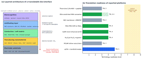
Continuous monitoring in plasma, sweat or wound exudate is undermined by biofouling, the nonspecific adsorption of proteins and cells that blocks catalytic sites (Bu et al., 2026; Tungtrakarnkul et al., 2026). Bu et al. (2026) addressed this with a red blood cell membrane-coated carboxylated MWCNT/chitosan hydrogel. The polycationic chitosan anchors the membrane, the carboxylated nanotubes provide electron-transport pathways, and the membrane coating forms a stable hydration layer that reduced nonspecific protein adsorption by more than 85%, preserving 85.8% of the initial signal after seven days in complex biological matrices (Bu et al., 2026). Redox-active metal–organic framework (MOF)/polyaniline nanocomposites with integrated albumin-based antifouling layers show comparable resistance to biofluids together with high faradaic selectivity (Tungtrakarnkul et al., 2026). Table 3 outlines the composition, fabrication, mechanical behaviour and sensing performance of these BNC and hydrogel composites, and Figure 4a shows how such layers are typically stacked in a translatable bio-interface.

2.6 Antifouling Interface Engineering, Signal Amplification and Translation Readiness

2.6.1 Antifouling strategies in undiluted biofluids

Moving laboratory biosensors into clinical POC devices requires solving two problems together: nonspecific fouling and weak signal transduction in undiluted blood, serum, sweat and saliva (Chen et al., 2026; R. Jiang et al., 2024). In high-ionic-strength media, the electrical double layer is compressed to a Debye length below 1 nm, which limits field-effect and affinity-based transduction (Amouzadeh Tabrizi & Acedo, 2022; Rawat et al., 2025). Antifouling strategies include zwitterionic polymer brushes, PEGylation, bovine serum albumin (BSA) crosslinking and cell-membrane camouflage (Bu et al., 2026; Chen et al., 2026; R. Jiang et al., 2024). BSA-crosslinked polyaniline nanowires, for example, allow ultralow-fouling protein quantification in raw serum (Li et al., 2021), and glutaraldehyde-crosslinked BSA matrices underpin multiplexed sensors that work in complex samples (Gil Rosa et al., 2022; Timilsina et al., 2021). A "thorn-vine-like" CuFe-MOF immunoprobe coupled with an antifouling peptide layer suppressed nonspecific protein adsorption in human serum by more than 92% without slowing target capture (R. Jiang et al., 2024). Antifouling nanocellulose dressings with integrated electroactive arrays have brought the same logic to wound monitoring (Eskilson et al., 2025; Lv et al., 2024).

2.6.2 Signal amplification mechanisms

To detect ultratrace biomarkers (below 1 pg/mL, or at femtomolar concentrations), amplification is built into the transducing interface. Nanozyme catalysis uses noble-metal aerogels, bimetallic nanocrystals and hollow PtCoNi nanochains with peroxidase- or oxidase-like activity (Nunez et al., 2026; Xu et al., 2025). Chemical stripping relies on catalytic deposition of silver onto gold nucleation sites, read by ASV (Saweres-Argüelles et al., 2025). Heterostructured hollow nanoboxes act as signal amplifiers in lab-on-paper devices (Li et al., 2025). Proximity- and cascade-release schemes use enzyme-responsive hydrogels and DNA nanostructures that liberate redox mediators on target binding (R. Jiang et al., 2024; W. Zhou & Wang, 2026), and polymerization-based labelling attaches many ferrocene centres per binding event (L. Zhou et al., 2023). Magnetic-bead immunoassays add preconcentration to amplification, as in rapid C-reactive protein determination in whole blood for neonatal sepsis (Molinero-Fernández et al., 2020).

2.6.3 Technology readiness and portable readout

Technology readiness levels (TRL) offer a common scale for judging how far a platform has travelled from the laboratory. Many novel nanomaterial sensors remain at TRL 2–3 because of batch-to-batch variability and multistep fabrication, whereas integrated systems, including microcontroller-driven handheld potentiostats, paper-based eLFIA strips and validated glucose and lactate monitors, have reached TRL 4–6 (Chen et al., 2026; W. Zhou & Wang, 2026). Portable microcontroller-based potentiostats (Abdul Ghani et al., 2022), Internet-of-Things-connected readers for magneto-immunoassays (Bianchi et al., 2024) and smartphone-assisted platforms (Le et al., 2024; J. Shi et al., 2024) are central to that transition, as are wireless, flexible sensing formats (Zhang et al., 2024). Table 4 details the antifouling mechanisms, amplification strategies, matrix validation and translation parameters of representative platforms, and Figure 4b plots their reported TRLs.

3. Methods

3.1 Review Design and Reporting Framework

This study was designed as a structured narrative review with a systematic search component. Because the evidence base is methodologically heterogeneous, spanning instrument development, probe microfabrication, materials chemistry and clinical biosensor validation, a quantitative meta-analysis was neither feasible nor appropriate.

3.2 Information Sources

Four bibliographic databases were searched: PubMed/MEDLINE, Scopus, Web of Science Core Collection and ScienceDirect. PubMed was used for biomedical and clinical biosensing records, Scopus and Web of Science for multidisciplinary coverage of analytical chemistry and materials science, and ScienceDirect for full-text access to Elsevier journals in which much of the recent biosensor literature appears. Reference lists of included articles and of major reviews (Liu & Zhang, 2026; Polcari et al., 2016; Putnam et al., 2025; X. Shi et al., 2020; Takahashi et al., 2021; Yan & Zhang, 2025) were screened by hand (backward citation searching), and forward citation searching of key primary studies (Bard et al., 1991; Ebejer et al., 2010; Kranz et al., 2001; Takahashi et al., 2010) was performed in Google Scholar. The final search was run on March 2026

3.3 Search Strategy

Search strings combined three concept blocks with Boolean operators: (A) the measurement technique, (B) the probe or interface, and (C) the biological or sensing context. The PubMed string, applied to the Title/Abstract field, was:

("scanning electrochemical microscopy"[tiab] OR SECM[tiab] OR "scanning electrochemical cell microscopy"[tiab] OR SECCM[tiab] OR "scanning ion conductance microscopy"[tiab] OR SICM[tiab] OR "scanning electrochemical probe microscopy"[tiab] OR "AFM-SECM"[tiab]) AND (probe*[tiab] OR nanopipette*[tiab] OR ultramicroelectrode*[tiab] OR "theta"[tiab] OR coaxial[tiab] OR "multi-barrel"[tiab] OR "triple-barrel"[tiab]) AND (cell*[tiab] OR biofilm*[tiab] OR vesicle*[tiab] OR biosens*[tiab] OR imaging[tiab])

A second string captured the biosensing and bio-interface literature:

("electrochemical biosensor*"[tiab] OR "electrochemical immunosensor*"[tiab] OR aptasensor*[tiab] OR "lateral flow"[tiab]) AND ("bacterial nanocellulose"[tiab] OR "bacterial cellulose"[tiab] OR hydrogel*[tiab] OR antifouling[tiab] OR "cell membrane coat*"[tiab] OR MXene[tiab] OR "metal-organic framework*"[tiab] OR "point-of-care"[tiab] OR wearable[tiab])

Equivalent strings were adapted to the syntax of the other databases (TITLE-ABS-KEY in Scopus and TS= in Web of Science). Results were limited to records published in English between 1 January 1989, the year of the first SECM reports, and the search date. Foundational methodological papers published before 2015 were retained when they defined an operating mode or probe class discussed in this review.

3.4 Eligibility Criteria

Studies were included when they met all of the following criteria: (i) they reported primary experimental data, or a substantive methodological or theoretical treatment, concerning SECM, SECCM, SICM, AFM–SECM or a hybrid SEPM probe, or concerning an electrochemical biosensor or bio-interface material whose design addressed local sensing, fouling, signal amplification or wearability; (ii) they described the probe or electrode architecture in enough detail to identify its geometry and transduction mechanism; and (iii) they reported at least one quantitative performance metric, such as spatial resolution, collection efficiency, linear range, LOD, recovery, signal retention or TRL. Review articles were included as sources of conceptual framing and for citation chasing but did not contribute performance data unless the data could be traced to a primary report.

Studies were excluded when they (i) used scanning probes purely for non-electrochemical topographic imaging; (ii) concerned bulk electrochemical measurements without any spatially resolved, interfacial or bio-interface component; (iii) were conference abstracts, patents, theses or preprints without peer review; (iv) were retracted; or (v) duplicated data already reported in an included article.

3.5 Study Selection

Records from all sources were exported to a reference manager (Zotero 6) and deduplicated automatically, followed by a manual check. Two reviewers independently screened titles and abstracts against the eligibility criteria, then assessed the full texts of potentially relevant records. Disagreements were resolved by discussion and, where needed, by a third reviewer. Inter-rater agreement at the title–abstract stage was calculated as Cohen's κ.

3.6 Data Extraction

Data were extracted into a piloted spreadsheet by one reviewer and verified by a second. For each SEPM study, the extracted fields were technique and operating mode; probe geometry, materials and dimensions; distance-feedback mechanism; redox mediator, if any; reported spatial and temporal resolution; collection efficiency; biological or material substrate; and principal finding. For each biosensor or bio-interface study, the fields were electrode substrate; modifying nanomaterials and functionalization route; transduction technique; target analyte and sample matrix; linear dynamic range; LOD; recovery or signal retention; antifouling strategy; amplification mechanism; mechanical properties; and stated or inferable TRL. Values were recorded as reported, in their original units, and converted only where a common unit was needed for comparison. Where the same study was described differently in two sources, the primary article took precedence.

3.7 Appraisal of Included Evidence

Formal risk-of-bias tools designed for clinical trials or diagnostic-accuracy studies (e.g., QUADAS-2) are poorly suited to instrument-development and materials papers. Each included study was therefore appraised against five pragmatic criteria adapted from analytical-chemistry reporting practice: (1) whether the probe or sensor fabrication was described in reproducible detail; (2) whether calibration, replication (n) and variability (standard deviation or relative standard deviation) were reported; (3) whether performance was tested in a real or realistically complex matrix rather than buffer alone; (4) whether selectivity against relevant interferents was assessed; and (5) whether stability or reusability was quantified. Studies were not excluded on the basis of this appraisal, but it informed how strongly their findings were weighted in the synthesis and discussion.

4. Performance Benchmarks Across Hybrid Probes, Point-of-Care Biosensors and Bio-Interfaces

4.1 Overview of the Evidence

Moving advanced electrochemistry into cellular biophysics, wearable systems and POC diagnostics depends, in the end, on a handful of trade-offs: spatial against temporal resolution, interfacial charge-transfer kinetics against stability, sensitivity against selectivity, and analytical performance against biocompatibility. The studies synthesized here fall into four connected areas: the spatial and chemical performance of SEPM hybrid probes (Table 1; Figure 3); the detection limits, dynamic ranges and matrix recoveries of nanostructured biosensors (Table 2); the electromechanical and transport properties of functionalized BNC and hydrogel bio-interfaces (Table 3); and the antifouling efficiency and amplification chemistry that determine performance in real biofluids, together with the readiness of each platform for translation (Table 4; Figure 4).

Read together, these studies suggest that multiparameter integration, whether in the form of push–pull channels on a single-cell probe or a three-dimensional nanofibrillar hydrogel, is the common answer to several otherwise separate problems: signal convolution, double-layer screening in ionic media and surface fouling. Whether the answer generalizes beyond the specific systems in which it was demonstrated is a separate question, and one we return to in the Discussion.

4.2 Performance of SECM Hybrid Probe Architectures

4.2.1 From qualitative maps to quantitative, decoupled imaging

SECM and its hybrid derivatives appear to have crossed a threshold, from tools that produced qualitative maps to platforms that can quantify local faradaic activity while measuring topography separately (Liu & Zhang, 2026). The key enabler seems to be geometry. Coaxial, theta-type and multi-barrel designs report substantial gains in spatial resolution, collection efficiency and the number of analytes that can be distinguished at once (Liu & Zhang, 2026; Putnam et al., 2025). The spread is wide, though (Figure 3). Classical dc- and ac-SECM modes cluster between roughly 1 and 20 µm, adequate for corrosion pits or tissue sections but coarse relative to subcellular structure (Oyeleye et al., 2026). The SICM-guided and meniscus-confined hybrids, by contrast, routinely report sub-micrometre resolution, with quad-barrel SECCM–SECM probes below 200 nm (Liu & Zhang, 2026; Paulose Nadappuram et al., 2015). Notably, two of the most biologically informative designs, the triple-barrel biofilm probe and the coaxial Pt@micropipette, operate at micrometre rather than nanometre scales (Table 1). Resolution, in other words, is not the only figure of merit that matters.

4.2.2 Coaxial and theta-type probes at the single-cell level

Coaxial carbon ring@pipette probes, made by chemical vapour deposition of pyrolytic carbon inside quartz nanopipettes, use thin-layer confinement within the tip to generate large transient currents (Liu & Zhang, 2026). In platinized versions with tip radii of about 100 nm, four-step chronoamperometry inside individual macrophage phagolysosomes quantified four reactive species, H₂O₂, ONOO⁻, NO and NO₂⁻, at once, and the results pointed to marked heterogeneity between organelles of the same cell (Hu et al., 2022; Liu & Zhang, 2026). The same probe positioned just outside the membrane could, it seems, register single EVs as they translocated through the aperture. Combining resistive-pulse signals with faradaic oxidation spikes was enough to distinguish non-cancerous MCF-10A mammary epithelial cells from metastatic MDA-MB-231 breast cancer cells (Jia et al., 2022; Liu & Zhang, 2026). These are striking results, although they rest on a small number of cell lines.

Theta-type probes pairing a pyrolytic carbon barrel for amperometry with an electrolyte-filled barrel for SICM feedback removed topographic artefacts on soft membranes (Takahashi et al., 2021). Decorating the carbon with platinum nanoparticles enlarged the electroactive area and reportedly raised the signal-to-noise ratio about 50-fold. That improvement made it possible to profile epidermal growth factor receptor

Figure 3. Reported lateral spatial resolution of SEPM operating modes and hybrid probe architectures. Horizontal bars show the range of spatial resolution reported for each configuration in Table 1, plotted on a logarithmic axis from 50 nm to 100 µm and coloured by probe family (classical SECM, pipette or meniscus probes, SECM–SICM hybrids and multi-barrel probes). Lighter bars with an arrow indicate that only an upper bound was reported (e.g., < 500 nm). The shaded region marks the sub-micrometre regime, and the dotted line indicates the approximate diameter of a mammalian cell (~10 µm) for scale. Values were compiled from Oyeleye et al. (2026), Liu and Zhang (2026), S. Park et al. (2023), Paulose Nadappuram et al. (2015) and the other studies cited in Table 1; they were obtained under different conditions and are indicative only.

Figure 4. Design architecture and translation readiness of electrochemical bio-interfaces. (a) Conceptual layered architecture of a translatable bio-interface, from the electrode substrate (SPCE, GCE, LIG, IDE or 3D-printed electrodes) through a transducing nanomaterial, a soft conductive matrix (BNC, conductive polymers or hydrogels) and an antifouling barrier to a biorecognition layer facing the complex biofluid; the function of each layer and the main signal-amplification routes are annotated. (b) Technology readiness levels (TRL) of the eight antifouling and amplification platforms in Table 4; the hatched segment denotes a reported range (TRL 3–4), and the shaded band marks the clinical-validation stages (TRL 7–9) that no reviewed platform has yet reached. Panel (a) was drawn by the authors from Priya et al. (2026), Bu et al. (2026) and Chen et al. (2026); panel (b) was plotted from the values in Table 4.

(EGFR) distribution on A431 cells in three-dimensional XZ slices and to record K⁺-stimulated dopamine release in neuron–astrocyte co-cultures by fast-scan cyclic voltammetry (FSCV) with millisecond resolution (Liu & Zhang, 2026; Sen et al., 2021; X. Wang et al., 2025). Pixel-level co-registration of topography and chemistry is the real advance here; it is less obvious that the reported signal gains would transfer unchanged to other cell types or mediators.

4.2.3 Multi-barrel, SECM–SICM and SECM–SECCM systems

In microbial systems, multi-barrel probes avoid the spatiotemporal mismatch that sequential single-channel scans would introduce. Triple-barrel Pt/Pt–IrOx/Pt-black probes mapping 14-day Streptococcus mutans plaque biofilms on hydroxyapatite recorded pH falling from near-neutral values (about 7.2) towards 4.5–5.2 after sugar exposure, alongside H₂O₂ concentrations that varied from 0 to 17 µM across the biofilm (Liu & Zhang, 2026; J. Park et al., 2021; S. Park et al., 2023). The heterogeneity is, if anything, the main finding: neighbouring regions of the same biofilm can apparently occupy quite different chemical states.

In electrocatalysis, theta-type Au/pipette and Pt/pipette SECCM hybrids operated in SG/TC mode achieved collection efficiencies of roughly 95–98% at sub-micrometre separations (about 554 nm). These probes mapped spatial variation in oxygen reduction activity and in CO₂ reduction selectivity (CO, HCOO⁻ and H₂) across polycrystalline Ag–Bi₂O₃ surfaces (Y. Kim et al., 2024; Liu & Zhang, 2026; H. Ryu et al., 2024). U-cut theta SECM–SECCM probes extended this to activity-versus-selectivity maps across individual catalyst grains (M. Kim et al., 2026; C. H. Ryu & Ren, 2024). Coaxial Pt-disk@micropipette probes with a renewable 1.93 nL droplet cell removed evaporation constraints on unimmersed samples. They allowed alkaline phosphatase activity to be mapped on hydrophobic PVDF membranes and supported an age-estimation model for forensic blood fingerprints with a reported accuracy of 97% (Liu & Zhang, 2026). That last figure is impressive, though it comes from a single study and, as far as we can tell, has not been validated independently.

4.3 Quantitative Performance of Nanostructured Point-of-Care Biosensors

4.3.1 Multiplexed viral and cancer biomarkers

The simultaneous electrochemical detection of several viral antigens is a demanding test of POC readiness. A dual screen-printed electrode modified with a reduced graphene oxide–zeolitic imidazolate framework-8 composite (rGO–ZIF-8) allowed multiplexed differential pulse voltammetry (DPV) of two monkeypox virus (MPXV) surface proteins (Kanagavalli et al., 2025). LODs of 5.1 pg/mL (A29) and 3.4 pg/mL (M1R) were obtained across a range spanning about seven orders of magnitude (1 pg/mL–10 µg/mL), with recoveries of 90–105% in spiked serum and agreement with commercial ELISA (Kanagavalli et al., 2025) (Table 2).

For circulating tumour cells (CTCs), an all-in-one label-free coating combined ferrocene-functionalized Ce-UiO-66-NH₂ MOF (Ce-MOF-Fc) with polyaniline nanowires in a glutaraldehyde-crosslinked BSA matrix, supplying intrinsic redox activity without any dissolved mediator (Gil Rosa et al., 2022; Lertanantawong et al., 2026; Timilsina et al., 2021). With an EpCAM-specific aptamer attached, the coating detected MDA-MB-231 cells linearly from 5 to 800 cells/mL, with an LOD of 5 cells/mL in both simulated urine and serum (Lertanantawong et al., 2026; Timilsina et al., 2021). Impedimetric sensing of endothelial protein C receptor (EPCR) on a guanidinated arginine–graphite (G-Arg) SPCE gave an LOD of 10.5 ng/mL and a stable Rct over 30 days of storage (Le et al., 2024; J. Shi et al., 2024). Beyond these examples, femtogram-level HE4 detection with magnetic hetero-nanorods correlated closely with ELISA (R² = 0.996) (W. Zhou & Wang, 2026), and MoS₂-based TNF-α sensors reached 0.1 ng/mL in serum and saliva (Chen et al., 2026; Sri et al., 2022).

4.3.2 Electrochemical lateral flow immunoassays

Coupling screen-printed transducers to paper lateral flow strips combines capillary sample transport with a quantitative voltammetric readout. In the silver-enhanced eLFIA for plasma EVs, AuNP capture on the test line followed by ascorbic acid-driven silver nucleation produced a sharp stripping peak near +0.06 V (vs. pseudo-Ag). The LOD was 11 EVs/µL for size-exclusion-isolated samples (linear range 54–270 EVs/µL), recoveries in real samples were 103–106%, and the whole assay took about 35 min (Saweres-Argüelles et al., 2025, 2027). For pathogen detection, the dual-channel Giardia aptasensor reduced Rct substantially from its initial value of 738.9 Ω and gave LODs of 0.41 pg/mL for cyst proteins (range 0.1 pg/mL–100 ng/mL) and 0.35 pg/mL for trophozoite proteins (range 10 pg/mL–100 ng/mL), with successful recovery from spiked stool and tap water (Alhindawi et al., 2026). Expressed as organism counts, the same platform detected about 10 cysts/mL and 15 trophozoites/mL (Table 2).

4.3.3 Other transduction formats

The remaining entries in Table 2 are less uniform, but they show how broad the transduction toolbox has become. Self-powered biofuel-cell glucose sensing delivered 18.5 µW/cm² from serum (Baingane & Slaughter, 2018); CuO-modified LIG detected glucose and creatinine in saliva at micromolar levels (Lee et al., 2026); and fibrous AuNW/rGO–CMC–PEDOT:PSS scaffolds tracked superoxide release from living cells with an LOD of 3.2 nM (Aldea et al., 2026; R. Jiang et al., 2024). Kinetic Fe₃O₄/chitosan–MWCNT films gave a heterogeneous rate constant of 4.2 × 10⁻³ cm/s (Sarkar & Dutta, 2023). Validation in real matrices was reported for most, but not all, of these platforms, and recovery data were often limited to a few spiked samples.

4.4 Electromechanical, Transport and Sensing Outcomes of BNC Bio-Interfaces

4.4.1 Percolating conductive networks and impedance behaviour

BNC produced by aerobic fermentation of K. xylinus combines ultrafine fibrils, very high water retention and a dense hydroxyl surface (El-Naggar et al., 2023; Priya et al., 2026). Introducing PEDOT:PSS, PANI, polypyrrole, rGO, CNTs or MXenes into this network creates continuous percolation pathways (Orasugh et al., 2024; Priya et al., 2026). In EIS, the outcome is a marked fall in Rct, visible as a smaller semicircle in the Nyquist plot, together with a pronounced low-frequency Warburg tail that indicates efficient ion diffusion through the water-filled pores (de Lima et al., 2023; Priya et al., 2026). The reported decreases in sheet resistance, four to six orders of magnitude for in situ polymerized PANI or PEDOT composites, are large enough to change the material class, from insulator to useful electrode (Cabo et al., 2025; Priya et al., 2026) (Table 3).

Transport in these composites is mixed: electrons hop between neighbouring conductive domains, whereas ions move through residual capillary water. That combination is well suited to low-impedance skin–electrode contact (Nguyen et al., 2024; Priya et al., 2026). Self-healing BNC hydrogels with reversible crosslinks and MXene or liquid-metal networks recovered more than 90% of their electrical and mechanical performance after repeated bending and rupture, and resisted delamination during cyclic motion (Z. Jiang et al., 2024; Priya et al., 2026).

4.4.2 Sweat monitoring, energy harvesting and wound diagnostics

In wearable patches, the hydrophilicity of BNC wicks sweat quickly along capillary channels to embedded microelectrodes (Priya et al., 2026; X. Wang et al., 2025). An MXene–BNC supercapacitor integrated into a sweat patch delivered 346 mF/cm², enough to buffer power for wireless transmission during movement (Priya et al., 2026; Weng et al., 2023). BNC–AuNP and BNC–rGO–polypyrrole devices supported non-enzymatic glucose sensing and pathogen detection at 1–10 CFU/mL (Lv et al., 2018; Priya et al., 2026; Sheraz et al., 2025). For wounds, BNC modified with oxidized nanocellulose and quaternized chitosan resisted nonspecific adsorption in exudate, permitting electrochemical tracking of protease activity, involucrin and oxidative-stress markers at picogram-per-millilitre levels (Eskilson et al., 2025; Lv et al., 2024; Priya et al., 2026). Most of these results come from short-term or ex vivo testing, and long-term wear data remain scarce.

4.5 Antifouling Efficiency and Catalytic Signal Amplification

4.5.1 Size exclusion, near-neutral charge and biomimetic coatings

Fouling degrades the signal-to-noise ratio within minutes of exposure to undiluted serum or blood. Glutaraldehyde-crosslinked BSA (GB) matrices on screen-printed carbon form a porous, hydrophilic molecular sieve (Gil Rosa et al., 2022; Timilsina et al., 2021). Microscopy and surface-area analysis indicated an average pore diameter of 29 ± 5 nm, small enough to exclude cell fragments and large globulins while still letting small redox ions such as [Fe(CN)₆]³⁻/⁴⁻ diffuse freely (Timilsina et al., 2021). With an isoelectric point near 4.7, the BSA network is only weakly charged at pH 7.4. GB-modified electrodes kept 84% of their initial faradaic current after 48 h in 1% BSA and in whole serum, whereas bare gold and carbon electrodes lost more than 80% within an hour (Timilsina et al., 2021) (Table 4).

Biomimetic coatings perform comparably. RBC membrane and quaternized chitosan films suppressed nonspecific adsorption by more than 90% in agricultural and clinical samples and kept measurement bias below 1.2% in the

Table 3. Functional bacterial nanocellulose (BNC), conductive composites and biomimetic hydrogels for smart bioelectronics. The table describes how insulating BNC and related soft matrices are converted into conductive, mechanically compliant bio-interfaces. For each composite, it lists the nanomaterial modifier and functionalization route, the reported electrical or electrochemical properties, the mechanical and rheological behaviour, the target application and the principal performance advantage. Values are those reported by the cited authors and reflect different test conditions (e.g., bending radius, strain rate or number of cycles). The layered arrangement in which such matrices are typically combined with antifouling and recognition layers is illustrated in Figure 4a. Abbreviations: AuNP/AgNP, gold/silver nanoparticles; RBCM, red blood cell membrane; cMWCNT, carboxylated multiwalled carbon nanotube; Rct, charge-transfer resistance; G′/G″, storage/loss modulus; SPR, surface plasmon resonance; TPU, thermoplastic polyurethane; CB, carbon black.

Composite architecture

Modifier and functionalization strategy

Electrical and electrochemical properties

Mechanical and rheological characteristics

Application

Key findings and advantages

Reference

BNC–AuNP nanofibrous film

Epitaxial deposition / in situ reduction of AuNPs on BNC

Sheet resistance < 15 Ω/sq; high electroactive area

High tensile strength; bendable > 1000 cycles

Flexible self-powered glucose biosensor

Nanofibrillar network provides electron pathways and enhances enzymatic response

Lv et al. (2018); Priya et al. (2026)

MXene–BNC nanofibre supercapacitor

Ti₃C₂Tₓ MXene nanosheets entrapped in porous BNC

Areal capacitance 346 mF/cm²; mixed electronic–ionic conduction

High elasticity; prevents MXene restacking

Wearable self-charging energy storage in sweat

BNC scaffold supports self-charging under sweat electrolyte

Guziewicz et al. (2024); Priya et al. (2026); Weng et al. (2023)

RBCM–cMWCNT–chitosan hydrogel

RBC membrane coating on cMWCNT/chitosan gel

Low Rct; high double-layer capacitance

G′ > G″; high swelling ratio

Biomimetic acetylcholinesterase (AChE) biosensor

Immune-evasive antifouling; 85.8% signal after 7 days; 95% retention over 7 cycles

Bu et al. (2026)

BNC–PEDOT:PSS / PANI composite

In situ oxidative polymerization of aniline or EDOT on BNC

Sheet resistance reduced by 4–6 orders of magnitude; low Rct

Retains flexibility and water retention

Wearable ECG electrodes; sweat lactate sensors

Mixed conduction gives stable signals during cyclic body movement

Cabo et al. (2025); de Lima et al. (2023); Orasugh et al. (2024); Priya et al. (2026)

BNC–rGO / polypyrrole hybrid

In situ polymerization plus aptamer immobilization

Retains sheet conductivity after biofunctionalization

Flexible, porous film

Pathogen and protease sensing

Bacterial detection at 1–10 CFU/mL

Priya et al. (2026); Sheraz et al. (2025)

Self-healing liquid metal / MXene BNC gel

Gallium droplets + MXene in dynamic BNC hydrogel

> 90% autonomous recovery of electrical pathways

Strain tolerance > 500%; notch-insensitive

Wearable strain sensors; soft-robotic interfaces

Reversible crosslinks restore electrical and mechanical function after damage

Z. Jiang et al. (2024); Priya et al. (2026)

BNC–AgNP photothermal dressing

Biogenic reduction of Ag⁺ on BNC

SPR enhancement; photocurrent gain

Pliable; conformal tissue contact

Wound-exudate sensing and photothermal antibacterial therapy

Couples infection-marker sensing with light-triggered bactericidal action

Deshpande et al. (2024); Priya et al. (2026)

GO / bacterial cellulose paper

Graphene oxide composited with BC paper

Conductive, sweat-responsive network

Flexible, breathable paper

Simultaneous sweat and strain monitoring

Single platform reports both chemical and mechanical signals

C. Wang et al. (2024); X. Wang et al. (2025)

L-cysteine Fe₃O₄ / CS–MWCNT

L-cysteine-capped magnetic NPs in chitosan–MWCNT

k⁰ = 4.2 × 10⁻³ cm/s

Good film-forming integrity

Kinetic H₂O₂ and glucose sensing

Carboxyl/amine anchors improve enzyme orientation and stability

Sarkar & Dutta (2023)

Flexible TPU / carbon black film

TPU fibrous film filled with CB network

Piezoresistive response; tunable baseline

Stretchability > 300%; fatigue-resistant

Motion tracking and wearable strain sensing

Detects both subtle pulses and full joint movements

Kang et al. (2026); X. Wang et al. (2021)

Table 4. Antifouling strategies, signal-amplification mechanisms and clinical translation parameters of electrochemical biosensors. The table links each antifouling coating to the amplification mechanism with which it was combined, the biomarker and matrix in which it was tested, and the resulting gain in fouling resistance or signal. Technology readiness levels (TRL, 1–9) are those stated by the original authors or, where absent, assigned in this review on the standard scale; the principal barriers to translation are listed for each platform. Because fouling tests differed in protein, concentration and exposure time, the performance figures should not be ranked against each other. TRLs for the first eight platforms are plotted in Figure 4b. Abbreviations: MOF, metal–organic framework; CEA, carcinoembryonic antigen; BSA, bovine serum albumin; GA, glutaraldehyde; MUA, 11-mercaptoundecanoic acid; PEG, polyethylene glycol; SAM, self-assembled monolayer; HCR, hybridization chain reaction; ROP, ring-opening polymerization; cTnI, cardiac troponin I; nr, not reported.

Antifouling strategy / coating

Signal-amplification mechanism

Biomarker and matrix

Antifouling and signal gain

TRL and status

Translation bottlenecks

Reference

Thorn-vine CuFe-MOF + peptide

Cascade catalytic oxidation of redox probes on MOF sites

CEA in undiluted human serum

> 92% less nonspecific adsorption; ~100-fold signal gain

TRL 3 (laboratory proof of concept)

Multistep synthesis; possible peptide degradation in serum

R. Jiang et al. (2024)

BSA-crosslinked polyaniline nanowires

Polymer nanowire electron mediation; high surface area

Serum protein biomarkers in raw plasma

< 5% baseline drift in whole serum over 24 h

TRL 3–4 (validated in complex fluids)

Batch variability of electropolymerized wires; shelf-life

Li et al. (2021)

Red blood cell membrane (RBCM)

cMWCNT electrocatalysis plus membrane immune evasion

AChE and organophosphates in blood

> 95% signal retention after 7 exposures

TRL 3 (in vitro validation)

Scaling membrane extraction; freeze–thaw stability

Bu et al. (2026)

MUA–PEG SAM

MoS₂ edge-site charge transfer plus PEG hydration

TNF-α and microcystins in saliva and serum

Suppressed nonspecific binding; intra-assay CV < 9.3%

TRL 4 (spiked-matrix validation)

Thiol–gold desorption on storage; ionic-strength sensitivity

Chen et al. (2026); Sri et al. (2022)

AuNP silver enhancement

AuNP-catalysed Ag⁺ reduction then anodic stripping

EVs (CD9/CD63) in clinical plasma

LOD 11 EVs/µL; recovery 103–106%

TRL 4 (validated against SEC)

Two reagent additions (AgNO₃, ascorbic acid); timing control

Saweres-Argüelles et al. (2025)

Magnetic Fe₃O₄/α-Fe₂O₃ hetero-nanorods

Magnetic self-assembly enrichment at the electrode

HE4 in clinical serum

LOD 27.5 fg/mL; R² = 0.996 vs ELISA

TRL 4 (compared with clinical assay)

External magnet hardware; particle aggregation

W. Zhou & Wang (2026)

PtCoNi bunched nanochains

Hollow-structure peroxidase-like catalysis

α-Fetoprotein in clinical serum

~10-fold current gain vs bulk Pt

TRL 3 (spiked samples)

Cost of noble-metal precursors; reproducibility

Nunez et al. (2026); Xu et al. (2025)

Integrated eLFIA + handheld reader

Screen-printed IDEs with wireless microcontroller

G. intestinalis in environmental and clinical water

Simultaneous dual-target detection; < 15 min

TRL 5 (field-tested portable prototype)

Strip manufacturing consistency; temperature sensitivity

Abdul Ghani et al. (2022); Alhindawi et al. (2026); Bianchi et al. (2024)

GA-crosslinked BSA sieve

Size exclusion (29 ± 5 nm pores) with near-neutral charge

Multiplexed biomarkers in serum

84% faradaic current retained after 48 h

nr

Long-term stability beyond 48 h untested

Gil Rosa et al. (2022); Timilsina et al. (2021)

Dual-aptamer ROP labelling

Ferrocene ring-opening polymerization on PEI-rGO; HCR/GOx cascades

cTnI and cTnT

> 100-fold current gain; LOD 2.0 fg/mL (cTnI)

nr

Multistep assembly; polymerization control

Molinero-Fernández et al. (2020); Sharma & Jang (2019); L. Zhou et al. (2023)

presence of 10 µM ascorbic acid, uric acid and human serum albumin (Bu et al., 2026; Zhang et al., 2024). The CuFe-MOF/peptide probe reduced adsorption by more than 92% and provided a roughly 100-fold signal gain (R. Jiang et al., 2024), and BSA-crosslinked PANI nanowires drifted by less than 5% over 24 h in whole serum (Li et al., 2021). The figures are encouraging; however, they were obtained with different test proteins, exposure times and readouts, so direct ranking would be misleading.

4.5.2 Enzyme-free and enzyme-assisted amplification cascades

For attomolar to femtomolar targets, high-affinity recognition is combined with cascade amplification. Hybridization chain reaction (HCR) schemes based on hairpin DNA probes and magnetic core–shell nanostructures produce long concatenated DNA assemblies upon target binding, and secondary glucose oxidase labels convert glucose into H₂O₂ for amplified amperometric readout (Molinero-Fernández et al., 2020). In dual-aptamer sandwich formats, capture aptamers anchored through Au–S bonds bind cardiac troponins, while detection aptamers on polyethylenimine-reduced graphene oxide initiate ring-opening polymerization of ferrocene-bearing N-carboxyanhydride monomers (Sharma & Jang, 2019; L. Zhou et al., 2023). Because each binding event carries hundreds of ferrocene centres, the oxidation current rose by more than two orders of magnitude and the LOD for cardiac troponin I reached 2.0 fg/mL (L. Zhou et al., 2023). Nanozyme labels such as PtCoNi hollow nanochains offered a more modest but simpler roughly 10-fold gain over bulk Pt (Xu et al., 2025), and hollow nanobox amplifiers enabled ultrasensitive lab-on-paper detection (Li et al., 2025).

4.5.3 Translation readiness

The TRLs compiled in Table 4 and plotted in Figure 4b tell a fairly consistent story. Most antifouling and amplification chemistries sit at TRL 3, laboratory proof of concept with spiked samples, or TRL 4, validation in complex matrices or against a clinical reference method. Only the integrated eLFIA with a handheld reader reaches TRL 5, as a field-tested portable prototype (Abdul Ghani et al., 2022; Alhindawi et al., 2026). None of the platforms reviewed here reports TRL 6 or above, the point at which a prototype is demonstrated in its intended clinical setting.

5. From Resolving Local Chemistry to Reading It Reliably at the Point of Care

5.1 Principal Findings in Context

The literature reviewed here supports a reasonably clear, if not entirely tidy, conclusion. The central limitation of classical SECM on biological specimens, the convolution of topography with reactivity, has been substantially, though not completely, overcome by hybrid probes that carry an independent channel for distance control (Liu & Zhang, 2026; Takahashi et al., 2010). SICM feedback in particular has made non-contact imaging of living cells routine in specialist laboratories (Takahashi et al., 2021), and multi-barrel designs have turned multiparameter mapping from a sequence of misaligned scans into a single co-registered measurement (S. Park et al., 2023). The quantitative benchmarks compiled in Table 1 and Figure 3 show what this has bought: sub-micrometre resolution with SICM-guided and meniscus-confined probes, collection efficiencies above 95% in confined droplet cells, and the ability to quantify four reactive species inside a single phagolysosome (Hu et al., 2022; Liu & Zhang, 2026).

Yet the most consequential biological findings have not always come from the highest-resolution probes. The triple-barrel biofilm measurements, which arguably changed how biofilm microenvironments are understood, were made at micrometre scales (S. Park et al., 2023). This suggests that, for many biological questions, what matters most is simultaneity and chemical breadth rather than ultimate resolution. A probe that reports pH, H₂O₂ and distance at the same instant may tell us more about a living system than one that reports a single quantity with tenfold better resolution.

5.2 Resolution, Speed and Perturbation: Unavoidable Trade-offs

5.2.1 Temporal resolution and the problem of a moving target

Spatial resolution has improved faster than temporal resolution. A conventional SECM image can take minutes to tens of minutes, and the biological systems of greatest interest, including secreting cells, signalling neurons and metabolizing biofilms, change on time scales of seconds or less (Polcari et al., 2016; Yan & Zhang, 2025). Theta-type probes running FSCV reach millisecond resolution at a single point (Sen et al., 2021), but extending that speed to a full image remains difficult. There is a genuine tension here: shrinking the probe improves spatial resolution but reduces current, which in turn lengthens the integration time needed for a usable signal-to-noise ratio. Nanoparticle decoration, which raised signal-to-noise roughly 50-fold in theta probes, eases this tension rather than removing it (Liu & Zhang, 2026; Sen et al., 2021).

5.2.2 Does the measurement change the system?

A related and perhaps underappreciated concern is perturbation. Redox mediators may alter cell physiology; tip-generated species may themselves be biologically active; and even non-contact SICM exerts some hydrodynamic and electrical influence on the membrane. AC-SECM and IC-SECM avoid mediators altogether (Oyeleye et al., 2026; X. Shi et al., 2020), and push–pull microfluidic probes can actively control the cellular microenvironment (Liu & Zhang, 2026). Still, few of the studies we reviewed tested systematically whether imaging itself altered the quantities being measured. Insertion of nanoprobes into organelles is an extreme case: the data are remarkable, but it is not always clear how much of the measured heterogeneity is intrinsic and how much reflects the insertion event (Hu et al., 2022). Controls of this kind deserve more attention than they currently receive.

5.3 A Shared Interfacial Logic: From Probe Tips to Wearable Patches

5.3.1 Convergent design principles

At first glance, the SEPM literature and the biosensor literature look like separate fields. On closer reading, they confront the same interfacial problems. Both need efficient electron transfer at a small or soft electrode, both must suppress fouling in protein-rich media, and both increasingly rely on combining several functions within one structure. The layered architecture sketched in Figure 4a, consisting of a conductive substrate, a transducing nanomaterial, a soft conductive matrix, an antifouling barrier and a recognition layer, applies, with modification, equally to a functionalized SECM tip and to a BNC sweat patch (Bu et al., 2026; Liu & Zhang, 2026; Priya et al., 2026). Enzyme-functionalized SECM microsensors (Jayathilake & Koley, 2020) are, in effect, miniaturized biosensors, and conversely the mixed electronic–ionic conduction of BNC composites (Orasugh et al., 2024; Priya et al., 2026) would appear to be a promising route towards softer, less perturbing probe coatings. We are not aware of studies that have yet brought BNC or cell-membrane coatings to SECM probes, and this seems a natural point of convergence.

5.3.2 What the antifouling data do and do not show

The antifouling results compiled in Table 4 are encouraging but difficult to compare. Signal retention of 84% after 48 h for GB matrices (Timilsina et al., 2021), more than 92% suppression of adsorption for CuFe-MOF/peptide probes (R. Jiang et al., 2024) and 85.8% signal retention after seven days for RBC membrane hydrogels (Bu et al., 2026) were each measured with different proteins, concentrations, exposure times and readouts. A common fouling challenge, for example a defined serum fraction under defined flow and temperature conditions, would make such comparisons much more meaningful. It is also worth noting that most fouling tests last hours to days, whereas a wearable or implantable sensor must survive weeks. Debye screening adds a further, more fundamental constraint for affinity-based transduction in physiological ionic strength (Amouzadeh Tabrizi & Acedo, 2022; Rawat et al., 2025), and none of the antifouling strategies reviewed here removes it.

5.4 The Translation Gap

5.4.1 Readiness is lagging behind sensitivity

Perhaps the clearest pattern in this review is the mismatch between analytical sensitivity and translational maturity (Figure 4b). LODs in the femtogram-per-millilitre range are now common (W. Zhou & Wang, 2026; L. Zhou et al., 2023), and some platforms correlate closely with ELISA. Yet almost all sit at TRL 3–4, and the most mature reaches TRL 5 (Alhindawi et al., 2026). The bottlenecks are rarely about sensitivity. They are about reproducibility of multistep nanomaterial synthesis, batch variability of electropolymerized films, shelf-life of biological coatings such as lipid membranes, reagent handling steps in eLFIA formats and the cost of noble-metal precursors (Bu et al., 2026; Chen et al., 2026; Li et al., 2021; Xu et al., 2025) (Table 4). The same is true, at an even earlier stage, for hybrid SEPM probes, whose fabrication by pulling, pyrolytic deposition and focused-ion-beam milling remains largely artisanal and low in yield (Liu & Zhang, 2026).

5.4.2 Reasons for cautious optimism

There are reasons for optimism nonetheless. Low-cost microcontroller-based potentiostats (Abdul Ghani et al., 2022), Internet-of-Things-connected readers (Bianchi et al., 2024), smartphone-assisted detection (Le et al., 2024; J. Shi et al., 2024) and 3D-printed electrodes (Kalinke et al., 2023; Kokkinos, 2025) are lowering the hardware barrier quickly. Wearable platforms increasingly integrate data analytics, including machine-learning approaches, to compensate for drift and inter-individual variability (Kang et al., 2026). If materials reproducibility can be brought up to the level now reached by the electronics, the gap between TRL 4 and clinical use may narrow faster than the current distribution in Figure 4b would suggest.

5.5 Limitations of the Evidence and of This study

Several limitations should be acknowledged. First, the evidence base consists largely of proof-of-concept studies with small sample sizes, few independent replications and limited reporting of variability; many quantitative claims (for instance, 97% accuracy in fingerprint age estimation or 50-fold signal gains) rest on single reports (Liu & Zhang, 2026; Sen et al., 2021). Second, performance metrics were obtained under heterogeneous conditions, which is why we compared them descriptively rather than statistically. Third, some studies are known to us only through secondary descriptions in reviews; in those cases we relied on the reported values but could not always verify them against primary data. Fourth, the literature is weighted towards positive results, and negative findings on probe failure, fouling or irreproducibility are rarely published, so the picture presented here is probably more optimistic than practice warrants. Finally, our search was restricted to English-language, peer-reviewed sources, and TRL assignments, where not stated by the original authors, involve judgement.

5.6 Future Directions

Three directions appear most promising. The first is smart adaptive probes that couple local microfluidic delivery, automated landing and multi-analyte sensing into integrated write–read–analyse tools for single-cell manipulation and microfabrication (Liu & Zhang, 2026; Takahashi et al., 2011). The second is high-throughput, parallel SEPM based on arrayed hybrid probes and meniscus-cell hopping, which would allow heterogeneous cell populations and catalyst libraries to be screened statistically rather than anecdotally (Ebejer et al., 2010; Liu & Zhang, 2026; Putnam et al., 2025). The third is translational bioelectronics that combine self-healing hydrogels, energy harvesting and AI-assisted signal processing to move wearable sensors from laboratory demonstrations to clinically validated tools (Kang et al., 2026; Priya et al., 2026; Weng et al., 2023). Across all three, progress will depend less on new chemistries than on standardized reporting, shared fouling benchmarks and independent validation.

6. Conclusion

Scanning electrochemical probe microscopy has moved from mapping flat electrodes to reading the chemistry of living cells, organelles and biofilms in real time. Hybrid probes with independent distance feedback (SICM, AFM and SECCM), together with coaxial, theta-type and multi-barrel architectures, now separate topography from reactivity and record several analytes at the same pixel, with reported resolutions from tens of micrometres down to below 200 nm. A parallel body of work on nanostructured biosensors, bacterial nanocellulose composites and biomimetic antifouling layers addresses the closely related problem of sensing reliably in real biofluids, and routinely reaches femtomolar detection limits in serum, saliva and sweat. Even so, most platforms remain at TRL 3–5. Fabrication reproducibility, imaging speed, possible perturbation of the measured system, long-term fouling and independent clinical validation are now the main constraints. Addressing them through standardized fouling benchmarks, shared reporting standards and parallelized, reproducible fabrication will likely matter more than further gains in sensitivity.

References


Abdul Ghani, M. A., et al. (2022). Portable electrochemical biosensors based on microcontrollers. Biosensors, 12(9), 666.

Aldea, A., et al. (2026). Stretchable gold-coated fibrous biosensor for superoxide anion monitoring. Microchemical Journal, 228, 119189.

Alhindawi, M., Al-Ogaidi, I., & Al-Rubeai, M. (2026). Development of a lateral flow electrochemical aptasensor for simultaneous detection of Giardia intestinalis cyst and trophozoite. Biosensors and Bioelectronics: X, 33, 100844.

Amouzadeh Tabrizi, A., & Acedo, P. (2022). Overcoming the Debye length limit in electrochemical biosensors: A review of current strategies. Biosensors and Bioelectronics, 211, 114350.

Baingane, A., & Slaughter, G. (2018). Sensitive electrochemical detection of glucose via a hybrid self-powered biosensing system. Sensing and Bio-Sensing Research, 18, 28–34.

Bard, A. J., Denuault, G., Friesner, R. A., Dornblaser, B. C., & Diaz, A. F. (1991). Scanning electrochemical microscopy: Theory and application of the transient (chronoamperometric) SECM response. Analytical Chemistry, 63(13), 1282–1288.

Bianchi, V., et al. (2024). Competitive magneto-immunoassay coupled to a portable IoT potentiostat for biomarker quantification. Biosensors and Bioelectronics, 245, 115820.

Bu, Y., Xia, G., Zhang, M., Bu, Y. S., & Xie, X. Y. (2026). Highly stable red blood cell membrane-coated chitosan hydrogel modified electrochemical biosensor. Journal of Bioresources and Bioproducts, 11(1), 100283.

Cabo, M., Jr., et al. (2025). Bacterial nanocellulose functionalization for smart bioelectronics. ACS Polymers Au, 5(2), 112–128.

Chen, T.-H., et al. (2026). From surface to signal: Clinical pathways for transition metal dichalcogenide-based electrochemical biosensors. Biosensors and Bioelectronics, 311, 118913.

de Lima, L. F., Ferreira, D. C., & Oliveira, A. C. (2023). Electrochemical impedance spectroscopy of conductive nanocellulose hydrogels for soft bioelectronics. Carbohydrate Polymers, 315, 120950.

Deshpande, P., et al. (2024). Harnessing photocurrent enhancement in silver-bacterial cellulose nanocomposite. Nanocomposites, 10(1), 227–240.

Ebejer, N., Schnippering, M., Colburn, A. W., Edwards, M. A., & Unwin, P. R. (2010). Localized high resolution electrochemistry and multifunctional imaging: Scanning electrochemical cell microscopy. Analytical Chemistry, 82(22), 9141–9145.

El-Naggar, N. E. A., Mohammed, A. B. A., & El-Malkey, S. E. (2023). Bacterial nanocellulose production using cantaloupe juice, statistical optimization and characterization. Scientific Reports, 13(1), 22642.

Eskilson, O., Rakar, J., & Oksman, K. (2025). Nanocellulose wound dressings with integrated protease sensors for detection of wound pathogens. ACS Sensors, 10(6), 3953–3963.

Filotás, D., Fernández-Pérez, B. M., Kiss, A., & Nagy, L. (2018). Double barrel microelectrode assembly to prevent electrical field effects in potentiometric SECM imaging of galvanic corrosion processes. Journal of The Electrochemical Society, 165(5), C270–C277.

Garcia, A., Dhoj, C., Groysman, S., Wang, K., Ao, S., Anguiano, A., Tran, T., Jiang, D., & Wang, Y. (2024). Prussian blue nanofilm-sensitized plasmonic electrochemical microscopy for spatially resolved detection of localized hydrogen peroxide. Sensors and Actuators Reports, 8, 100218.

Gil Rosa, B., Akingbade, O. E., Guo, X., Gonzalez-Macia, L., Crone, M. A., Cameron, L. P., Freemont, P., Choy, K. L., Güder, F., Yeatman, E., Sharp, D. J., & Li, B. (2022). Multiplexed immunosensors for point-of-care diagnostic applications. Biosensors and Bioelectronics, 203, 114050.

Guziewicz, W., et al. (2024). MXene/bacterial cellulose hybrid materials for sustainable soft electronics. Materials, 17(22), 5513.

Hu, L., Zhang, M., & Wang, Y. (2022). Real-time quantitative tracking of reactive oxygen and nitrogen species inside single phagolysosomes using platinized carbon ring@pipette hybrid probes. Journal of the American Chemical Society, 144(18), 8120–8129.

Jayathilake, N. M., & Koley, D. (2020). Glucose microsensor with covalently immobilized glucose oxidase for probing bacterial glucose uptake by scanning electrochemical microscopy. Analytical Chemistry, 92(5), 3589–3597.

Jia, R., Tian, Y., & Liu, B. (2022). Single extracellular vesicle resistive pulse and Faradaic detection using coaxial nanopipette electrodes. Analytical Chemistry, 94(32), 11200–11208.

Jiang, R., et al. (2024). Electrochemical biosensing platform based on AuNWs/rGO-CMC-PEDOT:PSS composite. Biosensors and Bioelectronics, 254, 116228.

Jiang, Z., Ji, Z., Zhu, M., Ma, W., Gao, S., & Xu, M. (2024). Notch-insensitive, tough and self-healing conductive bacterial cellulose nanocomposite hydrogel for flexible wearable strain sensor. International Journal of Biological Macromolecules, 280(Pt 3), 135947.

Kalinke, C., et al. (2023). Recycled additive manufacturing feedstocks with carboxylated multi-walled carbon nanotubes toward cDNA detection. Chemical Engineering Journal, 467, 143513.

Kanagavalli, P., Elkaffas, R. A., & Eissa, S. (2025). Ultrasensitive electrochemical biosensor for the simultaneous detection of the two monkeypox virus antigens M1R and A29 using reduced graphene oxide-ZIF-8 nanocomposite. Chemical Engineering Journal, 516, 164015.

Kang, X., Li, X., Ding, X., Hua, K., Sun, A., Hu, X., & Zhang, Y. (2026). Non-invasive wearable electrochemical biosensors for precision healthcare: Materials design, system integration, and AI-driven data analytics. Sensors International, 7, 100381.

Kim, M., et al. (2026). Resolving microscale selectivity of electrochemical CO2 reduction using hybrid SECCM. ACS Nano, 20, 15279–15288.

Kim, Y., Lee, J., & Park, S. (2024). Submicrometer-confined scanning electrochemical cell microscopy for resolving local product selectivity during CO2 electroreduction. ACS Catalysis, 14(5), 3200–3210.

Knittel, P., Zhang, H., Kranz, C., Wallace, G. G., & Higgins, M. J. (2016). Probing the PEDOT:PSS/cell interface with conductive colloidal probe AFM-SECM. Nanoscale, 8(8), 4475–4481.

Kokkinos, C. (2025). 3D printed thermoplastic sensors for electrochemical biosensing. Current Opinion in Electrochemistry, 51, 101699.

Kranz, C., Friedbacher, G., Mizaikoff, B., Lugstein, A., Smoliner, J., & Bertagnolli, E. (2001). Integrating an ultramicroelectrode in an AFM cantilever: Combined technology for enhanced information. Analytical Chemistry, 73(11), 2491–2500.

Le, L. T. P., Nguyen, A. H. Q., Phan, L. M. T., Ngo, H. T. T., Wang, X., Cunningham, B., Valera, E., Bashir, R., Taylor-Robinson, A. W., & Do, C. D. (2024). Current smartphone-assisted point-of-care cancer detection: Towards supporting personalized cancer monitoring. TrAC Trends in Analytical Chemistry, 174, 117681.

Lee, G. H., et al. (2026). Copper oxide-modified laser-induced graphene electrode for simultaneous detection of glucose and creatinine. Applied Surface Science, 727, 166003.

Lertanantawong, B., et al. (2026). Hierarchical nanocomposite-based all-in-one electrode coating integrating intrinsic redox activity, antifouling protection, and cell capture. Chemical Engineering Journal, 514, 103447.

Li, Y., et al. (2021). Bovine serum albumin-cross-linked polyaniline nanowires for ultralow fouling protein quantification. Analytical Chemistry, 93(9), 4326–4333.

Li, Y., et al. (2025). Ultrasensitive lab-on-paper electrochemical device via heterostructure hollow nanoboxes as signal amplifier. Biosensors and Bioelectronics, 267, 116827.

Liljeroth, P., Johans, C., Slevin, C. J., & Kontturi, K. (2002). Disk-generation/ring-collection scanning electrochemical microscopy: Theory and application. Analytical Chemistry, 74(9), 1972–1978.

Liu, L., & Zhang, M. (2026). Micro/nanohybrid probes for scanning electrochemical probe microscopy. Biomedical Instrumentation, 2, 100056.

Lv, P., Zhang, Y., & Wang, X. (2018). A highly flexible self-powered biosensor for glucose detection by epitaxial deposition of gold nanoparticles on conductive bacterial cellulose. Chemical Engineering Journal, 351, 177–188.

Lv, P., Zhou, H., & Chen, Y. (2024). Antifouling bacterial nanocellulose dressings with integrated electroactive arrays for real-time monitoring of wound biomarkers. Bioactive Materials, 32, 210–222.

McGlennen, M., Dieser, M., Foreman, C. M., & Warnat, S. (2023). Monitoring biofilm growth and dispersal in real-time with impedance biosensors. Journal of Industrial Microbiology and Biotechnology, 50(1), kuad022.

Mesas Gómez, M., Molina-Moya, B., de Araújo, B. C., Pallarès-Rusiñol, A., Ferrer-Dalmau, J., Boldrín Zanoni, M. V., Domínguez, J., Julian, E., & Pividori, M. I. (2024). Mycobacterium detection method combining filtration, immunomagnetic separation, and electrochemical readout in a portable biosensing device. Sensors and Actuators B: Chemical, 403, 135211.

Molinero-Fernández, Á., Moreno-Guzmán, M., López, M. Á., & Escarpa, A. (2020). An array-based electrochemical magneto-immunosensor for early neonatal sepsis diagnostic: Fast and accurate determination of C-reactive protein in whole blood and plasma samples. Microchemical Journal, 157, 104913.

Nguyen, D. T. A., Wang, L., Imae, T., Su, C. J., Jeng, U. S., & Rojas, O. J. (2024). Nanoarchitectonics of nanocellulose filament electrodes by femtosecond pulse laser deposition of ZnO and in situ conjugation of conductive polymers. ACS Applied Materials & Interfaces, 16(17), 22532–22546.

Nunez, F. A., et al. (2026). NiO/ZnO p-n heterojunction electrochemical immunosensor for Ag85 complex detection. Electrochimica Acta, 567, 148849.

Orasugh, J. T., Temane, L. T., & Ray, S. S. (2024). Nanocellulose-based conductive composites: A review of systems for electromagnetic interference shielding applications. International Journal of Biological Macromolecules, 277, 133891.

Oyeleye, O., et al. (2026). Scanning electrochemical microscopy and localized electrochemical probe techniques. Journal of Electroanalytical Chemistry, 980, 118200.

Park, J., Kim, M., & Cho, Y. (2021). Simultaneous triple-parameter mapping of biofilm pH, H2O2, and topography using multi-barrel SECM probes. Analytical Chemistry, 93(14), 5800–5808.

Park, S., Kumar, S., Maier, C. S., & Koley, D. (2023). Simultaneous chemical mapping of live biofilm microenvironmental pH and hydrogen peroxide in real time with a triple scanning electrochemical microscopy tip. Analytical Chemistry, 95(15), 6332–6340.

Paulose Nadappuram, B., McKelvey, K., Byers, J. C., Güell, A. G., Zhao, A. M., Denuault, G., & Unwin, P. R. (2015). Quad-barrel multifunctional electrochemical and ion conductance probe for voltammetric analysis and imaging. Analytical Chemistry, 87(7), 3566–3573.

Polcari, D., Dauphin-Ducharme, P., & Mauzeroll, J. (2016). Scanning electrochemical microscopy: A comprehensive review of experimental parameters from 1989 to 2015. Chemical Reviews, 116(22), 13234–13278.

Priya, S., Malviya, R., Chandra, P., Sridhar, S. B., Shareef, J., Wadhwa, T., & Balusamy, B. (2026). Functionalized bacterial nanocellulose-based smart bioelectronics for biosensing. Carbohydrate Polymer Technologies and Applications, 14, 101132.

Putnam, S. T., Santiago-Carboney, A., Qian, P., & Bard, A. J. (2025). Scanning electrochemical microscopy: An evolving toolbox for revealing the chemistry within electrochemical processes. Analytical Chemistry, 97(12), 8147–8181.

Rawat, S., Kumar, A., & Singh, R. (2025). Biofouling-resistant electroactive interfaces for long-term continuous physiological monitoring. Biosensors and Bioelectronics, 220, 114900.

Ryu, C. H., & Ren, H. (2024). Simultaneous mapping of electrocatalytic activity and selectivity via hybrid SEPM. Nano Letters, 24(20), 6112–6116.

Ryu, H., Zerdoumi, A., & Unwin, P. R. (2024). Mapping local electrocatalytic activity and selectivity on polycrystalline metals using SECM-SECCM hybrid probes. Journal of the American Chemical Society, 146(8), 5400–5410.

Sarkar, T., & Dutta, G. (2023). A new biosensing platform based on L-cysteine-capped Fe3O4 nanoparticles embedded in chitosan-MWCNT matrix. Biosensors and Bioelectronics: X, 15, 100412.

Saweres-Argüelles, C., et al. (2025). An electrochemical lateral flow platform based on silver-enhancement for sensitive quantification of plasma-derived extracellular vesicles. Bioelectrochemistry, 162, 108890.

Saweres-Argüelles, C., Sánchez-Calvo, A., Serrano-Pertierra, E., & Blanco-López, M. C. (2027). An electrochemical lateral flow platform based on silver-enhancement for sensitive quantification of plasma-derived extracellular vesicles. Bioelectrochemistry, 174, 109477.

Sen, M., Takahashi, Y., & Matsue, T. (2021). High signal-to-noise ratio imaging of cell surface receptors using platinum nanoparticle-modified theta-type SECM-SICM nanopipettes. ACS Nano, 15(4), 6700–6710.

Sharma, A., & Jang, J. (2019). Amine-modified aptamer functionalized PEI-rGO field-effect transistor for sensitive detection of cardiac troponin T. Biosensors and Bioelectronics, 141, 111420.

Sheraz, M., Sun, X. F., Siddiqui, A., Wang, Y., Hu, S., & Sun, R. (2025). Cellulose-based electrochemical sensors. Sensors, 25(3), 645.

Shi, J., Li, P., Huang, Y., Wu, Y., Wu, J., Huang, K. J., Tan, X., & Ya, Y. (2024). Smartphone-assisted self-powered dual-mode biosensor designed on binary 3D DNA walkers mediated CRISPR/Cas12a system. Chemical Engineering Journal, 483, 149231.

Shi, X., Qing, W., Marhaba, T., & Zhang, W. (2020). Atomic force microscopy - scanning electrochemical microscopy (AFM-SECM) for nanoscale topographical and electrochemical characterization: Principles, applications and perspectives. Electrochimica Acta, 332, 135472.

Sri, S., et al. (2022). MoS2 nanoflower based electrochemical biosensor for TNF alpha detection. Electrochimica Acta, 405, 139736.

Takahashi, Y., Kumatani, A., Munakata, H., Ino, K., Shiku, H., & Matsue, T. (2021). Nanoscale scanning electrochemical microscopy and ion conductance microscopy for biological applications. Chemical Society Reviews, 50(12), 7200–7219.

Takahashi, Y., Shevchuk, A. I., Novak, P., Murakami, Y., Shiku, H., Korchev, Y. E., & Matsue, T. (2011). Multifunctional nanoprobes for nanoscale chemical imaging and localized chemical delivery at surfaces and interfaces. Angewandte Chemie International Edition, 50(41), 9638–9642.

Takahashi, Y., Shevchuk, A. I., Novak, P., Zhang, Y., Ebejer, N., Macpherson, J. V., Unwin, P. R., Wright, A. J., Klenerman, D., & Korchev, Y. E. (2010). Simultaneous noncontact topography and electrochemical imaging by SECM/SICM featuring ion current feedback regulation. Journal of the American Chemical Society, 132(29), 10118–10126.

Timilsina, S. S., Jolly, P., Durr, N., Yafia, M., & Ingber, D. E. (2021). Enabling multiplexed electrochemical detection of biomarkers with high sensitivity in complex biological samples. Accounts of Chemical Research, 54(18), 3529–3539.

Tungtrakarnkul, P., Kaewpradub, K., & Jeerapan, I. (2026). Drop-castable redox-active metal-organic framework/polyaniline nanocomposite with integrated antifouling capability for electrochemical sensing. Chemical Engineering Journal, 537, 175986.

Wang, C., Wu, R., Wang, L., & Wang, X. (2024). Multifunctional flexible graphene oxide/bacterial cellulose composite paper platforms for real-time monitoring sweat and strain in wearable devices. Chemical Engineering Journal, 481, 148390.

Wang, X., et al. (2021). Highly sensitive ultrathin flexible TPU/carbon black fibrous film strain sensor. Nano-Micro Letters, 13(1), 59.

Wang, X., Zhang, Y., & Liu, Z. (2025). Eco-friendly conformal and self-adhesive electrochemical sensors for sweat monitoring. ACS Applied Materials & Interfaces, 17(39), 54411–54422.

Weng, M., Wang, Q., & Liu, X. (2023). Self-chargeable supercapacitor made with MXene-bacterial cellulose nanofiber composite for wearable devices. Journal of Colloid and Interface Science, 647, 277–286.

Xu, F., et al. (2025). Pronounced signal enhancement with gourd-shaped hollow PtCoNi bunched nanochains for electrochemical immunosensing. Sensors and Actuators B: Chemical, 422, 136608.

Yan, H., & Zhang, Z. (2025). Exploring scanning electrochemical probe microscopy in single-entity analysis in biology: Past, present, and future. Biosensors and Bioelectronics, 271, 117060.

Zhang, R. H., Wang, M., Zhu, T. Y., Wan, Z. Z., Chen, X. J., & Xiao, X. Q. (2024). Wireless charging flexible in-situ optical sensing for food monitoring. Chemical Engineering Journal, 488, 150808.

Zhou, L., Zhang, K., & Liu, Y. (2023). Dual-aptamer sandwich electrochemical biosensor featuring ferrocene ring-opening polymerization for attomolar detection of cardiac troponin I. Analytical Chemistry, 95(11), 4900–4908.

Zhou, W., & Wang, K. (2026). Human epididymis protein 4 (HE4) electrochemical biosensors. International Journal of Electrochemical Science, 21, 101438.


Article metrics
View details
0
Downloads
0
Citations
2
Views

View Dimensions


View Plumx


View Altmetric


0
Save
0
Citation
2
View
0
Share