2.1 From Ensemble Averages to Single-Entity Measurement
Traditional electrochemical detection records the collective response of an enormous number of nanoparticles or electroactive molecules reacting simultaneously across an electrode surface (Anderson & Zhang, 2016; Wang & Tao, 2015). Such macroscale measurements remain indispensable for thermodynamic and kinetic characterisation, but they mask heterogeneity between individual particles in catalytic activity, morphology and interfacial charge transfer (Anderson & Zhang, 2016). Dense nanoparticle arrays add a further complication, since overlapping diffusion profiles make it hard to attribute a measured current to any particular particle (Anderson & Zhang, 2016). Single-entity electrochemistry, encompassing single-particle impact electrochemistry (SPIE) and nano-impact electrochemistry, emerged as a means of interrogating individual nanoscale entities, cells, viruses and single biomolecular binding events in real time (Cheng & Compton, 2014; Ganesh et al., 2025; Wang & Tao, 2015).
2.2 Stochastic Collision Dynamics and Transduction Modes
A typical impact experiment immerses a UME or nanoelectrode in a dilute suspension of electroactive or catalytic entities, which diffuse freely and collide with the electrode one at a time (Cheng & Compton, 2014; Sokolov et al., 2017). The resulting current transients depend on how the particle interacts with the surface (Figure 1). Catalytic or conductive nanoparticles that strike an otherwise inert electrode under an applied potential generate Faradaic spikes by locally catalysing the oxidation or reduction of a solution redox species, the electro-oxidation of hydrazine being a widely used example (Anderson & Zhang, 2016; Bai et al., 2020). Brief contact produces "hit-n-run" spikes, whereas permanent adsorption yields "hit-n-stand" staircase steps (Wang & Tao, 2015) (Figure 1A, B). When non-conductive entities such as polystyrene beads or liposomes collide with an active electrode, they transiently obstruct the mass transport of the redox mediator and produce characteristic step-down blockades (Anderson & Zhang, 2016; Wang & Tao, 2015) (Figure 1C). Statistical analysis of event frequency, amplitude and duration then yields particle size distributions, number concentrations, diffusion coefficients and single-particle catalytic turnover rates (Sokolov et al., 2017). Representative single-entity platforms and their transduction mechanisms are compiled in Table 1.
2.3 Spatial Transport Heterogeneity and Signal Fidelity
The elegance of the collision experiment is tempered by heterogeneity in local mass transport. Standard disk UMEs have a non-uniform primary current density, with both electric field and diffusive flux markedly enhanced at the disk perimeter relative to its centre (Kanokkanchana & Tschulik, 2022; Sokolov et al., 2017) (Figure 1E). Particles that land near the edge therefore produce larger spikes or blockades than those landing near the centre, giving bimodal amplitude distributions that complicate size deconvolution (Kanokkanchana & Tschulik, 2022). Several electrode designs have been introduced to counter this effect, including recessed Pt ring UMEs that maintain a uniform radial current density, carbon-coated nanopipettes that promote complete particle oxidation within a confined geometry, and interfaces between two immiscible electrolyte solutions (ITIES) (Anderson & Zhang, 2016; Wang & Tao, 2015). Instrumental artefacts are a separate concern. Fast-scan cyclic voltammetry (FSCV) at sweep rates of the order of 400 V/s has been used to record complete voltammograms of individual colliding particles (Anderson & Zhang, 2016), and SPICE equivalent-circuit simulations now allow capacitive charging, resistive and diffusional contributions to be separated, so that filter-induced amplitude attenuation and phase shifts can be corrected (Kanokkanchana & Tschulik, 2022).
2.4 Functional Nanomaterials and Three-Dimensional Conductive Scaffolds
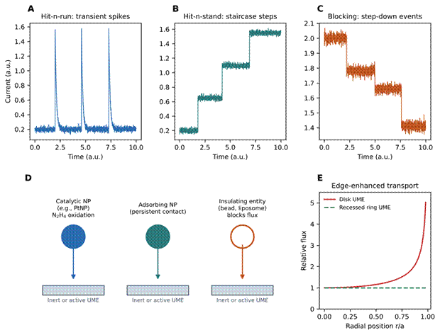
Figure 1. Transduction modes and transport heterogeneity in single-entity collision electrochemistry. (A–C) Simulated, illustrative current–time traces for the three principal collision signatures: hit-n-run transient spikes produced by brief contact of catalytic or conductive nanoparticles with an inert ultramicroelectrode (A); hit-n-stand staircase steps arising from permanent particle adsorption (B); and step-down blockades caused by insulating entities that obstruct redox-mediator flux at an active electrode (C). (D) Schematic of the corresponding particle–electrode interactions, including electrocatalytic hydrazine (N2H4) oxidation at PtNPs. (E) Qualitative radial flux profile across a disk UME, showing edge-enhanced transport that produces bimodal amplitude distributions, compared with the near-uniform flux targeted by recessed ring geometries. Traces are schematic and not reproduced experimental data. Based on Anderson and Zhang (2016), Wang and Tao (2015), Sokolov et al. (2017) and Kanokkanchana and Tschulik (2022).
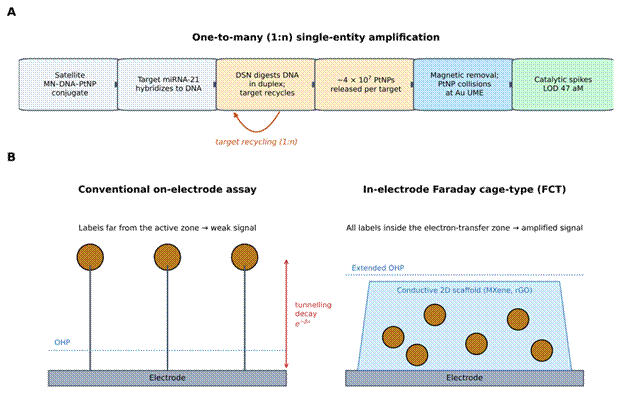
Figure 2. Nanomaterial-enabled signal amplification: one-to-many release and in-electrode Faraday cage-type architecture. (A) Workflow of the one-to-many (1:n) single-entity biosensor, in which hybridisation of target miRNA-21 to satellite magnetic nanoparticle–DNA–PtNP conjugates allows duplex-specific nuclease (DSN) to digest the DNA linker and release the target for further cycles; approximately 4 × 10⁷ PtNPs are liberated per target, separated magnetically and counted by catalytic collisions at a gold UME (LOD 47 aM). (B) Comparison of a conventional on-electrode assay, in which labels at the distal end of long recognition complexes suffer exponential electron-tunnelling decay, with a Faraday cage-type design, in which a conductive two-dimensional scaffold (MXene, rGO) encloses the complex, extends the outer Helmholtz plane and places all labels within the electron-transfer zone. Based on Bai et al. (2020), Arab et al. (2027), Nie et al. (2022) and Chen et al. (2021).
Engineered nanomaterials address what is, in practice, the central constraint on electrochemical biosensing: the need to detect scarce biomarkers without extensive sample preparation (Arab et al., 2027; Natarajan et al., 2024). AuNPs, PtNPs, MOFs, MXenes and carbon nanostructures function variously as nanoreporters, high-capacity nanocarriers and high-turnover electrocatalysts (Arab et al., 2027; Natarajan et al., 2024) (Table 3). DNA-templated bimetallic silver-platinum nanoclusters (DNA-Ag/Pt NCs), formed on single-stranded DNA scaffolds, illustrate the catalytic route particularly well. They mimic peroxidase, catalysing the reduction of hydrogen peroxide (H2O2) coupled with oxidation of 3,3′,5,5′-tetramethylbenzidine (TMB) to its blue oxidised form (Mu et al., 2023; Shateri et al., 2026). Platinum deposition onto the silver core quenches the intrinsic fluorescence of the silver cluster while raising peroxidase-like activity well above that of the monometallic counterparts, producing a tag that can be read optically or electrochemically (Mu et al., 2023; Shateri et al., 2026).
Three-dimensional conductive interfaces pursue a complementary goal, namely faster electron transfer and lower background capacitance (Arab et al., 2027). In-electrode Faraday cage-type architectures embed probe-label complexes within porous, highly conductive scaffolds such as Ag-doped Ti3AlC2 MXene nanosheets or single-atom Au/MXene heterostructures (Arab et al., 2027; Nie et al., 2022). Because the reporters sit inside the conductive network rather than at the distal end of a long recognition complex, the outer Helmholtz plane (OHP) is effectively extended, electron-tunnelling distances are shortened and a larger fraction of labels participates in the signal (Figure 2B). This design has supported near amplification-free microRNA detection (Arab et al., 2027; Nie et al., 2022) and, in MXene-based electrochemiluminescence (ECL) immunosensors, added a catalytic contribution to the geometric one (Chen et al., 2021). Three-dimensional porous graphene networks offer a related benefit by preventing π–π restacking of two-dimensional sheets and preserving accessible surface area (Natarajan et al., 2024).
2.5 One-to-Many Signal Amplification
A conceptual turning point in single-entity biosensing was the shift from 1:1 to one-to-many (1:n) signal transduction (Bai et al., 2020). In a standard single-collision bioassay, each target can generate no more than one collision or cleavage event, which places a hard limit on sensitivity. Bai et al. (2020) removed this limit with satellite magnetic nanoparticle-DNA-Pt nanoparticle (MN-DNA-PtNP) conjugates coupled to duplex-specific nuclease (DSN) target recycling (Figure 2A). Hybridisation of a single miRNA-21 strand allows DSN to digest the DNA linker in the resulting duplex, after which the target is freed to hybridise again. Repeated cycles release approximately 4 × 10⁷ catalytic PtNPs per target molecule; these are separated magnetically from the intact conjugates and counted through their catalytic hydrazine-oxidation collisions at a gold UME, giving a detection limit of 47 aM (Bai et al., 2020) (Table 1).
2.6 Programmable CRISPR-Cas Systems and Nanointerface Cleavage Kinetics
CRISPR and its associated Cas endonucleases, including Cas12a, Cas13a and Cas9, have changed molecular diagnostics by uniting programmable sequence recognition with enzymatic amplification (Schoch et al., 2026). Guided by a customisable crRNA, the Cas protein binds its target nucleic acid to form an active ternary ribonucleoprotein complex (Dong et al., 2024; Schoch et al., 2026). For class 2, type V effectors such as Cas12a, target recognition switches on non-specific single-stranded DNA (ssDNA) trans-cleavage that degrades surrounding reporters with high turnover (Dong et al., 2024; Schoch et al., 2026), an activity that has also been resolved at the single-molecule level for SARS-CoV-2 gene detection (Ji & Koh, 2023). The principal CRISPR-integrated platforms reviewed here are summarised in Table 2.
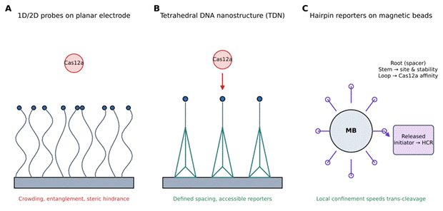
When this chemistry is moved onto an electrode, nanoscale spatial organisation becomes decisive (Dong et al., 2024; Zhao et al., 2025). Conventional electrochemical CRISPR sensors immobilise linear ssDNA or two-dimensional hairpin probes directly on planar electrodes, where flexible strands become entangled, collapse locally and crowd the surface, restricting enzyme access and slowing trans-cleavage (Dong et al., 2024) (Figure 3A). Tetrahedral DNA nanostructures, self-assembled from four complementary oligonucleotides, were introduced as rigid three-dimensional scaffolds that anchor to the electrode with nanometre-scale spacing (Dong et al., 2024) (Figure 3B). The rigid framework prevents probe intertwining, resists non-specific protein adsorption and presents pendant ssDNA reporters to solution, accelerating Cas12a cleavage and enabling detection of microRNAs and circulating tumour DNA (ctDNA) at sub-femtomolar levels (Dong et al., 2024).
Table 1. Single-entity electrochemistry (SEE) and nanoparticle collision platforms for ultrasensitive sensing. The table compares representative single-entity platforms according to the nanoparticle tag, electrode architecture, target and matrix, analytical performance, and the mechanism by which an individual collision or adsorption event is converted into a measurable current. Entries span catalytic spike counting after one-to-many (1:n) target recycling, in situ formation and cathodic impact of Hg2Cl2 nanoparticles, fast-scan cyclic voltammetry of single Pt particles, recessed-ring geometries designed to remove edge effects, and hit-n-run versus hit-n-stand transients. Detection limits are reported as given in the original studies. Abbreviations: UME, ultramicroelectrode; MN, magnetic nanoparticle; DSN, duplex-specific nuclease; CFE, carbon fibre microelectrode; FSCV, fast-scan cyclic voltammetry; LOD, limit of detection.
|
Biosensing system / technique
|
Nanoparticle / nanomaterial tag
|
Electrode / interface architecture
|
Target analyte & matrix
|
LOD & range
|
Signal transduction & amplification mechanism
|
Reference
|
|
One-to-many (1:n) single-entity impact biosensor
|
Satellite magnetic nanoparticle (MN)-DNA-PtNP conjugates (4.5 nm PtNPs)
|
Gold ultramicroelectrode (Au UME)
|
MicroRNA-21 (miRNA-21) in cancer cell lines (MCF-7, HeLa, A549)
|
LOD: 47 aM; Range: 50 aM – 5 nM
|
Target miRNA binds ssDNA on satellite MNs; DSN selectively digests the DNA to release ≈4 × 10⁷ PtNPs per target. Released PtNPs collide with the Au UME and catalyse hydrazine electro-oxidation, generating discrete impact current spikes.
|
Bai et al., 2020
|
|
Single-entity in situ impact electrochemistry
|
Mercurous chloride (Hg2Cl2) nanoparticles
|
Carbon ultramicroelectrode (C-UME)
|
Mercuric ions (Hg2+) in aqueous samples
|
LOD: sub-nanomolar; exponential frequency correlation
|
A potential-driven in situ reaction converts trace Hg2+ into Hg2Cl2 nanoparticles, which undergo cathodic impact reduction at the C-UME; spike frequency correlates with ion concentration.
|
Ganesh et al., 2025
|
|
Fast-scan cyclic voltammetry (FSCV) collision
|
Platinum nanoparticles (d = 4–80 nm)
|
Carbon fibre microelectrode (CFE) modified with AgCl
|
Model metal nanoparticles and electrocatalysts
|
Resolution: single-particle chemical kinetics
|
Ultra-fast voltage sweeps (400 V/s) during stochastic collisions yield complete single-particle cyclic voltammograms, resolving interfacial electron-transfer kinetic constants.
|
Anderson & Zhang, 2016
|
|
Recessed ring UME impact counting
|
Non-conductive polystyrene latex particles
|
Recessed Pt ring ultramicroelectrode
|
Nanoparticle size profiling and counting
|
Capability: uniform step-down amplitude resolution
|
Removes the edge effects and non-uniform current density inherent to disk UMEs, giving uniform current blockades during stochastic collisions.
|
Lemay and co-workers†
|
|
Single-particle catalytic impact electrochemistry
|
Platinum and silver nanoparticles
|
Mercury-amalgamated Pt UME (Hg/Pt UME)
|
Single-particle electrocatalysis and size analysis
|
Sensitivity: single-nanoparticle resolution
|
Hit-n-run spikes represent brief contact, whereas hit-n-stand staircase steps signify permanent adsorption; Hg amalgamation poisons Pt surface reactivity to reset the baseline.
|
Wang & Tao, 2015
|
Table 2. CRISPR-Cas integrated electrochemical and optical biosensors. The table summarises CRISPR-Cas biosensors according to effector system, target and disease context, sensing interface, upstream amplification cascade, analytical performance and the key mechanistic feature that distinguishes each design. Platforms include Cas12a acting on tetrahedral DNA nanostructures, PAM-free toehold-mediated strand displacement for SARS-CoV-2, Cas13a-triggered hybridisation chain reaction with electrochemiluminescent readout, magnetic-bead separation in serum, digital microfluidic automation and nanozyme-based colorimetric detection of a cancer protein. Optical entries are included where their interfacial or nanomaterial design informs electrochemical implementation. Abbreviations: TDN, tetrahedral DNA nanostructure; E-RCA, endonuclease-mediated rolling circle amplification; TSDR, toehold-mediated strand displacement reaction; HCR, hybridisation chain reaction; SDR, strand displacement reaction; RPA, recombinase polymerase amplification; DMF, digital microfluidics; EWOD, electrowetting-on-dielectric.
|
CRISPR effector system
|
Target analyte & disease context
|
Sensing interface / nanostructure
|
Amplification / cascade strategy
|
Analytical performance
|
Key mechanistic features
|
Reference
|
|
CRISPR/Cas12a
|
MicroRNA-155 (miRNA-155) and ctDNA (KRAS mutant) in cancer
|
Tetrahedral DNA nanostructure (TDN) on AuNPs/GCE
|
Endonuclease-mediated rolling circle amplification (E-RCA)
|
LOD: 0.5 fM (miRNA-155); Range: 10 fM – 1 nM
|
Target activates E-RCA to produce ssDNA activators, triggering Cas12a trans-cleavage of methylene blue-modified pendant probes on rigid 3D TDNs and attenuating the MB signal.
|
Dong et al., 2024
|
|
PAM-free PfTSDR-CRISPR/Cas12a
|
SARS-CoV-2 viral RNA fragments (RdRp, ORF1ab, N genes)
|
Gold electrode modified with methylene blue hairpin probes (MB-HP)
|
Toehold-mediated strand displacement reaction (TSDR)
|
LOD: 40 aM; Range: 100 aM – 10 pM
|
The toehold displacement region is physically separated from the Cas12a/gRNA recognition domain, enabling universal PAM-free detection across viral genes without redesigning the crRNA.
|
Shi et al., 2023
|
|
CRISPR/Cas13a
|
MicroRNA-21 (miRNA-21) in acute kidney injury urine
|
Streptavidin-modified magnetic beads (SAMBs)
|
Hybridisation chain reaction (HCR) with Ru(phen)32+
|
LOD: 0.53 fM; Range: 1 fM – 10 nM
|
Target-activated Cas13a cleaves pre-triggers into mature triggers that initiate HCR on magnetic beads; intercalated Ru(phen)32+ provides the electrochemiluminescence (ECL) readout.
|
Xu et al., 2023
|
|
CRISPR/Cas12a
|
MicroRNA-21 (miRNA-21) in 1% human blood serum
|
Streptavidin magnetic beads (MBs)
|
Strand displacement reaction (SDR) with auxiliary probe
|
LOD: 2.42 fM (buffer) / 12.12 fM (serum); Range: 8 fM – 250 nM
|
SDR releases trigger strand A3; magnetic separation isolates reaction products, and free A3 activates Cas12a trans-cleavage to restore the fluorescence signal.
|
Zhang et al., 2023
|
|
DMF-CRISPR/Cas12a
|
Staphylococcus aureus in urine and milk
|
Digital microfluidic (DMF) chip with EWOD actuation
|
Recombinase polymerase amplification (RPA)
|
LOD: 32 CFU/mL; Range: 102–107 CFU/mL
|
Automated, closed-system droplet operations (cell lysis, RPA and Cas12a trans-cleavage) completed in 55 min using 6.6 µL of total reagent.
|
Lu et al., 2023
|
|
CRISPR/Cas12a
|
Carcinoembryonic antigen (CEA), cancer protein
|
Peroxidase-mimicking DNA-Ag/Pt nanoclusters
|
Rolling circle amplification (RCA) and Nb.BbvCI nicking
|
LOD: 0.85 ng/mL (clinical serum)
|
CEA binding releases a primer that initiates RCA, generating Cas12a activators that degrade DNA-Ag/Pt NCs and diminish their peroxidase catalysis of the H2O2–TMB colour reaction.
|
Mu et al., 2023
|
Magnetic bead (MB) nanointerfaces offer a different handle on the same problem. By varying the root (spacer), stem and loop lengths of MB-anchored hairpin reporters, Zhao et al. (2025) showed that both the cleavage rate and the cleavage site can be tuned (Figure 3C). Colocalising hairpins on the bead surface raises local reactant density and accelerates cleavage relative to unconfined linear ssDNA in solution (Zhao et al., 2025). Because Cas12a binds single-stranded loops more readily than double-stranded stems, adjusting stem stability allows cleavage to release defined single-stranded initiator strands, which can then drive hybridisation chain reactions (HCR) on the bead without thermal denaturation, linking CRISPR recognition to dynamic DNA nanotechnology (Xu et al., 2023; Zhao et al., 2025).
2.7 PAM-Free Activation, Isothermal Circuits and Non-Nucleic-Acid Targets
Native Cas12a requires a protospacer adjacent motif (PAM), typically 5′-TTTN-3′, on double-stranded DNA targets, which restricts the regions that can be selected within short viral or mutated sequences (Hu et al., 2026; Shi et al., 2023). PAM-free activation strategies circumvent this requirement by constructing toehold-mediated duplexes (toe-dsDNA) or artificial bubble structures in the seed region, allowing the guide RNA to hybridise with the target strand directly (Hu et al., 2026; Shi et al., 2023). Coupling PAM-free TSDR with Cas12a (PfTSDR-CRISPR) creates a dual isothermal cascade in which target RNA drives recycling displacement cycles that generate bubble-containing dsDNA activators; these in turn trigger cleavage of methylene blue-modified hairpin probes (MB-HP) on a gold electrode (Shi et al., 2023). Because the displacement region is physically separated from the Cas12a/gRNA recognition domain, the same gRNA serves several SARS-CoV-2 gene fragments (RdRp, ORF1ab and N) with a detection limit of 40 aM (Shi et al., 2023) (Table 2).
Enzyme-driven signal converters have extended CRISPR sensing beyond nucleic acids to cofactors and proteins (Schoch et al., 2026). Ligation-driven designs use E. coli or T4 DNA ligase to seal a single-stranded gap only in the presence of NAD+ or ATP, generating an intact dsDNA activator (Schoch et al., 2026). Aptamer-mediated switches have been used for prostate-specific antigen (PSA) (Taghdisi et al., 2022), and combining RCA with Cas12a cleavage of DNA-Ag/Pt NCs has enabled visual detection of CEA (Mu et al., 2023). Electrochemical platforms for NGAL, an acute kidney injury (AKI) biomarker, have likewise drawn on nanostructured transducers and dual-signal readouts (Liang et al., 2022; Natarajan et al., 2024).
2.8 Diagnostic Translation in Complex Matrices and Microfluidic Integration
Integrated single-entity and CRISPR-assisted sensors have now been tested in a range of complex biological and environmental matrices (Table 4). Immunomagnetic separation, antifouling nanocomposites and bead-mediated enrichment have allowed analytes to be isolated from diluted human serum, urine, food and environmental water (Duan et al., 2023; Lee et al., 2024; Zhang et al., 2023). Reported demonstrations include miRNA-21 quantification in urine from AKI patients (Xu et al., 2023), detection of Salmonella Typhimurium and Staphylococcus aureus in food and clinical fluids (Duan et al., 2023; Lu et al., 2023), monitoring of Hg2+ through single-particle impact reduction of in situ-formed Hg2Cl2 (Ganesh et al., 2025) and viral gene profiling (Lee et al., 2024; Shi et al., 2023).
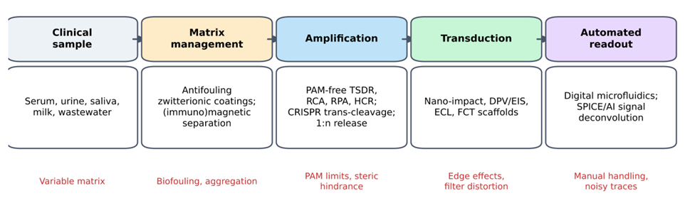
Translation to the point of care nevertheless remains incomplete (Schoch et al., 2026). In whole blood or undiluted plasma, protein adsorption and high ionic strength reduce diffusion, coat the electrode and suppress Faradaic current (Lee et al., 2024; Schoch et al., 2026). Zwitterionic antifouling nanocomposites, biomimetic interfaces and integrated pre-treatment modules are the main responses so far (Arab et al., 2027; Lee et al., 2024). Digital microfluidic (DMF) chips that combine recombinase polymerase amplification (RPA) with Cas12a cleavage automate lysis, enrichment, mixing and readout within a closed device (Lu et al., 2023), and AI/ML methods are beginning to be proposed for baseline correction, deconvolution of stochastic impact traces and pattern recognition across multiplexed arrays (Kanokkanchana & Tschulik, 2022; Schoch et al., 2026). Figure 4 summarises how these elements might be assembled into a single diagnostic workflow.