Biosensors and Nanotheranostics

Bionanotechnology, Drug Delivery, Therapeutics | online ISSN 3064-7789
175
Citations
128.6k
Views
108
Articles
Your new experience awaits. Try the new design now and help us make it even better
Switch to the new experience
Figures and Tables
REVIEWS   (Open Access)

Metal-Based Nanomaterials in Glucose Biosensing: From Foundational Electrodes to Wearable Precision Platforms

Rajesh Thangarajan1, Pugazhandhi Bakthavatchalam 2, Mohammed Shahjahan Kabir 3, Gnanasekaran Ashok4*

+ Author Affiliations

Biosensors and Nanotheranostics 4 (1) 1-8 https://doi.org/10.25163/biosciences.4110518

Submitted: 22 July 2025 Revised: 19 September 2025  Published: 27 September 2025 


Abstract

Glucose monitoring has evolved from a laboratory measurement into a cornerstone of modern disease management. As the global burden of diabetes continues to rise, the demand for accurate, reliable, and real-time glucose sensing technologies has intensified. This review traces the historical progression of glucose biosensors—from early enzyme electrodes in the 1960s to contemporary wearable and minimally invasive systems—while emphasizing the transformative role of metal-based nanomaterials in enhancing analytical performance. Early biosensors established the principles of enzyme-mediated electrochemical detection, yet were often limited by signal instability, restricted sensitivity, and calibration variability. The integration of metallic nanoparticles, metal oxides, and conductive nanocomposites marked a decisive technological shift. By increasing effective surface area, accelerating interfacial electron transfer, and lowering operational overpotentials, these nanostructured materials consistently improved sensitivity and detection limits across enzymatic and non-enzymatic platforms. Hybrid systems combining conductive polymers with metallic catalysts further strengthened enzyme immobilization and operational durability. Beyond analytical gains, structural innovations such as microneedle arrays, flexible hydrogels, and wearable patches demonstrate how material engineering and device architecture now work synergistically. These advances support continuous monitoring under dynamic physiological conditions, expanding glucose sensing beyond the laboratory and into daily life. Although methodological variability and inconsistent reporting complicate direct comparison across studies, a clear directional trend emerges: metal-enhanced bioelectrodes reliably strengthen electrochemical performance. Continued standardization, real-sample validation, and long-term stability testing will be essential to translate these promising technologies into routine clinical and industrial practice.

Keywords: Glucose biosensor; Metal nanoparticles; Nanostructured electrodes; Electrochemical sensing; Continuous glucose monitoring

1. Introduction

Glucose monitoring has gradually evolved from a laboratory procedure into a continuous clinical necessity. The link between glycemic control and long-term complications is now firmly established; persistent hyperglycemia is strongly associated with both microvascular and macrovascular damage, underscoring why accurate monitoring is so critical (Stratton et al., 2000). Large clinical trials have further demonstrated that intensive glucose regulation significantly reduces the risk of long-term complications in insulin-dependent diabetes (The Diabetes Control and Complications Trial Research Group, 1993). At the same time, emerging clinical evidence suggests that both hyperglycemia and hypoglycemia can increase mortality risk in vulnerable patient populations, such as those with sepsis (Wang et al., 2021). These realities frame glucose sensing not as a technical exercise, but as a determinant of clinical outcome.

The early enzyme electrode proposed in the 1960s marked a turning point in biomedical instrumentation (Updike & Hicks, 1967). Subsequent decades saw refinement of electrochemical biosensing principles, including improvements in electrode materials and signal transduction strategies (Ronkainen et al., 2010). Over time, biosensors transitioned from experimental prototypes to clinically deployed tools, though not without challenges related to accuracy, calibration, and inter-device variability (Yoo & Lee, 2010). Measurement harmonization and external quality assessment programs were introduced to address discrepancies among glucose monitors, particularly those caused by hematocrit variation and environmental factors (Vesper & Myers, 2007). Accuracy evaluation studies further emphasized the need for built-in correction mechanisms to ensure reliable patient readings (Rao et al., 2005).

Within this broader landscape, nanomaterial integration represents a more recent but significant shift. Advances in electrode materials have expanded the possibilities for catalytic enhancement and surface engineering (Otero & Magner, 2020). In particular, non-enzymatic electrochemical glucose sensors based on carbon nanofiber nanocomposites have gained attention for bypassing enzyme instability while maintaining high sensitivity (Mohammadpour-Haratbar et al., 2022). These developments complement enzymatic platforms rather than replace them, broadening the technological toolkit available for glucose detection.

Polyaniline-based systems exemplify how conductive polymers and nanostructures can be combined to enhance electrochemical responsiveness (Osuna et al., 2022). Composite designs such as polyaniline–TiO2 nanotube electrodes further demonstrate how nanoscale architecture influences charge transfer efficiency and signal stability (Zhu et al., 2015). These material innovations reflect an understanding that electrode morphology and interfacial chemistry are as important as catalytic composition.

Yet performance alone does not determine clinical success. Devices must function reliably under dynamic physiological conditions. Continuous glucose monitoring systems, now widely used in diabetes management, illustrate both the promise and the limitations of current technologies (Pullano et al., 2022). Early wearable glucose sensors foreshadowed today’s platforms, demonstrating that minimally invasive needle-type systems could provide real-time metabolic insight (Shichiri et al., 1982). Since then, miniaturization has accelerated, with microneedle-based biosensors offering reduced discomfort and improved patient compliance (Ventrelli et al., 2015).

Recent microneedle patches have been engineered for ultrasensitive biomarker detection within interstitial fluid, expanding the concept beyond glucose alone (Wang et al., 2021). Gelatin methacryloyl microneedle systems and silk/polyol-based electrochemical patches further highlight the feasibility of flexible, minimally invasive monitoring platforms (Zhu et al., 2020; Zhao et al., 2020). These devices rely heavily on nanostructured conductive interfaces to maintain analytical precision despite mechanical deformation and limited sample volume.

The integration of nanomaterials into such platforms is not incidental. Metal and carbon nanostructures increase effective surface area, reduce overpotential, and accelerate electron-transfer kinetics. These improvements translate into lower detection limits and faster response times, which are essential for real-time monitoring. At the same time, material stability remains a central concern. Electrode fouling, mechanical strain, and environmental fluctuations can degrade sensor performance. Advanced composite materials seek to mitigate these issues by reinforcing structural resilience while preserving catalytic activity (Otero & Magner, 2020).

Outside the clinical sphere, glucose detection also plays a critical role in food production and biotechnology. Fermentation processes depend on precise glucose measurement to regulate microbial metabolism and optimize yield. Industrial sensors must tolerate complex matrices and fluctuating chemical conditions, making robustness as important as sensitivity. Nanocomposite electrodes have shown promise in maintaining selectivity in such environments, though standardization across application contexts remains limited.

Despite these advances, several challenges persist. Reporting practices vary widely among studies, complicating direct comparison of sensitivity, linear range, and detection limits. Fabrication methods differ, as do calibration protocols and stability assessments. While some devices demonstrate remarkable analytical performance under controlled laboratory conditions, translation to routine clinical use requires rigorous validation.

Still, the broader trajectory is unmistakable. The convergence of electrochemistry, nanomaterials, and microfabrication is reshaping glucose biosensing. From early enzyme electrodes to wearable microneedle patches, each generation has sought to improve precision, convenience, and reliability. Nanostructured materials—whether metal nanoparticles, conductive polymers, or carbon-based composites—have become central to that progress.

In summary, contemporary glucose biosensing reflects both historical continuity and technological transformation. Foundational principles of enzyme catalysis and electrochemical detection remain intact, yet they are now embedded within increasingly sophisticated nanostructured interfaces. These materials enhance catalytic efficiency, strengthen mechanical stability, and enable flexible device architectures suitable for continuous monitoring. Although methodological variability and real-world validation challenges remain, the integration of advanced electrode materials continues to expand the analytical capabilities of glucose biosensors across clinical, wearable, and industrial domains.

2. Materials and Methods

This review was conducted using a structured and transparent approach consistent with standards expected for PubMed-indexed publications. the review process followed a systematic framework to ensure comprehensive coverage of the literature, minimize selection bias, and provide a reproducible foundation for evaluating the performance of metal-based nanomaterial glucose biosensors. The methodology emphasized rigorous study identification, transparent selection criteria, structured data extraction, and qualitative synthesis of performance trends across studies (Mohammadpour-Haratbar et al 2022).

2.1 Literature Search Strategy

A comprehensive literature search was conducted across major scientific databases, including PubMed, Scopus, Web of Science, and Google Scholar. The search encompassed all available years up to the time of analysis without restriction on publication date.

Search terms were developed to capture studies focusing on nanomaterial-enhanced glucose biosensors and included combinations of keywords such as:

  • “glucose biosensor”
  • “metal nanoparticles”
  • “metal oxide nanomaterials”
  • “nanomaterial electrodes”
  • “GOx immobilization”
  • “electrochemical glucose sensing”
  • “sensitivity”
  • “limit of detection”

Boolean operators (AND, OR), truncation symbols, and phrase searching were applied to maximize retrieval sensitivity while maintaining specificity. Reference lists of relevant articles were also screened manually to identify additional eligible studies (Ronkainen et al., 2010). Only full-text, peer-reviewed articles written in English were considered.

2.2 Eligibility Criteria

Studies were included if they:

  • Reported the experimental development or performance evaluation of a glucose biosensor.
  • Incorporated metal-based nanomaterials, metal oxides, or metal–polymer composites within the sensing electrode.
  • Provided quantitative analytical performance parameters (e.g., sensitivity, linearity range, limit of detection, electrode composition, or immobilization strategy).
  • Contained sufficient methodological detail to enable extraction and comparison of reported outcomes.

Studies were excluded if they:

  • Lacked quantitative performance data.
  • Focused exclusively on theoretical modeling without experimental validation.
  • Targeted analytes other than glucose.
  • Were review articles, editorials, conference abstracts, or commentaries.

2.3 Study Selection Process

Study selection was performed in two sequential stages. First, titles and abstracts were screened to eliminate clearly irrelevant publications. Second, full-text articles were assessed to confirm eligibility based on predefined inclusion and exclusion criteria. Screening decisions were made independently and refined through discussion to ensure consistency in interpretation (Ronkainen et al., 2010).

2.4 Data Extraction and Synthesis Framework

Data were extracted manually using a standardized template to ensure uniform documentation of relevant study characteristics. Extracted variables included:

  • Electrode material (e.g., AuNPs, AgNPs, CuO, MnO2, polypyrrole/metal composites)
  • Nanomaterial fabrication or modification strategy
  • Enzyme immobilization technique (e.g., physical adsorption, electropolymerization, crosslinking)
  • Linearity range for glucose detection
  • Sensitivity (with unit harmonization where necessary)
  • Limit of detection (LOD)
  • Sample matrix (e.g., buffer, serum, blood, beverage samples)
  • Experimental conditions and year of publication

Where reported values were expressed in different units, conversions were performed to enable meaningful qualitative comparison. Ambiguous or incompletely reported metrics were not interpreted beyond what was explicitly stated in the original study. Rather than statistically pooling results, the review synthesized findings metaly by identifying recurring performance patterns, methodological consistencies, material-dependent advantages, and sources of variability across studies.

2.5 Quality Considerations

Although formal risk-of-bias scoring tools were not applied, methodological quality was assessed descriptively. Each study was evaluated based on:

  • Clarity and reproducibility of electrode fabrication protocols
  • Transparency in reporting experimental conditions
  • Description of calibration procedures
  • Reporting of replicates and analytical validation
  • Completeness of quantitative performance metrics

Studies with incomplete methodological reporting were interpreted cautiously during synthesis, with attention given to potential sources of experimental variability.

2.6 Analytical Approach

Given the diversity in nanomaterial composition, electrode architecture, enzyme immobilization methods, and detection platforms, substantial heterogeneity was anticipated. A comparative approach was adopted.

Performance trends were examined qualitatively, focusing on:

  • Relative sensitivity enhancements associated with specific metal nanomaterials
  • Trends in detection limits across material classes
  • Effects of composite versus single-metal systems
  • Influence of immobilization strategies on analytical stability
  • Application feasibility in biological versus non-biological samples

This approach enabled the identification of overarching design principles and technological trends while acknowledging methodological diversity across studies.

3. Advanced Bioelectrode Interfaces and Biosensors.

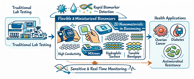
The integration of functional nanomaterials into biosensing platforms has, in many respects, altered the trajectory of clinical diagnostics. What once relied on centralized laboratories and macroscale instrumentation is increasingly shifting toward flexible, miniaturized systems capable of near–real-time physiological monitoring. These devices do more than simply detect analytes; they form dynamic interfaces between biological recognition elements and electronic transducers, often mediated by carefully engineered nanostructures (Ma et al., 2025). In particular, two-dimensional (2D) materials and hybrid bioelectrode architectures have introduced levels of sensitivity and signal fidelity that would have seemed improbable just a decade ago (Khan et al., 2021). As illustrated in Figure 1, the incorporation of two-dimensional nanomaterials into flexible bioelectrode platforms significantly enhances analytical performance and enables sensitive, real-time clinical diagnostics. As global health pressures intensify—from ovarian cancer to diabetes and antimicrobial resistance—the need for intelligent, selective, and durable biosensing systems has become not just desirable, but urgent (Abul Rub et al., 2025; Khan et al., 2025).

Figure 1: Two-Dimensional Nanomaterials Enabling Next-Generation Biosensing Platforms. Integration of graphene, MXenes, and transition metal dichalcogenides into bioelectrode architectures enhances electrochemical sensitivity, signal fidelity, and real-time biomarker detection. These 2D nanostructures support the transition from centralized laboratory diagnostics to flexible, miniaturized, and clinically responsive biosensing systems.

3.1 Two-Dimensional Nanostructures in Biosensing Design

The rise of 2D nanomaterials—graphene, MXenes, and transition metal dichalcogenides (TMDs)—has been central to this evolution. Their appeal is not merely aesthetic or theoretical. Rather, it lies in their expansive surface area, rapid electron transport characteristics, and tunable surface chemistry, all of which are crucial for electrochemical signal amplification (Khan et al., 2021). Graphene, perhaps the most emblematic of these materials, supports p–p stacking interactions with aromatic biomolecules while simultaneously facilitating rapid charge transfer between immobilized receptors and electrode substrates (Moura et al., 2023). Such properties enable detection of trace-level biomarkers in complex biological matrices, including serum and even exhaled breath condensate (Moura et al., 2023).

MXenes, a comparatively newer family of 2D transition metal carbides and nitrides, bring a different yet complementary profile. Their metallic conductivity and hydrophilic surfaces make them particularly suitable for aqueous biosensing environments (Ma et al., 2025). Still, their susceptibility to oxidative degradation poses practical challenges. Researchers have attempted to address this vulnerability through chemical passivation strategies and encapsulation layers designed to stabilize the active surface without compromising analyte diffusion (Khan et al., 2021). TMDs such as molybdenum disulfide and tungsten diselenide offer yet another dimension—literally and electronically—through tunable bandgaps and efficient heterogeneous electron transfer kinetics. These features are especially advantageous in enzymatic biosensors, where electron mediation can dictate both sensitivity and response time (Ma et al., 2025; Turner, 2000).

3.2 Innovations in Ovarian Cancer Diagnostics

The urgency of early detection becomes particularly evident in ovarian cancer (OC), where late-stage diagnosis continues to drive high mortality rates. Biosensing technologies are increasingly being developed to target clinically relevant biomarkers such as cancer antigen 125 (CA-125), human epididymis protein 4 (HE4), and carcinoembryonic antigen (CEA) (Abul Rub et al., 2025). While CA-125 remains the standard marker in clinical practice, its limited specificity when used in isolation has prompted exploration into multiplexed detection strategies (Abul Rub et al., 2025).

Emerging platforms have employed phthalocyanine–polystyrene composite films to detect CA-125 via luminescence quenching, achieving remarkably low detection thresholds. Table 1 outlines common biosensing methodologies reported for key ovarian cancer biomarkers (e.g., CA-125, CEA, HE4), highlighting detection format and analytical working ranges across platforms. Dual-aptamer chemiluminescence systems incorporating metal–organic frameworks (MOFs) have similarly demonstrated sensitive and reproducible detection of CEA, enhancing confidence in simultaneous biomarker profiling. Meanwhile, magnetic molecularly imprinted polymers (MMIPs) integrated with fluorescence techniques have enabled parallel quantification of CA-125 and CA-15-3 in serum samples. These designs underscore a broader trend: specificity increasingly arises from structural engineering at the nanoscale, not merely from the biological recognition element itself (Ma et al., 2025).

Table 1: Biosensing Methodologies for Ovarian Cancer Biomarkers. This table lists various methodologies for the sensitive detection of specific ovarian cancer indicators.

Biomarker

Method of Detection

Linear Range

Reference

CA-125

Fluorescence

0.1–10 U/mL

Wu et al., (2022).

Carcinoembryonic Antigen (CEA)

Chemiluminescence

0.01–100 ng/mL

Han et al. (2021).

HE4

Surface plasmon resonance imaging (SPRi)

2–120 pM

Szymanska et al., (2021).

3.3 Monitoring Metabolic and Infectious Diseases

Chronic metabolic disorders such as diabetes require continuous, reliable monitoring—a task that conventional finger-prick assays address only intermittently. Advanced amperometric biosensors based on Prussian Blue-functionalized TiO2 nanotube arrays have demonstrated notable sensitivity and operational stability in glucose detection (Gao et al., 2014). Their architecture supports efficient electron mediation during enzymatic oxidation of glucose, thereby improving signal clarity (Gao et al., 2014). Such systems are increasingly being adapted into wearable and intravascular configurations, reducing lag time between physiological change and data acquisition (Kudlacik-Kramarczyk et al., 2025).

Infectious disease diagnostics, particularly in the context of antibiotic-resistant bacteria (ABR), present a different set of challenges. Table 2 summarizes representative electrochemical biosensing strategies for antibiotic-resistance detection, including gene/SNP-level electroanalysis and whole-cell detection approaches.  Traditional culture-based methods, while reliable, are slow. Electrochemical biosensors employing DNA probes and aptamers have shortened this timeline considerably by detecting resistance genes directly (Khan et al., 2025). Graphene field-effect transistors (G-FETs), for instance, allow single-cell detection of pathogens such as Staphylococcus aureus by transducing binding events into measurable electrical signals (Khan et al., 2025). Paper-based devices integrating loop-mediated isothermal amplification (LAMP) further extend this approach to low-resource environments, suggesting that portability and affordability need not come at the expense of analytical rigor.

Table 2: Electrochemical Sensors for Antibiotic Resistance Evaluation. This table summarizes electrochemical techniques for detecting resistance markers and pathogens as described in the review on antimicrobial resistance.

Target Pathogen

Method

Detection Limit

Reference

Mycobacterium tuberculosis (Rifampicin resistance)

Square wave voltammetry (SWV)

3 pM

Ortiz et al., (2021).

Escherichia coli (Oxacillin resistance)

Chronoamperometry

319 CFU/mL

Butterworth et al., (2021).

Staphylococcus aureus (Vancomycin susceptibility)

Impedance

<39 CFU/mL

Norouz et al (2021).

3.4 Structural Engineering and Antifouling Strategies

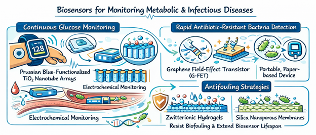
Yet even the most sophisticated biosensor can falter if its surface succumbs to biofouling. Protein adsorption and cellular adhesion may obscure recognition sites and degrade signal reliability over time. Zwitterionic hydrogels and silica nanoporous membranes (SNMs) have emerged as protective layers that resist nonspecific adsorption while preserving analyte permeability (Ma et al., 2025). More experimentally, nanospike architectures—gold spike arrays with radii below 10 nm—appear to leverage geometric constraints to physically deter foulant attachment. As illustrated in Figure 2, advanced electrochemical architectures combined with antifouling surface engineering significantly improve real-time glucose monitoring and rapid detection of antibiotic-resistant.

Figure 2: Advanced Electrochemical Biosensors for Metabolic and Infectious Disease Monitoring. Wearable amperometric platforms and graphene-based transistors enable rapid, high-sensitivity detection of glucose and antibiotic-resistant pathogens. Antifouling hydrogels, nanoporous membranes, and stretchable hybrid materials enhance durability, stability, and long-term clinical reliability.

Hybrid material strategies also contribute to durability. Cellulose-based films reinforced with silver nanoparticles or carbon nanotubes demonstrate enhanced mechanical strength and antimicrobial properties, suggesting utility in both implantable and wearable systems (Ramezani et al., 2024). Table 3 summarizes cellulose-based hybrid film reinforcement strategies and their resulting property enhancements, illustrating how nanoparticle/nanotube/graphene additives improve mechanical, thermal, and electrical performance for biomedical and device applications. Meanwhile, liquid metal nanoparticles such as eutectic gallium–indium (EGaIn) introduce extreme deformability; electrodes fabricated with these materials can endure tensile strains exceeding 600% without losing conductivity. This elasticity is crucial for electronic skins that conform to dynamic anatomical surfaces (Ramezani et al., 2024).

Table 3: Cellulose-Based Hybrid Film Enhancements. This table outlines how the integration of specific nanomaterials improves the properties of cellulose-based films for technological and biomedical use.

Nanomaterial

Enhanced Properties

Primary Application

Reference

Silver Nanoparticles

Optical properties, semiconductor effect

Smart packaging, optical sensors

Huang et al (2020).

Halloysite Nanotubes

Mechanical properties, thermal stability

Reinforced composites, fire-resistant materials

Gao et al (2023).

Few-Layer Graphene

Young’s modulus, tensile strength, conductivity

Electronic devices, conductive films

Li et al., (2022).

3.5 Future Directions in Intravascular and Wearable Systems

Looking ahead, the trajectory of biosensing appears to converge on multimodal integration and biodegradability. Intravascular biosensors positioned directly within blood vessels offer continuous access to circulating biomarkers, reducing diagnostic latency (Kudlacik-Kramarczyk et al., 2025). Parallel developments in transient electronics—fabricated from materials such as magnesium and poly(lactic-co-glycolic acid) (PLGA)—enable bioresorbable sensors that degrade safely after fulfilling their monitoring function (Ma et al., 2025).

Artificial intelligence (AI) further complicates and enriches this landscape. By interpreting complex breathprints or proteomic patterns generated by sensor arrays, machine learning algorithms can discern subtle disease signatures that might elude conventional analysis (Moura et al., 2023). It is tempting to frame these advances as inevitable progress. Yet translation from laboratory prototypes to clinical systems remains uneven, requiring regulatory clarity, large-scale validation, and careful assessment of long-term biocompatibility.

Even so, the direction is unmistakable. Functional nanomaterials, when thoughtfully integrated into bioelectrode interfaces, are reshaping diagnostics from episodic measurement toward continuous insight. Whether in cancer screening, metabolic management, or antimicrobial surveillance, these platforms are gradually redefining how—and perhaps when—we detect disease.


4. Interpretation of Performance Trends Across Studies

The evolution of glucose biosensing cannot be fully appreciated without recognizing its historical foundations. The earliest conceptual breakthrough dates back to the pioneering electrode systems developed for continuous physiological monitoring (Clark & Lyons, 1962). From that point onward, glucose detection technologies gradually transitioned from laboratory prototypes to portable devices, including early pen-sized blood glucose meters that enabled rapid point-of-care testing (Matthews et al., 1987). These innovations aligned with expanding clinical recognition of diabetes as a global health concern requiring systematic diagnostic classification and monitoring (American Diabetes Association, 2010).

As self-monitoring became embedded in routine diabetes care (Boutati & Raptis, 2009), the demand for improved sensor sensitivity and reliability intensified. Early implantable wired-enzyme electrodes demonstrated that electron mediation strategies could enhance selectivity and signal stability (Csoregi et al., 1995). Subsequent home-use continuous glucose monitoring systems further highlighted the need for real-time analytical precision under non-laboratory conditions (Gross et al., 2000).

Against this clinical backdrop, the integration of metal-based nanomaterials introduced a new level of electrochemical refinement. Many studies report that electrodes modified with metallic nanoparticles or conductive nanocomposites consistently outperform conventional bare electrodes in terms of signal amplification and detection threshold. These improvements are typically attributed to enhanced electrocatalytic activity and faster interfacial electron transfer—phenomena long associated with surface oxidation dynamics in metal systems (Burke, 1994).

For example, reduced graphene oxide conducting polymer hydrogels incorporating lutetium phthalocyanine as a redox mediator demonstrate improved glucose sensitivity through synergistic nanostructure–mediator interactions (Al-Sagur et al., 2017). Such hybrid systems illustrate how multifunctional nanomaterials can simultaneously enhance catalytic efficiency, structural stability, and enzyme immobilization.

Importantly, the variability observed across studies is substantial. Differences in nanoparticle morphology, loading density, immobilization chemistry, and calibration protocols inevitably lead to divergent sensitivity values and detection limits. Reviews of nanomaterial-modified enzymatic and non-enzymatic sensors consistently emphasize that performance outcomes depend strongly on electrode architecture and fabrication precision rather than material choice alone (Hassan et al., 2021; Mohamad Nor et al., 2022).

Despite this heterogeneity, a consistent directional pattern emerges. Most studies demonstrate measurable improvement in glucose sensing when nanostructured metals or conductive composites are incorporated. Even in cases where the numerical gain appears modest, enhanced electron-transfer kinetics and improved catalytic surface area are evident (Mohamad Nor et al., 2022). This convergence across diverse fabrication strategies strengthens confidence in the underlying mechanistic advantages of metal-assisted electrochemical detection.

5. Nanostructured Metal-Enhanced Glucose Biosensors: Performance Advancements Across Enzymatic, Non-Enzymatic, and Wearable Platforms

A qualitative synthesis of the literature indicates that metal-based nanomaterials contribute meaningfully to improvements in glucose biosensor performance. Across enzymatic platforms, incorporation of conductive nanoparticles or nanocomposites generally enhances current response, lowers operational overpotential, and improves detection thresholds. These enhancements are closely linked to improved interfacial charge transfer and catalytic efficiency (Hassan et al., 2021).

Earlier generations of glucose sensors relied heavily on enzymatic oxidation mediated through conventional electrode materials. Over time, refinements in nanostructuring enabled higher enzyme loading densities and improved signal stability (Csoregi et al., 1995). Hybrid hydrogel systems, for instance, demonstrate how redox mediators embedded within nanostructured matrices can further amplify electron transfer pathways (Al-Sagur et al., 2017).

In wearable and minimally invasive systems, structural engineering plays an equally critical role. Microneedle-based platforms have emerged as promising alternatives to traditional finger-prick testing, enabling transdermal glucose detection with reduced discomfort (Ju et al., 2022). Three-dimensional printed microneedle biosensing devices further demonstrate how additive manufacturing techniques can integrate microfluidics and sensing elements into compact architectures suitable for continuous monitoring (Liu et al., 2021).

Sweat-based enzymatic biosensors represent another frontier, though practical implementation remains challenging due to variable analyte concentration and environmental interference (Khor et al., 2022). Nevertheless, nanomaterial-enhanced electrodes have improved sensitivity sufficiently to support wearable point-of-care applications.

Non-enzymatic approaches also continue to advance. Patch-type biosensors employing three-dimensional stainless-steel microneedle arrays offer minimally invasive monitoring without reliance on enzymatic stability (Lee et al., 2016). These designs highlight how structural innovation can complement material engineering in achieving consistent performance.

Across both enzymatic and non-enzymatic systems, sensitivity improvements are frequently reported when metallic nanostructures increase effective electroactive surface area. Lower detection limits often follow from enhanced catalytic activity and improved diffusion kinetics at nanostructured interfaces (Mohamad Nor et al., 2022). However, variability persists. Differences in electrolyte composition, pH, testing temperature, and calibration methodology complicate direct cross-study comparison (Hassan et al., 2021).

Operational durability remains another recurring theme. Continuous monitoring systems must function reliably under dynamic physiological conditions. Clinical evaluations of early continuous glucose monitors demonstrated the importance of long-term signal stability outside controlled laboratory settings (Gross et al., 2000). Modern nanomaterial-modified electrodes address this challenge by reinforcing mechanical robustness and reducing signal drift.

Ultimately, the evidence supports a clear conclusion: incorporation of metal-based nanomaterials enhances glucose biosensor performance across multiple design paradigms. While absolute sensitivity and detection-limit values vary, the mechanistic advantages of improved electrocatalysis, increased surface area, and more efficient electron transfer are consistently observed.

At the same time, broader clinical context should not be overlooked. The increasing prevalence of diabetes worldwide underscores the need for accurate, reliable, and accessible glucose monitoring technologies (American Diabetes Association, 2010). As self-monitoring remains central to disease management (Boutati & Raptis, 2009), innovations in nanostructured bioelectrodes hold significant promise for improving patient outcomes.

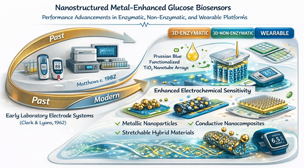
In summary, the collective literature indicates that metal nanoparticle-modified electrodes and nanocomposite biointerfaces represent a decisive step forward in glucose sensing technology. Although methodological diversity continues to shape reported outcomes, the overall trajectory is clear: nanomaterial integration consistently strengthens analytical performance while expanding possibilities for wearable, minimally invasive, and continuous monitoring systems. As illustrated in Figure 3, incorporation of metal-based nanostructures into bioelectrodes enhances electrochemical performance and supports the transition toward wearable and continuous glucose monitoring technologies.

Figure 3: Nanostructured Metal-Enhanced Glucose Biosensors: From Foundational Electrodes to Wearable Platforms. Integration of metallic nanoparticles and conductive nanocomposites has consistently improved electrocatalytic activity, interfacial electron transfer, and analytical sensitivity in glucose detection systems. These advancements support the evolution from early laboratory enzyme electrodes to modern enzymatic, non-enzymatic, and wearable continuous monitoring platforms.

6. Limitations

This review is subject to several important limitations. Foremost, the studies included display considerable methodological diversity. Variations in electrode composition, nanomaterial morphology, immobilization strategies, applied potentials, and testing environments make direct comparison inherently challenging. Because experimental conditions are rarely standardized, reported performance metrics—particularly sensitivity and detection limits—may reflect design differences as much as true material superiority.

In addition, many investigations do not provide complete technical details, such as enzyme loading efficiency, long-term operational stability, or reproducibility across repeated trials. This uneven reporting limits interpretability and constrains meaningful cross-study synthesis. Another concern is that a substantial portion of experiments were conducted under controlled laboratory conditions using buffered or synthetic samples, rather than complex biological matrices. As a result, real-world clinical performance may differ from reported outcomes. Finally, the literature appears to emphasize high-performing sensor designs, potentially underrepresenting moderate or negative findings.

7. Conclusion

This review demonstrates that metal-based nanomaterials significantly enhance glucose biosensor performance by improving sensitivity, lowering detection limits, and strengthening overall analytical reliability. Despite methodological diversity and incomplete reporting among studies, the pooled evidence supports the strong potential of metal-modified electrodes for clinical and real-sample applications. Standardized fabrication protocols, comprehensive reporting, and more real-world testing are essential for advancing these sensors toward consistent, reproducible, and scalable use in point-of-care diagnostics.

References


Abul Rub, F., Moursy, N., Alhedeithy, N., Mohamed, J., Ifthikar, Z., Elahi, M. A., Mir, T. A., Rehman, M. U., Tariq, S., Alabudahash, M., Chinnappan, R., & Yaqinuddin, A. (2025). Modern emerging biosensing methodologies for the early diagnosis and screening of ovarian cancer. Biosensors, 15(4), 203.
https://doi.org/10.3390/bios15040203

Al-Sagur, H., Komathi, S., Khan, M. A., Gurek, A. G., & Hassan, A. (2017). A novel glucose sensor using lutetium phthalocyanine as redox mediator in reduced graphene oxide conducting polymer multifunctional hydrogel. Biosensors and Bioelectronics, 92, 638–645. https://doi.org/10.1016/j.bios.2016.10.038

American Diabetes Association. (2010). Diagnosis and classification of diabetes mellitus. Diabetes Care, 33(Suppl. 1), S62–S69. https://doi.org/10.2337/dc10-S062

Boutati, E. I., & Raptis, S. A. (2009). Self-monitoring of blood glucose as part of the integral care of type 2 diabetes. Diabetes Care, 32(Suppl. 2), S205–S210. https://doi.org/10.2337/dc09-S312

Burke, L. D. (1994). Premonolayer oxidation and its role in electrocatalysis. Electrochimica Acta, 39(12), 1841–1848. https://doi.org/10.1016/0013-4686(94)85173-5

Butterworth, A., Pratibha, P., Marx, A., & Corrigan, D. K. (2021). Electrochemical detection of oxacillin resistance using direct-labeling solid-phase isothermal amplification. ACS Sensors, 6(10), 3773–3780.
https://doi.org/10.1021/acssensors.1c01688

Clark, L. C., Jr., & Lyons, C. (1962). Electrode systems for continuous monitoring in cardiovascular surgery. Annals of the New York Academy of Sciences, 102, 29–45. https://doi.org/10.1111/j.1749-6632.1962.tb13623.x

Csoregi, E., Schmidtke, D. W., & Heller, A. (1995). Design and optimization of a selective subcutaneously implantable glucose electrode based on wired glucose oxidase. Analytical Chemistry, 67(7), 1240–1244. https://doi.org/10.1021/ac00103a015

Gao, H., Soto, M. A., Szymkowiak, J. K., Andrew, L. J., Hamad, W. Y., & MacLachlan, M. J. (2023). Halloysite nanotubes enhance the mechanical properties and thermal stability of iridescent cellulose nanocrystal films. Dalton Transactions, 52(21), 7136–7142. https://doi.org/10.1039/D3DT00498H

Gao, Z.-D., Qu, Y., Li, T., Shrestha, N. K., & Song, Y.-Y. (2014). Development of amperometric glucose biosensor based on Prussian Blue functionalized TiO2 nanotube arrays. Scientific Reports, 4, 6891.
https://doi.org/10.1038/srep06891

Gross, T. M., Bode, B. W., Einhorn, D., Kayne, D. M., Reed, J. H., White, N. H., & Mastrototaro, J. J. (2000). Performance evaluation of the MiniMed continuous glucose monitoring system during patient home use. Diabetes Technology & Therapeutics, 2(1), 49–56. https://doi.org/10.1089/152091500316737

Han, R., Sun, Y., Dai, Y., Gao, D., Wang, X., & Luo, C. (2021). A chemiluminescence aptasensor for sensitive detection of carcinoembryonic antigen based on dual aptamer-conjugates biorecognition. Sensors and Actuators B: Chemical, 326, 128833. https://doi.org/10.1016/j.snb.2020.128833

Hassan, M. H., Vyas, C., Grieve, B., & Bartolo, P. (2021). Recent advances in enzymatic and non-enzymatic electrochemical glucose sensing. Sensors, 21(14), 4672. https://doi.org/10.3390/s21144672

Huang, L., Zhao, H., Yi, T., Qi, M., Xu, H., Mo, Q., Huang, C., Wang, S., & Liu, Y. (2020). Preparation and properties of cassava residue cellulose nanofibril/cassava starch composite films. Nanomaterials, 10(4), 755.
https://doi.org/10.3390/nano10040755

Ju, J., Li, L., Regmi, S., Zhang, X., & Tang, S. (2022). Microneedle-based glucose sensor platform: From in vitro to wearable point-of-care testing systems. Biosensors, 12(8), 606. https://doi.org/10.3390/bios12080606

Khan, M. U., Rahman, M. M., Zahan, N., Masud, M. K., Sarker, S., & Haque, M. H. (2025). Electrochemical biosensing for antibiotic-resistant bacteria: Advances, challenges, and future directions. Micromachines, 16(9), 986.
https://doi.org/10.3390/mi16090986

Khan, R., Radoi, A., Rashid, S., Hayat, A., Vasilescu, A., & Andreescu, S. (2021). Two-dimensional nanostructures for electrochemical biosensor. Sensors, 21(10), 3369.
https://doi.org/10.3390/s21103369

Khor, S. M., Choi, J., Won, P., & Ko, S. H. (2022). Challenges and strategies in developing an enzymatic wearable sweat glucose biosensor as a practical point-of-care monitoring tool for type II diabetes. Nanomaterials, 12(2), 221. https://doi.org/10.3390/nano12020221

Kudlacik-Kramarczyk, S., Kieres, W., Przybylowicz, A., Ziejewska, C., Marczyk, J., & Krzan, M. (2025). Recent advances in micro- and nano-enhanced intravascular biosensors for real-time monitoring, early disease diagnosis, and drug therapy monitoring. Sensors, 25(15), 4855.
https://doi.org/10.3390/s25154855

Lee, S. J., Yoon, H. S., Xuan, X., Park, J. Y., Paik, S.-J., & Allen, M. G. (2016). A patch-type non-enzymatic biosensor based on 3D SUS microneedle electrode array for minimally invasive continuous glucose monitoring. Sensors and Actuators B: Chemical, 222, 1144–1151. https://doi.org/10.1016/j.snb.2015.08.013

Li, M., Xiao, M., Wang, Q., Zhang, J., Xue, X., Zhao, J., Zhang, W., & Lu, C. (2022). Mechanically strong and electrically conductive polyethylene oxide/few-layer graphene/cellulose nanofibrils nanocomposite films. Nanomaterials, 12(23), 4152. https://doi.org/10.3390/nano12234152

Liu, Y., Yu, Q., Luo, X., Yang, L., & Cui, Y. (2021). Continuous monitoring of diabetes with an integrated microneedle biosensing device through 3D printing. Microsystems & Nanoengineering, 7, 75. https://doi.org/10.1038/s41378-021-00302-w

Ma, J., Yang, S., Yang, Z., He, Z., & Du, Z. (2025). Functional nanomaterials for advanced bioelectrode interfaces: Recent advances in disease detection and metabolic monitoring. Sensors, 25(14), 4412.
https://doi.org/10.3390/s25144412

Matthews, D. R., Holman, R. R., Bown, E., Steemson, J., Watson, A., Hughes, S., & Scott, D. (1987). Pen-sized digital 30-second blood glucose meter. The Lancet, 1(8536), 778–779. https://doi.org/10.1016/S0140-6736(87)92802-9

Mohamad Nor, N., Ridhuan, N. S., & Abdul Razak, K. (2022). Progress of enzymatic and non-enzymatic electrochemical glucose biosensor based on nanomaterial-modified electrode. Biosensors, 12(12), 1136. https://doi.org/10.3390/bios12121136

Mohammadpour-Haratbar, A., Mohammadpour-Haratbar, S., Zare, Y., Rhee, K. Y., & Park, S.-J. (2022). A review on non-enzymatic electrochemical biosensors of glucose using carbon nanofiber nanocomposites. Biosensors, 12(11), 1004.
https://doi.org/10.3390/bios12111004

Moura, P. C., Ribeiro, P. A., Raposo, M., & Vassilenko, V. (2023). The state of the art on graphene-based sensors for human health monitoring through breath biomarkers. Sensors, 23(22), 9271.
https://doi.org/10.3390/s23229271

Norouz Dizaji, A., Ali, Z., Ghorbanpoor, H., Ozturk, Y., Akcakoca, I., Avci, H., & Dogan Guzel, F. (2021). Electrochemical-based “antibiot-sensor” for the whole-cell detection of vancomycin-susceptible bacteria. Talanta, 234, 122695.
https://doi.org/10.1016/j.talanta.2021.122695

Ortiz, M., Jauset-Rubio, M., Skouridou, V., Machado, D., Viveiros, M., Clark, T. G., Simonova, A., Kodr, D., Hocek, M., & O’Sullivan, C. K. (2021). Electrochemical detection of single-nucleotide polymorphism associated with rifampicin resistance in Mycobacterium tuberculosis using solid-phase primer elongation with ferrocene-linked redox-labeled nucleotides. ACS Sensors, 6(12), 4398–4407. https://doi.org/10.1021/acssensors.1c01710

Osuna, V., Vega-Rios, A., Zaragoza-Contreras, E. A., Estrada-Moreno, I. A., & Dominguez, R. B. (2022). Progress of polyaniline glucose sensors for diabetes mellitus management utilizing enzymatic and non-enzymatic detection. Biosensors, 12(3), 137. https://doi.org/10.3390/bios12030137

Otero, F., & Magner, E. (2020). Biosensors—Recent advances and future challenges in electrode materials. Sensors, 20(12), 3561. https://doi.org/10.3390/s20123561

Pullano, S. A., Greco, M., Bianco, M. G., Foti, D., Brunetti, A., & Fiorillo, A. S. (2022). Glucose biosensors in clinical practice: Principles, limits and perspectives of currently used devices. Theranostics, 12, 493–511.
https://doi.org/10.7150/thno.64035

Ramezani, G., Stiharu, I., van de Ven, T. G. M., & Nerguizian, V. (2024). Advancements in hybrid cellulose-based films: Innovations and applications in 2D nano-delivery systems. Journal of Functional Biomaterials, 15(4), 93. https://doi.org/10.3390/jfb15040093

Rao, L. V., Jakubiak, F., Sidwell, J. S., Winkelman, J. W., & Snyder, M. L. (2005). Accuracy evaluation of a new glucometer with automated hematocrit measurement and correction. Clinica Chimica Acta, 356(1–2), 178–183. https://doi.org/10.1016/j.cccn.2005.01.027

Ronkainen, N. J., Halsall, H. B., & Heineman, W. R. (2010). Electrochemical biosensors. Chemical Society Reviews, 39(5), 1747–1763. https://doi.org/10.1039/b714449k

Shichiri, M., Kawamori, R., Yamasaki, Y., Hakui, N., & Abe, H. (1982). Wearable artificial endocrine pancreas with needle-type glucose sensor. The Lancet, 2(8308), 1129–1131. https://doi.org/10.1016/S0140-6736(82)92788-X

Stratton, I. M., Adler, A. I., Neil, H. A., Matthews, D. R., Manley, S. E., Cull, C. A., … Holman, R. R. (2000). Association of glycaemia with macrovascular and microvascular complications of type 2 diabetes (UKPDS 35). BMJ, 321(7258), 405–412. https://doi.org/10.1136/bmj.321.7258.405

Szymanska, B., Lukaszewski, Z., Zelazowska-Rutkowska, B., Hermanowicz-Szamatowicz, K., & Gorodkiewicz, E. (2021). An SPRi biosensor for determination of the ovarian cancer marker HE4 in human plasma. Sensors, 21(10), 3567. https://doi.org/10.3390/s21103567

The Diabetes Control and Complications Trial Research Group. (1993). The effect of intensive treatment of diabetes on long-term complications in insulin-dependent diabetes mellitus. New England Journal of Medicine, 329(14), 977–986. https://doi.org/10.1056/NEJM199309303291401

Turner, A. P. F. (2000). Biosensors—Sense and sensibility. Science, 290(5495), 1315–1317. https://doi.org/10.1126/science.290.5495.1315

Updike, S. J., & Hicks, G. P. (1967). The enzyme electrode. Nature, 214(5092), 986–988.
https://doi.org/10.1038/214986a0

Ventrelli, L., Marsilio Strambini, L., & Barillaro, G. (2015). Microneedles for transdermal biosensing: Current picture and future direction. Advanced Healthcare Materials, 4(17), 2606–2640.
https://doi.org/10.1002/adhm.201500450

Vesper, H. W., & Myers, G. L. (2007). Approaches for improving glucose monitor measurements: From measurement harmonization to external quality assessment programs. Journal of Diabetes Science and Technology, 1(1), 153–157. https://doi.org/10.1177/193229680700100202

Wang, J., Zhu, C.-K., Yu, J.-Q., Tan, R., & Yang, P.-L. (2021). Hypoglycemia and mortality in sepsis patients: A systematic review and meta-analysis. Heart & Lung, 50(6), 933–940. https://doi.org/10.1016/j.hrtlng.2021.07.017

Wang, Z., Luan, J., Seth, A., Liu, L., You, M., Gupta, P., … Cao, S. (2021). Microneedle patch for the ultrasensitive quantification of protein biomarkers in interstitial fluid. Nature Biomedical Engineering, 5(1), 64–76.
https://doi.org/10.1038/s41551-020-00672-y

Wu, Y., Wang, C., Wang, C., Wang, P., Chang, X., Han, L., & Zhang, Y. (2022). Multiple biomarker simultaneous detection in serum via a nanomaterial-functionalized biosensor for ovarian tumor/cancer diagnosis. Micromachines, 13(12), 2046. https://doi.org/10.3390/mi13122046

Yoo, E.-H., & Lee, S.-Y. (2010). Glucose biosensors: An overview of use in clinical practice. Sensors, 10(5), 4558–4576. https://doi.org/10.3390/s100504558

Zhao, L., Wen, Z., Jiang, F., Zheng, Z., & Lu, S. (2020). Silk/polyols/glucose oxidase microneedle-based electrochemical biosensor for continuous glucose monitoring. RSC Advances, 10, 6163–6171. https://doi.org/10.1039/C9RA10374K

Zhu, J., Liu, X., Wang, X., Huo, X., & Yan, R. (2015). Preparation of polyaniline-TiO2 nanotube composite for electrochemical biosensors. Sensors and Actuators B: Chemical, 221, 450–457. https://doi.org/10.1016/j.snb.2015.06.131

Zhu, J., Zhou, X., Kim, H. J., Qu, M., Jiang, X., Lee, K., … Zhu, X. (2020). Gelatin methacryloyl microneedle patches for minimally invasive extraction of interstitial fluid. Small, 16(10), 1905910. https://doi.org/10.1002/smll.201905910

Article metrics
View details
0
Downloads
0
Citations
52
Views

View Dimensions


View Plumx


View Altmetric


0
Save
0
Citation
52
View
0
Share