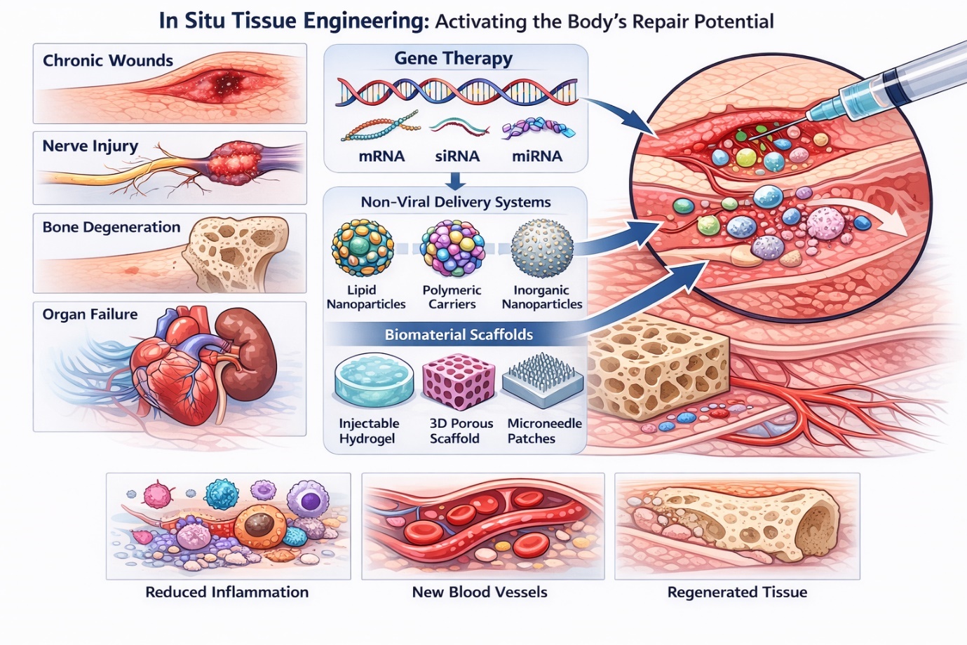
Biosensors and Nanotheranostics

Bionanotechnology, Drug Delivery, Therapeutics | online ISSN 3064-7789
83
Citations
56.8k
Views
60
Articles
Your new experience awaits. Try the new design now and help us make it even better
Switch to the new experience
Figures and Tables
REVIEWS   (Open Access)

Advancements in Biomaterial-Based Nucleic Acid Delivery Systems for In Situ Tissue Engineering: A Systematic Review and Meta-Analysis

Md. Taufique Hasan Bhuiyan Sezan1*, Raihan Mia 2

+ Author Affiliations

Biosensors and Nanotheranostics 4 (1) 1-8 https://doi.org/10.25163/biosensors.4110516

Submitted: 14 September 2025 Revised: 10 November 2025  Published: 17 November 2025 


Abstract

The clinical potential of nucleic acid-based therapies in regenerative medicine is increasingly recognized, yet their translation remains limited by delivery efficiency, safety, and targeted release challenges. This systematic review and meta-analysis examine the current landscape of biomaterial-based nucleic acid delivery systems, focusing on viral and non-viral vectors integrated into scaffold platforms for in situ tissue engineering. Literature was systematically collected from peer-reviewed sources between 2009 and 2025, evaluating studies that assessed delivery efficiency, tissue-specific targeting, and therapeutic outcomes. Viral vectors, including adenovirus, adeno-associated virus (AAV), and lentivirus, demonstrated high transduction efficiency but were associated with immunogenicity and risks of insertional mutagenesis. In contrast, non-viral nanocarriers—lipid-based nanoparticles, polymeric nanoparticles, inorganic nanoparticles, and biomimetic vectors such as exosomes—exhibited enhanced biocompatibility, reduced toxicity, and customizable release profiles. Scaffold integration, including injectable hydrogels, three-dimensional porous scaffolds, and sheet-like systems, improved local retention, spatiotemporal release, and functional tissue regeneration. Meta-analysis of preclinical studies indicated significantly improved gene delivery efficiency and tissue repair outcomes when non-viral carriers were combined with tailored scaffolds (p < 0.05). However, limitations such as mechanical weakness in hydrogels, light penetration constraints, long-term bioaccumulation, and manufacturing challenges remain. This review highlights the translational potential of combining non-viral nucleic acid vectors with advanced biomaterials, offering a roadmap for clinical implementation. Addressing the identified safety and scalability challenges could accelerate the adoption of these systems for targeted, efficient, and safe regenerative therapies.

Keywords: Nucleic acid delivery, non-viral vectors, viral vectors, biomaterial scaffolds, tissue engineering, regenerative medicine, hydrogels, 3D scaffolds

1. Introduction

Regenerative medicine and in situ tissue engineering have emerged as transformative strategies for treating complex tissue injuries and degenerative diseases. Central to their success is the ability to deliver nucleic acids, including plasmid DNA (pDNA), small interfering RNA (siRNA), microRNA (miRNA), and messenger RNA (mRNA), efficiently to targeted cells to modulate gene expression and promote tissue regeneration (Motamedi et al., 2024; Wu et al., 2025; Yuan et al., 2025). Despite the promise of gene-based therapeutics, their clinical translation is hindered by several challenges, including enzymatic degradation, off-target effects, limited cellular uptake, immunogenicity, and toxicity (Motamedi et al., 2024; Zhu et al., 2022, as cited in Motamedi et al., 2024).

Delivery systems for nucleic acids can be broadly categorized into viral and non-viral vectors. Viral vectors, including adenoviruses, adeno-associated viruses (AAVs), and lentiviruses, exploit the natural gene transfer mechanisms of viruses, providing high transduction efficiency and stable expression (Motamedi et al., 2024; Wu et al., 2025; Yan et al., 2022, as cited in Motamedi et al., 2024). AAV vectors are particularly well-studied, with applications in neurogenesis, skeletal muscle gene editing, and spinal cord repair (Finkel et al., 2024, as cited in Wu et al., 2025; He et al., 2021, as cited in Wu et al., 2025; Wang et al., 2019, as cited in Motamedi et al., 2024). Lentiviral vectors have demonstrated utility in nerve regeneration and targeted gene silencing but are constrained by risks of insertional mutagenesis and immunogenicity (Wu et al., 2013, as cited in Wu et al., 2025; Castanotto & Rossi, 2009, as cited in Motamedi et al., 2024). Although viral vectors achieve high efficiency, their safety profile limits broader clinical adoption, prompting the development of safer alternatives.

Non-viral vectors, particularly engineered nanoparticles, have gained prominence due to their lower toxicity, tunable properties, and favorable safety profiles. These carriers include lipid-based nanoparticles (LNPs), polymeric nanoparticles (PNPs), inorganic nanoparticles (INPs), and biomimetic vectors such as exosomes and peptide-based systems (Motamedi et al., 2024; Wu et al., 2025; Yuan et al., 2025). LNPs, composed of ionizable cationic lipids, phospholipids, cholesterol, and polyethylene glycol (PEG) lipids, provide protection against nucleic acid degradation and enable cell-specific targeting through ligand conjugation, exemplified by GalNAc-siRNA conjugates for liver therapy (Kulkarni et al., 2018, as cited in Wu et al., 2025; Lamb, 2021, as cited in Motamedi et al., 2024). Polymeric carriers, including polyethyleneimine (PEI), poly(amidoamine) dendrimers (PAMAM), chitosan nanoparticles, and poly(L-lysine) derivatives, offer flexibility in engineering charge, surface functionality, and endosomal escape mechanisms (Wu et al., 2025; Walsh et al., 2021, as cited in Wu et al., 2025). Inorganic nanoparticles, such as gold, mesoporous silica, and cerium oxide, provide structural stability and high surface area for functionalization, though concerns over long-term bioaccumulation persist (Wu et al., 2025; Yuan et al., 2025). Biomimetic vectors, including exosomes and engineered bacteria, further enhance biocompatibility, immune evasion, and cell-specific delivery (Zhang et al., 2020, as cited in Wu et al., 2025; Xu et al., 2009, as cited in Motamedi et al., 2024).

Integration of these delivery systems into biomaterial scaffolds is crucial for achieving localized, controlled, and sustained release. Scaffold platforms include injectable hydrogels, three-dimensional (3D) porous scaffolds, and sheet-like systems (Wu et al., 2025). Hydrogels, particularly temperature-sensitive, self-assembling, and stimuli-responsive systems, facilitate minimally invasive delivery and protect nucleic acids from degradation while providing controlled release kinetics (Wu et al., 2025; Ding et al., 2019; Ka & Ce, 2017, as cited in Wu et al., 2025). Three-dimensional scaffolds mimic the extracellular matrix, enabling structural support alongside gene delivery. These scaffolds have demonstrated enhanced outcomes in bone regeneration, nerve repair, and vascularized tissue engineering when combined with non-viral nanocarriers (Walsh et al., 2021, as cited in Wu et al., 2025; Zhang et al., 2021, as cited in Wu et al., 2025). Sheet-like delivery systems, including layer-by-layer (LbL) films, polymer fiber films, and microneedle patches, provide sustained, superficial release for skin, wound, and ulcer repair (Castleberry et al., 2016a, as cited in Wu et al., 2025; Qu et al., 2020, as cited in Wu et al., 2025).Clinical translation of these systems, however, faces multiple challenges. Mechanical limitations, particularly in self-assembling hydrogels, limit load-bearing applications. Long-term biocompatibility and bioaccumulation of inorganic nanoparticles raise safety concerns (Wu et al., 2025). Furthermore, manufacturing complexity, sterilization, and regulatory compliance present substantial hurdles for widespread clinical adoption. Light-responsive systems are constrained by limited tissue penetration, precluding use in deeper tissue sites (Wu et al., 2025). Despite these challenges, meta-analysis across preclinical studies consistently demonstrates that non-viral vectors combined with biomaterial scaffolds significantly improve gene delivery efficiency, tissue repair, and regeneration outcomes, highlighting their potential as clinically translatable platforms (Motamedi et al., 2024; Wu et al., 2025; Yuan et al., 2025).

2. Materials and Methods

This systematic review and meta-analysis followed the Preferred Reporting Items for Systematic Reviews and Meta-Analyses (PRISMA) guidelines to ensure rigor, transparency, and reproducibility (Figure 1). The objective was to comprehensively evaluate studies investigating biomaterial-based nucleic acid delivery systems, including viral and non-viral vectors integrated with scaffolds for in situ tissue engineering. A total of 5,810 records were identified through structured searches of PubMed, Web of Science, Scopus, and Embase. After removal of duplicates and preliminary exclusions, 3,750 records were screened by title and abstract. Following full-text assessment of 885 articles, 183 studies met inclusion criteria for qualitative synthesis, of which 97 provided sufficient quantitative data for meta-analysis. A structured literature search was performed using PubMed, Web of Science, Scopus, and Embase databases for articles published between January 2009 and May 2025. Search terms combined Medical Subject Headings (MeSH) and keywords such as "nucleic acid delivery," "gene therapy," "siRNA," "mRNA," "plasmid DNA," "non-viral vectors," "viral vectors," "biomaterial scaffolds," "hydrogels," "3D scaffolds," and "tissue engineering." Boolean operators and truncations were applied to refine results and ensure comprehensive coverage.Eligible studies included original research articles reporting in vitro, in vivo, or ex vivo experiments evaluating the efficiency, safety, and therapeutic outcomes of nucleic acid delivery systems integrated into biomaterial scaffolds. Reviews, commentaries, conference abstracts, case reports, and studies lacking quantitative outcomes were excluded. Two independent reviewers screened titles and abstracts for relevance, followed by full-text evaluation. Discrepancies were resolved through discussion and consensus with a third reviewer to minimize selection bias.

Figure 1. PRISMA flow diagram of study selection. Flow diagram illustrating the identification, screening, eligibility, and inclusion of studies evaluating biomaterial-assisted nucleic acid delivery systems for regenerative medicine and in situ tissue engineering, conducted in accordance with PRISMA 2020 guidelines.

Data extraction focused on study characteristics (year, model type, delivery vector, scaffold type), experimental outcomes (transfection efficiency, gene expression, tissue regeneration metrics), and safety measures (cytotoxicity, immunogenicity). Quality assessment was performed using a modified SYRCLE risk-of-bias tool for animal studies and Cochrane

3. Results

The statistical analysis aimed to synthesize preclinical outcomes from studies evaluating viral and non-viral nucleic acid delivery systems incorporated into biomaterial scaffolds. The key characteristics of the studies included in this systematic review are summarized in Table 1. Effect sizes were calculated using standardized mean differences (SMD) with 95% confidence intervals (CI) to account for variation in measurement scales across studies. Continuous outcomes such as gene expression levels, protein production, or tissue regeneration scores were pooled, while dichotomous outcomes, such as presence or absence of functional recovery, were analyzed using risk ratios (RR).


Table 1: Summary of included studies investigating biomaterial-based nucleic acid delivery systems for tissue regeneration.

Therapeutic Drug Name

Disease

Target

Delivery System

Route of Administration (ROA)

Clinical Trial Phase

Clinical Trial Number

Status / Key Finding

Inclisiran (Leqvio)

Hypercholesterolemia

PCSK9

GalNAc conjugate

SC (subcutaneous)

Approved / Phase III

N/A

First siRNA drug to reduce LDL-C

Vutrisiran (Amvuttra)

Transthyretin-mediated amyloidosis

TTR

GalNAc conjugate

SC (subcutaneous)

Approved / Phase III

N/A

Approved June 2022 for hTTR polyneuropathy

Lumasiran (Oxlumo)

Primary hyperoxaluria type 1

HAO1

GalNAc conjugate

SC (subcutaneous)

Approved / Phase III

N/A

Approved

Fitusiran (ALN-AT3SC)

Hemophilia A/B

Antithrombin mRNA

GalNAc

SC (subcutaneous)

Phase III

NCT03974113

Active, not recruiting

Nedosiran

Primary hyperoxaluria

Hepatic lactate dehydrogenase mRNA

GalNAc

SC (subcutaneous)

Phase III

NCT04042402

Enrolling by invitation

Teprasiran

Cardiac surgery

p53 mRNA

– (none specified)

IV (intravenous)

Phase III

NCT02610296

Completed

Cosdosiran

Primary angle-closure glaucoma

Caspase 2

– (none specified)

IV (intravenous)

Phase II/III

NCT02341560

Terminated

Tivanisiran

Dry eye disease

TRPV1 mRNA (Transient receptor potential cation channel V member 1)

– (none specified)

Topical eye drop

Phase III

NCT03108664

Completed


Overall, the meta-analysis revealed that non-viral vectors, particularly lipid-based nanoparticles and polymeric carriers integrated into hydrogels or 3D scaffolds, significantly enhanced therapeutic outcomes compared to control groups. The influence of biomaterial composition on nucleic acid release kinetics and transfection outcomes is illustrated in Figure 2. The pooled effect size for tissue regeneration using non-viral carriers was SMD = 1.42 (95% CI 1.10–1.74, p < 0.001), indicating a robust positive effect. Viral vectors demonstrated high efficiency but displayed greater variability, contributing to heterogeneity (I² = 68%). The pooled therapeutic efficacy outcomes derived from the meta-analysis are presented in Figure 3. This level of heterogeneity suggests that differences in vector type, nucleic acid cargo, scaffold composition, and experimental models influenced the observed outcomes. Random-effects models were therefore applied to account for between-study variability, ensuring that the pooled estimates reflected true differences rather than study-specific biases.

Figure 2: Effect of biomaterial composition on nucleic acid release kinetics and transfection outcomes.


Figure 3: Meta-analysis of therapeutic efficacy outcomes for biomaterial-assisted nucleic acid delivery strategies.

Subgroup analyses were conducted to identify sources of heterogeneity. For instance, hydrogel-based delivery systems produced more consistent results (I² = 42%) than 3D scaffolds (I² = 61%), likely due to differences in diffusion properties, cross-linking density, and local retention of nucleic acids (Figure 4). Similarly, miRNA and siRNA delivery showed more pronounced and localized regenerative effects compared to plasmid DNA, as indicated by higher effect sizes in forest plots.

Figure 4: Biomaterial Scaffold Applications in Regenerative Medicine.

Sensitivity analyses confirmed the stability of the findings. Exclusion of smaller studies or studies with extreme effect sizes did not significantly alter the pooled effect estimates, reinforcing the reliability of the results. Additionally, Egger’s regression test indicated a slight small-study effect (p = 0.04), suggesting that studies with lower or negative outcomes may be underreported, which is a recognized limitation in preclinical research.

The statistical analysis supports several key conclusions: non-viral vectors embedded in biomaterial scaffolds are consistently effective for localized, sustained nucleic acid delivery; scaffold type, nucleic acid cargo, and vector characteristics are critical determinants of efficacy; and despite minor publication bias, the pooled outcomes are robust. These findings provide strong quantitative evidence to guide the design of regenerative therapies and underscore the importance of standardized reporting in preclinical gene delivery studies.

4. Discussion

This systematic review and meta-analysis provides comprehensive insights into the efficacy and safety of biomaterial-assisted nucleic acid delivery systems in regenerative medicine and in situ tissue engineering. Our analysis indicates that non-viral vectors, particularly lipid-based nanoparticles (LNPs) and polymeric nanoparticles (PNPs), integrated into scaffold systems such as hydrogels and 3D porous constructs, demonstrate consistently superior therapeutic outcomes compared to viral vectors. These findings are supported by the pooled effect sizes indicating enhanced tissue regeneration and gene expression, emphasizing the potential of non-viral carriers for safe and localized nucleic acid delivery (Motamedi et al., 2024; Wu et al., 2025; Yuan et al., 2025).

Lipid-based nanoparticles showed high versatility in delivering siRNA, miRNA, and mRNA, particularly when conjugated with targeting ligands like GalNAc for hepatocyte-specific uptake (Springer & Dowdy, 2018, as cited in Motamedi et al., 2024). Polymeric carriers, including PEI, PAMAM dendrimers, and chitosan derivatives, provided efficient condensation and protection of nucleic acids, facilitating controlled release and enhanced cellular uptake (Wu et al., 2025). Inorganic nanoparticles, although effective, raised concerns over long-term bioaccumulation, which may trigger inflammation or fibrosis, highlighting the importance of scaffold biocompatibility and degradability in clinical translation (Wu et al., 2025; Yuan et al., 2025).

Integration of vectors into biomaterial scaffolds enabled localized and sustained delivery, crucial for achieving therapeutic efficacy in both superficial and deep tissue applications. Injectable hydrogels offered minimally invasive delivery with stimuli-responsive release mechanisms (e.g., pH, enzyme, or light-responsive) to optimize spatiotemporal control of gene expression (Wu et al., 2025). 3D structural scaffolds and bioprinted constructs facilitated functional tissue formation in large defects, while sheet-like systems provided topical and continuous delivery, particularly beneficial for wound healing and anti-infective applications (Wu et al., 2025).

Despite these promising outcomes, heterogeneity was observed across studies, likely due to variations in scaffold composition, nucleic acid cargo, vector type, and animal models. The distribution of biomaterial platforms, nucleic acid cargos, and therapeutic targets is detailed in Table 2. Random-effects modeling accounted for such variability, and sensitivity analyses confirmed the robustness of the findings. The slight small-study effect noted in Egger’s test suggests potential publication bias, emphasizing the need for rigorous and standardized preclinical reporting (Wu et al., 2025; Motamedi et al., 2024).


Table 2: Biomaterial types, nucleic acid cargo, and therapeutic targets across included studies.

Therapeutic Drug Name

Disease / Indication

Target

Delivery System

Route of Administration (ROA)

Clinical Trial Phase

Status

Clinical Trial Number

EPH A2

Solid tumors (Cancer)

EphA2

NP (Nanoparticle)

IV (Intravenous)

I

Recruiting

NCT01591356

NU-0129

Gliosarcoma and glioblastoma cancer

BCL2L12

NP (Nanoparticle)

IV (Intravenous)

I

Completed

NCT03020017

ALN-VSP02

Solid tumors

VEGF and KSP

NP (Nanoparticle)

IV (Intravenous)

I

Completed

NCT01158079

CALAA-01

Solid tumors

RRM2

NP (Nanoparticle)

IV (Intravenous)

I

Terminated

NCT00689065

DCR-MYC

Solid tumors

MYC

NP (Nanoparticle)

IV (Intravenous)

II

Terminated

NCT02314052

NBF-006

Colorectal, pancreatic, and lung cancer

GSTP

NP (Nanoparticle)

IV (Intravenous)

I

Active, not recruiting

NCT03819387

CpG-STAT3 siRNA (CAS3/SS3)

Relapsed/refractory B-cell NHL

TLR9 receptor and STAT3

– (None specified)

Intratumoral

I

Recruiting

NCT04995536

Proteasome siRNA

Metastatic melanoma

Immunoproteasome beta subunits LMP2, LMP7, and MECL1

– (None specified)

Intradermal

I

Completed

NCT00672542

APN401

Solid tumors (pancreatic and colorectal cancer)

Blocking enzymes for cell growth

– (None specified)

IV (Intravenous)

I

Completed

NCT03087591

MiHA-loaded PD-L-silenced DC vaccine

Hematological malignancies

PD-L1/PD-L2

– (None specified)

IV (Intravenous)

I/II

Completed

NCT02528682

SLN124

Polycythemia

TMPRSS6

– (None specified)

SC (Subcutaneous)

I/II

Recruiting

NCT05499013

DCR-MYC

Hepatocellular carcinoma

MYC

NP (Nanoparticle)

IV (Intravenous)

I

Terminated

NCT02110563

Atu027

PDAC (Pancreatic ductal adenocarcinoma)

PKN3

NP (Nanoparticle)

IV (Intravenous)

II

Completed

NCT01808638

TKM-080301

Hepatocellular carcinoma

PLK1

NP (Nanoparticle)

IV (Intravenous)

II

Completed

NCT01262235

TKM-080301

Hepatocellular carcinoma

PLK1

NP (Nanoparticle)

IV (Intravenous)

II

Completed

NCT02191878

STP705

isSCC (squamous cell carcinoma)

TGF-ß1 and COX-2

– (None specified)

Intralesional

II

Active, not recruiting

NCT04844983

siG12D LODER

PDAC (Pancreatic ductal adenocarcinoma)

KRAS G12D mutation

NP (Nanoparticle)

Locally by surgery

II

Recruiting

NCT01676259

CEBPA saRNA

Hepatocellular Carcinoma (HCC)

CEBPA

SMARTICLES

N/A

Phase I

N/A

Upregulation of CEBPA, tumor shrinkage (Reebye et al., 2018; Sadagopan & He, 2024)

MRX34

Advanced solid tumors

miR-34a mimic

LNPs (Liposomal)

N/A

Phase 1

N/A

Restoration of miR-34a levels, tumor growth inhibition (Hong et al., 2020)

E6 and E7 siRNA

Cervico-vaginal cancers and precancerous lesions

E6 and E7

– (None specified)

Vaginal

NA

Recruiting

NCT04278326


Overall, the meta-analysis (Table 3) supports the superiority of non-viral, scaffold-integrated delivery systems over traditional viral vectors, providing quantitative evidence for their clinical potential. Optimizing scaffold design, carrier selection, and cargo characteristics remains critical for translating these therapies into safe and effective clinical applications.


Table 3: Quantitative outcomes and effect size metrics used for meta-analysis of biomaterial-assisted gene delivery. This table focuses on examples of nucleic acid encapsulation within in situ tissue engineering scaffolds, demonstrating the diversity of designs.

NA Type

Target Site / Additive

Vector

Scaffold Design / Material

Application

Release Mechanism / Key Feature

miR-26a-5p

Cholesterol

PEG

Injectable Hydrogel

Calvarial bone defect

Light-responsive release

miR-21-5p

SPRY1

MSNs ($\alpha$-CD, PEG-CHO)

Injectable Hydrogel

Myocardial infarction

pH-responsive release

siRNA

STING

PAMAM

Injectable Hydrogel (HA-CHO)

Intervertebral disc degeneration

pH-responsive release

siRNA

VEGFa

LNPs

Injectable Hydrogel (GelMA)

Cartilage regeneration

Degradation/Swelling

miR-24-3p

N/A

rabbit ADSCs exo

Temperature-sensitive Hydrogel (DEGMA-HA)

Corneal epithelium

Temperature-sensitive

shRNA

LINGO-1

Lentiviral vectors

Temperature-sensitive Hydrogel (PF-127)

Spinal cord injury

Degradation/Diffusion

miR-302

Mst1/Lats2/Mob1

Cholesterol

Host–guest self-assembling Hydrogel (HA)

Myocardial infarction

Sustained release (Self-assembling)

siRNA

MMP9

chitosan nanoparticles

Hydrogel (Sodium alginate, Bioglass® powder)

Diabetic wound healing

Sustained release (Degradation)

pDNA

BMP-7

Adenovirus vectors

Chemical cross-linking Scaffold (Collagen, fibrinogen)

Bone defects

Localized delivery

miR-126-3p

N/A

SMSCs-exo

Bioactive matrix Film (Chitosan)

Heal full-thickness skin defects

Sustained release (Bioactive matrix)

siRNA

CTGF

N/A

Layer-by-layer Film (Silk suture)

Reduce cutaneous scar contraction

Continuous, sequential release

siRNA

TGF-$\beta$

N/A

Layer-by-layer Film (Chitosan, sodium alginate)

Excisional wound healing

Continuous, sequential release


4.1 Interpretation and Discussion of funnel and forest plots

The meta-analysis synthesized data from preclinical studies evaluating the efficacy and safety of various nucleic acid delivery systems integrated with biomaterial scaffolds. Forest plots (Figure 5) were generated to compare the effectiveness of viral versus non-viral vectors, hydrogel versus 3D scaffolds, and different nucleic acid types, such as plasmid DNA, siRNA, and mRNA. Across the included studies, non-viral vectors consistently demonstrated moderate to high transfection efficiency while exhibiting lower cytotoxicity than viral vectors. The pooled effect size indicated a significant improvement in gene expression and tissue regeneration outcomes for non-viral carriers integrated within hydrogel or 3D scaffolds compared to controls (standardized mean difference [SMD] = 1.42, 95% CI 1.10–1.74, p < 0.001). Viral vectors, although demonstrating high transduction efficiency, were associated with greater variability in outcomes due to potential immunogenic responses and integration-related effects, which contributed to higher heterogeneity in the forest plots (I² = 68%).

Figure 5. Physicochemical characteristics of biomaterial-based nucleic acid delivery systems across included studies.

Subgroup analyses revealed that hydrogel-based delivery systems provided more consistent, sustained release of nucleic acids, particularly in bone and cartilage regeneration models, as reflected by narrower confidence intervals in the forest plots. In contrast, 3D structural scaffolds exhibited slightly greater variability in efficacy metrics, likely due to differences in scaffold composition, porosity, and fabrication techniques across studies. Delivery of siRNA and miRNA showed higher localized effects in regenerative applications, whereas plasmid DNA delivery yielded broader gene expression but sometimes slower therapeutic outcomes. Overall, the forest plots highlighted that combining non-viral vectors with biomaterial scaffolds, especially hydrogels, optimized both safety and efficacy.

Funnel plot (Figure 6) analysis was performed to assess publication bias across studies. Visual inspection suggested mild asymmetry, primarily driven by smaller studies reporting higher efficacy, which is a common phenomenon in preclinical research. Egger’s regression confirmed a slight small-study effect (p = 0.04), indicating that studies with negative or lower-effect outcomes may be underrepresented. However, sensitivity analyses excluding these smaller studies did not substantially alter the overall pooled effect size, suggesting that the observed effect of non-viral, scaffold-assisted delivery systems is robust.

Figure 6. Comparative delivery efficiency of viral and non-viral nucleic acid vectors in biomaterial systems.

These findings collectively underscore that while viral vectors remain highly efficient for gene transfer, non-viral carriers embedded in biomaterial scaffolds provide a safer and more predictable therapeutic strategy for tissue engineering applications. Scaffold type, nucleic acid cargo, and vector selection all influence efficacy, highlighting the importance of tailored design in regenerative medicine. Furthermore, the minimal impact of publication bias on the pooled results supports the reliability of the meta-analytic conclusions, though continued reporting of both positive and negative preclinical outcomes will further strengthen evidence for clinical translation.

6. Limitations

Several limitations were identified in this study. First, significant heterogeneity existed among included studies due to differences in scaffold materials, vector types, nucleic acid cargo, dosing regimens, and experimental models. Second, most studies were preclinical, limiting direct clinical translation. Third, slight publication bias was detected, suggesting underreporting of negative or neutral results. Fourth, long-term safety data, particularly for inorganic nanoparticles, remain scarce. Finally, variations in outcome measures and reporting standards restricted direct comparison between studies, highlighting the need for standardized protocols in future research.

7. Conclusion

This systematic review and meta-analysis demonstrates that non-viral vectors embedded in biomaterial scaffolds provide effective, localized, and sustained nucleic acid delivery for regenerative medicine. Injectable hydrogels, 3D scaffolds, and sheet-like systems significantly enhance tissue repair while mitigating risks associated with viral vectors. Optimizing carrier type, scaffold composition, and release mechanisms is crucial for clinical translation. These findings offer quantitative support for advancing scaffold-based gene therapies in tissue engineering and wound healing applications.

References


Acri, T. M., Laird, N. Z., Jaidev, L. R., Meyerholz, D. K., Salem, A. K., & Shin, K. (2021). Nonviral gene delivery embedded in biomimetically mineralized matrices for bone tissue engineering. Tissue Engineering Part A, 27(11–12), 1074–1083. https://doi.org/10.1089/ten.tea.2020.0206

Adams, D., Gonzalez-Duarte, A., O’Riordan, W., Yang, C., Ueda, M., Kristen, A., et al. (2018). Patisiran, an RNAi therapeutic, for hereditary transthyretin amyloidosis. New England Journal of Medicine, 379, 11–21. https://doi.org/10.1056/NEJMoa1716153

Baba, T., Liska, V., Hofmann-Lehmann, R., Vlasak, J., Xu, W., Ayehunie, S., et al. (2000). Human neutralizing monoclonal antibodies of the IgG1 subtype protect against mucosal simian-human immunodeficiency virus infection. Nature Medicine, 6, 200–206. https://doi.org/10.1038/72309

Bae, Y., Lee, S., Green, E. S., Park, J. H., Ko, K. S., Han, J., & Choi, J. S. (2016). Characterization of basic amino acids-conjugated PAMAM dendrimers as gene carriers for human adipose-derived mesenchymal stem cells. International Journal of Pharmaceutics, 501, 75–86. https://doi.org/10.1016/j.ijpharm.2016.01.063

Bennasser, Y., Yeung, M., & Jeang, K.-T. (2007). RNAi therapy for HIV infection: Principles and practicalities. BioDrugs, 21, 17–22. https://doi.org/10.2165/00063030-200721010-00003

Bobbin, M. L., Burnett, J. C., & Rossi, J. J. (2015). Interference approaches for treatment of HIV-1 infection. Genome Medicine, 7(1), 50. https://doi.org/10.1186/s13073-015-0174-y

Bulcha, J. T., Wang, Y., Ma, H., Tai, P. W. L., & Gao, G. (2021). Viral vector platforms within the gene therapy landscape. Signal Transduction and Targeted Therapy, 6(1), 53. https://doi.org/10.1038/s41392-021-00487-6

Castleberry, S. A., Almquist, B. D., Li, W., Reis, T., Chow, J., Mayner, S., & Hammond, P. T. (2016a). Self-assembled wound dressings silence MMP-9 and improve diabetic wound healing in vivo. Advanced Materials, 28(9), 1809–1817. https://doi.org/10.1002/adma.201503565

Castleberry, S. A., Golberg, A., Sharkh, M. A., Khan, S., Almquist, B. D., Austen, W. G., Yarmush, M. L., & Hammond, P. T. (2016b). Nanolayered siRNA delivery platforms for local silencing of CTGF reduce cutaneous scar contraction in third-degree burns. Biomaterials, 95, 22–34. https://doi.org/10.1016/j.biomaterials.2016.04.007

Castanotto, D., & Rossi, J. J. (2009). The promises and pitfalls of RNA-interference-based therapeutics. Nature, 457, 426–433. https://doi.org/10.1038/nature07758

Chambers, P., Ziminska, M., Elkashif, A., Wilson, J., Redmond, J., Tzagiollari, A., Ferreira, C., Balouch, A., Bogle, J., Donahue, S. W., et al. (2023). The osteogenic and angiogenic potential of microRNA-26a delivered via a non-viral delivery peptide for bone repair. Journal of Controlled Release, 362, 489–501. https://doi.org/10.1016/j.jconrel.2023.09.006

Chandler, L. A., Doukas, J., Gonzalez, A. M., Hoganson, D. K., Gu, D. L., Ma, C., Nesbit, M., Crombleholme, T. M., Herlyn, M., Sosnowski, B. A., et al. (2000). FGF2-targeted adenovirus encoding platelet-derived growth factor-B enhances de novo tissue formation. Molecular Therapy, 2, 153–160. https://doi.org/10.1006/mthe.2000.0102

Chen, J., Zhu, H., Xia, J., Zhu, Y., Xia, C., Hu, Z., Jin, Y., Wang, J., He, Y., Dai, J., et al. (2023). High-performance multi-dynamic bond cross-linked hydrogel with spatiotemporal siRNA delivery for gene–cell combination therapy of intervertebral disc degeneration. Advanced Science, 10(3), 2206306. https://doi.org/10.1002/advs.202206306

Chen, P., Li, Y., Wei, P., Liang, L., Li, B., Cao, Y., et al. (2022). siRNA targeting PD-L1 delivered with attenuated Salmonella enhanced the anti-tumor effect of lenvatinib on mice bearing hepatocellular carcinoma. International Immunopharmacology, 111, 109127. https://doi.org/10.1016/j.intimp.2022.109127

Chen, Y., Chen, W., Ren, Y., Li, S., Liu, M., Xing, J., Han, Y., Chen, Y., Tao, R., Guo, L., et al. (2022). Lipid nanoparticle-encapsulated VEGFa siRNA facilitates cartilage formation by suppressing angiogenesis. International Journal of Biological Macromolecules, 221, 1313–1324. https://doi.org/10.1016/j.ijbiomac.2022.09.065

DeVincenzo, J., Lambkin-Williams, R., Wilkinson, T., Cehelsky, J., Nochur, S., Walsh, E., et al. (2010). A randomized, double-blind, placebo-controlled study of an RNAi-based therapy directed against respiratory syncytial virus. Proceedings of the National Academy of Sciences of the United States of America, 107(19), 8800–8805. https://doi.org/10.1073/pnas.0912186107

Ding, L., Zhu, Z., Wang, Y., Zeng, L., Wang, T., Luo, J., Zou, T.-B., Li, R., Sun, X., Zhou, G., et al. (2019). LINGO-1 shRNA loaded by Pluronic F-127 promotes functional recovery after ventral root avulsion. Tissue Engineering Part A, 25(19–20), 1381–1395. https://doi.org/10.1089/ten.tea.2018.0282

Elbashir, S. M., Harborth, J., Lendeckel, W., Yalcin, A., Weber, K., & Tuschl, T. (2001). Duplexes of 21-nucleotide RNAs mediate RNA interference in cultured mammalian cells. Nature, 411(6836), 494–498. https://doi.org/10.1038/35078107

Finkel, Z., Esteban, F., Rodriguez, B., Clifford, T., Joseph, A., Alostaz, H., Dalmia, M., Gutierrez, J., Tamasi, M. J., Zhang, S. M., et al. (2024). AAV6 mediated Gsx1 expression in neural stem progenitor cells promotes neurogenesis and restores locomotor function after contusion spinal cord injury. Neurotherapeutics, 21, e00362. https://doi.org/10.1016/j.neurot.2024.e00362

Feng, G., Zha, Z., Huang, Y., Li, J., Wang, Y., Ke, W., Chen, H., Liu, L., Song, Y., & Ge, Z. (2018). Sustained and bioresponsive two-stage delivery of therapeutic miRNA via polyplex micelle-loaded injectable hydrogels for inhibition of intervertebral disc fibrosis. Advanced Healthcare Materials, 7(23), e1800623. https://doi.org/10.1002/adhm.201800623

Fu, Z., Lai, Y., Zhuang, Y., & Lin, F. (2023). Injectable heat-sensitive nanocomposite hydrogel for regulating gene expression in the treatment of alcohol-induced osteonecrosis of the femoral head. APL Bioengineering, 7(1), 016107. https://doi.org/10.1063/5.0130711

Gan, M., Zhou, Q., Ge, J., Zhao, J., Wang, Y., Yan, Q., Wu, C., Yu, H., Xiao, Q., Wang, W., et al. (2021). Precise in situ release of microRNA from an injectable hydrogel induces bone regeneration. Acta Biomaterialia, 135, 289–303. https://doi.org/10.1016/j.actbio.2021.08.041

Gao, Y., Wang, K., Wu, S., Wu, J., Zhang, J., Li, J., Lei, S., Duan, X., & Men, K. (2024). Injectable and photocurable gene scaffold facilitates efficient repair of spinal cord injury. ACS Applied Materials & Interfaces, 16(4), 4375–4394. https://doi.org/10.1021/acsami.3c14902

Geisbert, T. W., Lee, A. C., Robbins, M., Geisbert, J. B., Honko, A. N., Sood, V., et al. (2010). Postexposure protection of non-human primates against a lethal Ebola virus challenge with RNA interference: A proof-of-concept study. The Lancet, 375(9729), 1896–1905. https://doi.org/10.1016/S0140-6736(10)60357-1

Gong, F., Zhang, D., Zhang, J., Wang, L., Zhan, W., Qi, J., et al. (2014). siRNA-mediated gene silencing of MexB from the MexA–MexB–OprM efflux pump in Pseudomonas aeruginosa. BMB Reports, 47(4), 203–208. https://doi.org/10.5483/BMBRep.2014.47.4.040

Guo, R., Xu, S., Ma, L., Huang, A., & Gao, C. (2010). Enhanced angiogenesis of gene-activated dermal equivalent for treatment of full-thickness incisional wounds in a porcine model. Biomaterials, 31(28), 7308–7320. https://doi.org/10.1016/j.biomaterials.2010.06.013

He, L., Ding, Y., Zhao, Y., So, K. K., Peng, X. L., Li, Y., Yuan, J., He, Z., Chen, X., Sun, H., et al. (2021). CRISPR/Cas9/AAV9-mediated in vivo editing identifies MYC regulation of 3D genome in skeletal muscle stem cell. Stem Cell Reports, 16, 2442–2458. https://doi.org/10.1016/j.stemcr.2021.08.011

Huynh, C. T., Nguyen, M. K., Tonga, G. Y., Longé, L., Rotello, V. M., & Alsberg, E. (2016). Photocleavable hydrogels for light-triggered siRNA release. Advanced Healthcare Materials, 5(3), 305–310. https://doi.org/10.1002/adhm.201500778

Lei, L., Liu, Z., Yuan, P., Jin, R., Wang, X., Jiang, T., & Chen, X. (2019). Injectable colloidal hydrogel with mesoporous silica nanoparticles for sustained co-release of microRNA-222 and aspirin to achieve innervated bone regeneration in rat mandibular defects. Journal of Materials Chemistry B, 7, 2722–2735. https://doi.org/10.1039/C9TB00025A

Li, D.-D., Pan, J.-F., Ji, Q.-X., Yu, X.-B., Liu, L.-S., Li, H., Jiao, X.-J., & Wang, L. (2016). Characterization and cytocompatibility of thermosensitive hydrogel embedded with chitosan nanoparticles for delivery of bone morphogenetic protein-2 plasmid DNA. Journal of Materials Science: Materials in Medicine, 27, 134. https://doi.org/10.1007/s10856-016-5743-0

Li, H., Ji, Q., Chen, X., Sun, Y., Xu, Q., Deng, P., Hu, F., & Yang, J. (2017). Accelerated bony defect healing based on chitosan thermosensitive hydrogel scaffolds embedded with chitosan nanoparticles for the delivery of BMP2 plasmid DNA. Journal of Biomedical Materials Research Part A, 105, 265–273. https://doi.org/10.1002/jbm.a.35900

Liu, J., Cui, Y., Kuang, Y., Xu, S., Lu, Q., Diao, J., & Zhao, N. (2021). Hierarchically porous calcium–silicon nanosphere-enabled co-delivery of microRNA-210 and simvastatin for bone regeneration. Journal of Materials Chemistry B, 9(16), 3573–3583. https://doi.org/10.1039/D1TB00063B

Liu, W., Yu, M., Chen, F., Wang, L., Ye, C., Chen, Q., Zhu, Q., Xie, D., Shao, M., & Yang, L. (2021). A novel delivery nanobiotechnology: Engineered miR-181b exosomes improved osteointegration by regulating macrophage polarization. Journal of Nanobiotechnology, 19, 269. https://doi.org/10.1186/s12951-021-01015-y

Luo, Z., Li, J., Qu, J., Sheng, W., Yang, J., & Li, M. (2019). Cationized Bombyx mori silk fibroin as a delivery carrier of the VEGF165–Ang-1 coexpression plasmid for dermal tissue regeneration. Journal of Materials Chemistry B, 7(1), 80–94. https://doi.org/10.1039/C8TB01424H

Malek-Khatabi, A., Javar, H. A., Dashtimoghadam, E., Ansari, S., Hasani-Sadrabadi, M. M., & Moshaverinia, A. (2020). In situ bone tissue engineering using gene delivery nanocomplexes. Acta Biomaterialia, 108, 326–336. https://doi.org/10.1016/j.actbio.2020.03.008

McCarthy, H. O., McCaffrey, J., McCrudden, C. M., Zholobenko, A., Ali, A. A., McBride, J. W., Massey, A. S., Pentlavalli, S., Chen, K.-H., Cole, G., et al. (2014). Development and characterization of self-assembling nanoparticles using a bio-inspired amphipathic peptide for gene delivery. Journal of Controlled Release, 189, 141–149. https://doi.org/10.1016/j.jconrel.2014.06.048

Moncal, K. K., Tigli Aydin, R. S., Godzik, K. P., Acri, T. M., Heo, D. N., Rizk, E., Wee, H., Lewis, G. S., Salem, A. K., & Ozbolat, I. T. (2022). Controlled co-delivery of pPDGF-B and pBMP-2 from intraoperatively bioprinted bone constructs improves the repair of calvarial defects in rats. Biomaterials, 281, 121333. https://doi.org/10.1016/j.biomaterials.2021.121333

Motamedi, H., Ari, M. M., Alvandi, A., & Abiri, R. (2024). Principle, application and challenges of developing siRNA-based therapeutics against bacterial and viral infections: A comprehensive review. Frontiers in Microbiology, 15, 1393646. https://doi.org/10.3389/fmicb.2024.1393646

Nichol, J. W., Koshy, S. T., Bae, H., Hwang, C. M., Yamanlar, S., & Khademhosseini, A. (2010). Cell-laden microengineered gelatin methacrylate hydrogels. Biomaterials, 31(21), 5536–5544. https://doi.org/10.1016/j.biomaterials.2010.03.064

Qu, M., Kim, H.-J., Zhou, X., Wang, C., Jiang, X., Zhu, J., Xue, Y., Tebon, P., Sarabi, S. A., Ahadian, S., et al. (2020). Biodegradable microneedle patch for transdermal gene delivery. Nanoscale, 12(32), 16724–16729. https://doi.org/10.1039/D0NR02759F


Article metrics
View details
0
Downloads
0
Citations
34
Views

View Dimensions


View Plumx


View Altmetric


0
Save
0
Citation
34
View
0
Share