2.1 Conceptual Foundations and Classification of Biomedical Nanomaterials
2.1.1 Defining Nanomedicine
Nanotechnology can be defined as the intentional design, synthesis, characterisation, and application of structures, devices, and systems by controlling shape and size at the nanometre scale (Durgam & Oroszi, 2025; Ma et al., 2024). At this scale, materials exhibit quantum confinement, elevated surface area-to-mass ratios, heightened reactivity, and distinctive optical, magnetic, and electronic behaviour (Hannah, 2008; Maynard, 2006). Nanomedicine, the application of these properties to health care, is therefore distinguished less by any single product than by a shared capacity: engineered nanomaterials operate at the same spatial dimension as proteins and nucleic acids, allowing them to cross restrictive biological barriers and interact directly with subcellular structures (European Group on Ethics, 2007; Ma et al., 2024). Ethical commentators have noted that this capacity is precisely what makes nanomedicine both promising and contentious (Ebbesen et al., 2006; Resnik & Tinkle, 2007). The overall relationship between these defining properties, their clinical benefits, their hazards, and the governance responses they demand is summarised in a conceptual framework (Figure 1).
2.1.2 Classes of Biomedical Nanomaterials
By chemical composition and spatial dimensionality, biomedical nanomaterials fall into four broad groups (Figure 1). Organic constructs include liposomes, polymeric nanoparticles, solid lipid carriers, dendrimers, and extracellular vesicles; inorganic platforms include gold, iron oxide, mesoporous silica, and quantum dots; carbon-based nanostructures include single- and multi-walled carbon nanotubes, fullerenes, and graphene; and hybrid nanocomposites combine elements of these classes (Durgam & Oroszi, 2025; Ma et al., 2024; Mitchell et al., 2021). The European Group on Ethics (2007) characterised nanomedicine across analytical tools, nanoimaging, novel therapeutics and drug delivery systems, nanomaterials and nanodevices, and clinical, regulatory, and toxicological assessment. While these platforms offer unprecedented clinical precision, their rapid integration into health care has exposed significant gaps in nanotoxicological knowledge, regulatory oversight, and socio-ethical governance (Maynard, 2006; Mehlich & Thiele, 2014).
2.2 Clinical Applications of Nanomedicine
Nanomedicine spans a broad range of clinical modalities that can be grouped into targeted drug and gene delivery, diagnostic imaging and biosensing, theranostics, regenerative tissue engineering, and nanovaccines for infectious disease control (Durgam & Oroszi, 2025; Ma et al., 2024). These domains, their representative platforms, and their translational status are mapped in Figure 2 and detailed in Table 1.
2.2.1 Targeted Drug Delivery and Precision Pharmacotherapy
Systemic administration of conventional small-molecule therapeutics is routinely limited by poor aqueous solubility, non-specific distribution, rapid clearance, and off-target toxicity (Durgam & Oroszi, 2025; Ma et al., 2024). Engineered nanocarriers mitigate these limitations by encapsulating hydrophobic or labile agents within biocompatible matrices, protecting them from enzymatic degradation, prolonging their circulation half-life, and enhancing local bioavailability (Blanco et al., 2015; Mitchell et al., 2021). In oncology, passive targeting relies on the enhanced permeability and retention (EPR) effect, a phenomenon driven by leaky tumour vasculature and impaired lymphatic drainage, which allows nanoformulations to accumulate preferentially in malignant tissue (Chaturvedi et al., 2019; Durgam & Oroszi, 2025). Active targeting refines this localisation by functionalising nanoparticle surfaces with ligands such as antibodies, peptides, or aptamers that bind receptors overexpressed on diseased cells (Mitchell et al., 2021).
Translational success is exemplified by liposomal doxorubicin (Doxil®) and nanoparticle albumin-bound paclitaxel (Abraxane®), which increase intratumoural drug concentration while reducing systemic toxicity (Durgam & Oroszi, 2025; Ma et al., 2024). Nano-delivery systems have also shown capacity to surmount formidable physiological barriers. Functionalised metallic, polymeric, and lipid-based nanoparticles loaded with
Table 1. Mainstream clinical applications of biomedical nanotechnology: platforms, mechanisms, benefits, and translational status. The table summarises the five principal domains in which engineered nanomaterials (ENMs) are applied in health care. For each domain it lists the representative nanomaterial platforms, the physiological mechanisms and delivery routes through which they act, and the main clinical advantages over conventional approaches. The translational column distinguishes products already approved by regulators from those in clinical trials or preclinical development, illustrating the uneven maturity of the field. Abbreviations: BBB, blood-brain barrier; CT, computed tomography; ECM, extracellular matrix; EPR, enhanced permeability and retention; LNP, lipid nanoparticle; MHC, major histocompatibility complex; MIP, molecularly imprinted polymer; MOF, metal-organic framework; MRI, magnetic resonance imaging; NIR, near-infrared; NP, nanoparticle; PDT, photodynamic therapy; PET, positron emission tomography; SPION, superparamagnetic iron oxide nanoparticle; VLP, virus-like particle.
|
Application category
|
Key nanomaterial platforms and constructs
|
Physiological mechanisms and delivery routes
|
Key clinical benefits and advantages
|
Translational and regulatory status
|
Primary references
|
|
Targeted drug delivery and precision oncology
|
Liposomes (Doxil®), polymeric and albumin-bound NPs (Abraxane®), solid lipid NPs, dendrimers, mesoporous silica
|
Passive targeting via the EPR effect; active ligand-receptor targeting (e.g., SPARC, CD44); crossing biological barriers including the BBB
|
Encapsulation of hydrophobic drugs; reduced systemic off-target toxicity; enhanced intratumoural accumulation; prolonged biological half-life
|
Approved and in clinical trials: liposomal doxorubicin and albumin-bound paclitaxel approved by the FDA; brain-targeted gold and lipid formulations in Phase I/II trials
|
Durgam & Oroszi (2025); European Group on Ethics (2007); Ma et al. (2024); Mitchell et al. (2021)
|
|
Bioimaging and molecular diagnostics
|
SPIONs, gold NPs (e.g., CNA35-conjugated), quantum dots, carbon nanotubes, MIPs
|
Enhanced contrast resolution in MRI, CT, and PET; intravascular biosensing; single-molecule detection of circulating biomarkers (miRNA, proteins) in blood or urine
|
Sub-symptomatic disease detection; high signal-to-noise ratio; prolonged vascular pool imaging; ultrasensitive point-of-care screening
|
Approved and in clinical trials: SPIONs approved as MRI contrast agents; AuNP-CT probes and quantum dot cytosensors in preclinical and clinical evaluation
|
Butt & Bach (2024); Ma et al. (2024); Naseri et al. (2018); Occhiutto et al. (2020)
|
|
Theranostics and smart platforms
|
Stimuli-responsive polymeric micelles, hybrid MOFs, photosensitising NPs, magnetic NPs
|
Integration of diagnostic imaging with pH-, temperature-, enzyme-, or NIR light-triggered drug release; localised hyperthermia and PDT
|
Real-time monitoring of therapeutic efficacy; reduced dosing frequency; adaptive personalised treatment; combined chemo-immunotherapy
|
Clinical trials and investigational: NIR-activated photosensitisers, hydrogels, and magnetic hyperthermia platforms in Phase I/II trials
|
Durgam & Oroszi (2025); Jia et al. (2023); Ma et al. (2024); Mehlich & Thiele (2014)
|
|
Tissue engineering and regenerative medicine
|
Nanofibrous scaffolds, carbon nanotube composites, nanostructured hydrogels, bio-nanoprinted matrices
|
Mimicry of nanoscale ECM architecture; promotion of stem cell adhesion, proliferation, and lineage-specific differentiation
|
Regeneration of bone, dental, cartilage, neural, and cardiovascular tissue; improved osteointegration and stent re-endothelialisation
|
Clinical trials and preclinical: nanocomposite bone implants and nanocoated vascular stents in trials; 3D bio-nanoprinting in preclinical development
|
Durgam & Oroszi (2025); Ma et al. (2024); Patel et al. (2016); Ruan et al. (2024)
|
|
Nanovaccines and infectious disease control
|
LNPs, VLPs, polymeric and protein nanocages, nanoantibiotics
|
Encapsulation and protection of fragile mRNA/DNA; dual MHC class I and II antigen presentation; dendritic cell activation; mucosal and intranasal delivery
|
Rapid vaccine development; enhanced antigen stability; robust humoral and cellular immunity; potential to overcome antibiotic resistance
|
Approved: mRNA-LNP COVID-19 vaccines approved globally; nanoantibiotics and antiviral LNPs in clinical trials
|
Chaturvedi et al. (2019); Durgam & Oroszi (2025); Wilson & Geetha (2022)
|
Table 2. Biological mechanisms of nanotoxicity, in vivo hazards, occupational exposure, and ecotoxicology. The table organises the reported hazards of engineered nanomaterials by level of biological organisation, from subcellular injury through organ-system accumulation to occupational and environmental exposure. For each level it identifies the nanomaterial classes most often implicated, the molecular and cellular mechanisms of injury, and the resulting pathological outcomes. The fifth column highlights the physicochemical variables that modify toxicity and the mitigation strategies proposed in the literature, underlining that much of the hazard is design-dependent. Abbreviations: Ag, silver; CNS, central nervous system; CuO, copper oxide; HEPA, high-efficiency particulate air; LCA, life-cycle assessment; LMP, lysosomal membrane permeabilisation; MWCNT/SWCNT, multi-/single-walled carbon nanotube; NLRP3, NOD-like receptor protein 3; PPE, personal protective equipment; RES, reticuloendothelial system; ROS, reactive oxygen species; TiO2, titanium dioxide; ZnO, zinc oxide.
|
Hazard level / exposure domain
|
Primary nanomaterial classes
|
Molecular and cellular mechanisms
|
Pathological outcomes and physiological effects
|
Key safety variables and mitigation strategies
|
Primary references
|
|
Intracellular and subcellular injury
|
Metallic NPs (Ag, CuO, ZnO), silica NPs, quantum dots, fullerenes (C60)
|
Overproduction of ROS; mitochondrial membrane depolarisation; DNA strand breakage; lipid peroxidation
|
Apoptosis, necrosis, oxidative stress-induced cytotoxicity, genotoxicity, loss of membrane integrity
|
Size and surface: toxicity increases as particle size decreases; surface coating/PEGylation reduces ROS generation
|
Hannah (2008); Ma et al. (2024); Maynard (2006); Setyawati et al. (2014)
|
|
Autophagic dysfunction and lysosomal stress
|
Silver NPs, amine-modified NPs, silica NPs, TiO2 NPs
|
Defective autophagic flux; autophagosome accumulation; LMP; inhibition of hydrolytic enzymes
|
Autophagic cell death; NLRP3 inflammasome activation; persistent intracellular toxicity; impaired protein degradation
|
Biopersistence: non-biodegradable particles resist lysosomal degradation; surface modification modulates autophagic flux
|
Ma et al. (2024); Mao et al. (2016); Wu et al. (2020); Zhao et al. (2013)
|
|
Organ-system accumulation and biopersistence
|
High-aspect-ratio carbon nanotubes (SWCNTs, MWCNTs), inorganic metal oxides
|
Translocation across endothelial barriers; accumulation in the RES (liver, spleen, bone marrow); asbestos-like frustrated phagocytosis
|
Interstitial pulmonary fibrosis, chronic granulomas, pleural mesothelioma, systemic vascular thrombosis, hepatic lobular injury
|
Aspect ratio and route: inhalation poses severe pulmonary risk; high-aspect-ratio fibres require strict occupational exposure limits
|
Ebbesen et al. (2006); IEEE (2009); Lam et al. (2006); Schulte & Salamanca-Buentello (2007)
|
|
Occupational exposure
|
Airborne nanofibres, metal oxide powders, unbound nanoparticles
|
Deep alveolar penetration; translocation via blood capillaries; retrograde migration along the olfactory nerve into the CNS
|
Pulmonary inflammation, neurotoxicity, skin sensitisation, keratinocyte apoptosis, systemic microvascular dysfunction
|
Occupational controls: sealed glove boxes, HEPA filtration, specialised PPE, updated safety data sheets
|
Choi & Ramachandran (2009); Maynard & Kuempel (2005); Schulte & Salamanca-Buentello (2007)
|
|
Environmental life cycle and ecotoxicity
|
Dispersed Ag NPs, TiO2 NPs, fullerenes (C60), carbon nanofibres
|
Environmental aging (sulfidation, aggregation, altered surface charge); uptake by filter-feeders; root cell wall penetration in staple crops
|
Aquatic bioaccumulation across food webs; inhibition of algal photosynthesis; delayed crop flowering and reduced rice/wheat yield; high global warming footprint
|
LCA: safety-by-design engineering; sustainable industrial waste disposal; tracking of aged environmental states
|
Hannah (2008); Ma et al. (2024); Ok & Benneyan (2009); Purohit et al. (2017)
|
neurotherapeutic agents can cross the blood-brain barrier (BBB), offering non-invasive strategies for central nervous system malignancies and neurodegenerative disorders such as Alzheimer's and Parkinson's diseases (Blanco et al., 2015; Gao et al., 2025). Comparable approaches are being explored for sustained ocular delivery in medical and surgical glaucoma therapy (Occhiutto et al., 2020).
2.2.2 Diagnostic Imaging and Biosensing
Beyond therapeutics, nanotechnology has substantially improved medical diagnostics by enhancing contrast resolution, signal stability, and biomarker sensitivity (European Group on Ethics, 2007; Ma et al., 2024). Superparamagnetic iron oxide nanoparticles (SPIONs) act as contrast agents in magnetic resonance imaging (MRI), improving visualisation of soft-tissue lesions, liver pathology, and inflammatory sites (Butt & Bach, 2024; Naseri et al., 2018). Gold nanoparticles (AuNPs) and quantum dots (QDs) serve as optical and computed tomography (CT) contrast probes; AuNPs functionalised with the collagen-binding adhesion protein CNA35, for example, permit prolonged CT visualisation of myocardial scar tissue after infarction (Butt & Bach, 2024; Naseri et al., 2018). Metal-organic frameworks have further been explored to enhance resolution in CT and positron emission tomography (PET) (Ma et al., 2024). At the point of care, nanostructured arrays, including nanowires, carbon nanotubes, and molecularly imprinted polymers (MIPs), incorporated into electrochemical and optical biosensors can lower detection thresholds by several orders of magnitude, enabling single-molecule detection of circulating tumour cells, microRNAs, cardiac biomarkers, and viral pathogens in blood or urine (European Group on Ethics, 2007; Ma et al., 2024).
2.2.3 Theranostics and Stimuli-Responsive Platforms
Diagnostic precision converges with therapeutic delivery in theranostics, in which a single multifunctional platform enables simultaneous diagnosis, targeted payload delivery, and real-time treatment tracking (Durgam & Oroszi, 2025; Ma et al., 2024). Theranostic nanocarriers release their cargo in response to internal triggers, such as acidic tumour pH, hypoxia, or elevated enzyme levels, or to external stimuli including near-infrared (NIR) light, magnetic fields, and ultrasound (Jia et al., 2023; Ma et al., 2024). Nanotechnology-enabled photodynamic and photothermal therapies, increasingly combined with immunotherapy, illustrate how such platforms might create a feedback loop that allows clinicians to adjust treatment to individual patient profiles (Jia et al., 2023; Mehlich & Thiele, 2014).
2.2.4 Regenerative Medicine and Tissue Engineering
In tissue engineering, biomimetic nanostructured scaffolds replicate the nanoscale architecture of the native extracellular matrix (ECM), providing mechanical support and guiding cell behaviour (Durgam & Oroszi, 2025; Ma et al., 2024). Nanofibrous hydrogels, nanocomposites, and surface-modified carbon nanotube scaffolds promote stem cell adhesion, proliferation, and lineage-specific differentiation (Patel et al., 2016). These strategies have produced encouraging advances in neural, dental, skin, cardiovascular, and bone regeneration, with nanocomposite orthopaedic implants and nanocoated vascular stents now under clinical investigation (Ruan et al., 2024).
2.2.5 Nanovaccines and Infectious Disease Control
Nanotechnology has also reshaped vaccinology. Nanovaccines use lipid nanoparticles (LNPs), virus-like particles (VLPs), or polymeric constructs to encapsulate fragile genetic cargo such as mRNA, protecting it from extracellular ribonucleases and delivering it efficiently to antigen-presenting cells (Durgam & Oroszi, 2025; Wilson & Geetha, 2022). The global deployment of mRNA-LNP COVID-19 vaccines demonstrated the clinical viability of this approach (Wilson & Geetha, 2022). Nanoparticle platforms can enhance antigen stability, facilitate dual MHC class I and class II antigen presentation, promote dendritic cell maturation, and prolong lymph node retention, thereby inducing durable humoral and cellular immunity (Chaturvedi et al., 2019; Durgam & Oroszi, 2025).
2.3 Biological Mechanisms of Nanotoxicity
Despite these advances, wider clinical implementation is constrained by biological and toxicological challenges (Durgam & Oroszi, 2025; Ma et al., 2024; Maynard, 2006). The principal pathways of injury are illustrated in Figure 3 and summarised in Table 2.
2.3.1 Cellular Entry and Oxidative Stress
Because of their minute scale, inhaled, ingested, or injected nanoparticles can bypass primary anatomical defences and cross cell membranes by endocytosis or direct penetration, reaching intracellular compartments
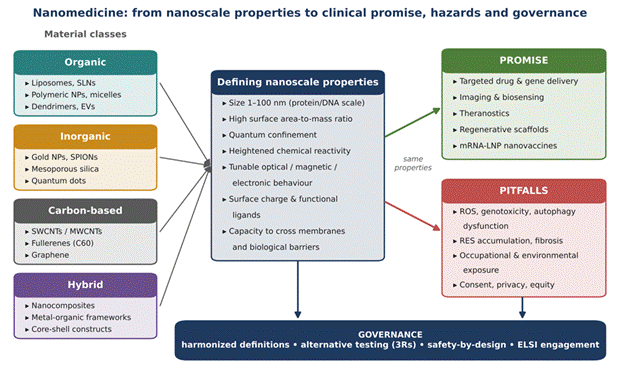
Figure 1. Conceptual framework linking the nanoscale properties of biomedical nanomaterials to their clinical promise, potential hazards, and governance needs. Organic, inorganic, carbon-based, and hybrid nanomaterials (left) share defining nanoscale properties (centre), including a size of 1-100 nm, a high surface area-to-mass ratio, quantum confinement, heightened reactivity, and the capacity to cross biological barriers. These same properties give rise both to clinical benefits (upper right) and to toxicological, environmental, and socio-ethical pitfalls (lower right). Both outcomes converge on a common set of governance responses (bottom). The figure synthesises Sections 2.1-2.6 and underpins the cross-domain analysis presented in the Discussion. Abbreviations: EVs, extracellular vesicles; LNP, lipid nanoparticle; MWCNT/SWCNT, multi-/single-walled carbon nanotube; NPs, nanoparticles; RES, reticuloendothelial system; ROS, reactive oxygen species; SLNs, solid lipid nanoparticles; SPIONs, superparamagnetic iron oxide nanoparticles.
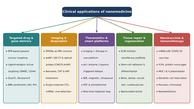
Figure 2. Principal clinical applications of nanomedicine across five therapeutic and diagnostic domains. Each column represents one application domain, with its key mechanisms, representative platforms, and clinical examples. Targeted delivery exploits passive (EPR) and active (ligand-receptor) targeting, exemplified by Doxil® and Abraxane®, and enables passage across the blood-brain barrier; imaging and diagnostics rely on SPIONs, gold nanoparticles, quantum dots, and nanostructured biosensors; theranostic platforms couple imaging with stimulus-triggered release; regenerative scaffolds mimic the extracellular matrix; and nanovaccines, led by mRNA-LNP COVID-19 vaccines, enhance antigen delivery and presentation. Translational status for each domain is detailed in Table 1. Abbreviations: AD, Alzheimer's disease; BBB, blood-brain barrier; CNT, carbon nanotube; CTC, circulating tumour cell; ECM, extracellular matrix; MIP, molecularly imprinted polymer; NIR, near-infrared; PD, Parkinson's disease; PDT, photodynamic therapy; QD, quantum dot; VLP, virus-like particle.
such as mitochondria, lysosomes, and the nucleus (IEEE, 2009; Resnik & Tinkle, 2007). A unifying paradigm in nanotoxicology is oxidative stress driven by overproduction of reactive oxygen species, including hydroxyl radicals, superoxide anions, and hydrogen peroxide (Hannah, 2008; Ma et al., 2024). Excess ROS overwhelms endogenous antioxidant defences, causing lipid peroxidation, membrane degradation, mitochondrial depolarisation, and DNA strand breakage (Ma et al., 2024; Wu et al., 2020). Secondary genotoxicity may follow when nanoparticle-induced inflammation activates macrophages and neutrophils, whose oxidative burst inflicts sustained genomic damage on surrounding tissue (Allon et al., 2016; Ma et al., 2024). Toxicity generally increases as particle size decreases, whereas surface coatings such as polyethylene glycol (PEG) can attenuate ROS generation (Maynard, 2006; Setyawati et al., 2014).
2.3.2 Autophagic Dysfunction and Lysosomal Failure
A second major pathway involves autophagy dysfunction and lysosomal failure (Mao et al., 2016; Wu et al., 2020). When internalised nanoparticles overwhelm autophagosomes or accumulate in lysosomes, they can induce lysosomal membrane permeabilisation (LMP), leakage of hydrolytic enzymes into the cytosol, blockade of autophagic flux, and activation of the NLRP3 inflammasome (Mao et al., 2016; Setyawati et al., 2014; Wu et al., 2020). The consequences include apoptotic or necrotic cell death and tissue-level injury; titanium dioxide nanoparticles, for instance, have been shown to induce autophagy in primary human keratinocytes, linking this pathway to dermal toxicity (Zhao et al., 2013).
2.3.3 Organ Accumulation and Fibre Pathogenicity
Structural factors strongly influence toxicological severity. High-aspect-ratio nanomaterials such as single-walled carbon nanotubes (SWCNTs) share structural features with asbestos fibres; when inhaled or systemically distributed, rigid SWCNTs can provoke frustrated phagocytosis in macrophages, persistent pulmonary inflammation, interstitial fibrosis, granuloma formation, and pleural mesothelioma (Ebbesen et al., 2006; IEEE, 2009; Lam et al., 2006; Schulte & Salamanca-Buentello, 2007). Non-biodegradable inorganic nanoparticles, including silver, titanium dioxide, and silica, may persist within the reticuloendothelial system (RES), concentrating in the liver, spleen, bone marrow, and lymph nodes, which raises concern about chronic organ toxicity and immune dysfunction (Ebbesen et al., 2006; Ma et al., 2024; Resnik & Tinkle, 2007).
2.4 Occupational Hazards and Environmental Life-Cycle Risks
2.4.1 Occupational Exposure
Toxicological risks extend beyond patients to manufacturing plants, research laboratories, and waste-processing facilities, where workers may be exposed through inhalation, dermal contact, or accidental ingestion (Maynard & Kuempel, 2005; Schulte & Salamanca-Buentello, 2007). Airborne nanoparticles can penetrate deep into the pulmonary alveoli, translocate into the bloodstream, and migrate along the olfactory nerve into the central nervous system (Maynard & Kuempel, 2005). Dermal contact with metallic nanoparticles, such as the copper oxide and zinc oxide used in antimicrobial textiles and cosmetics, may breach compromised epidermal barriers, causing keratinocyte apoptosis, sensitisation, and systemic absorption (Ma et al., 2024; Zhao et al., 2013). Existing occupational oversight frameworks were not designed with nanomaterials in mind, and their adequacy has been questioned (Choi & Ramachandran, 2009). The full life cycle of medical nanomaterials and its exposure points are depicted in Figure 4.
2.4.2 Environmental Fate and Ecotoxicity
At the ecosystem level, free nanoparticles released through industrial effluents or medical waste undergo environmental "aging", interacting with organic matter, undergoing sulfidation, and altering their surface charge and aggregation state (Hannah, 2008; Ma et al., 2024). Released nanoscale silver, titanium dioxide, and fullerenes (C60) show acute toxicity towards algae, Daphnia, filter-feeders, and fish, disrupting membrane integrity, inhibiting photosynthesis, and accumulating along food webs (IEEE, 2009; Ma et al., 2024). In agriculture, carbon-based nanomaterials can penetrate plant root cell walls, increasing vulnerability to chemical pollutants and reducing yields of staple crops such as rice and wheat (Purohit et al., 2017). Life-cycle analyses further suggest that producing high-purity carbon nanofibres may carry an environmental toxicity and global warming footprint up to 100 times greater per unit mass than conventional materials such as steel or aluminium (Ma et al., 2024; Purohit et al., 2017). The literature on non-toxicological nano-environmental, health, and safety (EHS) risk analysis remains comparatively thin
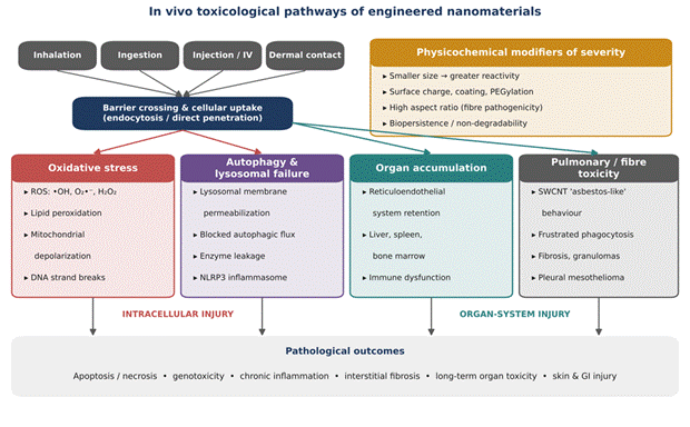
Figure 3. In vivo toxicological pathways of engineered nanomaterials, from routes of exposure to pathological outcomes. Nanomaterials entering by inhalation, ingestion, injection, or dermal contact cross biological barriers and are taken up by cells through endocytosis or direct membrane penetration. Injury then proceeds along two intracellular routes, oxidative stress and autophagic-lysosomal failure, and two organ-level routes, reticuloendothelial accumulation and fibre-like pulmonary toxicity of high-aspect-ratio carbon nanotubes. Physicochemical modifiers (upper right), including size, surface chemistry, aspect ratio, and biopersistence, shape the severity of each pathway. The convergent pathological outcomes (bottom) are summarised with supporting sources in Table 2. Abbreviations: LMP, lysosomal membrane permeabilisation; NLRP3, NOD-like receptor protein 3; PEG, polyethylene glycol; ROS, reactive oxygen species; SWCNT, single-walled carbon nanotube.
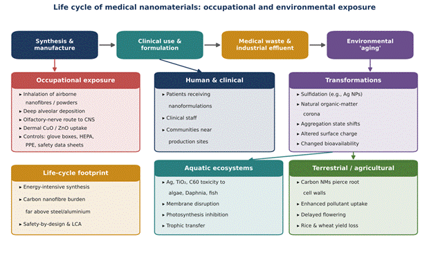
Figure 4. Life cycle of medical nanomaterials, showing occupational, clinical, and environmental exposure points. Nanomaterials move from synthesis and manufacture through clinical use to disposal as medical waste and industrial effluent, after which they undergo environmental aging. Workers may be exposed during production through inhalation and dermal contact, and exposure may reach the central nervous system via the olfactory nerve. Once released, particles are transformed by sulfidation, organic-matter coronas, and aggregation, affecting algae, Daphnia, fish, and staple crops such as rice and wheat. The life-cycle footprint panel notes that carbon nanofibre production carries an environmental burden far exceeding that of conventional materials. Abbreviations: C60, fullerene; CNS, central nervous system; CuO, copper oxide; HEPA, high-efficiency particulate air; LCA, life-cycle assessment; NMs, nanomaterials; PPE, personal protective equipment; TiO2, titanium dioxide; ZnO, zinc oxide.
(Ok & Benneyan, 2009).
2.5 Ethical, Legal, and Societal Implications (ELSI)
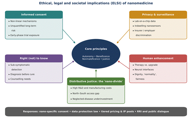
The integration of nanotechnology into clinical medicine challenges classical bioethical frameworks grounded in autonomy, beneficence, nonmaleficence, and justice (Ebbesen et al., 2006; Resnik & Tinkle, 2007). Whether these challenges are genuinely new or simply more acute versions of familiar problems is itself debated (Allon et al., 2016; Kermisch, 2012). The main domains are mapped in Figure 5 and summarised in Table 3.
2.5.1 Informed Consent and Autonomy
Because nanomedicines have complex, non-linear biological mechanisms and long-term risks that cannot yet be quantified, clinicians struggle to communicate potential hazards, and patients find the novel risks difficult to grasp (Kermisch, 2012; Ma et al., 2024). The problem is most acute in early-phase clinical trials, where participants may be exposed to experimental nanomaterials before their biopersistence, metabolic fate, or teratogenic potential is understood (Resnik & Tinkle, 2007).
2.5.2 Privacy, Data Ownership, and Bio-Surveillance
Highly sensitive nano-diagnostics, point-of-care lab-on-a-chip platforms, and indwelling nanosensors can generate continuous streams of personal physiological and genetic data (Allon et al., 2016; Kermisch, 2012). Unauthorised access to, or commercial exploitation of, such data by insurers, employers, or state bodies could lead to genetic discrimination, stigmatisation, and erosion of privacy (Baran, 2016; Kato, 2009).
2.5.3 The Right to Know and the Right Not to Know
Sub-symptomatic screening creates a moral tension between the "right to know" and the "right not to know", particularly when ultra-early detection identifies conditions for which no effective treatment yet exists (Allon et al., 2016; Desai, 2012; Kato, 2009).
2.5.4 Therapy Versus Enhancement
Ethical debate also concerns a possible shift from therapy, which restores normal function, to human physical and cognitive enhancement, which alters baseline capacities (Allon et al., 2016; Durgam & Oroszi, 2025; Ebbesen et al., 2006). Implantable neural interface nanodevices, nano-enabled muscle augmentation, and memory-enhancing nanotherapeutics would blur this boundary (Gordijn, 2006). Critics argue that non-therapeutic enhancement could threaten human dignity, unsettle prevailing notions of "normality", and divide enhanced from non-enhanced individuals (Ebbesen et al., 2006; Resnik & Tinkle, 2007).
2.5.5 Distributive Justice and the "Nano-Divide"
High research and development expenditure and complex manufacturing raise the cost of nanomedicines (Ma et al., 2024). Access may consequently be confined to affluent individuals and nations, widening global health disparities, a phenomenon described as the "nano-divide" (Allon et al., 2016; Baran, 2016; Kermisch, 2012). Developing nations bear a heavy burden of infectious and neglected diseases that low-cost nano-diagnostics or vaccines could mitigate, yet commercial investment remains concentrated in lucrative markets in industrialised countries (Choi, 2005; Sahoo et al., 2010; Uskokovic, 2021).
2.5.6 Public Engagement and Responsible Innovation
Several authors have warned that public backlash, comparable to that seen with genetically modified foods, could follow if social concerns are ignored or if benefits are overstated (Mills & Fleddermann, 2005; Tegart, 2008). Proposals to address this include structured approaches to ethical analysis during research and development (van de Poel, 2008), early and transparent public dialogue (Mills & Fleddermann, 2005), and Responsible Research and Innovation (RRI) frameworks that assign explicit responsibilities to researchers and institutions (Oftedal, 2014).
2.6 Regulatory Landscapes and Alternative Testing Strategies
2.6.1 Statutory Oversight
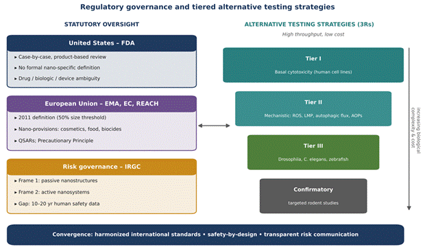
Regulating nanomedicine presents statutory and operational challenges (Baran, 2016; Durgam & Oroszi, 2025). Because nanoscale products frequently combine mechanical, chemical, optical, and biological modes of action, whether a hybrid product should be regulated as a drug, a biological agent, or a medical device often remains ambiguous (Hogle, 2009; Ma et al., 2024). The US Food and Drug Administration (FDA) regulates nanotechnology under existing statutory authorities on a case-by-case, product-based basis, without categorical nano-specific definitions (Resnik & Tinkle, 2007; Yeagle, 2007). The European Union, by contrast, has adopted a formal definition of nanomaterial (European Commission, 2011), integrated nano-specific provisions into legislation on cosmetics, biocides, and food, and relies on the Registration, Evaluation, Authorisation and Restriction of Chemicals (REACH) framework, which incorporates quantitative structure-activity relationships (QSARs) to evaluate hazard (Baran, 2016; Mehlich & Thiele, 2014). At a more conceptual level, risk governance scholars have distinguished passive nanostructures from active, integrated nanosystems, arguing that each warrants a different governance frame (Renn & Roco, 2006). The governance landscape is summarised in Figure 6 and Table 4.
2.6.2 Methodological Gaps in Safety Assessment
A key bottleneck is the lack of standardised toxicological testing protocols tailored to nanomaterials (Mehlich & Thiele, 2014; Ok & Benneyan, 2009; Resnik & Tinkle, 2007). Conventional cytotoxicity assays may produce false-positive or false-negative results because nanoparticles physically interfere with assay reagents (Mehlich & Thiele, 2014; Vinken & Blaauboer, 2017). Long-term human safety data spanning 10-20 years are unavailable for most novel formulations (Ma et al., 2024). Moreover, evaluating the enormous diversity of manufactured nanomaterials with traditional vertebrate models is economically and ethically problematic; such an undertaking has been estimated to cost over US$1 billion, take more than 50 years, and require millions of animals (Wu et al., 2020).
2.6.3 Alternative Testing Strategies and the 3Rs
To address these obstacles, regulatory science is moving towards alternative testing strategies (ATS) aligned with the 3Rs principle of reduction, refinement, and replacement, building on the vision of twenty-first-century toxicity testing (Krewski et al., 2010; National Research Council, 2007). These strategies combine mechanism-based high-throughput screening in human cell lines, focusing on early markers such as ROS generation, autophagic flux disruption, and LMP, with adverse outcome pathway (AOP) reasoning and validated non-mammalian whole-organism models (Vinken & Blaauboer, 2017; Wu et al., 2020). Drosophila melanogaster, Caenorhabditis elegans, and zebrafish (Danio rerio) embryos offer ethically acceptable, cost-effective, high-throughput in vivo test beds (Ecovoiu et al., 2016; Johnston et al., 2018). Direct microtransfer of nanomaterials into Drosophila embryos, together with somatic mutation and comet assays, permits rapid evaluation of tissue-specific toxicity, genotoxicity, and developmental delay, bridging the gap between cell assays and rodent studies (Ecovoiu et al., 2016; Vega-Alvarez et al., 2014). Ultimately, harmonised international characterisation standards, transparent risk communication, safety-by-design engineering, and proactive public engagement, modelled on the ELSI programme of the Human Genome Project, will be needed if nanomedicine is to fulfil its potential safely, ethically, and equitably (Mills & Fleddermann, 2005; Renn & Roco, 2006).