Data Modeling

Mathematical and Computational Data Modeling
1
Citations
7k
Views
37
Articles
Your new experience awaits. Try the new design now and help us make it even better
Switch to the new experience
Figures and Tables
REVIEWS   (Open Access)

Controlling Outbreaks of Acute Contagious Conjunctivitis Infection: A Diagnostic Review and Proposal for Mobile-Based Screening

Kamruzzaman Mithu 1*

+ Author Affiliations

Data Modeling 1 (1) 1-14 https://doi.org/10.25163/data.1110859

Submitted: 09 September 2026 Revised: 01 November 2020  Published: 11 November 2020 


Abstract

Conjunctivitis is, in some ways, an unassuming diagnosis — red eyes, some discharge, a bit of grittiness — yet it turns out to be one of the more frequently encountered conditions across both primary care and ophthalmology settings worldwide, carrying a financial burden that is anything but small. This review set out, somewhat modestly, to pull together the existing evidence on how conjunctivitis is recognized, classified, and managed, with a particular eye toward the persistent difficulty of telling viral, bacterial, and allergic subtypes apart at the bedside. A structured literature search was conducted across three major databases using a focused set of keywords, restricted to publications from 2013 through early 2020, from which ten studies were ultimately retained for synthesis. What emerged was a fairly consistent picture of diagnostic uncertainty: clinical heuristics long treated as reliable — discharge type, reaction pattern, symptom timing — proved considerably less trustworthy than textbooks tend to suggest, and this ambiguity appears to feed directly into patterns of antibiotic overprescribing, particularly outside specialist care. At the same time, a smaller subset of findings pointed toward conjunctivitis occasionally serving as a visible marker of broader systemic disease, underscoring the value of a careful history even in seemingly routine cases. Building on these findings, this review also outlines a proposed smartphone-based screening tool intended to make earlier, more accessible triage possible, especially for populations with limited access to eye care. Taken together, the evidence suggests that improving diagnostic confidence — whether through better clinical training or emerging screening technology — may be one of the more practical levers available for reducing unnecessary treatment and limiting the spread of conjunctivitis outbreaks at a community level.

Keywords: conjunctivitis; pink eye; red eye; controlling outbreaks of conjunctivitis; spread control of pink eye; solutions for controlling conjunctivitis.

1. Introduction

Anyone who has sat in a crowded waiting room during conjunctivitis season knows the drill: red, weepy eyes, a box of tissues passed hand to hand, and a low hum of worry about who will catch it next. It seems like such a small complaint on paper — an inflamed conjunctiva, some discharge, maybe a little grit under the eyelid — and yet conjunctivitis manages to be one of the most frequently seen conditions in both general medicine and ophthalmology clinics worldwide. The symptoms themselves are not mysterious: discomfort, engorged blood vessels, ocular discharge, and swelling of the conjunctival tissue. What is less obvious, at least until you look closely at who is actually diagnosing and treating these cases, is how much of this burden falls outside specialist care altogether. Non-ophthalmologists — nurse practitioners, pediatricians, internists, family doctors, and other frontline clinicians — are responsible for diagnosing more than 80% of all acute conjunctivitis cases (Shekhawat et al., 2017). That figure alone reframes conjunctivitis less as an "eye disease" in the narrow sense and more as an everyday primary-care problem, one that nearly every type of clinician will eventually have to manage.

And the costs, it turns out, are not trivial either. Bacterial conjunctivitis alone is estimated to cost the United States somewhere around $857 million annually (Smith & Waycaster, 2009) — a figure that becomes easier to understand once you account for how many office visits, prescriptions, and missed workdays accumulate behind it. Much of that expense seems to trace back to prescribing habits that do not always match the underlying cause. Antibiotic eye drops are prescribed in roughly 60% of acute conjunctivitis cases, and — somewhat counterintuitively — most of those prescriptions come from clinicians who are not ophthalmologists (Shekhawat et al., 2017). The disparity is even more pronounced depending on setting: only 36% of patients evaluated by an ophthalmologist left with antibiotic drops, compared with 68% of those seen in an emergency department (Shekhawat et al., 2017). Whether this reflects genuine diagnostic uncertainty, time pressure, or simply a lower threshold for "just in case" prescribing is hard to say with certainty, but the pattern itself is striking. Socioeconomic status appears to play a role too; patients from higher socioeconomic backgrounds were more likely both to obtain a prescription and to fill and complete it (Shekhawat et al., 2017), which raises its own questions about access that go somewhat beyond a purely clinical discussion.

Part of the difficulty, arguably, is that "conjunctivitis" is not really one disease but a loose umbrella term covering a fairly wide range of underlying processes. It can be organized along several axes at once — cause, severity, duration, or how much surrounding tissue is involved — and none of these schemes is mutually exclusive. Broadly, etiology splits into infectious and non-infectious categories: allergic reactions and toxic or irritant exposures dominate the non-infectious side, while viral and bacterial pathogens account for the overwhelming majority of infectious cases. This distinction matters in practice, since appropriate management — and whether antibiotics are indicated at all — hinges on getting the classification right, or at least reasonably close.

Time course offers another useful, if somewhat blunt, way of thinking about the condition. Conjunctivitis is generally described as acute when it comes on quickly and resolves within about four weeks, subacute when it stretches somewhat beyond that window, and chronic once it persists past the four-week mark (Ryder & Benson, 2020). Severity adds a further layer of nuance: cases with pronounced symptoms and heavy mucopurulent discharge are typically labeled severe, in contrast to the milder, more self-limited presentations that make up the bulk of everyday clinical encounters. The disease can also spread — not in the contagious sense, but anatomically — to involve neighboring structures, producing blepharoconjunctivitis when the eyelid margins are affected or keratoconjunctivitis when the cornea becomes involved as well.

It would be a mistake, too, to think of conjunctivitis purely as an isolated ocular event, disconnected from the rest of the body. In a meaningful subset of patients, it turns out to be one visible piece of a larger systemic picture. Immune-mediated conditions are a good example: keratoconjunctivitis sicca can accompany rheumatoid arthritis, and conjunctival involvement is well recognized in both Stevens-Johnson syndrome and Reiter's syndrome. Nutritional deficiency, particularly of vitamin A, is another recognized contributor. Even certain rare congenital metabolic disorders — Richner-Hanhart syndrome and porphyria among them — have been linked to conjunctival findings (de Laet et al., 2013; Sati et al., 2013), a reminder that what looks, at first glance, like a simple red eye can occasionally be the first clue to something considerably more systemic. None of this is to say that every case warrants an extensive workup — most decidedly do not — but it does underscore why a careful history and a willingness to look beyond the obvious remain important, even for a condition as ostensibly mundane as pink eye.

There is also the matter of simply telling conjunctivitis apart from everything else that can make an eye look red — a task that sounds trivial until you actually try it at the bedside. "Red eye" is one of those catch-all presentations that can account for as much as 1% of all primary care visits (Narayana & McGee, 2015), and it covers everything from a harmless subconjunctival hemorrhage to sight-threatening conditions such as keratitis, scleritis, and anterior uveitis. For a long time, clinical teaching held that visual disturbance, severe pain, and photophobia were the red flags separating benign conjunctival disease from something more dangerous (Narayana & McGee, 2015). More recent work has refined that picture somewhat: a large meta-analysis found that the combination of anisocoria and mild photophobia was strongly associated with serious eye disease, correctly flagging roughly 59% of patients with conditions like keratitis and anterior uveitis (Narayana & McGee, 2015). It is not a perfect screening tool — nothing at the bedside ever quite is — but it does give busy clinicians a reasonably efficient shortcut for deciding who needs urgent referral and who can be managed more conservatively.

Getting the history right seems to matter just as much as the exam itself, maybe more. A focused ocular history typically covers onset and duration, laterality, any change in vision, contact lens use, itching, associated symptoms like sinusitis or upper respiratory infection, prior episodes, known allergies, current medications, and exposure to chemical irritants — together with constitutional symptoms and any known contact with an infected person, all of which help narrow the differential. On exam, palpating the periauricular and submandibular lymph nodes is a small step that is easy to skip but genuinely useful, and slit-lamp examination remains the most reliable way to characterize discharge and inspect the ocular surface, including corneal opacities, infiltrates, and the palpebral conjunctiva for follicles, papillae, pseudomembranes, or symblepharon.

Even with a careful history and exam, though, distinguishing infectious from non-infectious conjunctivitis — and, within the infectious category, viral from bacterial — turns out to be trickier than the textbooks sometimes suggest. Clinical clues traditionally used to flag infectious disease include ocular discharge, conjunctival injection, redness, eyelashes matted together on waking, a gritty sensation, lid or conjunctival swelling, and a history of contact with an infected person (Everitt & Little, 2002). Allergic conjunctivitis, meanwhile, is thought to be underdiagnosed and undertreated relative to how common it actually is (La Rosa et al., 2013); when significant corneal involvement is absent, it tends to present with itching, chemosis, and redness, with conjunctival edema often outpacing conjunctival hyperemia in severity (Friedlaender, 2011). Two more specific allergic phenotypes are worth flagging: vernal keratoconjunctivitis, marked by large papillae on the superior tarsal conjunctiva alongside intense itching, and atopic keratoconjunctivitis, more often confirmed by conjunctival scarring and anterior subcapsular cataract (Bielory & Bielory, 2010). Chronic toxic conjunctivitis rounds out this picture, typically presenting with watery discharge, punctate epithelial erosions, an initial follicular reaction that shifts toward a papillary one, and eyelid dermatitis (Wilson-Holt & Dart, 1989; Soparkar et al., 1997; van Ketel & Melzer-van Riemsdijk, 1980).

Distinguishing viral from bacterial conjunctivitis on clinical grounds alone, unfortunately, tends to be even less reliable than clinicians would like to believe. One study found that even experienced ocular surface disease centers correctly identified adenoviral conjunctivitis only 48% of the time based on presentation, with bacterial pathogens confirmed in only about half of cases labeled clinically as bacterial, and bacteria turning up in as many as 52% of cases initially suspected to be viral (Woodland et al., 1992). Textbook heuristics have long circulated regardless: second-eye involvement within 24 to 48 hours pointing toward bacterial disease, involvement after 48 hours with enlarged periauricular nodes suggesting a viral etiology, follicular reactions leaning viral, and papillary or pseudomembranous reactions leaning bacterial (Everitt & Little, 2002). These associations, once treated almost as diagnostic gospel, have not held up especially well under closer scrutiny. A meta-analysis pooling data from 622 patients across three trials found that purulent discharge and mild-to-moderate redness were, if anything, associated with a lower likelihood of benefiting from topical antibiotics (Friedlaender, 2011) — which rather undercuts the intuitive assumption that "more purulent equals more bacterial." One somewhat more durable finding did emerge from a 2013 study: eyelid gluing on waking, combined with age over 50, was associated with a meaningfully higher likelihood of a positive bacterial culture (Bielory & Frohman, 1992) — a small but useful anchor amid an otherwise murky diagnostic landscape.

All of this — the diagnostic uncertainty, the overlapping presentations, the sheer frequency with which non-specialists are asked to make these calls without reliable bedside tools — is really what motivates the present work. If clinicians cannot easily and confidently tell viral, bacterial, and allergic conjunctivitis apart from history and examination alone, then antibiotic overuse, delayed appropriate treatment, and unnecessary outbreak spread become almost inevitable byproducts of an otherwise well-intentioned system. This paper attempts to pull together the existing evidence on conjunctivitis classification, diagnosis, and management, and to use it as the foundation for something more concrete: a proposed mobile application intended to help patients and clinicians alike screen for conjunctivitis more quickly, more cheaply, and with wider reach — particularly in rural or underserved areas where access to ophthalmology is limited. The hope, admittedly a modest one, is that better tools for early recognition might translate into more appropriate treatment decisions and, ultimately, more effective control of conjunctivitis outbreaks at the community level.
2. Methods

2.1 Study Design and Rationale

This paper is, at its core, a narrative review — not a systematic review in the formal PRISMA sense, though we tried to borrow enough of that discipline to make the search process transparent and, ideally, repeatable by another team working from the same starting point. Conjunctivitis, as outlined in the Introduction, is not a single disease so much as a cluster of overlapping presentations (viral, bacterial, allergic, toxic, and occasionally systemic), and the literature reflects that same fragmentation — scattered across ophthalmology journals, primary care literature, and the occasional case report tucked into a specialty journal nobody thinks to search first. Given that spread, a narrow, single-database search seemed unlikely to capture the full picture, so we opted for a somewhat broader, multi-database strategy instead.

2.2 Information Sources

Three databases were searched: PubMed, the ISI Web of Knowledge, and the Cochrane Library. The choice was fairly deliberate — PubMed for its depth of biomedical coverage, Web of Knowledge to pick up citation-linked material that PubMed sometimes misses, and Cochrane specifically to check for any systematic reviews or meta-analyses that might already have synthesized parts of this literature more rigorously than we could on our own. No single database, in our experience, reliably covers everything on a topic this heterogeneous, so the redundancy across the three was intentional rather than an oversight.

2.3 Search Strategy

The search itself relied on a small, purpose-built set of keyword combinations: "therapy of bacterial conjunctivitis," "treatment of viral conjunctivitis," "allergic conjunctivitis," and "viral conjunctivitis." These terms were chosen to map onto the major etiological categories discussed above — infectious (viral and bacterial) and non-infectious (principally allergic) — since the intent was to build a picture broad enough to support differential diagnosis, not just treatment of one subtype in isolation. No language restrictions were applied at the search stage; we would rather have screened out a non-English article for other reasons than miss it entirely because of the language field. In hindsight, a more exhaustive strategy might have added subject headings (MeSH terms) or truncation/wildcard variants to catch spelling differences (e.g., "conjunctivitis" versus regional variants), and we would recommend that refinement to anyone attempting to reproduce or extend this search.

2.4 Eligibility Criteria

Time boundaries mattered here almost as much as the keywords. We restricted eligible publications to those appearing between March 2013 and February 2020, reasoning that older evidence — while not necessarily wrong — was less likely to reflect current diagnostic practices, current antibiotic-resistance patterns, or contemporary classification schemes such as the acute/subacute/chronic framework described by Ryder and Benson (2020). Articles were further screened for methodological robustness; case reports and low-evidence sources were deprioritized in favor of the most robust evidence-based articles available on each subtopic, though — and this is worth being candid about — "most robust" was applied as a qualitative judgment by the reviewing author rather than through a formal quality-scoring instrument like GRADE or the Cochrane risk-of-bias tool. That is a limitation, and one we would encourage future reviewers to address more formally.

2.5 Study Selection Process

Titles and abstracts identified through the search were screened first, with full texts retrieved for anything that appeared potentially relevant to the diagnosis, classification, or management of conjunctivitis. Selection was performed by a single reviewer, which is, admittedly, a departure from the dual-reviewer standard recommended for systematic reviews — a point we flag here rather than gloss over, since transparency about that limitation is part of what makes a methods section actually useful to someone trying to replicate or critique the work.

2.6 Data Charting and Synthesis

From the pool of retrieved articles, ten publications ultimately met the inclusion criteria and were carried forward into the synthesis presented in this review. The earliest of these was published in 2013, and the most recent in 2020 — consistent with, and in fact defining, the search window described above. Rather than pooling quantitative data (which would have required considerably more homogeneous outcome measures than these ten studies offered), we synthesized findings narratively, organizing them around the clinical questions that seemed most pressing to practicing clinicians: how to recognize conjunctivitis, how to distinguish it from other causes of red eye, how to differentiate viral from bacterial disease, and how systemic conditions might present with conjunctival involvement.

2.7 Reproducibility Considerations

For anyone hoping to reproduce or update this search — and we would genuinely encourage that, given how quickly the underlying evidence base can shift — the essential parameters are these: the three databases named above, the four keyword strings, no language filter, and a publication window that would now need to be re-anchored to a later cutoff date. We would also suggest, as a methodological improvement over our own approach, registering the review protocol in advance (for instance through PROSPERO) and applying a structured critical-appraisal tool during full-text screening, neither of which was done here but both of which would strengthen the rigor of any follow-up effort.

3. Results

3.1 Overview of Included Evidence

Ten studies, spanning 2013 to 2020, ultimately formed the evidentiary backbone of this review — a modest number, admittedly, but one that turned out to cover a surprisingly wide swath of the clinical picture: how red eye is recognized, how conjunctivitis is distinguished from more serious mimics, how the major subtypes differ from one another, and what, if anything, can realistically be done to interrupt transmission. What follows is organized roughly along those same lines, moving from bedside recognition through laboratory confirmation and finally toward the technology-based screening approach proposed here.

3.2 Distinguishing Conjunctivitis from Other Causes of Red Eye

Before conjunctivitis can even be diagnosed, it first has to be separated from a fairly long list of other conditions that can make an eye look red — some of them harmless, several of them genuinely urgent. This differential, laid out across the underlying conditions and their typical clinical features, is not a small undertaking; the reasoning behind it is summarized in (Table 1), which draws together conjunctivitis, subconjunctival hemorrhage, keratitis, scleritis, and anterior uveitis under a single comparative framework. Somewhat more granular differentials — dry eye, blepharitis, pterygium, angle-closure glaucoma, and so on — are captured separately in (Table 4), since these conditions rarely masquerade convincingly as conjunctivitis but still belong on a clinician's radar when a patient's presentation does not quite fit the expected pattern.

A structured approach to history-taking helps narrow this list considerably faster than symptom review alone. Alarming features — decreased vision, severe pain, a poorly reacting pupil, anisocoria, orbital signs — point toward uveitis, scleritis, keratitis, or, less commonly, orbital or parasellar pathology; chronicity and recurrence patterns, meanwhile, help separate infectious and allergic conjunctivitis from conjunctivitis tied to systemic disease or chronic toxic exposure. This decision logic, organized around alarming signs, chronicity, associated systemic symptoms, and drug history, is set out in (Table 3).

3.3 Differentiating Viral, Bacterial, and Allergic Conjunctivitis

Once conjunctivitis itself has been confirmed — or at least assumed, in the absence of alarming features — the next question is almost always etiological: viral, bacterial, or allergic. Here the evidence is, frankly, a little humbling. Discharge character, conjunctival reaction pattern, laterality, and associated findings each carry some diagnostic weight, but none of them, on their own, are especially reliable; (Table 2) lays these features out side by side, and the overlap between columns is arguably as instructive as the differences. Viral conjunctivitis tends to

Table 1: Common Causes and Clinical Features of "Red Eye."This table compares the major underlying causes of red eye — conjunctivitis, subconjunctival hemorrhage, keratitis, scleritis, and anterior uveitis — alongside their typical clinical presentations and relative severity. It is intended to help clinicians quickly distinguish benign, self-limited conditions from those requiring urgent ophthalmologic referral. Notably, the combination of anisocoria and mild photophobia identifies roughly 59% of patients with serious eye disorders such as keratitis and anterior uveitis, underscoring the value of these two findings as red flags during initial assessment.

Underlying Condition

Typical Clinical Features

Relative Severity

Conjunctivitis

Diffuse conjunctival injection, discharge, mild discomfort, minimal pain

Benign / self-limited (most cases)

Subconjunctival hemorrhage

Localized, sharply demarcated redness; no discharge or pain

Benign

Keratitis

Pain, photophobia, corneal opacity or infiltrate, visual impairment

Serious — requires urgent referral

Scleritis

Severe, boring pain; deep redness; may involve systemic disease

Serious — requires urgent referral

Anterior uveitis

Photophobia, anisocoria, ciliary flush, pain

Serious — requires urgent referral

Table 2: Differentiating Viral, Bacterial, and Allergic Conjunctivitis by Clinical Feature. This table outlines how discharge character, conjunctival reaction pattern, itching, laterality/onset, and associated findings vary across the three principal conjunctivitis subtypes. It is meant to support — though not replace — clinical differentiation, since associations based on signs and symptoms alone carry limited diagnostic accuracy and should be confirmed with laboratory testing when the diagnosis remains uncertain or treatment is not responding as expected.

Feature

Viral

Bacterial

Allergic

Discharge

Watery / mucoid

Purulent / mucopurulent

Watery, stringy mucus

Conjunctival reaction

Follicular

Papillary or pseudomembranous

Papillary

Itching

Uncommon

Typically absent

Prominent

Laterality / onset

Often starts unilateral, second eye involved after >48 h with lymphadenopathy

Second eye often involved within 24–48 h

Usually bilateral

Associated findings

Preauricular lymphadenopathy, recent URI (historically reported, low evidence)

Eyelids glued shut on waking, especially age > 50

Seasonal pattern, allergic history

present with watery or mucoid discharge and a follicular reaction, often starting unilaterally before the second eye becomes involved more than 48 hours later, sometimes with preauricular lymphadenopathy. Bacterial conjunctivitis, by contrast, tends to produce purulent or mucopurulent discharge with a papillary or pseudomembranous reaction, second-eye involvement typically arriving sooner — within 24 to 48 hours — and, in older patients particularly, the classic complaint of eyelids "glued shut" on waking. Allergic conjunctivitis sits somewhat apart from both, marked by prominent itching, a watery, stringy mucus discharge, and a bilateral pattern that usually tracks with a seasonal or allergic history.

3.4 Laboratory Confirmation

Clinical impression alone, as the preceding section makes clear, only goes so far, which is presumably why laboratory testing retains a role — albeit a fairly selective one. Conjunctival cultures are not routine; they tend to be reserved for suspected neonatal conjunctivitis, recurrent or treatment-resistant disease, cases with severe purulent discharge, or a suspicion of gonococcal or chlamydial infection (Bielory & Bielory, 2010). Swabs are best collected before antimicrobial treatment begins, then plated across different growth media depending on what is suspected: Sabouraud agar for fungal pathogens in immunocompromised patients or those with persistent blepharitis, and anaerobic culture for patients with a recent history of ocular surgery or trauma (Wilson-Holt & Dart, 1989). None of this is exotic microbiology, but it is easy to overlook in a busy clinic, and getting the sequencing right — swab first, treat second — matters more than it might seem.

3.5 Classification of Conjunctivitis by Etiology

Pulling all of this together, conjunctivitis can reasonably be organized into five broad categories: viral, bacterial, allergic, toxic, and nonspecific, with viral disease accounting for the majority of infectious cases across both adults and children. Within viral conjunctivitis, the recognized subtypes include adenoviral conjunctivitis (by far the most common), herpetic conjunctivitis, acute hemorrhagic conjunctivitis, and a smaller miscellaneous group. Bacterial conjunctivitis includes methicillin-resistant S. aureus conjunctivitis, chlamydial conjunctivitis, and gonococcal conjunctivitis — the latter two carrying particular urgency given their potential for corneal involvement if left untreated. Allergic conjunctivitis, meanwhile, splits further still into seasonal and perennial allergic conjunctivitis, vernal and atopic keratoconjunctivitis, giant papillary conjunctivitis, and simple contact allergy. This full classification scheme, together with its infectious and non-infectious branches, is depicted in (Figure 1) — a visual that, admittedly, looks more like a family tree than a flowchart, but seems to capture the relationships reasonably well.

3.6 Strategies for Interrupting Transmission

Because both bacterial and viral conjunctivitis spread readily from person to person, a meaningful part of "controlling outbreaks" turns out to have very little to do with medication and almost everything to do with hygiene — unglamorous, perhaps, but evidently effective. For patients who already have conjunctivitis, the core recommendations include frequent handwashing (at least twenty seconds, soap and warm water, or an alcohol-based sanitizer with at least 60% alcohol when soap is unavailable), particular care before and after administering eye drops or cleaning an affected eye, avoiding rubbing or touching the eyes, using separate eye-drop bottles for each eye if only one is affected, laundering pillowcases and towels in hot water, discontinuing contact-lens wear until cleared by an eye care provider, and — perhaps easiest to forget — never sharing personal items such as washcloths, cosmetics, or lens cases. For those simply in close contact with an infected person, the same basic principles apply in reverse: frequent handwashing, avoiding contact between unwashed hands and the eyes, and refraining from sharing any item that might have touched the infected eye. Once the infection clears, a final round of precautions — discarding any cosmetic brushes or disposable lenses used while infected, and thoroughly cleaning eyeglasses and reusable items — helps guard against reinfection. None of this is particularly novel guidance, but its consistent application appears to be where a fair amount of outbreak control quietly succeeds or fails.

3.7 Proposed Mobile Screening Application

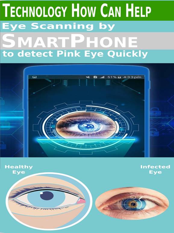
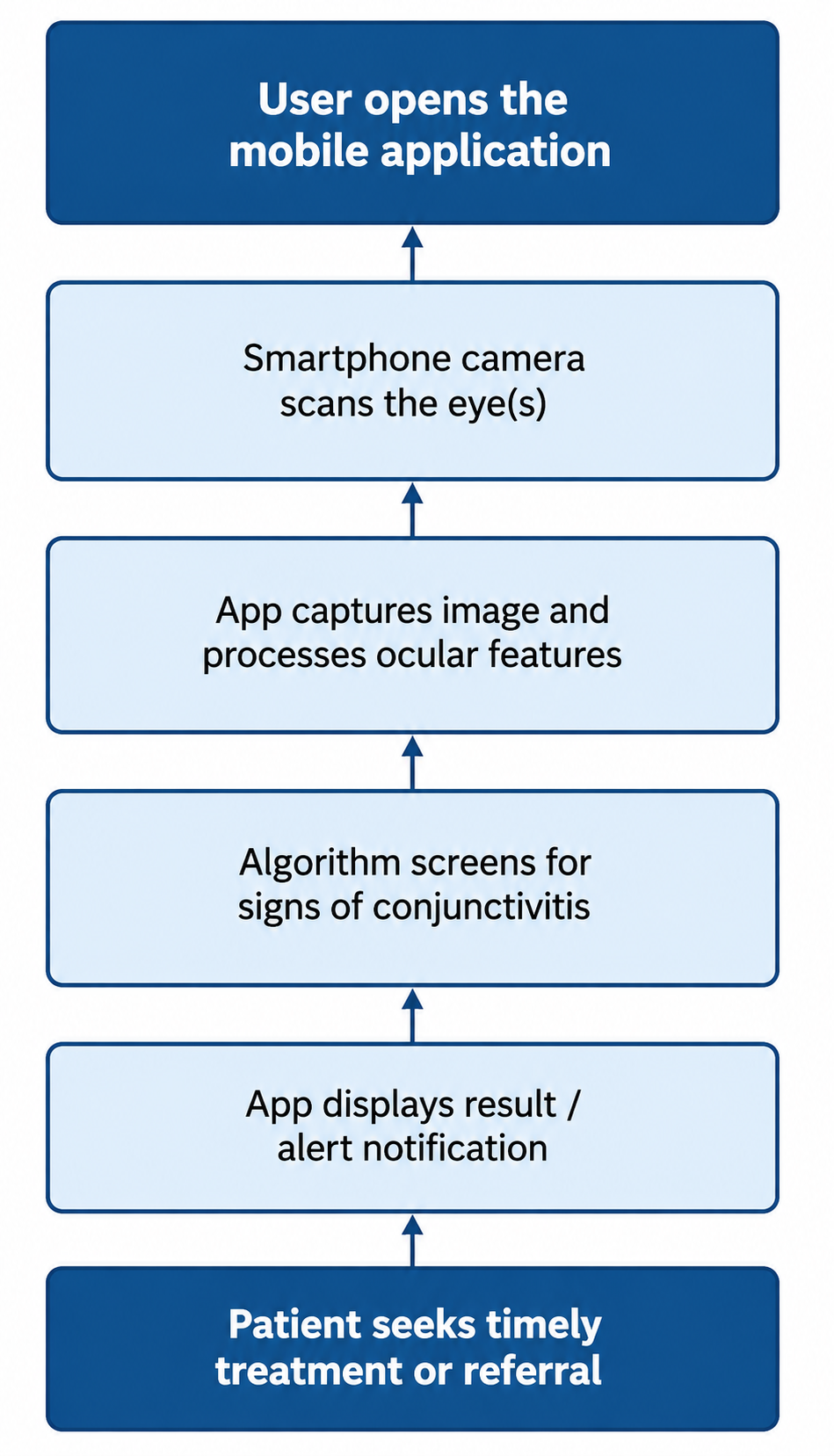
Building on this diagnostic and preventive framework, the review's applied component centers on a proposed mobile application designed to bring faster, more accessible conjunctivitis screening to smartphone users — a population that, at this point, covers more than half the world. The intended workflow is fairly simple in concept, even if the underlying image-recognition engineering is not: a user holds their smartphone up so the camera can scan the eye, the application processes that image, and a notification is returned indicating whether findings are consistent with conjunctivitis and, ideally, which subtype seems most likely. This proposed workflow is illustrated in (Figure 2), while a broader conceptual illustration of the intended use case — summarized under the working phrase "detect quick, control outbreak, protect yourself and others" — appears in (Figure 3). The appeal of this approach, at least in theory, lies less in replacing clinical judgment than in shortening the interval between symptom onset and appropriate care-seeking, particularly for people in rural or remote areas where same-day access to an eye care provider is often not realistic (Smith & Waycaster, 2009).

4. Discussion

4.1 Interpreting the Diagnostic Picture

Stepping back from the individual findings, what emerges from this review is a condition that looks deceptively simple from the outside and turns out to be considerably messier once you actually try to pin it down. Conjunctivitis is common enough that most clinicians — ophthalmologists and non-ophthalmologists alike — will encounter it regularly, and yet the evidence gathered here suggests that confident, symptom-based differentiation between its major subtypes remains genuinely difficult, even for experienced eyes. The overlapping features summarized in (Table 2) make this point almost better than any narrative could: discharge character, reaction pattern, and laterality each shift the probability of a diagnosis somewhat, but none of them shift it decisively, and it is easy to imagine two clinicians looking at the same patient and reasonably arriving at different conclusions. That ambiguity, more than any single statistic, seems to be the real story here.

4.2 Why Misdiagnosis Persists

It is tempting to assume that better training alone would fix this — that clinicians simply need to memorize the textbook associations more thoroughly. But the evidence complicates that assumption rather than supporting it. Several of the heuristics long taught in ophthalmology curricula (second-eye timing, follicular versus papillary reaction, presence or absence of itching) were, at some point, elevated to something close to diagnostic doctrine, used not just at the bedside but to select participants for clinical trials. And yet, when tested directly, these associations have not consistently held up. Facilities with genuine expertise in ocular surface disease still misclassified a substantial share of adenoviral cases, bacterial pathogens turned up in roughly half of clinically diagnosed bacterial conjunctivitis, and — perhaps most tellingly — bacteria were isolated in over half of cases initially suspected to be viral. If specialists working from a full clinical picture struggle with this distinction, it seems almost unreasonable to expect the same accuracy from a rushed primary care visit or an emergency department encounter, which may partly explain the prescribing disparities noted earlier in this paper. This is, in a sense, less a failure of individual clinicians than a limitation baked into the disease itself.

4.3 The Antibiotic Overuse Problem

This diagnostic uncertainty does not exist in a vacuum; it has a fairly direct downstream consequence, and that consequence is antibiotic overprescribing. When roughly 60% of acute conjunctivitis cases receive antibiotic eye drops — and when the majority of that prescribing comes from non-ophthalmologists working with limited diagnostic confidence — it becomes hard to avoid the conclusion that a meaningful share of these prescriptions are, in effect, hedges against uncertainty rather than responses to confirmed bacterial disease. The financial cost of this pattern is not trivial (Smith & Waycaster, 2009), but the clinical cost may matter more: unnecessary antibiotic exposure carries its own risks, from resistance development to disruption of the ocular microbiome, and it does nothing to address the viral or allergic cases that make up the bulk of conjunctivitis presentations in the first place. There is also something worth sitting with in the finding that purulent discharge and worse-appearing redness were, if anything, associated with lower antibiotic benefit — a result that runs directly counter to prescribing intuition, and one that probably deserves more attention than it typically receives in clinical training.

4.4 Conjunctivitis as a Marker of Systemic Disease

A further complication, easy to overlook amid the focus on viral-versus-bacterial distinctions, is that conjunctivitis is sometimes not a standalone diagnosis at all but a visible marker of something happening elsewhere in the body. Immune-mediated conditions, nutritional deficiencies, and rare congenital metabolic disorders can all produce conjunctival findings that, superficially, resemble ordinary infectious or allergic disease. This is not the common case

Figure 1: Classification of Conjunctivitis by Etiology. This figure depicts the overall classification of conjunctivitis into five major categories — viral, bacterial, allergic, toxic, and nonspecific — along with their principal recognized subtypes (e.g., adenoviral and herpetic conjunctivitis; chlamydial and gonococcal conjunctivitis; seasonal, perennial, vernal, and atopic allergic subtypes). It provides a visual reference for understanding how the various forms of conjunctivitis relate to one another within the broader infectious and non-infectious framework.

Table 3. Guideline for Differentiating the Major Etiologies of Conjunctivitis Based on History and Examination. This table organizes clinical history and examination findings — including alarming signs, chronicity, associated systemic symptoms, and relevant drug history — into a structured framework for narrowing the differential diagnosis. It is designed to help clinicians flag cases warranting urgent referral (e.g., uveitis, scleritis, glaucoma) versus those more consistent with infectious, allergic, or systemic-disease-associated conjunctivitis, including acute reactions such as Stevens-Johnson syndrome and toxic epidermal necrolysis.

Clinical history and exam findings

Most probable etiologies

Alarming signs and symptoms


Decreased vision, severe pain, painful pupillary reaction, anisocoria, orbital signs

Uveitis, scleritis, keratitis, glaucoma, orbital, or parasellar pathology

Chronicity


Sudden onset, lasting less than four weeks

Infectious conjunctivitis, allergic conjunctivitis, acute systemic reactions (SJS/TEN)

Insidious onset, chronic course

Conjunctivitis associated with systemic diseases, toxic conjunctivitis, allergic conjunctivitis

Recurrent course

Allergic conjunctivitis, conjunctivitis associated with systemic diseases

Associated symptoms


Skin lesions, arthropathy, genito-perineal involvement, oropharyngeal lesions

Conjunctivitis associated with systemic diseases, infectious diseases

Drug history


Long-term eye drop usage

Toxic conjunctivitis, allergic conjunctivitis

Recent initiation of a systemic medication

Acute systemic reactions (SJS/TEN)

Table 4. Selected Non-Conjunctivitis Etiologies of Red Eye and Their Distinguishing Features. This table catalogs conditions that can mimic conjunctivitis — including dry eye, blepharitis, pterygium, corneal abrasion, scleritis, iritis, angle-closure glaucoma, carotid-cavernous fistula, endophthalmitis, and orbital cellulitis — alongside their characteristic symptoms and exam findings. It serves as a broader differential checklist for clinicians evaluating red eye presentations that do not clearly fit a straightforward conjunctivitis pattern.

Differential diagnosis

Symptoms

Exam findings

Dry eyes

Burning and FB sensation. Symptoms are usually transient, worse with reading or watching TV due to decreased blinking. Symptoms are worse in dry, cold, and windy environments due to increased evaporation

Bilateral redness, superficial punctate keratopathy, meibomian glands dysfunction, decreased tear break-up time, small tear meniscus

Blepharitis

Similar to dry eyes

Redness greater at the margins of eyelids, inflammation, telangiectasia, and crust around eyelashes

Pterygium

Recurrent ocular redness

Visible conjunctival extension over the cornea

Hordeolum, chalazion

Eyelid pain and swelling

Palpable eyelid mass, may be tender or not

Anterior segment tumors

Variable

Variable

Corneal abrasion, keratitis, corneal foreign body

FB sensation, relevant history including contact lens usage and occupational exposure

Corneal epithelial defects, corneal infiltration, corneal FB

Contact lens overwear

Relevant history

Corneal epithelial defect

Subconjunctival hemorrhage

Ocular redness

Blood under conjunctiva

Scleritis

Decreased vision, moderate to severe pain

Redness, bluish scleral hue

Iritis

Photophobia, pain, blurred vision. Symptoms are usually bilateral

Decreased vision, poorly reacting pupils, constant eye pain radiating to temple and brow. Redness, severe photophobia, presence of inflammatory cells in the anterior chamber

Angle closure glaucoma

Headaches, nausea, vomiting, ocular pain, decreased vision, light sensitivity, and seeing haloes around lights. Symptoms are usually unilateral

Firm eye upon palpation, ocular redness with limbal injection. Appearance of a hazy/steamy cornea, moderately dilated pupils that are unreactive to light

Carotid cavernous fistula

Chronic red eye, may have a history of head trauma

Dilated tortuous vessels (corkscrew vessels), bruits upon auscultation with a stethoscope

Endophthalmitis

Severe pain, photophobia, may have a history of eye surgery or ocular trauma

Redness, pus in the anterior chamber and photophobia

Figure 2: Proposed Mobile Application Workflow for Rapid Conjunctivitis Screening. This figure illustrates the intended user workflow for the proposed smartphone-based screening tool, from image capture through automated analysis to a notification advising the user on next steps. It is meant to convey how the application could shorten the interval between symptom onset and appropriate care-seeking, particularly for users in rural or underserved areas with limited access to eye care specialists.

Figure 3: Conceptual Illustration — "Detect Quick, Control Outbreak, Protect Yourself and Others."This figure presents a broader conceptual overview of the proposed screening tool's public health rationale, linking early detection to outbreak containment and personal/community protection. It is intended to communicate the tool's underlying value proposition at a glance, rather than to depict specific technical or diagnostic details.

— most red eyes are exactly what they appear to be — but the possibility is worth holding onto, particularly for patients whose presentation is atypical, chronic, or simply refuses to respond to standard treatment. The alarming-features framework summarized in (Table 3) offers a reasonably practical way of flagging these cases without requiring an extensive workup for every patient who walks through the door.

4.5 Rethinking the Role of Laboratory Testing

Given how unreliable clinical impression alone appears to be, one might expect laboratory confirmation to feature more prominently in everyday practice than it currently does. In reality, culture and sensitivity testing remain reserved for a fairly narrow set of circumstances — neonatal disease, recurrent or resistant infection, severe purulent discharge, or suspected gonococcal or chlamydial involvement (Bielory & Bielory, 2010) — largely because testing every case of suspected conjunctivitis would be neither practical nor cost-effective given how self-limited most presentations turn out to be. Still, the gap between how uncertain clinical diagnosis actually is and how infrequently that uncertainty gets resolved through testing is worth noting. It is not obviously a failure of practice so much as a reasonable trade-off between diagnostic rigor and resource constraints — but it is a trade-off, and trade-offs are worth naming rather than assuming away.

4.6 Implications for Public Health and Outbreak Control

Because both viral and bacterial conjunctivitis are readily transmissible, the diagnostic ambiguity discussed throughout this section has a public health dimension that goes beyond any individual patient encounter. If clinicians cannot reliably determine whether a given case is contagious viral conjunctivitis or something more benign, appropriate isolation and hygiene counseling may be inconsistently applied — and inconsistent application, multiplied across a school, workplace, or household, is precisely how localized outbreaks take hold. The hygiene-based prevention measures outlined earlier in this paper are, encouragingly, largely diagnosis-agnostic; handwashing, avoiding shared personal items, and discontinuing contact lens use during active infection all reduce transmission risk regardless of whether the underlying cause turns out to be viral or bacterial. That said, hygiene guidance is only useful if it reaches patients promptly, which returns us, somewhat circularly, to the diagnostic delay problem this review keeps circling back to.

4.7 The Case for a Mobile Screening Tool

It is against this backdrop — diagnostic uncertainty, antibiotic overuse, delayed care-seeking, and a transmission risk that does not wait for a clinic appointment — that the proposed mobile screening application, illustrated in (Figure 2) and conceptually framed in (Figure 3), starts to make more sense as something other than a novelty. The tool does not claim to replace clinical judgment, and it probably should not try to; what it offers instead is a way of shortening the interval between symptom onset and some form of triage, particularly for the substantial portion of the global population with smartphone access but limited proximity to ophthalmology care. Whether image-based screening can achieve meaningful diagnostic accuracy for distinguishing viral, bacterial, and allergic conjunctivitis remains, admittedly, an open question — one this review cannot answer on its own, since no validation data for the proposed application currently exist. That is arguably the most honest limitation to name here: this is a proposal grounded in a genuine clinical gap, not yet a proven solution.

4.8 Limitations

Several limitations of this review are worth stating plainly rather than glossing over. The underlying evidence base is derived from just ten included studies, a single-reviewer selection process rather than the dual-reviewer standard typically expected of systematic reviews, and no formal quality appraisal beyond qualitative judgment of methodological robustness. The search window, ending in February 2020, also means that any more recent developments in rapid antigen testing, point-of-care diagnostics, or updated antibiotic stewardship guidance fall outside this review's scope. None of these limitations invalidate the findings, exactly, but they do mean the conclusions drawn here should be read as a reasonably well-supported synthesis rather than a definitive, exhaustive account.

4.9 Directions for Future Work

Looking ahead, two threads seem worth pursuing. The first is more rigorous evaluation of point-of-care and rapid diagnostic tools — including, eventually, the kind of mobile screening approach proposed in this paper — against confirmed microbiological outcomes, since intuition and textbook heuristics have repeatedly proven insufficient on their own. The second is a closer look at how prescribing behavior might be shifted through better bedside decision support, given how strongly diagnostic uncertainty appears to drive antibiotic overuse. Neither of these is a small undertaking, but given how common and how consequential conjunctivitis turns out to be — both clinically and economically (Smith & Waycaster, 2009) — the effort seems justified.

5. Limitations

This review has several limitations worth acknowledging honestly rather than glossing over. The evidence base rests on just ten included studies, which, while informative, is a fairly narrow foundation for sweeping clinical conclusions. Study selection was carried out by a single reviewer rather than the dual-reviewer process typically expected of more rigorous systematic reviews, and no formal quality-appraisal tool was applied — judgments about methodological robustness were, admittedly, qualitative rather than standardized. The search itself was restricted to three databases and a defined keyword set, which may have missed relevant literature indexed elsewhere or described using different terminology. The publication window closed in early 2020, meaning more recent developments in rapid diagnostics or antibiotic stewardship fall outside this review's scope. Finally, the proposed mobile screening application remains conceptual; no validation data yet exist to confirm its real-world diagnostic accuracy, which is perhaps the most important limitation of all.

6. Conclusion

Taken as a whole, this review suggests that conjunctivitis is a far less straightforward diagnosis than its everyday familiarity might imply. Clinical signs alone — discharge character, reaction pattern, timing of second-eye involvement — carry real but limited diagnostic weight, and relying on them too heavily seems to contribute meaningfully to antibiotic overuse, particularly among non-specialist clinicians working under time pressure. A careful history, thoughtful use of laboratory testing where genuinely indicated, and ongoing attention to the possibility of underlying systemic disease all appear to matter more than any single bedside heuristic. Consistent hygiene practices remain, perhaps unglamorously, one of the more effective tools available for limiting transmission. Building on this evidence, the proposed mobile screening application offers a potentially useful, low-cost complement to existing clinical pathways — not a replacement for clinical judgment, but a way of narrowing the gap between symptom onset and appropriate care, especially where specialist access is limited.

Author Contribution

K.M. conceived and designed the review, conducted the structured literature search and synthesis across the retained studies, developed the proposed smartphone-based screening tool concept, and wrote, reviewed, and approved the final manuscript.

Acknowledgement

The author K.M. would like to thank the institution affiliated with the author for providing the resources and support necessary to complete this review.

Competing Financial Interests

The author K.M. declares no competing financial interests.

References


Bielory, B., & Bielory, L. (2010). Atopic dermatitis and keratoconjunctivitis. Immunology and Allergy Clinics of North America, 30(3), 323–336. https://doi.org/10.1016/j.iac.2010.06.004

Bielory, L., & Frohman, L. P. (1992). Allergic and immunologic disorders of the eye. Journal of Allergy and Clinical Immunology, 89(1), 1–15. https://doi.org/10.1016/S0091-6749(05)80033-8

de Laet, C., Dionisi-Vici, C., Leonard, J. V., McKiernan, P., Mitchell, G., Monti, L., Voit, T., Cochat, P., Roberts, N. B., Watson, A. R., Pintos-Morell, G., Santer, R., & Beauchamp, G. R. (2013). Recommendations for the management of tyrosinaemia type 1. Orphanet Journal of Rare Diseases, 8, Article 8. https://doi.org/10.1186/1750-1172-8-8

Everitt, H., & Little, P. (2002). How do GPs diagnose and manage acute infective conjunctivitis? A GP survey. Family Practice, 19(6), 658–660. https://doi.org/10.1093/fampra/19.6.658

Friedlaender, M. H. (2011). Ocular allergy. Current Opinion in Allergy and Clinical Immunology, 11(5), 477–482. https://doi.org/10.1097/ACI.0b013e32834a9652

La Rosa, M., Lionetti, E., Reibaldi, M., Russo, A., Longo, A., Leonardi, S., Tomarchio, S., Avitabile, T., & Reibaldi, A. (2013). Allergic conjunctivitis: A comprehensive review of the literature. Italian Journal of Pediatrics, 39, Article 18. https://doi.org/10.1186/1824-7288-39-18

Narayana, S., & McGee, S. (2015). Bedside diagnosis of the 'red eye': A systematic review. American Journal of Medicine, 128(11), 1220–1224. https://doi.org/10.1016/j.amjmed.2015.06.026

Ryder, E. C., & Benson, S. (2020). Conjunctivitis. In StatPearls. StatPearls Publishing.

Sati, A., Sangwan, V. S., & Basu, S. (2013). Porphyria: Varied ocular manifestations and management. BMJ Case Reports, 2013, Article bcr2013009496. https://doi.org/10.1136/bcr-2013-009496

Shekhawat, N. S., Shtein, R. M., Blachley, T. S., & Stein, J. D. (2017). Antibiotic prescription fills for acute conjunctivitis among enrollees in a large United States managed care network. Ophthalmology, 124(8), 1099–1107. https://doi.org/10.1016/j.ophtha.2017.04.034

Smith, A. F., & Waycaster, C. (2009). Estimate of the direct and indirect annual cost of bacterial conjunctivitis in the United States. BMC Ophthalmology, 9, Article 13. https://doi.org/10.1186/1471-2415-9-13

Soparkar, C. N., Wilhelmus, K. R., Koch, D. D., Wallace, G. W., & Jones, D. B. (1997). Acute and chronic conjunctivitis due to over-the-counter ophthalmic decongestants. Archives of Ophthalmology, 115(1), 34–38. https://doi.org/10.1001/archopht.1997.01100150036004

van Ketel, W. G., & Melzer-van Riemsdijk, F. A. (1980). Conjunctivitis due to soft lens solutions. Contact Dermatitis, 6(5), 321–324. https://doi.org/10.1111/j.1600-0536.1980.tb04956.x

Wilson-Holt, N., & Dart, J. K. (1989). Thiomersal keratoconjunctivitis, frequency, clinical spectrum and diagnosis. Eye, 3, 581–587. https://doi.org/10.1038/eye.1989.91

Woodland, R. M., Darougar, S., Thaker, U., Cornell, L., Siddique, M., Wania, J., & Wania, S. (1992). Causes of conjunctivitis and keratoconjunctivitis in Karachi, Pakistan. Transactions of the Royal Society of Tropical Medicine and Hygiene, 86(3), 317–320. https://doi.org/10.1016/0035-9203(92)90328-A


Article metrics
View details
0
Downloads
0
Citations
5
Views
📖 Cite article

View Dimensions


View Plumx


View Altmetric


0
Save
0
Citation
5
View
0
Share