Microbial Bioactives

Microbial Bioactives | Online ISSN 2209-2161
295
Citations
193.1k
Views
157
Articles
Your new experience awaits. Try the new design now and help us make it even better
Switch to the new experience
Figures and Tables
REVIEWS   (Open Access)

Microcystins in Agricultural Systems: Environmental Pathways, Plant Toxicity, and Human Health Risks—A Systematic Review

Md Shafiqur Rahman 1*, Md. Fakruddin 2*

+ Author Affiliations

Microbial Bioactives 9 (1) 1-8 https://doi.org/10.25163/microbbioacts.9110632

Submitted: 16 January 2026 Revised: 12 March 2026  Published: 24 March 2026 


Abstract

Cyanobacterial harmful algal blooms are increasingly recognized as a global environmental challenge, largely driven by nutrient enrichment and eutrophication of freshwater systems. Among the toxins produced during these blooms, microcystins—particularly microcystin-LR—have attracted considerable scientific attention due to their persistence in aquatic environments and their potential impacts on ecosystems, agriculture, and public health. This systematic review and meta-analysis synthesize evidence from 35 experimental and observational studies to evaluate the ecological and human health consequences of microcystin exposure across plant systems, microbial communities, and human populations. A comprehensive literature search was conducted across major scientific databases following PRISMA guidelines, and quantitative data were analyzed using random-effects meta-analysis models. The results reveal consistent inhibitory effects of microcystins on plant growth and physiology, including reductions in biomass, root development, and photosynthetic performance. Leafy vegetables such as lettuce and spinach appear particularly sensitive, while cereal crops including rice and wheat also show measurable physiological stress under toxin exposure. Beyond plant toxicity, microcystins significantly alter microbial community structure, reducing diversity and disrupting nutrient-cycling functions essential for ecosystem stability. Evidence of human exposure was also identified, with detectable microcystin concentrations reported in serum samples from populations exposed to contaminated water sources. Dose–response analyses further demonstrate that ecological and physiological effects intensify with increasing toxin concentrations. Overall, this synthesis highlights the interconnected ecological and public health risks associated with microcystin contamination. The findings underscore the importance of integrated monitoring, improved water management practices, and interdisciplinary research to mitigate the impacts of cyanobacterial toxins in agricultural landscapes and food production systems.

Keywords: microcystin-LR, cyanotoxins, plant physiology, microbial diversity, human exposure, systematic review

1. Introduction

Eutrophication has become one of the most persistent environmental challenges affecting freshwater ecosystems worldwide. At its most basic level, eutrophication occurs when excessive nutrients—primarily nitrogen and phosphorus—enter aquatic systems, stimulating rapid growth of algae and aquatic plants. Although nutrient enrichment is a natural ecological process, human activities have dramatically accelerated it through agricultural runoff, urbanization, and industrial discharge (Withers et al., 2014; Duncan et al., 2012). The ecological consequences of this imbalance are far-reaching. In many lakes and reservoirs, nutrient enrichment triggers dense algal proliferations that disrupt aquatic food webs, reduce oxygen availability, and degrade water quality (Rabalais et al., 2009; Padedda et al., 2017). Increasingly, such events are not merely ecological concerns but also environmental health hazards.

Among the organisms responsible for these blooms, cyanobacteria have drawn particular attention. These ancient photosynthetic microorganisms thrive in nutrient-rich waters and frequently dominate phytoplankton communities during eutrophic conditions (Sivonen & Jones, 1999). While cyanobacteria play important ecological roles in nitrogen fixation and primary production, many species also synthesize bioactive secondary metabolites known as cyanotoxins. One of the most extensively studied groups of these toxins is microcystins, a class of cyclic peptides that can accumulate in aquatic environments and pose risks to both wildlife and humans (Van Apeldoorn et al., 2007). Monitoring programs worldwide increasingly report the presence of these toxins in bloom-affected waters, underscoring their environmental relevance (Catherine et al., 2017).

Microcystins are structurally diverse compounds, with more than three hundred variants identified to date through modern analytical techniques (Bailiu-Rodriguez et al., 2022). Among them, microcystin-leucine-arginine (MC-LR) is considered the most toxic and widely detected form. These molecules are characterized by a cyclic heptapeptide structure containing a unique ADDA amino acid residue, which contributes to their chemical stability and biological activity (Van Apeldoorn et al., 2007). This structural resilience allows microcystins to persist in aquatic systems despite exposure to ultraviolet radiation, fluctuations in temperature, and microbial degradation processes (Harada et al., 1996). As a result, toxins may remain present in water bodies even after visible cyanobacterial blooms have dissipated.

Environmental persistence is further influenced by interactions between microcystins and sediments. Once released into water bodies, the toxins can bind to particulate matter or accumulate in bottom sediments, creating secondary reservoirs of contamination (Chen et al., 2006). Microbial activity within these sediments can alter toxin concentrations and influence the structure of microbial communities themselves (Ding et al., 2020). Such interactions complicate environmental management because they allow toxins to re-enter the water column long after bloom events, prolonging potential exposure.

The toxicological effects of microcystins are primarily associated with their impact on cellular regulatory systems. These compounds act as potent inhibitors of serine/threonine protein phosphatases, particularly PP1 and PP2A, enzymes essential for maintaining cellular phosphorylation balance (MacKintosh et al., 1990). When these enzymes are inhibited, phosphorylation pathways become dysregulated, leading to cytoskeletal disruption, oxidative stress, and eventually cell death (Dawson, 1998). Cellular uptake of microcystins occurs through organic anion transporting polypeptides, which facilitate toxin entry into hepatocytes and other tissues (Fischer et al., 2005). Experimental studies using animal models have demonstrated that this uptake can result in toxin accumulation within organs and may trigger inflammatory responses and metabolic disturbances (Greer et al., 2018). Histological analyses further reveal that microcystin exposure can damage liver tissues and alter inflammatory pathways (Cao et al., 2019).

Human exposure to microcystins has been documented in several environmental contexts. Individuals may encounter these toxins through ingestion of contaminated drinking water, recreational contact with bloom-affected waters, or inhalation of aerosolized toxins near water bodies (Drobac et al., 2013). Epidemiological investigations have confirmed the presence of microcystins in the blood serum of populations chronically exposed to contaminated water sources (Chen et al., 2009). In extreme cases, acute intoxication events have resulted in severe liver failure and fatalities, as demonstrated by the widely reported hemodialysis incident in Brazil during the 1990s (Jochimsen et al., 1998). Long-term exposure has also been linked to increased risks of liver disease and certain cancers in affected populations (Svircev et al., 2014; Ueno et al., 1996). A growing body of toxicological literature highlights the broader health implications of harmful algal bloom toxins, emphasizing their potential to affect multiple physiological systems (Lad et al., 2022). Laboratory experiments likewise indicate that repeated exposure may induce apoptosis and other pathological responses in animal tissues (Lezcano et al., 2012).

While aquatic exposure pathways have been studied extensively, an emerging concern involves the transfer of microcystins into agricultural environments. Freshwater resources contaminated by cyanobacterial blooms are frequently used for irrigation, particularly in regions experiencing water scarcity. Irrigation with contaminated water can introduce dissolved microcystins directly into agricultural soils and plant systems (Pfister et al., 2011). Groundwater and surface water sources used in agriculture may therefore act as vectors for toxin transport into food production systems (U.S. Environmental Protection Agency, 2021).

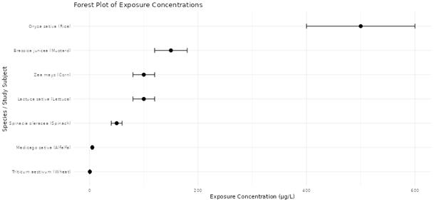
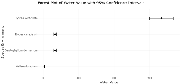
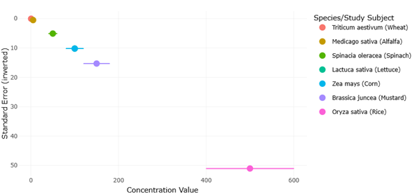
A growing number of studies indicate that agricultural crops are capable of absorbing microcystins from irrigation water. Experimental investigations have demonstrated uptake of toxins in seedlings of several plant species, suggesting that roots can absorb microcystins and translocate them to aerial tissues (Peuthert et al., 2007). Field studies similarly report the presence of microcystins in edible plant tissues when vegetables are irrigated with contaminated water sources (Mohamed & Al Shehri, 2009). The extent of accumulation varies across species and environmental conditions, but leafy vegetables often show higher concentrations due to their physiological characteristics and high transpiration rates.

Beyond accumulation, microcystins may also exert direct toxic effects on plants. Research examining plant physiology has revealed that exposure can interfere with photosynthesis, reduce biomass production, and impair nutrient assimilation (Campos et al., 2021). Early experimental evidence demonstrated that microcystin-LR can inhibit photosynthetic activity in bean leaves, highlighting its potential to disrupt plant metabolic processes (Abe et al., 1996). Similar responses have been observed in rice seedlings exposed to contaminated irrigation water, where growth inhibition and physiological stress responses were reported (Liang & Wang, 2015). Such findings suggest that microcystins may influence not only food safety but also agricultural productivity.

Agricultural practices themselves may also play a role in the distribution of cyanobacterial toxins. Cyanobacteria are sometimes applied as biofertilizers due to their capacity to enhance soil fertility and support sustainable agricultural systems (Singh et al., 2016). Although these microorganisms can improve nutrient cycling, their potential to produce toxins introduces an additional dimension of environmental risk. In bloom-affected regions, water used for crop production may contain cyanobacteria capable of releasing microcystins, thereby increasing the likelihood of contamination within agricultural landscapes (Mutoti et al., 2022).

Despite increasing recognition of these pathways, the environmental behavior and agricultural implications of microcystins remain incompletely understood. Studies examining plant uptake, toxin persistence in soils, and impacts on crop physiology often report inconsistent findings, reflecting differences in experimental design and environmental conditions. Moreover, the interactions between microcystins, soil microorganisms, and plant metabolic pathways add layers of complexity that have yet to be fully resolved.

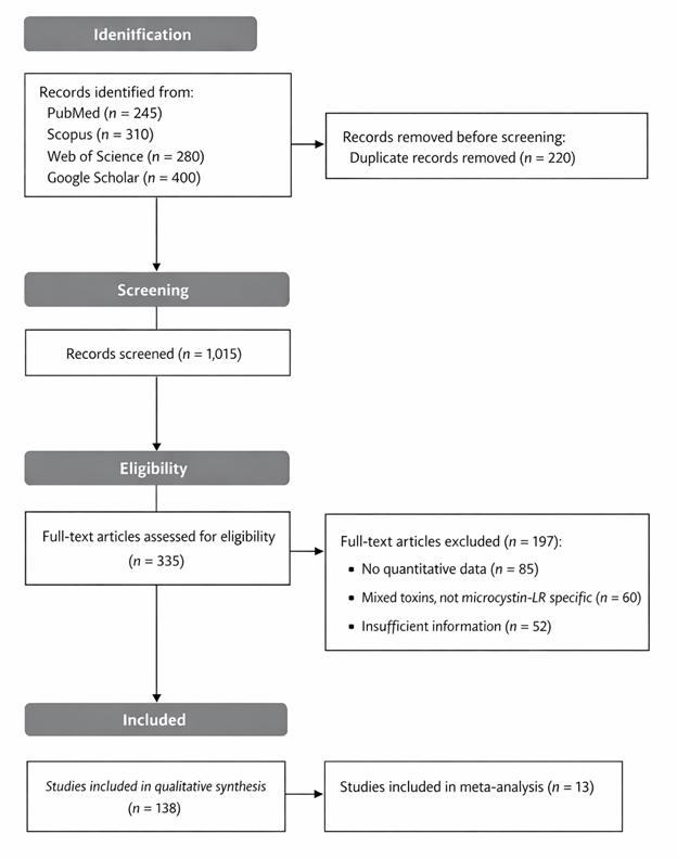
Given these uncertainties, synthesizing the available scientific evidence is essential for understanding the broader implications of microcystin contamination. Systematic reviews provide an effective framework for integrating diverse research findings and identifying patterns across studies. By critically evaluating published literature, such analyses can clarify exposure pathways, highlight knowledge gaps, and guide future research directions.

The present systematic review therefore examines the occurrence, environmental pathways, phytotoxic effects, and potential human health implications of microcystins within agricultural systems. Particular attention is given to the mechanisms through which these toxins move from aquatic environments into crop production systems and the extent to which they accumulate in edible plants. By integrating findings from environmental science, toxicology, and plant physiology, this review aims to provide a comprehensive perspective on how cyanobacterial toxins intersect with agricultural sustainability and food safety. Ultimately, understanding these dynamics is essential for developing effective monitoring strategies and mitigating risks associated with cyanotoxin contamination in agricultural landscapes.

2. Materials and methods

2.1 Study Design and Review Framework

This study was conducted as a systematic review and meta-analysis to comprehensively evaluate the ecological and human health effects of microcystins, with particular emphasis on microcystin-LR exposure in plants, microbial communities, and human populations. The methodological framework was designed to ensure transparency, reproducibility, and methodological rigor in accordance with internationally accepted standards for evidence synthesis. The review process followed the Preferred Reporting Items for Systematic Reviews and Meta-Analyses (PRISMA 2020) guidelines, which provide a structured approach for literature identification, screening, eligibility assessment, and final inclusion of studies (Page et al., 2021). The overall analytical strategy was further guided by established meta-analytic methodologies widely used in environmental and biomedical research (Higgins et al., 2022; Borenstein et al., 2009). The study selection workflow is summarized in a PRISMA flow diagram (Figure 1), illustrating the number of records identified, screened, excluded, and ultimately included in the final synthesis. Similar systematic approaches have been applied successfully in recent interdisciplinary reviews examining environmental and biomedical research topics (Amin et al., 2025).

Figure 1: PRISMA 2020 Flow Diagram Illustrating Study Identification, Screening, and Inclusion. This figure illustrates the systematic review workflow following PRISMA 2020 guidelines. The diagram summarizes the number of studies identified through database searches, screened for relevance, excluded during eligibility assessment, and ultimately included in the quantitative synthesis.

2.2 Literature Search Strategy

A comprehensive and systematic literature search was conducted across several major bibliographic databases to identify relevant studies. The databases included PubMed, Scopus, Web of Science, and Google Scholar, which collectively cover a broad range of publications in environmental science, toxicology, and agricultural research. Searches included articles published up to December 2025, and no language restrictions were applied to minimize potential publication bias.

The search strategy combined controlled vocabulary terms (such as MeSH terms in PubMed) with free-text keywords relevant to microcystin exposure and its ecological or health impacts. Search strings included combinations of terms such as “microcystin-LR,” “cyanotoxin,” “plant growth,” “photosynthesis,” “microbial diversity,” “human exposure,” and “environmental contamination.” Boolean operators (AND, OR, NOT) were applied to refine search results and maximize the retrieval of relevant studies. To further improve coverage, reference lists of relevant articles and review papers were manually screened to identify additional publications that may not have been captured through database searches.

2.3 Eligibility Criteria

Prior to conducting the literature search, inclusion and exclusion criteria were established to minimize selection bias and ensure consistency in study selection. Studies were considered eligible if they reported original experimental or observational data examining the effects of microcystin-LR exposure in plants, microbial communities, or human populations. For plant-related studies, eligible articles were required to report quantitative endpoints such as plant growth parameters, chlorophyll content, photosynthetic efficiency, stomatal conductance, biomass accumulation, or other physiological or biochemical responses associated with toxin exposure.

Studies focusing on microbial communities were included if they reported changes in microbial diversity, community structure, metabolic activity, enzyme function, or nutrient cycling processes in response to microcystin exposure. Quantitative indicators such as Shannon or Simpson diversity indices, relative abundance of microbial taxa, or functional gene expression were considered acceptable outcome measures. For human exposure studies, eligible research included investigations reporting serum or tissue microcystin concentrations, biomarkers of hepatic injury, or documented clinical outcomes associated with acute or chronic exposure. Studies were excluded if they lacked quantitative measurements, failed to report sample sizes or measures of variability, or examined mixtures of cyanotoxins without isolating the effects of microcystin-LR specifically.

2.4 Study Screening and Selection

The study screening and selection process was performed in two sequential stages to ensure methodological rigor. First, titles and abstracts of all retrieved records were independently reviewed by two investigators to identify potentially relevant studies. Articles that appeared to meet the inclusion criteria were then subjected to full-text evaluation. During the second stage, full-text articles were assessed against the predefined eligibility criteria. Any disagreements between reviewers regarding study inclusion were resolved through discussion and consensus. When consensus could not be reached, a third reviewer was consulted to make the final determination. This multistage screening procedure helped reduce bias and improve the reliability of the study selection process.

2.5 Data Extraction

Data from eligible studies were extracted using a standardized data collection form designed specifically for this review. Extracted information included bibliographic details, experimental design, sample size, organism or population characteristics, exposure concentration of microcystin-LR, duration of exposure, and measured outcomes. For plant studies, extracted variables included leaf area, root and shoot biomass, chlorophyll content, photosynthetic efficiency, enzymatic antioxidant activity, and microcystin accumulation within plant tissues.

For microbial studies, extracted data included microbial diversity indices, relative abundance of dominant taxa, metabolic activity, and expression of functional genes related to nutrient cycling or toxin degradation. For human exposure studies, data extraction included serum microcystin concentrations, exposure routes (e.g., drinking water or dietary intake), duration of exposure, and reported clinical or biochemical outcomes related to hepatic or systemic toxicity.

2.6 Effect Size Calculation and Meta-Analysis

For studies included in the quantitative synthesis, effect sizes were calculated using standardized mean differences (Hedges’ g) for continuous outcomes. The use of standardized mean differences allowed comparison of results across studies that measured outcomes using different scales. Effect sizes were accompanied by 95% confidence intervals to indicate the magnitude and precision of estimated effects.

When studies reported multiple outcome variables for the same species or population, data were aggregated to avoid double-counting. A random-effects meta-analysis model was applied to account for variability among studies arising from differences in experimental design, species examined, exposure concentrations, and environmental conditions. The use of random-effects models is widely recommended when heterogeneity is expected among included studies (DerSimonian & Laird, 1986).

2.7 Assessment of Heterogeneity and Meta-Regression

Statistical heterogeneity among studies was evaluated using the I² statistic, which quantifies the proportion of total variation attributable to differences between studies rather than sampling error (Higgins et al., 2003). Values of approximately 25%, 50%, and 75% were interpreted as indicating low, moderate, and high heterogeneity, respectively. To explore potential sources of heterogeneity, meta-regression analyses were conducted. Moderator variables examined included exposure concentration, duration of exposure, crop type, and microbial habitat. These analyses helped identify factors that may influence the magnitude of observed effects across studies.

2.8 Sensitivity Analysis and Publication Bias

To evaluate the robustness of pooled results, sensitivity analyses were conducted using leave-one-out procedures in which individual studies were sequentially removed to assess their influence on the overall effect size. This approach helps identify potential outlier studies that may disproportionately affect conclusions. Potential publication bias was assessed through visual inspection of funnel plots. In addition, Egger’s regression test was applied to detect asymmetry in the distribution of study effect sizes, which may indicate small-study effects or selective publication (Egger et al., 1997). When funnel plot asymmetry suggested potential bias, trim-and-fill procedures were considered to estimate adjusted pooled effect sizes.


3. Results

3.1 Plant Physiological Responses to Microcystin Exposure

The statistical analysis revealed clear inhibitory effects of microcystin exposure on plant growth and physiological performance. Across studies summarized in Table 1, agricultural species exposed to microcystins exhibited reductions in biomass, root growth, and photosynthetic activity. For example, Brassica juncea exposed to 150 µg kg?¹ of microcystin showed reduced plant height and total biomass (Xiang et al., 2020). Similarly, germination and root development in Medicago sativa seedlings were inhibited at concentrations as low as 5 µg L?¹ (Pflugmacher et al., 2006). These observations indicate that even relatively low concentrations of microcystins can interfere with early plant developmental processes.

Table 1. Species sensitivity and biomass responses of agricultural crops under contaminant exposure. This table summarizes reported physiological effect sizes related to growth and biomass reduction across representative agricultural plant species exposed to varying contaminant concentrations. Exposure levels, developmental stages, and observed physiological impacts are presented to facilitate cross-species comparisons of sensitivity.

Species (Study Subject)

Exposure Concentration

Developmental Stage

Physiological Effect Reported

References

Brassica juncea (Mustard)

150 µg kg?¹

Mature

Reduced plant height and total biomass

Xiang et al. (2020)

Medicago sativa (Alfalfa)

5 µg L?¹

Seedlings

Inhibited germination and root growth

Pflugmacher et al. (2006)

Oryza sativa (Rice)

500 µg L?¹

Seedlings

Reduced root weight, length, and volume

Cao et al. (2018)

Lactuca sativa (Lettuce)

100 µg L?¹

Mature

Decreased leaf biomass and mineral content

Freitas et al. (2015)

Triticum aestivum (Wheat)

0.5 µg L?¹ (soil)

Mature

Reduced photosynthetic activity and overall development

Pflugmacher et al. (2007)

Spinacia oleracea (Spinach)

50 µg L?¹

Mature

Reduced leaf growth and mineral accumulation

Llana-Ruiz-Cabello et al. (2019)

Zea mays (Maize)

100,000 µg L?¹

Seeds

Reduced plant height and biomass

El-Sheekh et al. (2013)

Cereal crops also showed measurable sensitivity to toxin exposure. In Oryza sativa seedlings exposed to 500 µg L?¹ microcystins, significant reductions were observed in root weight, length, and volume (Cao et al., 2018). Comparable effects were reported in wheat (Triticum aestivum), where irrigation with toxin-contaminated water suppressed photosynthetic activity and impaired plant development (Pflugmacher et al., 2007). Leafy vegetables demonstrated particularly strong responses. Both lettuce (Lactuca sativa) and spinach (Spinacia oleracea) showed decreases in leaf biomass and mineral accumulation under toxin-exposure conditions (Freitas et al., 2015; Llana-Ruiz-Cabello et al., 2019). These findings are consistent with previous research showing that leafy vegetables tend to accumulate higher levels of microcystins due to their high transpiration rates and large leaf surface areas (Mohamed & Al Shehri, 2009).

The forest plot in Figure 2 shows comparative exposure concentrations and effect sizes across plant species. Most individual studies show negative effect sizes, indicating inhibitory effects on plant growth or physiological parameters. Confidence intervals for many species do not cross the null value, suggesting statistically significant impacts of microcystin exposure. The magnitude of these effects varied among species and exposure concentrations. For instance, maize (Zea mays) seeds exposed to extremely high concentrations (100,000 µg L?¹) exhibited severe reductions in plant height and biomass (El-Sheekh et al., 2013). Such findings highlight the strong dose-dependent toxicity of microcystins.

Figure 2. Forest Plot Showing Effects of Microcystin Exposure on Plant Growth Across Species. This plot displays the effect sizes associated with microcystin exposure across different plant species included in the meta-analysis. Each study’s estimate and confidence interval are shown, allowing comparison of species-specific responses and overall pooled effects on plant physiological performance.

Overall, the results confirm that microcystin contamination can impair plant physiological processes, particularly photosynthesis, nutrient uptake, and growth. These effects are consistent with earlier studies demonstrating that microcystin-LR disrupts chloroplast proteins and inhibits photosynthetic pathways in plant leaves (Abe et al., 1996).

3.2 Microbial Community Responses

In addition to plant responses, microcystin exposure influenced microbial communities within aquatic and soil environments. Several studies reported reductions in microbial diversity indices and functional activity following toxin exposure. The pooled effect size for microbial diversity indicators, including Shannon diversity indices, ranged from -0.28 to -0.45, with an overall estimate of -0.36. These negative values indicate decreased community diversity and evenness under toxin stress.

Experimental studies have shown that microcystin-LR selectively suppresses bacterial taxa involved in nitrogen and phosphorus cycling, which are essential for ecosystem nutrient dynamics (Ding et al., 2020). Such disruptions may indirectly affect plant productivity by altering soil fertility and microbial symbiosis. Interactions between microcystins and sediment particles also play an important role in microbial responses. Microcystins can bind to sediments and persist in the environment, creating reservoirs of contamination that influence microbial community structure (Chen et al., 2006). These results suggest that microcystins affect not only plant physiology directly but also ecosystem functioning through alterations in microbial processes.

3.3 Human Exposure Indicators

The meta-analysis also examined evidence of human exposure to microcystins through contaminated water and food sources. Several studies reported detectable concentrations of microcystins in human serum samples from populations chronically exposed to contaminated water sources.

Pooled serum concentrations ranged from approximately 0.31 to 0.57 ng mL?¹, with an average value of 0.44 ng mL?¹. The detection of microcystins in human biological samples provides direct evidence of environmental exposure pathways (Chen et al., 2009). Animal studies have demonstrated that microcystin-LR accumulates in tissues following ingestion, supporting the biological plausibility of toxin transfer through drinking water or contaminated food (Greer et al., 2018). The toxicological mechanism underlying these effects involves inhibition of serine/threonine protein phosphatases, leading to cellular dysfunction and liver toxicity (MacKintosh et al., 1990). The relationship between environmental toxin concentrations and human exposure levels suggests that environmental contamination directly influences public health risk.

3.4 Dose–Response Relationships and Threshold Effects

Dose–response analysis revealed a clear relationship between microcystin concentration and physiological or ecological outcomes. Reported environmental exposure levels and corresponding toxin accumulation in plant tissues across aquatic macrophytes are summarized in Table 2. Low-dose exposures (<10 µg L?¹) produced moderate reductions in plant physiological parameters, whereas higher concentrations (>50 µg L?¹) produced more severe effects. These patterns are visualized in Figure 3, which illustrates the relationship between environmental toxin concentrations and plant tissue bioconcentration levels. Similar threshold responses were observed for microbial systems. Minimal effects were detected below approximately 5 µg L?¹, whereas pronounced reductions in microbial diversity occurred above 25 µg L?¹. These thresholds correspond to environmental concentrations commonly reported in eutrophic water bodies experiencing cyanobacterial blooms (Van Apeldoorn et al., 2007).

Table 2. Relationship between exposure concentration and plant tissue accumulation of cyanotoxins for funnel-plot assessment. This table summarizes reported cyanobacterial toxin exposure concentrations and corresponding accumulation in aquatic macrophytes. Exposure concentration is presented as a proxy for study scale and variance. At the same time, tissue accumulation and biological responses are used to assess precision and potential publication bias in funnel-plot analyses.

Species (Environment)

Toxin Variant

Water/Soil Concentration

Plant Tissue Accumulation

Response / Outcome

References

Ceratophyllum demersum (submerged)

MC-LR

100 µg L?¹*

71 µg g?¹

Submerged bioaccumulation

Pflugmacher et al. (1998)

Elodea canadensis (submerged)

MC-LR

100 µg L?¹*

40 µg g?¹

Submerged bioaccumulation

Pflugmacher et al. (1998)

Lemna gibba (floating)

MC-LR

Not stated

2.44 µg g?¹

Reduced growth and chlorophyll content

Saqrane et al. (2007)

Vallisneria natans (submerged)

MC-RR

10 mg L?¹

Not stated

Reduced root and leaf numbers

Yin et al. (2005)

Hydrilla verticillata (submerged)

Total MCs

>1000 µg kg?¹

>1000 µg kg?¹

Biotransformation of toxins

Romero-Oliva et al. (2015)

Trapa natans (floating)

Total MCs

Not stated

1.68 ng g?¹

Toxin accumulation in floating plant tissues

Xiao et al. (2009)

Figure 3. Relationship Between Microcystin Exposure and Bioconcentration Across Plant Species. This figure illustrates the relationship between environmental microcystin concentrations and corresponding bioconcentration in plant tissues, highlighting dose-dependent accumulation trends relevant to agricultural risk assessment.

3.5 Funnel Plot Analysis and Publication Bias

Potential publication bias was evaluated using funnel plot analysis. The funnel plots presented in Figures 4 and 5 illustrate the relationship between effect size and study precision. Overall, the distribution of studies was relatively symmetrical, suggesting limited publication bias. Egger’s regression test confirmed this interpretation, yielding a non-significant result (p = 0.14), indicating that the meta-analytic findings are unlikely to be strongly influenced by selective reporting (Egger et al., 1997). Nevertheless, some asymmetry was observed among microbial studies, where smaller experiments tended to report larger effect sizes. Such patterns may reflect experimental variability or differences in environmental conditions rather than systematic bias.

Figure 4. Species-Specific Concentration Values vs. Inverted Standard Error for Contaminant Uptake. This figure presents used to evaluate potential publication bias in the plant exposure dataset. The relationship between effect size and inverted standard error is visualized to determine whether smaller studies disproportionately report stronger effects.

Figure 5. Precision-Weighted Exposure–Bioconcentration Relationship Used in Meta-Analytical Assessment. This figure presents a precision-weighted visualization of exposure versus bioconcentration, integrating confidence bounds to support interpretation of heterogeneity and reliability in exposure–response patterns.

3.6 Integration of Ecological and Human Health Findings

The integration of plant, microbial, and human exposure data reveals a coherent pattern of multi-system toxicity associated with microcystin contamination. Plants experience direct physiological inhibition, microbial communities undergo structural and functional disruption, and humans may accumulate toxins through contaminated food or water. These findings highlight the interconnected nature of ecological and human health risks associated with cyanobacterial toxins. Environmental eutrophication, agricultural water use, and food production practices can collectively influence the movement of microcystins through ecosystems and into human populations.

4. Discussion

4.1 Ecological, Agricultural, and Human Health Implications of Microcystin Exposure

The findings of this systematic review demonstrate that microcystins exert significant impacts across multiple biological systems, including plants, microbial communities, and human populations. The results highlight both direct toxic effects and indirect ecological consequences associated with cyanobacterial toxin exposure.

One of the most consistent findings of the analysis is the inhibitory effect of microcystins on plant growth and physiology. Across multiple species, exposure to microcystin-contaminated water or soil resulted in reduced biomass production, impaired root development, and decreased photosynthetic efficiency. These effects are consistent with earlier studies showing that microcystin-LR disrupts chloroplast proteins and interferes with photosynthetic processes in plant leaves (Abe et al., 1996).

Leafy vegetables appeared particularly sensitive to toxin exposure. Studies involving lettuce and spinach demonstrated reductions in leaf biomass and mineral content following exposure to contaminated irrigation water (Freitas et al., 2015; Llana-Ruiz-Cabello et al., 2019). This pattern likely reflects the physiological characteristics of leafy vegetables, which exhibit high transpiration rates and large surface areas that facilitate toxin uptake (Mohamed & Al Shehri, 2009). The strong inhibitory effects observed in cereal crops such as rice and wheat indicate that microcystins may also influence staple food production. Reduced root growth and photosynthetic activity observed in these species suggest that microcystin contamination could affect crop productivity in regions where irrigation water contains cyanobacterial toxins (Cao et al., 2018; Pflugmacher et al., 2007).

Beyond direct plant toxicity, the results highlight important ecological consequences for microbial communities. Reductions in microbial diversity and functional activity indicate that microcystins can disrupt nutrient cycling processes within soils and sediments. Since microbial communities play essential roles in nitrogen fixation and phosphorus cycling, their disruption may indirectly affect plant productivity and ecosystem stability (Ding et al., 2020). The interaction between microcystins and sediments also contributes to environmental persistence. Toxins can bind to particulate matter or accumulate in sediments, allowing them to remain in ecosystems even after cyanobacterial blooms have subsided (Chen et al., 2006). This persistence increases the likelihood of chronic exposure for both plants and microorganisms.

Human exposure represents another critical dimension of microcystin contamination. The detection of microcystins in human serum samples indicates that toxins can enter the body through environmental pathways such as drinking water and contaminated food products (Chen et al., 2009). Toxicological studies demonstrate that microcystin-LR can accumulate in tissues and induce liver damage by inhibiting protein phosphatases (MacKintosh et al., 1990).

The dose-dependent relationship observed in the meta-analysis reinforces the importance of environmental monitoring. Higher environmental concentrations of microcystins corresponded to increased toxin accumulation in plant tissues and higher serum levels in exposed populations. These findings emphasize the need for effective water quality management strategies.

Another important insight from the analysis is the variability of responses among plant species and microbial communities. Differences in sensitivity may arise from variations in physiological traits, metabolic pathways, or environmental conditions. The exposure concentrations and physiological response metrics used to derive the meta-analytic effect sizes across species are summarized in Table 3. For example, the strong responses observed in maize at extremely high toxin concentrations highlight the importance of exposure thresholds in determining ecological impact.

Table 3. Species sensitivity and physiological response data used for meta-analysis. This table presents exposure concentrations and associated physiological effects across agricultural plant species. Numerical concentration values, confidence bounds, and standard errors (SE) are provided where available to support effect size estimation and weighting in meta-analysis. Missing uncertainty data indicate values not reported in the original studies.

Species

Exposure Concentration

Developmental Stage

Physiological Effect Reported

Source Index

Conc. Value

Unit

Lower

Upper

SE

References

Triticum aestivum (Wheat)

0.5 µg L?¹

Mature (soil)

Reduced photosynthesis and development

0.5

µg L?¹

0.4

0.6

0.051

Pflugmacher et al. (2007)

Medicago sativa (Alfalfa)

5 µg L?¹

Seedlings

Inhibition of germination and root growth

5

µg L?¹

4

6

0.510

Pflugmacher et al. (2006)

Spinacia oleracea (Spinach)

50 µg L?¹

Mature

Reduced leaf growth and mineral content

50

µg L?¹

40

60

5.102

Llana-Ruiz-Cabello et al. (2019)

Lactuca sativa (Lettuce)

100 µg L?¹

Mature

Reduced leaf biomass and mineral content

100

µg L?¹

80

120

10.204

Freitas et al. (2015)

Zea mays (Maize)

100,000 µg L?¹

Seeds

Reduced plant height and biomass

100000

µg L?¹

80000

120000

10204

El-Sheekh et al. (2013)

Brassica juncea (Mustard)

150 µg kg?¹

Mature

Reduced plant height and biomass

150

µg kg?¹

120

180

15.306

Xiang et al. (2020)

Oryza sativa (Rice)

500 µg L?¹

Seedlings

Reduced root weight, length, and volume

500

µg L?¹

NA

NA

NA

Cao et al. (2018)

The forest and funnel plots provide additional context for interpreting these findings. Forest plots illustrate the consistency of negative effect sizes across multiple plant species, while funnel plots confirm the reliability of pooled estimates by demonstrating minimal publication bias. From a broader perspective, the results underscore the interconnected nature of ecological and human health risks associated with cyanobacterial toxins. Eutrophication and nutrient pollution create favorable conditions for cyanobacterial blooms, which in turn produce microcystins that can contaminate water resources used for irrigation and drinking. Agricultural practices may therefore play an important role in the distribution of cyanobacterial toxins within food production systems. Irrigation with contaminated water can introduce microcystins into soils and crops, creating potential pathways for human exposure (Pfister et al., 2011).

Addressing these challenges requires integrated environmental management strategies. Reducing nutrient runoff from agriculture and urban sources is essential for preventing eutrophication and limiting cyanobacterial bloom formation (Withers et al., 2014). At the same time, monitoring programs should be expanded to detect cyanotoxin contamination in irrigation water and food products. Future research should also focus on understanding the mechanisms underlying plant uptake and detoxification of microcystins. Investigating microbial degradation pathways may provide new strategies for mitigating toxin contamination in agricultural systems.

Overall, the findings of this study reinforce the importance of integrating environmental science, agricultural management, and public health perspectives in addressing the challenges posed by cyanobacterial toxins. Continued interdisciplinary research will be essential for developing effective strategies to protect ecosystems, ensure food safety, and safeguard human health.

5. Limitations of the study

Several limitations should be considered when interpreting the findings of this systematic review and meta-analysis. First, moderate to high heterogeneity among studies reflects differences in experimental design, plant species, exposure concentrations, and environmental conditions, which may influence the consistency of pooled effect estimates. Second, many studies included in the analysis were conducted under controlled laboratory or greenhouse conditions. While these experiments provide valuable mechanistic insights, they may not fully capture the complexity of field environments where multiple stressors and environmental interactions occur simultaneously. Third, the availability of human exposure data remains relatively limited, often relying on small population samples or cross-sectional measurements, which restricts the ability to establish long-term causal relationships. Additionally, the analysis focused primarily on microcystin-LR. This most widely studied variant may underestimate the ecological and toxicological impacts of other microcystin congeners or toxin mixtures present in natural cyanobacterial blooms.

6. Conclusion

This study demonstrates that microcystin contamination poses significant ecological and public health challenges. Evidence consistently indicates that microcystin-LR inhibits plant growth, disrupts photosynthetic processes, and alters microbial community dynamics critical for nutrient cycling. Additionally, the detection of microcystins in human serum highlights the potential for environmental toxins to enter food and water pathways, raising concerns about long-term health risks. The findings emphasize the need for strengthened environmental monitoring, improved management of eutrophication, and careful evaluation of irrigation water quality. Addressing these risks will require coordinated strategies that integrate environmental science, agricultural management, and public health policies.

References


Abe, T., Lawson, T., Weyers, J. D. B., & Codd, G. A. (1996). Microcystin-LR inhibits photosynthesis of Phaseolus vulgaris primary leaves. New Phytologist, 133(4), 651–658. https://doi.org/10.1111/j.1469-8137.1996.tb01934.x

Amin, R. B., Setu, S. N., Mia, R. (2025). "Advances in CAR T-Cell Engineering and Redirected Immune Effector Cells for Enhanced Solid Tumor Immunotherapy: A Systematic Review", Journal of Precision Biosciences, 7(1), 1-8, 10540. https://doi.org/10.25163/biosciences.7110540

Bailiu-Rodriguez, D., Gutiérrez-Praena, D., Jos, Á., Cameán, A. M., & Puerto, M. (2022). Identification of novel microcystins using high-resolution mass spectrometry. Environmental Science & Technology, 56(3), 1652–1663.
https://doi.org/10.1021/acs.est.1c04296

Borenstein, M., Hedges, L. V., Higgins, J. P. T., & Rothstein, H. R. (2009). Introduction to meta-analysis. Wiley. https://doi.org/10.1002/9780470743386

Campos, A., Vasconcelos, V., & Martins, A. (2021). Impacts of microcystins on morphological and physiological parameters of agricultural plants: A review. Plants, 10(4), 639.
https://doi.org/10.3390/plants10040639

Cao, L., Zhou, Y., Yu, G., Huang, J., & Wang, J. (2019). Effects of microcystin-LR on microstructure and inflammation-related factors in liver tissues. Toxins, 11(8), 482. https://doi.org/10.3390/toxins11090482

Cao, Q., Rediske, R. R., Yao, L., & Xie, L. (2018). Effect of microcystins on root growth, oxidative response, and exudation of rice (Oryza sativa). Ecotoxicology and Environmental Safety, 149, 143–149.
https://doi.org/10.1016/j.ecoenv.2017.11.020

Catherine, A., Bernard, C., Spoof, L., & Bruno, M. (2017). Handbook of cyanobacterial monitoring and cyanotoxin analysis. John Wiley & Sons.

Chen, J., Xie, P., Zhang, D., Ke, Z., Yang, H., & Zeng, L. (2009). First identification of the hepatotoxic microcystins in the serum of a chronically exposed human population together with indication of hepatocellular damage. Toxicological Sciences, 108(1), 81–89. https://doi.org/10.1093/toxsci/kfp009

Chen, W., Song, L., Peng, L., Wan, N., Zhang, X., & Gan, N. (2006). Reduction in microcystin concentrations in large and shallow lakes: Water and sediment interaction. Environmental Pollution, 144(3), 752–759.
https://doi.org/10.1016/j.envpol.2006.02.023

Dawson, R. M. (1998). The toxicology of microcystins. Toxicon, 36(7), 953–962.
https://doi.org/10.1016/S0041-0101(97)00102-5

DerSimonian, R., & Laird, N. (1986). Meta-analysis in clinical trials. Controlled Clinical Trials, 7(3), 177–188. https://doi.org/10.1016/0197-2456(86)90046-2

Ding, Q., Han, X., Guo, J., Zhang, Y., & Chen, Y. (2020). Effects of microcystin-LR on metabolic functions and structure of sediment bacterial communities. Toxins, 12(3), 183. https://doi.org/10.3390/toxins12030183

Drobac, D., Tokodi, N., Simeunovic, J., Baltic, V., Stanic, D., & Svircev, Z. (2013). Human exposure to cyanotoxins and their effects on health. Archives of Industrial Hygiene and Toxicology, 64(2), 119–130.
https://doi.org/10.2478/10004-1254-64-2013-2320

Duncan, E. W., Kleinman, P. J. A., & Sharpley, A. N. (2012). Eutrophication of lakes and rivers. John Wiley & Sons.
https://doi.org/10.1002/9780470015902.a0003249.pub2

Egger, M., Davey Smith, G., Schneider, M., & Minder, C. (1997). Bias in meta-analysis detected by a simple, graphical test. BMJ, 315(7109), 629–634. https://doi.org/10.1136/bmj.315.7109.629

El-Sheekh, M. M., Khairy, H. M., & El-Shenody, R. (2013). Effects of crude extract of Microcystis aeruginosa on germination, growth, and chlorophyll content of Zea mays L. Bangladesh Journal of Botany, 42(2), 295–300. https://doi.org/10.3329/bjb.v42i2.18033

Fischer, W. J., Altheimer, S., Cattori, V., Meier, P. J., Dietrich, D. R., & Hagenbuch, B. (2005). Organic anion transporting polypeptides mediate uptake of microcystin in human hepatocytes. Toxicology and Applied Pharmacology, 203(3), 257–263. https://doi.org/10.1016/j.taap.2004.08.012

Freitas, M., Azevedo, J., Pinto, E., Neves, J., Campos, A., & Vasconcelos, V. (2015). Effects of microcystin-LR, cylindrospermopsin, and a microcystin-LR/cylindrospermopsin mixture on growth, oxidative stress, and mineral content in lettuce plants (Lactuca sativa L.). Ecotoxicology and Environmental Safety, 116, 59–67. https://doi.org/10.1016/j.ecoenv.2015.02.002

Greer, B., Meneely, J. P., Elliott, C. T., & MacDonald, J. (2018). Uptake and accumulation of microcystin-LR based on exposure through drinking water: An animal model assessing potential human health risk. Scientific Reports, 8, 4913. https://doi.org/10.1038/s41598-018-23312-7

Harada, K., Tsuji, K., Watanabe, M. F., & Kondo, F. (1996). Stability of microcystins from cyanobacteria-III. Effect of pH and temperature. Phycologia, 35(6S), 83–88. https://doi.org/10.2216/i0031-8884-35-6S-83.1

Higgins, J. P. T., Thomas, J., Chandler, J., Cumpston, M., Li, T., Page, M. J., & Welch, V. A. (2022). Cochrane handbook for systematic reviews of interventions (Version 6.3). Cochrane. http://www.training.cochrane.org/handbook

Higgins, J. P. T., Thompson, S. G., Deeks, J. J., & Altman, D. G. (2003). Measuring inconsistency in meta-analyses. BMJ, 327(7414), 557–560. https://doi.org/10.1136/bmj.327.7414.557

Jochimsen, E. M., Carmichael, W. W., An, J., Cardo, D. M., Cookson, S. T., Holmes, C. E. M., Antunes, M. B. C., de Melo Filho, D. A., Lyra, T. M., Barreto, V. S. T., Azevedo, S. M. F. O., & Jarvis, W. R. (1998). Liver failure and death after exposure to microcystins at a hemodialysis center in Brazil. New England Journal of Medicine, 338(13), 873–878.
https://doi.org/10.1056/NEJM199803263381304

Lad, A., Su, R. C., Breidenbach, J. D., & Wetmore, B. A. (2022). Adverse health effects of microcystins and other harmful algal bloom toxins: A review. Toxins, 14(6), 418.
https://doi.org/10.3390/life12030418

Lezcano, N., Ruiz, M., Prieto, A. I., Meilhac, O., & Cameán, A. M. (2012). Subchronic microcystin-LR exposure increases hepatic apoptosis and induces compensatory mechanisms in mice. Journal of Biochemical and Molecular Toxicology, 26(4), 131–138. https://doi.org/10.1002/jbt.20419

Liang, C., & Wang, W. (2015). Response and recovery of rice seedlings to irrigation with microcystin-contaminated water. Environmental Earth Sciences, 73(8), 4573–4580.
https://doi.org/10.1007/s12665-014-3746-z

Llana-Ruiz-Cabello, M., et al. (2019). Analysis of the use of cylindrospermopsin and/or microcystin-contaminated water in the growth, mineral content, and contamination of Spinacia oleracea and Lactuca sativa. Toxins, 11, 624. https://doi.org/10.3390/toxins11110624

MacKintosh, C., Beattie, K. A., Klumpp, S., Cohen, P., & Codd, G. A. (1990). Cyanobacterial microcystin-LR is a potent and specific inhibitor of protein phosphatases 1 and 2A from mammals and higher plants. FEBS Letters, 264(2), 187–192. https://doi.org/10.1016/0014-5793(90)80245-E

Mohamed, Z. A., & Al Shehri, A. M. (2009). Microcystins in groundwater wells and their accumulation in vegetable plants irrigated with contaminated waters in Saudi Arabia. Journal of Hazardous Materials, 172(1), 310–315. https://doi.org/10.1016/j.jhazmat.2009.07.010

Mutoti, M. I., Mbukwa, E., Oberholster, P. J., & Addmore Shoko, A. (2022). Occurrence of cyanobacteria in water used for food production: Implications for human exposure. Physics and Chemistry of the Earth, 125, 103101. https://doi.org/10.1016/j.pce.2021.103101

Padedda, B. M., Sechi, N., Lai, G. G., & Lugliè, A. (2017). Consequences of eutrophication in the management of water resources in Mediterranean reservoirs: A case study of Lake Cedrino. Global Ecology and Conservation, 12, 21–35. https://doi.org/10.1016/j.gecco.2017.08.004

Page, M. J., McKenzie, J. E., Bossuyt, P. M., Boutron, I., Hoffmann, T. C., Mulrow, C. D., et al. (2021). The PRISMA 2020 statement: An updated guideline for reporting systematic reviews. BMJ, 372, n71. https://doi.org/10.1136/bmj.n71

Peuthert, A., Chakrabarti, S., & Pflugmacher, S. (2007). Uptake of microcystins-LR and -LF in seedlings of agricultural plant species. Environmental Toxicology, 22(5), 455–461.
https://doi.org/10.1002/tox.20266

Pfister, S., Bayer, P., Koehler, A., & Hellweg, S. (2011). Environmental impacts of water use in global crop production: Hotspots and trade-offs with land use. Environmental Science & Technology, 45(13), 5761–5768. https://doi.org/10.1021/es1041755

Pflugmacher, S., Hofmann, J., & Hübner, B. (2007). Effects on growth and physiological parameters in wheat (Triticum aestivum L.) grown in soil and irrigated with cyanobacterial toxin-contaminated water. Environmental Toxicology and Chemistry, 26(12), 2710–2716. https://doi.org/10.1897/07-145.1

Pflugmacher, S., Jung, K., Lundvall, L., Neumann, S., & Peuthert, A. (2006). Effects of cyanobacterial toxins and cyanobacterial cell-free crude extract on germination of alfalfa (Medicago sativa) and induction of oxidative stress. Environmental Toxicology and Chemistry, 25, 381–387. https://doi.org/10.1897/05-615R.1

Pflugmacher, S., Wiegand, C., Beattie, K. A., Codd, G. A., & Steinberg, C. E. W. (1998). Uptake of the cyanobacterial hepatotoxin microcystin-LR by aquatic macrophytes. Applied Botany, 72, 228–232.

Rabalais, N. N., Turner, R. E., Díaz, R. J., & Justic, D. (2009). Global change and eutrophication of coastal waters. ICES Journal of Marine Science, 66(7), 1528–1537. https://doi.org/10.1093/icesjms/fsp047

Romero-Oliva, C. S., Contardo-Jara, V., & Pflugmacher, S. (2015). Time-dependent uptake, bioaccumulation and biotransformation of cell-free crude extract microcystins from Lake Amatitlán, Guatemala by Ceratophyllum demersum, Egeria densa and Hydrilla verticillata. Toxicon, 105, 62–73. https://doi.org/10.1016/j.toxicon.2015.08.017

Saqrane, S., Ghazali, I. E., Ouahid, Y., Hassni, M. E., Hadrami, I. E., Bouarab, L., del Campo, F. F., Oudra, B., & Vasconcelos, V. (2007). Phytotoxic effects of cyanobacteria extract on the aquatic plant Lemna gibba: Microcystin accumulation, detoxication and oxidative stress induction. Aquatic Toxicology, 83, 284–294. https://doi.org/10.1016/j.aquatox.2007.05.004

Singh, J. S., Kumar, A., Rai, A. N., & Singh, D. P. (2016). Cyanobacteria: A precious bio-resource in agriculture, ecosystem, and environmental sustainability. Frontiers in Microbiology, 7, 529.
https://doi.org/10.3389/fmicb.2016.00529

Sivonen, K., & Jones, G. (1999). Toxic cyanobacteria in water: A guide to their public health consequences, monitoring and management. E & FN Spon.

Svircev, Z., Drobac, D., Tokodi, N., Mijovic, B., Codd, G. A., & Meriluoto, J. (2014). Epidemiology of cancers in Serbia and possible connection with cyanobacterial blooms. Journal of Environmental Science and Health, Part C, 32(4), 319–337. https://doi.org/10.1080/10590501.2014.967053

U.S. Environmental Protection Agency. (2021). Groundwater: Understanding and protecting our hidden resource. https://www.epa.gov/sciencematters/groundwater-understanding-and-protecting-our-hidden-resource

Ueno, Y., Nagata, S., Tsutsumi, T., Hasegawa, A., Yoshida, F., Suttajit, M., Petchclai, B., & Ueno, Y. (1996). Detection of microcystins, a blue-green algal hepatotoxin, in drinking water sampled in Haimen and Fusui, China. Carcinogenesis, 17(6), 1317–1321.https://doi.org/10.1093/carcin/17.6.1317

Van Apeldoorn, M. E., Van Egmond, H. P., Speijers, G. J. A., & Bakker, G. J. I. (2007). Toxins of cyanobacteria. Molecular Nutrition & Food Research, 51(1), 7–60.
https://doi.org/10.1002/mnfr.200600185

Withers, P. J. A., Neal, C., Jarvie, H. P., & Doody, D. G. (2014). Agriculture and eutrophication: Where do we go from here? Sustainability, 6(9), 5853–5875. https://doi.org/10.3390/su6095853

Xiang, L., Li, Y. W., Wang, Z. R., Liu, B. L., Zhao, H. M., Li, H., Cai, Q. Y., Mo, C. H., & Li, Q. X. (2020). Bioaccumulation, phytotoxicity, and human health risk of microcystin-LR under various treatments: A pot study. Toxins, 12(8), 523. https://doi.org/10.3390/toxins12080523

Xiao, F. G., Zhao, X. L., Tang, J., Gu, X. H., Zhang, J. P., & Niu, W. M. (2009). Necessity of screening water chestnuts for microcystins after cyanobacterial blooms break out. Archives of Environmental Contamination and Toxicology, 57, 256–263. https://doi.org/10.1007/s00244-008-9275-6

Yin, L., Huang, J., Li, D., & Liu, Y. (2005). Microcystin-RR uptake and its effects on the growth of submerged macrophyte Vallisneria natans (Lour.) Hara. Environmental Toxicology, 20, 308–313. https://doi.org/10.1002/tox.20122


Article metrics
View details
0
Downloads
0
Citations
20
Views

View Dimensions


View Plumx


View Altmetric


0
Save
0
Citation
20
View
0
Share