Biosensors and Nanotheranostics

Bionanotechnology, Drug Delivery, Therapeutics | online ISSN 3064-7789
175
Citations
127.7k
Views
108
Articles
Your new experience awaits. Try the new design now and help us make it even better
Switch to the new experience
Figures and Tables
REVIEWS   (Open Access)

Biogenic Silver–Zinc Oxide Nanocomposites as Dual Antibacterial and Immunomodulatory Agents Against Multidrug-Resistant Pathogens: A Critical Review

Amin Malik Shah Abdul Majid 1*, Muhit Rana 3, Md Shamsuddin Sultan Khan 4


+ Author Affiliations

Biosensors and Nanotheranostics 4 (1) 1-8 https://doi.org/10.25163/biosensors.4111016

Submitted: 27 August 2025 Revised: 17 October 2025  Published: 25 October 2025 


Abstract

Antimicrobial resistance now claims more lives each year than many of the infections it complicates, and the antibiotic pipeline has not kept pace. Against this backdrop, biogenic silver–zinc oxide (Ag–ZnO) nanocomposites have drawn growing attention, partly because they are made with plant extracts or microbial secretomes rather than harsh reductants, and partly because they seem to do two jobs at once. This review brings together evidence on how these hybrids are synthesized, how they kill multidrug-resistant (MDR) bacteria, and how they interact with the host immune system. Phytochemicals such as polyphenols, flavonoids and terpenoids reduce metal precursors and cap the resulting particles, yielding structures of roughly 5–65 nm with negative surface charge and a slightly narrowed band gap. Silver deposited on wurtzite ZnO forms a Schottky-type junction that slows charge recombination, which helps explain the elevated generation of reactive oxygen species. Combined with sustained Ag+/Zn2+ release, electrostatic membrane damage and quorum-sensing interference, this produces MIC values of about 3–25 µg/mL against Gram-negative isolates and 12.5–100 µg/mL against Gram-positive strains, along with 80–90% reductions in mature biofilm. At sub-toxic doses, the composites (and their single-metal relatives) appear to dampen NF-κB-driven cytokine release and to favour M2 macrophage polarization; in rodent wound models, Ag–ZnO-loaded dressings achieved 95–99% closure within 12–14 days. Yet much of the immunological evidence comes from silver-only systems, assays are poorly standardized, and no controlled human trials have been reported. We argue that standardized extracts, harmonized susceptibility testing, long-term biodistribution studies and early clinical evaluation are now the priorities.

Keywords: antimicrobial resistance; green synthesis; silver–zinc oxide nanocomposite; biofilm; reactive oxygen species; macrophage polarization; wound healing

1. Introduction

Antimicrobial resistance (AMR) has become one of the defining public health problems of this century, threatening biosecurity and undermining much of what modern clinical medicine takes for granted (Murray et al., 2022; Tacconelli et al., 2018). The scale is now quantifiable: bacterial AMR was associated with millions of deaths in 2019 alone, and forecasts suggest the burden will keep rising toward 2050 unless the current trajectory changes (GBD 2021 Antimicrobial Resistance Collaborators, 2024; Murray et al., 2022). Behind these figures lies a familiar story. Antibiotics have been overused and misused across human healthcare, veterinary practice and agriculture, and this sustained selective pressure has favoured the emergence and spread of multidrug-resistant (MDR), extensively drug-resistant (XDR) and even pandrug-resistant bacteria (De Oliveira et al., 2020; Tang et al., 2023).

The pathogens of greatest concern tend to be grouped under the ESKAPE acronym, namely Enterococcus faecium, Staphylococcus aureus, Klebsiella pneumoniae, Acinetobacter baumannii, Pseudomonas aeruginosa and Enterobacter species. These organisms routinely evade β-lactams, fluoroquinolones, carbapenems and aminoglycosides, which leaves clinicians with shrinking options (De Oliveira et al., 2020; Tacconelli et al., 2018). The difficulty is compounded by their tendency to form biofilms on implants, catheters and chronic wound beds. Encased in a self-produced extracellular polymeric substance (EPS), biofilm communities resist drug penetration and escape immune clearance, so that infections become persistent rather than acute (Høiby et al., 2011; Koo et al., 2017). It is therefore not only a question of which drug kills a bacterium in suspension, but of whether any agent can reach and disrupt bacteria living within a protected, three-dimensional community.

The conventional pipeline offers limited reassurance. Development of new small-molecule antibiotics has largely stagnated, and most candidates are variations on existing scaffolds that remain vulnerable to established resistance mechanisms (Theuretzbacher & Piddock, 2019). This has redirected attention toward non-traditional strategies, among which inorganic nanomaterials, particularly metallic and metal oxide nanoparticles, have attracted sustained interest (Chakraborty et al., 2022; Hajipour et al., 2012; Slavin et al., 2017). Their appeal lies in how they act. Rather than binding a single molecular target, metal-based nanostructures attack bacteria through several physical and chemical routes at once, which in principle makes it far harder for resistance to arise through a single mutation (Dizaj et al., 2014; Lemire et al., 2013).

Silver nanoparticles (AgNPs) and zinc oxide nanoparticles (ZnONPs) are the two most thoroughly studied of these biocidal platforms (Franci et al., 2015; Sirelkhatim et al., 2015). Both, however, have typically been produced by physical or chemical methods that rely on hazardous reductants, toxic organic solvents and considerable energy input, with attendant ecological risks (Agarwal et al., 2017). Biogenic or "green" synthesis has emerged as a practical alternative. By using plant extracts from leaves, seeds, roots or fruits, or beneficial bacteria and fungi, these protocols tend to be cheaper, more environmentally benign and more readily scaled (Albrecht et al., 2006; Ovais et al., 2018). The phytochemicals involved, including polyphenols, flavonoids, alkaloids, terpenoids and structural proteins, reduce precursor salts such as AgNO3 and Zn(CH3COO)2 while also coating the nascent particles. This biomolecular corona limits irreversible agglomeration, stabilizes the colloid and, in several reports, lowers cytotoxicity toward mammalian cells compared with chemically produced equivalents (Ali et al., 2016; El-Kattan et al., 2022; Zare et al., 2019).

Combining the two metals appears to offer more than the sum of its parts. When silver is deposited on hexagonal wurtzite ZnO, the resulting Ag–ZnO nanocomposite shows altered electronic band structure, enhanced localized surface plasmon resonance (LSPR) and more sustained ion release than either component alone (Mott et al., 2012; Zare et al., 2019). Studies using turmeric and fenugreek extracts, for example, have reported physicochemical and biological performance superior to that of monometallic particles (Arya et al., 2023; Noohpisheh et al., 2020). The antibacterial activity of these hybrids is thought to stem from several converging mechanisms (Dakal et al., 2016; Jobe et al., 2022). Photo-induced charge separation in the ZnO lattice generates hydroxyl radicals, superoxide anions and hydrogen peroxide, while silver accelerates redox electron transfer, so that reactive oxygen species (ROS) accumulate inside the cell and damage lipids, proteins and DNA (Jalal et al., 2010; Lam et al., 2020; Vatansever et al., 2013). Positively charged Ag+ and Zn2+ ions bind electrostatically to lipopolysaccharides and teichoic acids, depolarizing the envelope and causing leakage of cytoplasmic contents (Cushnie et al., 2016; Ramasamy & Lee, 2016). Once inside, these ions bind thiol, carboxyl and amino groups on respiratory and ribosomal proteins (Lemire et al., 2013; Slavin et al., 2017). Small composite particles can also penetrate biofilm matrices and interfere with quorum-sensing pathways that regulate virulence (Abdelghafar et al., 2022; Husain et al., 2022; Rather et al., 2021). Gram-negative species tend to be more susceptible than Gram-positive ones, a difference usually attributed to their thinner peptidoglycan layer (Heidary et al., 2019; Zare et al., 2019).

What makes Ag–ZnO hybrids particularly interesting, though, is a second line of activity directed at the host rather than the pathogen. Infections and persistent biofilms provoke excessive inflammation, marked by high levels of tumour necrosis factor-α (TNF-α), interleukin-1β (IL-1β) and interleukin-6 (IL-6), which can itself delay healing (Hanley et al., 2009; Yang et al., 2012). Metal-based nanoparticles are known to interact with immune cells in complex, dose-dependent ways (Dai et al., 2016; Luo et al., 2015). Several studies suggest that, at appropriate concentrations, silver- and zinc-based nanostructures downregulate NF-κB and Toll-like receptor (TLR) signalling in macrophages and monocytes (Jiao et al., 2014; Sarkar et al., 2015), promote polarization toward the pro-healing M2 phenotype, and support VEGF-mediated angiogenesis, fibroblast proliferation and collagen deposition (Dai et al., 2016; Krishnan et al., 2020). Phytochemical capping layers may add intrinsic antioxidant activity, buffering excess ROS in host tissue (Arya et al., 2023; El-Kattan et al., 2022). Taken together, this dual profile has led to interest in Ag–ZnO composites for wound dressings, implant coatings and topical formulations (El-Kattan et al., 2022; Sathiyaseelan et al., 2023).

Despite this activity, the literature remains fragmented. Many studies examine biogenic silver or zinc oxide in isolation, and existing reviews have often focused on photocatalytic dye degradation or on monometallic systems, giving less attention to what happens at the interface between host, pathogen and nanomaterial. A consolidated account of biogenic Ag–ZnO nanocomposites that considers antibacterial efficacy against MDR pathogens alongside host immunomodulation has, to our knowledge, been lacking. This review aims to address that gap. Specifically, it sets out to (i) critically evaluate plant- and microorganism-mediated synthesis routes and the role of capping biomolecules in controlling size, morphology, surface charge and colloidal stability; (ii) clarify the antibacterial and anti-biofilm mechanisms of these composites against Gram-negative and Gram-positive MDR strains; (iii) examine how they influence macrophage polarization, cytokine expression and tissue repair in vitro and in vivo; and (iv) assess cytocompatibility, hemocompatibility and the regulatory, manufacturing and clinical barriers that stand between laboratory promise and patient use.

2. Biogenic Silver-Zinc Oxide Nanocomposites Combat Multidrug-Resistant Bacteria and Modulate Immunity to Promote Wound Healing

2.1 The Resistance Problem and the Case for Hybrid Nanomaterials

The global spread of MDR bacteria continues to erode the efficacy of standard antibiotic regimens, and recent burden estimates leave little doubt about the urgency of the problem (GBD 2021 Antimicrobial Resistance Collaborators, 2024). Clinically important pathogens such as methicillin-resistant Staphylococcus aureus (MRSA), extensively drug-resistant Pseudomonas aeruginosa, Klebsiella pneumoniae and Acinetobacter baumannii deploy a broad repertoire of survival strategies, including enzymatic drug degradation, target-site alteration, reduced membrane permeability and robust biofilm formation (Kakian et al., 2024; Zarepour et al., 2024). Biofilm-associated communities are especially problematic because they can tolerate up to a thousand-fold higher antibiotic concentrations than their planktonic counterparts (Ramasamy & Lee, 2016). In this context, nanobiotechnology has been proposed as a distinct therapeutic paradigm, and inorganic metal and metal oxide nanocomposites, notably biogenic Ag–ZnO heterostructures, offer multi-pathway modes of action that sidestep many established resistance mechanisms (Islam et al., 2022; Rehal et al., 2023).

Monometallic nanoparticles are not without drawbacks. They are prone to colloidal aggregation and can show dose-dependent toxicity toward mammalian cells, which narrows their therapeutic window. Bimetallic Ag–ZnO nanocomposites appear to mitigate some of these limitations through synergistic physicochemical and biological behaviour (Busila et al., 2023; Mohapatra et al., 2023). The coupling of silver's high electron affinity and membrane-permeabilizing capacity (Ag0/Ag+) with the wide band gap and sustained catalytic potential of ZnO (ZnO/Zn2+) produces hybrids that act as dual-action bioagents (Babu & Antony, 2019; Jin & Jin, 2021). Beyond direct broad-spectrum killing, more recent work has emphasized their ability to modulate host immune dynamics, temper excessive inflammation and accelerate tissue regeneration (Mendes et al., 2022; Ramachandran et al., 2024). The subsections that follow trace this literature from synthesis to mechanism to application, with the main lines of evidence summarized schematically in Figures 1–4 and in Tables 1–4.

2.2 Phyto- and Microbe-Mediated Green Synthesis of Ag–ZnO Nanocomposites

Green chemistry routes have increasingly replaced conventional physical and chemical synthesis, which frequently relies on toxic reducing agents such as sodium

Table 1. Green synthesis protocols, precursor salts, phytochemical profiles and morphological attributes of biogenic Ag–ZnO and related metallic nanocomposites. The table compiles plant-mediated and microbe-mediated routes, listing the biological source, metal precursor salts, the principal capping biomolecules each feedstock supplies, and the reaction conditions of temperature, pH and duration. Resulting particle morphology and crystallite size ranges are given alongside the primary biological outcome reported in each study. Entries span polyphenol-rich extracts such as Curcuma longa and Azadirachta indica, saponin- and flavonoid-rich Trigonella foenum-graecum, polysaccharide-rich Aloe vera gel, and microbial systems using Bacillus subtilis filtrate and Escherichia coli D8 biomass combined with chitosan or graphene oxide carriers. Together the entries illustrate how feedstock chemistry and reaction parameters jointly determine particle size, stability and downstream bioactivity.

Plant / Biological Source

Metal Precursor Salts

Key Phytochemical / Capping Biomolecules

Reaction Conditions (Temp, pH, Time)

Nanoparticle Morphology & Crystallite Size

Primary Biological & Functional Outcome

Reference

Curcuma longa (turmeric stem & leaf extract)

AgNO₃, Zn(CH₃COO)₂·2H₂O

Curcuminoids, polyphenols, phenolic –OH groups

60 °C, pH 8.0, 3 h stirring + calcination at 400 °C

Spherical heterostructures (20–50 nm)

High colloidal stability; broad-spectrum antibacterial efficacy against MDR isolates

Arya et al. (2023)

Azadirachta indica (neem leaf extract)

AgNO₃, Zn(NO₃)₂·6H₂O

Quercetin, nimbin, polyphenols, flavonoids

Hydrothermal, 120 °C, 4 h, pH 7.5

Granular wurtzite hexagonal (32–52 nm)

Enhanced electrical conductivity, band-gap tuning and potent bactericidal action

Slathia et al. (2021)

Trigonella foenum-graecum (fenugreek leaf extract)

AgNO₃, Zn(CH₃COO)₂

Saponins, flavonoids, alkaloids, amino acids

70 °C, pH 8.5, 2.5 h + drying at 100 °C

Spherical nanocomposite aggregates (18–35 nm)

Tri-functional antibacterial, antifungal and solar photocatalytic activity

Noohpisheh et al. (2020)

Aloe vera gel extract (AVGE)

Zn(CH₃COO)₂·2H₂O

Acemannan, aloin, polysaccharides, proteins

60 °C, ambient pH, 1.5 h + drying at 80 °C

Spherical to quasi-spherical NPs (25–40 nm)

Strong growth inhibition against opportunistic aquatic and clinical pathogens

Ali et al. (2016)

Catharanthus roseus leaf extract

Zn(NO₃)₂·6H₂O

Polyphenols, alkaloids, flavonoids

Ambient, pH 9.0, 4 h stirring

Hexagonal wurtzite spheres (22–45 nm)

Dose-dependent bactericidal action via enhanced cell wall penetration

Gupta et al. (2018)

Prosopis juliflora leaf extract

AgNO₃

Tannins, flavonoids, phenolic acids

60 °C, pH 7.0, 2 h

Spherical silver nanoparticles (12–28 nm)

High catalytic dye degradation and potent wound healing acceleration in vivo

Arya et al. (2019)

Propolis extract (bee product)

Zn(CH₃COO)₂, CuCl₂, AgNO₃

Caffeic acid phenethyl ester (CAPE), flavonoids

80 °C, pH 8.0, 3 h stirring

Ternary ZnO/CuO/Ag nanocomposites (15–40 nm)

Coated on surgical gauze; accelerated full-thickness wound contraction

Bayrami et al. (2020)

Bacillus subtilis ATCC 6633 filtrate

Zn(CH₃COO)₂, graphene oxide, chitosan

Extracellular reductases, secretomes, amino acids

37 °C biosynthesis + ambient cryogelation

Macroporous CS/GO/ZnO cryogel (25–50 nm core)

Effective nanoremediation of heavy metal ions and sustained bactericidal release

El-Zahed et al. (2024)

Escherichia coli D8 biomass extract

AgNO₃, chitosan matrix

Intracellular nitrate reductases, proteins

37 °C incubation, 24 h + biopolymer blending

Spherical chitosan/Ag nanocomposite (10–30 nm)

High biocompatibility, low cytotoxicity and broad-spectrum bactericidal activity

Baka et al. (2019); El-Zahed et al. (2023)

Prunus cerasus (sour cherry leaf extract)

AgNO₃, chitosan solution (0.1% w/v)

Polyphenolics, hydroxycinnamic acids

Room temperature, pH 5.5, 12 h

CS-AgNP bioconjugates (15–35 nm)

Low cytotoxicity on normal epithelial cells; effective inhibition of clinical MDR strains

Shinde et al. (2015)

Table 2. Comparative antibacterial susceptibility, zones of inhibition, MIC and MBC values, and anti-biofilm metrics of biogenic nanocomposites against multidrug-resistant pathogens. For each pathogen and resistance phenotype, the table reports the nanocomposite formulation tested, the measured zone of inhibition in millimetres, minimum inhibitory and bactericidal or fungicidal concentrations in µg/mL, percentage inhibition of biofilm or quorum-sensing-regulated phenotypes, and the principal mechanism inferred by the original authors. Coverage includes methicillin-resistant Staphylococcus aureus, multidrug-resistant Pseudomonas aeruginosa, extended-spectrum β-lactamase-producing Escherichia coli, carbapenem-resistant Klebsiella pneumoniae, fluconazole-resistant Candida albicans, vancomycin-resistant Enterococcus faecalis, and multidrug- and extensively drug-resistant acid-fast isolates. Values derive from heterogeneous assay protocols and should be read as indicative ranges rather than directly comparable benchmarks.

Pathogen Species & Resistance Profile

Nanocomposite / Formulation

Zone of Inhibition (ZOI, mm)

MIC / MBC Values (µg/mL)

Biofilm / Quorum-Sensing Inhibition (%)

Key Antibacterial Mechanism Observed

Reference

Methicillin-resistant Staphylococcus aureus (MRSA)

Biogenic Ag–ZnO nanocomposite

24.5 ± 0.8

MIC = 15.6; MBC = 31.2

84.5% biofilm biomass reduction

ROS-mediated lipid peroxidation, cell wall disruption and intracellular oxidative stress

Abdelghafar et al. (2022)

Pseudomonas aeruginosa (MDR clinical isolate)

Plant-mediated Ag–ZnO NCs

22.0 ± 1.1

MIC = 7.8; MBC = 15.6

88.2% inhibition of EPS synthesis & swarming

Downregulation of pyocyanin secretion and disruption of quorum-sensing signalling

Noohpisheh et al. (2020); Ahmed et al. (2020)

Escherichia coli (extended-spectrum β-lactamase)

Aloe vera-synthesized ZnO NPs

15.67 ± 1.2

MIC = 25.0; MBC = 50.0

76.0% reduction in crystal violet biofilm binding

LPS membrane destabilization, cytoplasmic leakage and K⁺ ion efflux

Ali et al. (2016); Gunalan et al. (2012)

Klebsiella pneumoniae (carbapenem-resistant)

Biogenic CS/GO/ZnO cryogel

21.3 ± 0.5

MIC = 12.5; MBC = 25.0

81.0% clearance of pre-formed mature biofilms

Electrostatic binding, pore formation and physical membrane deformation

El-Zahed et al. (2024); Elumalai et al. (2015)

Candida albicans (fluconazole-resistant yeast)

Biogenic Ag–ZnO nanocomposite

19.8 ± 0.7

MIC = 31.25; MFC = 62.5

79.4% yeast-to-hyphae transition suppression

Plasma membrane ergosterol binding, ROS accumulation and mitochondrial dysfunction

Dananjaya et al. (2018); Noohpisheh et al. (2020)

Aeromonas veronii & Stenotrophomonas maltophilia

Biogenic ZnO NPs (AVGE)

18.2 ± 0.9

MIC = 31.25; MBC = 62.5

72.5% inhibition of initial surface attachment

Disruption of respiratory electron transport chains and metabolic arrest

Ali et al. (2016)

Fastidious acid-fast bacterial isolates (MDR & XDR)

Ag–ZnO bimetallic NCs

Broth assay

MIC = 3.91 (MDR); 7.81 (XDR)

Synergistic suppression of intracellular survival

Cell wall mycolic acid layer penetration and genomic DNA cleavage

Heidary et al. (2019)

Enterococcus faecalis (vancomycin-resistant, VRE)

Biogenic green Ag–ZnO NPs

17.4 ± 0.6

MIC = 50.0; MBC = 100.0

68.0% disruption of established urinary biofilm

Inactivation of membrane-bound ATPase enzymes and respiratory depression

Gupta et al. (2018); Ahmed et al. (2020)


borohydride or hydrazine and on high-energy calcination steps that generate hazardous waste (Gharpure & Ankamwar, 2020; Mutukwa et al., 2024). Green synthesis instead exploits natural "bioreactors", chiefly plant extracts, algae and microbial culture filtrates, to reduce Ag+ and Zn2+ precursors into zero-valent or oxide nanostructures in a sustainable, often single-pot reaction (Albrecht et al., 2006; Okaiyeto et al., 2024). The overall workflow, from biological source to finished heterostructure, is illustrated in Figure 1, and representative protocols are compiled in Table 1.

2.2.1 Phytochemical Reduction and Capping

Plant-mediated synthesis draws on extracts rich in secondary metabolites such as polyphenols, flavonoids, tannins, alkaloids and terpenoids (Endeshaw et al., 2023; Haddi et al., 2024). Hydroxyl (–OH), carboxyl (–COOH), amino (–NH2) and carbonyl (–C=O) groups on these molecules serve two purposes: they donate electrons to reduce metal ions, and they act as steric and electrostatic capping agents once particles have formed (Babu & Antony, 2019; Vairamuthu et al., 2024). Methanolic stem and leaf extracts of Curcuma longa, rich in the polyphenol curcumin, support room-temperature synthesis of stable Ag–ZnO nanocomposites with core–shell or decorated morphologies (Arya et al., 2023; El-Kattan et al., 2022). Comparable results have been reported with Azadirachta indica (neem), Trigonella foenum-graecum (fenugreek), Rumex crispus, Lavandula stoechas and Aloe vera, all of which yield composites with good stability, high surface reactivity and little agglomeration (Ali et al., 2016; Bayisa et al., 2023; Kiani et al., 2024; Noohpisheh et al., 2020; Slathia et al., 2021). As Table 1 shows, these syntheses typically run at 25–120 °C and pH 5.5–9.0, sometimes followed by calcination, and the plant source strongly shapes the capping chemistry (Gupta et al., 2018; Arya et al., 2019).

2.2.2 Microbial Bio-Factories

Microorganisms offer an alternative route. Probiotic bacteria such as Bacillus species and endophytic fungal secretomes can serve as nanoparticle factories (Ovais et al., 2018). Extracellular oxidoreductases, exopolysaccharides and secreted proteins mediate controlled nucleation and capping, yielding monodisperse particles with intrinsic biocompatibility (Gahlawat & Choudhury, 2019). Examples include chitosan/Ag composites produced with Escherichia coli D8 biomass and chitosan/graphene oxide/ZnO cryogels made using Bacillus subtilis ATCC 6633 (Baka et al., 2019; El-Zahed et al., 2023, 2024), as well as chitosan–silver bioconjugates prepared from Prunus cerasus leaf extract (Shinde et al., 2015). These microbial and hybrid routes are listed alongside plant protocols in Table 1 and depicted in Figure 1.

2.2.3 Morphology and the Ag/ZnO Heterojunction

The physicochemical properties of the final product depend on precursor concentration, pH, temperature and botanical family (Aji et al., 2025). X-ray diffraction, transmission electron microscopy and field-emission scanning electron microscopy show that biogenic Ag–ZnO nanocomposites are mostly spherical, quasi-spherical, flower-like or hexagonal wurtzite, with mean sizes of about 5–65 nm (Ahmad et al., 2022; Jobe et al., 2022; Rafique et al., 2023). At the interface, metallic Ag deposited on mesoporous ZnO forms a Schottky barrier (Busila et al., 2023). This junction traps photogenerated electrons in the silver domain, reducing electron–hole recombination and raising catalytic reactivity under ambient light (Djidjeli et al., 2023; Hojjati-Najafabadi et al., 2021). A simplified band diagram of this junction is shown in Figure 1; its biological consequences are considered next.

2.3 Multifaceted Antibacterial and Anti-Biofilm Mechanisms Against MDR Pathogens

The antibacterial efficacy of biogenic Ag–ZnO nanocomposites arises from a multi-target attack on structural, metabolic and genetic components of the bacterial cell (Gudkov et al., 2021; Jiang et al., 2020). Because several pathways operate at once, resistance is thought to be considerably less likely than with single-target antibiotics (Mohammed et al., 2024; Rather et al., 2021). The four principal routes, namely electrostatic membrane damage, dual-ion release, ROS generation and biofilm/quorum-sensing interference, are summarized in Figure 2, and the corresponding cellular targets are tabulated in Table 3.

2.3.1 Dual-Metal Ion Release and Membrane Permeabilization

In aqueous and physiological media, Ag–ZnO nanocomposites dissolve gradually, releasing Ag+ and Zn2+ (Caron et al., 2024; Durán et al., 2016). These cations adhere electrostatically to the negatively charged teichoic acids of Gram-positive bacteria and to the

Figure 1. Biogenic synthesis of Ag–ZnO nanocomposites, from biological feedstock to finished heterostructure. Plant extracts (leaves, stems, seeds and gels of species such as Curcuma longa, Azadirachta indica, Trigonella foenum-graecum, Aloe vera, Rumex crispus, Lavandula stoechas and Urginea epigea) and microbial factories (Bacillus subtilis filtrates, Escherichia coli D8 biomass, endophytic and ligninolytic fungi) supply polyphenols, flavonoids, terpenoids, alkaloids and secreted proteins that reduce AgNO3 and zinc acetate or nitrate precursors while capping the growing particles. Reaction variables (pH 5.5–9.0, 25–120 °C, 1.5–24 h, optional calcination near 400 °C) govern the final architecture. The product is a wurtzite ZnO core decorated with 2–15 nm silver nanodomains inside a phytochemical capping shell (dashed outline), with typical diameters of 5–65 nm, zeta potentials of −11.0 to −28.5 mV, and a band gap narrowed from 3.28–3.35 eV to 3.18–3.24 eV. The inset band diagram shows the Schottky junction, where photogenerated electrons are trapped in the silver domain, limiting electron–hole recombination and raising surface reactivity.

Figure 2. Multitarget antibacterial and anti-biofilm actions of biogenic Ag–ZnO nanocomposites. Four converging mechanisms are shown. (1) Electrostatic adhesion: cationic Ag+ and Zn2+ bind anionic lipopolysaccharides and teichoic acids, depolarizing the envelope and causing pore formation with K+ and protein leakage. (2) Dual-ion release: sustained dissolution delivers Ag+ to thiol groups of NADH dehydrogenase and cytochromes while Zn2+ inhibits autolysin activity and protein synthesis, arresting ATP generation. (3) Reactive oxygen species burst: photo-excitation of ZnO generates electron–hole pairs that yield •OH, O2^{•−} and H2O2, driving lipid peroxidation, protein oxidation and DNA strand breaks, with silver trapping electrons to prevent recombination. (4) Biofilm and quorum-sensing interference: sub-inhibitory exposure disrupts acyl-homoserine lactone signalling, lowering extracellular polymeric substance output by roughly 84% and reducing pyocyanin, protease activity and swarming motility, with 81–92% reductions in mature biofilm biomass. The lower panel contrasts envelope thickness and typical susceptibility between Gram-negative and Gram-positive pathogens

lipopolysaccharide (LPS) outer membrane of Gram-negative bacteria (Jiang et al., 2020; Xu et al., 2020). Ag+ binds thiol (–SH) groups of respiratory enzymes such as NADH dehydrogenase, disrupting the electron transport chain, depolarizing the membrane and causing K+ leakage (Lok et al., 2006; Thakur & Neogi, 2021). Zn2+, for its part, enters the envelope and inhibits autolysin activity and protein synthesis (Caron et al., 2024; Ishida, 2017). Gram-negative strains (E. coli, P. aeruginosa, K. pneumoniae) are generally more susceptible than Gram-positive ones (S. aureus, B. subtilis) (Hojjati-Najafabadi et al., 2021; Zare et al., 2019), which has been attributed to their thinner peptidoglycan layer (about 2–8 nm, compared with 20–80 nm in Gram-positive walls) (Babu & Antony, 2019; Sirelkhatim et al., 2015).

2.3.2 Reactive Oxygen Species-Mediated Oxidative Stress

Light- and defect-induced ROS generation is a dominant contributor to bactericidal activity (Islam et al., 2022; Mohapatra et al., 2023). Under ambient light or photo-activation, electron–hole pairs (e-/h+) formed in ZnO react with adsorbed oxygen and water to produce superoxide (O2^{•−}), hydroxyl radicals (•OH) and hydrogen peroxide (H2O2) (Busila et al., 2023; Jalal et al., 2010). Metallic silver amplifies this process through plasmonic hot-electron transfer, which prevents charge recombination (Sharwani et al., 2022). Charged species such as O2^{•−} and •OH mostly damage outer membrane lipids, whereas uncharged H2O2 crosses the bilayer into the cytoplasm (Jalal et al., 2010; Sirelkhatim et al., 2015). Intracellular ROS then oxidize structural proteins, break double-stranded DNA and cleave ribosomal RNA, leading to metabolic arrest and lysis (Gharpure & Ankamwar, 2020; Gold et al., 2018).

2.3.3 Quorum-Sensing Inhibition and Biofilm Eradication

Biofilms, structured communities embedded in self-produced EPS, shield pathogens from antibiotics and host defences (Devanga Ragupathi et al., 2022; Ramasamy & Lee, 2016). Biogenic Ag–ZnO and zinc-based nanoparticles show potent anti-biofilm and anti-quorum-sensing activity against MDR P. aeruginosa and MRSA (Abdelghafar et al., 2022; Fulindi et al., 2023; Husain et al., 2022). At sub-inhibitory concentrations, they interfere with acyl-homoserine lactone (AHL) signalling and downregulate quorum-sensing-controlled virulence genes (Ahmad et al., 2024; Shah et al., 2019), suppressing EPS production, protease secretion and swarming motility (Ahmad et al., 2024). Their small size also allows them to penetrate biofilm channels and destabilize extracellular DNA and polysaccharides (Husain et al., 2022; Zarepour et al., 2024). Quantitative anti-biofilm data from these studies are collated in Table 2.

2.4 Host Immune Response and Immunomodulatory Dynamics

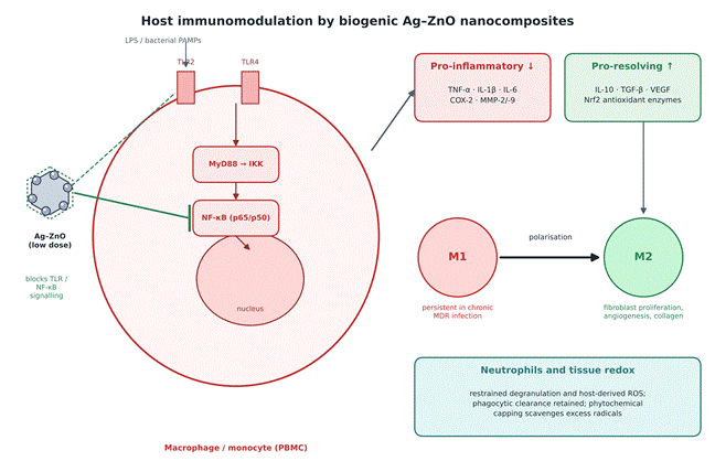
Beyond direct antimicrobial effects, biogenic Ag–ZnO nanocomposites can modulate host immunity, suppressing hyper-inflammatory responses while promoting tissue repair (Mendes et al., 2022; Perumalsamy et al., 2024). The main immunological pathways implicated are depicted in Figure 3.

2.4.1 Cytokine Regulation and NF-κB Signalling Suppression

Bacterial infection often triggers an exaggerated inflammatory cascade driven by TNF-α, IL-1β and IL-6 (Jiao et al., 2014; Soltan et al., 2024). Uncontrolled cytokine release contributes to local necrosis and chronic, non-healing wounds (Frankova et al., 2016; Singh et al., 2023). Silver- and zinc-based nanoparticles have been shown to downregulate pro-inflammatory gene expression in macrophages and peripheral blood mononuclear cells (PBMCs) (Bae et al., 2022; Vuković et al., 2021). At low doses, they block TLR-2/TLR-4 signalling and inhibit NF-κB nuclear translocation (Gliga et al., 2020; Perumalsamy et al., 2024), lowering TNF-α, IL-1β and cyclooxygenase-2 (COX-2) without overt immune-cell toxicity (Frankova et al., 2016; Soltan et al., 2024).

2.4.2 Macrophage Polarization and Neutrophil Regulation

Macrophages orchestrate tissue repair across a spectrum from pro-inflammatory M1 to pro-healing M2 phenotypes (Chen et al., 2020; Luo et al., 2015). Chronic MDR infection tends to lock macrophages in a persistent M1 state, aggravating tissue damage (Zarepour et al., 2024). Silver-based nanostructures have been shown to promote the M1-to-M2 transition (Chen et al., 2020), increasing transforming growth factor-β (TGF-β) and vascular endothelial growth factor (VEGF) and thereby stimulating fibroblast proliferation, endothelial migration and neovascularization (Krishnan et al., 2020; Mendes et al., 2022). These nanomaterials also appear to restrain excessive neutrophil degranulation and secondary oxidative damage while preserving phagocytic clearance (Bae et al., 2022; Vuković et al., 2021). The immunological


Table 3. Cellular targets, reactive oxygen species generation kinetics and dual immunomodulatory pathways of Ag–ZnO nanocomposites. Each row addresses one mechanistic category, identifying the primary cellular or molecular target, the specific biochemical or radical reaction involved, and the distinct contributions of the silver and zinc oxide components. The adjacent columns record the corresponding immunomodulatory or inflammatory consequence in host tissue and the resulting downstream biological outcome. Categories span ROS generation and oxidative stress, membrane disruption, enzyme and protein inactivation, genomic DNA cleavage, cytokine and NF-κB modulation, extracellular polymeric substance degradation, macrophage phenotype transition, and matrix metalloproteinase regulation with collagen deposition. The table thereby links each antibacterial mechanism to its parallel effect on the host.

Mechanism Category

Primary Cellular / Molecular Target

Specific Biochemical & ROS Reaction

Ag vs. ZnO Synergistic Contribution

Immunomodulatory & Inflammatory Impact

Downstream Biological Outcome

Reference

ROS generation & oxidative stress

Lipids, proteins, genomic DNA, respiratory chain

Photogenerated electron–hole pair generation; hydroxyl and superoxide radical production

ZnO acts as main ROS generator; Ag enhances electron–hole separation

Suppresses excessive neutrophil ROS burst in host tissue via radical scavenging

Lipid peroxidation, membrane degradation and bacterial cell death

Ahmed et al. (2020); Dananjaya et al. (2018)

Cell membrane disruption

Peptidoglycan wall, LPS outer layer, phospholipids

Electrostatic attraction between metallic ions and negatively charged cell walls

Ag⁺ disrupts LPS integrity; Zn²⁺ causes mechanical pore formation

Reduces local tissue damage caused by bacterial secretion

Dissipation of membrane potential, ion leakage, cell lysis

Ali et al. (2016); Elumalai et al. (2015)

Enzyme & protein inactivation

Sulfhydryl (–SH) thiol groups, ATPase, cytochromes

Metal cation coupling with thiol groups; disulfide bond disruption

Ag⁺ specifically targets thiol enzymes; Zn²⁺ displaces essential divalent ions

Inhibits bacterial protease secretion that degrades host extracellular matrix

Inactivation of respiratory chain enzymes and metabolic arrest

Gunalan et al. (2012); Heidary et al. (2019)

Genomic DNA cleavage

Nucleoid, plasmid DNA, replication enzymes

Intercalation of metallic cations with nucleic acid bases

Dual ion binding causes DNA condensation and loss of replication ability

Prevents horizontal gene transfer of antibiotic resistance plasmids

Blockage of DNA transcription/translation and cell division arrest

Zare et al. (2019); Gupta et al. (2018)

Cytokine & NF-κB modulation

Macrophages, monocytes, keratinocytes

Downregulation of NF-κB nuclear translocation

Phytochemical capping agents scavenge excess free radicals

Significant reduction of pro-inflammatory TNF-α, IL-1β and IL-6 levels

Resolution of chronic tissue inflammation and prevention of scarring

Batool et al. (2019); Bhowmick & Koul (2016)

Anti-biofilm EPS degradation

Extracellular DNA, polysaccharides, pilus

Disruption of EPS matrix and inhibition of quorum sensing

ZnO disrupts eDNA binding; Ag inhibits autoinducer synthesis

Promotes clearance of persistent bacterial reservoirs by host phagocytes

Eradication of established biofilms and restoration of antibiotic sensitivity

Husain et al. (2022); Abdelghafar et al. (2022)

Macrophage phenotype transition

Dermal macrophages, fibroblasts

Upregulation of Nrf2 expression and antioxidant defence enzymes

Dual metal core promotes balanced cell signalling

Shifts macrophages from pro-inflammatory M1 to pro-healing M2 phenotype

Acceleration of granulation tissue formation and neovascularization

Krishnan et al. (2020); El-Aassar et al. (2020)

MMP modulation & collagen deposition

Extracellular matrix, dermal fibroblasts

Regulation of MMP-2 and MMP-9 expression levels

Zn²⁺ acts as cofactor for host collagenase and tissue repair

Enhances fibroblast proliferation, migration and organized collagen synthesis

Rapid wound re-epithelialization and improved mechanical tissue strength

Kantipudi et al. (2018); Bayrami et al. (2020)

Table 4. Biomedical delivery platforms, biocompatibility profiles, cytotoxicity thresholds and wound-healing efficacy of biogenic Ag–ZnO and related nanocomposite formulations. For each application format, the table lists the composite matrix or carrier material, the biological testing model comprising cell line and in vivo wound model, the cytotoxicity threshold expressed as IC50 or percentage cell viability at a stated concentration, the wound closure rate achieved and the day on which it was recorded, and the key clinical or therapeutic observation. Platforms include polymeric dressing films, electrospun nanofibrous matrices, hydrogel scaffolds, antifungal patches, photo-responsive hydrogels, bioactive gauze, injectable composites, topical gels, macroporous cryogels and antimicrobial bioconjugate films. Comparison across rows indicates the breadth of carrier systems through which the therapeutic window has been evaluated.


Application / Delivery System

Composite Matrix / Carrier Material

Biological Testing Model (Cell Line / In Vivo)

Cytotoxicity Threshold (IC50 / Cell Viability)

In Vivo Regeneration / Wound Closure Rate (%)

Key Clinical & Therapeutic Highlights

Reference

Polymeric wound dressing film

PVA / starch blend containing biogenic AgNPs (Diospyros lotus)

Fibroblast cell line & full-thickness rat wound

High cytocompatibility (>85% viability at 50 µg/mL)

98.5% closure by day 14 vs. 72.0% control

Sustained Ag⁺ release over 96 h; antibacterial efficacy comparable to standard controls

Batool et al. (2019)

Electrospun nanofibrous matrix

Polygalacturonic / hyaluronic acid embedded AgNPs

Fibroblast cell line & full-thickness excision model

Non-cytotoxic up to 100 µg/mL; excellent cell adhesion

99.2% closure by day 12 with full re-epithelialization

Rapid blood coagulation, enhanced tissue granulation and dermal repair

El-Aassar et al. (2020)

Hydrogel tissue scaffold

PVA hydrogel scaffold encapsulating green AgNPs

Human dermal fibroblasts

Minimal cytotoxicity (>90% viability at 25 µg/mL)

96.0% re-epithelialization by day 14

Maintains moist wound bed; persistent microbicidal activity up to 96 h post-application

Bhowmick & Koul (2016)

Antifungal wound patch

Chitosan polymer matrix loaded with biogenic ZnO NPs

Fibroblast cell line & Candida-infected wound

Viability >82% at therapeutic MIC (75 µg/mL)

Accelerated fungal clearance and 94.5% closure by day 14

Synergistic antifungal activity of chitosan and ZnO; effective against resistant candidiasis

Dananjaya et al. (2018)

Photo-responsive hydrogel

Hydrogel embedded with Ag/Ag@AgCl/ZnO nanostructures

Human keratinocytes & infected wound model

Non-toxic under visible light (>88% viability at 40 µg/mL)

99.4% clearance of MRSA infection & closure by day 12

Dual light-inspired photothermal and ROS bactericidal action with accelerated tissue repair

Mao et al. (2017)

Bio-active gauze dressing

Propolis-coated surgical gauze with ZnO/CuO/Ag NCs

Fibroblast cell line & wound model

Safe biocompatibility profile (>80% cell survival)

97.8% wound area reduction by day 14

Natural propolis polyphenols synergize with metallic NCs for rapid wound healing

Bayrami et al. (2020)

Injectable nanocomposite

Green-synthesized Ag–ZnO composite NPs

Full-thickness cutaneous wound model

High biocompatibility; no systemic toxicity observed

96.5% complete wound contraction by day 14

Stimulates fibroblast migration, dense collagen deposition and rapid angiogenesis

Kantipudi et al. (2018)

Catalytic topical gel

Biosynthesized AgNPs (Prosopis juliflora) in carbopol gel

Fibroblast cell line & excision wound model

High cytocompatibility (>85% survival at 50 µg/mL)

95.2% closure by day 12

Strong catalytic antioxidant property reduces local oxidative damage during healing

Arya et al. (2019)

Macroporous bio-cryogel

Chitosan / graphene oxide / ZnO (CS/GO/ZnO) cryogel

Epithelial cell line

Non-cytotoxic (>88% viability at 100 µg/mL)

Effective local bacterial clearance in tissue model

High exudate absorption capacity, swelling index and sustained dual ion release

El-Zahed et al. (2024)

Bioconjugate antimicrobial film

Chitosan-AgNP bioconjugates (Prunus cerasus)

Epithelial cell line

Low hemolytic and cytotoxic activity on normal cells

Safe topical antimicrobial application

Environmentally friendly synthesis; broad-spectrum efficacy against clinical MDR isolates

Shinde et al. (2015)

consequences of each mechanistic pathway are cross-referenced in Table 3.

2.5 Synergistic Efficacy, Cytocompatibility and Wound-Healing Applications

2.5.1 Broad-Spectrum Synergy and MIC Reduction

Integrating silver and zinc oxide into a single framework yields a synergistic effect that lowers the minimum inhibitory concentration (MIC) needed to eradicate pathogens compared with the individual components (Heidary et al., 2019; Jobe et al., 2022). Biogenic Ag–ZnO nanocomposites, for instance, show several-fold lower MICs against XDR Mycobacterium tuberculosis, MRSA and carbapenem-resistant A. baumannii than pure ZnO or Ag nanoparticles (Heidary et al., 2019; Kakian et al., 2024). Lower total metal doses, in turn, reduce the risk of systemic accumulation and off-target cytotoxicity (Anvar et al., 2021; Xu et al., 2020). The comparative susceptibility data are presented in Table 2.

2.5.2 Biopolymer Scaffolds and Topical Wound Dressings

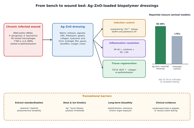
To move toward clinical use, Ag–ZnO and related particles have been incorporated into chitosan, sodium alginate, carboxymethyl cellulose (CMC), gelatin and collagen matrices (Osorio Echavarría et al., 2022; Ragab et al., 2024; Singh et al., 2023). These scaffolds mimic the native extracellular matrix, keep the wound moist, improve oxygen permeability and allow sustained release of Ag+ and Zn2+ (Liu et al., 2017; Ramachandran et al., 2024). In full-thickness cutaneous wound models in rats and mice, Ag–ZnO-loaded dressings closed wounds in about 10–12 days compared with more than 20 days in untreated controls (Kantipudi et al., 2018; Mao et al., 2017). Histology shows enhanced re-epithelialization, dense collagen deposition, rapid granulation tissue formation and suppressed microbial colonization (Bayrami et al., 2020; Ezhilarasu et al., 2020; Kantipudi et al., 2018). Figure 4 outlines this translational sequence, and Table 4 summarizes the delivery platforms and safety data reported to date.

2.6 Summary of the Literature and Critical Knowledge Gaps

2.6.1 What the Literature Establishes

Taken together, the literature supports biogenic Ag–ZnO nanocomposites as multifunctional agents that address both antimicrobial resistance and chronic tissue inflammation. Green synthesis via plant extracts and microbial secretomes offers an environmentally sound and potentially scalable production route. The Schottky heterojunction between Ag and ZnO amplifies ROS generation, supports sustained dual-ion release, destabilizes bacterial envelopes and dismantles biofilms across Gram-positive and Gram-negative MDR pathogens (Figure 2). In parallel, these nanostructures (or their silver- and zinc-based analogues) dampen NF-κB-mediated cytokine cascades and favour M2 polarization, producing a microenvironment conducive to tissue regeneration (Figure 3).

2.6.2 Outstanding Gaps

Several challenges nonetheless limit clinical translation. First, botanical extracts vary by season, geography and species, producing batch-to-batch differences in particle size, shape and capping density; standardized extraction and identification of the active biomolecules will be needed for regulatory approval (Aji et al., 2025; Ovais et al., 2018). Second, although short-term assays indicate safety in dermal fibroblasts and keratinocytes at therapeutic doses, data on long-term bioaccumulation, hepatic and renal clearance and chronic organ toxicity remain sparse (Hashempour et al., 2019; Xu et al., 2020). Third, most applications are still confined to in vitro assays and rodent wound models. Randomized, controlled clinical trials of Ag–ZnO dressings in chronic human wounds such as diabetic foot ulcers and venous leg ulcers are urgently needed (Ezhilarasu et al., 2020; GBD 2021 Antimicrobial Resistance Collaborators, 2024). These barriers are summarized in Figure 4.

3. Review Approach and Evidence Synthesis Framework

3.1 Scope and Source Material

This work was conceived as a critical narrative review rather than a formal meta-analysis. It draws on peer-reviewed experimental studies and reviews concerned with biogenic silver, zinc oxide and Ag–ZnO nanostructures, published up to early 2025, that report on at least one of four domains: synthesis and characterization, antibacterial or anti-biofilm activity against MDR organisms, host immune responses, and biocompatibility or wound-healing performance. Studies on closely related systems, such as biopolymer-embedded silver or zinc oxide particles and ternary ZnO/CuO/Ag composites, were retained where they informed mechanism or translation, and they are clearly identified as such in the tables.

3.2 Data Extraction and Organization of Evidence

From each eligible study, we extracted the biological source, precursor salts, reaction conditions, particle size and morphology (Table 1); target pathogens, zones of inhibition, MIC/MBC values and biofilm-inhibition percentages (Table 2); cellular targets, ROS pathways and immunological outcomes (Table 3); and delivery platform, cytotoxicity thresholds and in vivo wound-closure rates (Table 4). Because assay protocols, strains and units differed considerably between studies, values were not pooled statistically. Instead, ranges are reported and compared qualitatively, and the conceptual relationships among findings are summarized in Figures 1–4.

4. Multi-Target Killing, Biofilm Disruption and Immunomodulation Make Green Silver-Zinc Oxide Nanocomposites Promising Against Drug-Resistant Infections

4.1 Overview of the Evidence Base

Read together, the studies point to a reasonably consistent picture, though not a uniform one. Plant-assisted Ag–ZnO heterostructures do not behave as passive carriers; they appear to be intrinsically active against MDR isolates and established biofilms. Depositing silver on zinc oxide changes surface chemistry, optical band gap and biological reactivity in ways that seem to matter for both bacterial killing and host compatibility (Table 1; Figure 1). Across the antibacterial datasets, composites tend to achieve lower MICs and wider zones of inhibition than single-metal particles or unmodified oxides (Table 2). Mammalian cell studies, meanwhile, generally report acceptable cytocompatibility at antibacterial concentrations, alongside suppression of inflammatory cytokines and enhanced dermal repair (Tables 3 and 4). The sections below unpack each strand, noting where the evidence is strong and where it is, frankly, thinner than the enthusiasm surrounding it.

4.2 Biosynthesis Routes and Structural–Optical Tuning

4.2.1 Reduction, Capping and Reaction Conditions

The synthesis protocols compiled in Table 1 vary widely, yet they share a common logic. Polyphenols, flavonoids, alkaloids and soluble proteins in the extract reduce AgNO3 together with Zn(CH3COO)2, Zn(NO3)2 or ZnCl2, and then remain on the particle surface as a steric cap that discourages agglomeration (Slathia et al., 2021). Reaction temperatures range from ambient to about 120 °C, pH from 5.5 to 9.0, and reaction times from roughly 1.5 to 24 hours (Ali et al., 2016; Arya et al., 2023; Noohpisheh et al., 2020; Shinde et al., 2015). Some protocols add a calcination step at around 400 °C, which presumably improves crystallinity, although few studies directly compare calcined and non-calcined products (Arya et al., 2023). Microbial routes using Bacillus subtilis filtrate or Escherichia coli D8 biomass tend to run at 37 °C and are often coupled with chitosan or graphene oxide matrices (Baka et al., 2019; El-Zahed et al., 2023, 2024) (Table 1; Figure 1).

4.2.2 Band-Gap Narrowing and Surface Charge

The optical data are fairly consistent. Pure biogenic ZnO typically shows a band gap of about 3.28–3.35 eV, which limits absorption to the ultraviolet. When silver is deposited on the surface or substituted into the lattice, LSPR effects and inter-band electron transfer shift absorption toward longer wavelengths, narrowing the band gap to approximately 3.18–3.24 eV (Noohpisheh et al., 2020; Slathia et al., 2021). The shift is modest, but it is probably enough to improve visible-light photoreactivity and to accelerate charge separation, consistent with the Schottky-junction model (Figure 1). Zeta potentials fall between about −11.0 and −28.5 mV, indicating reasonable colloidal stability conferred by negatively charged phytochemical groups (Elemike et al., 2019; Nagajyothi et al., 2021). Whether the more negative end of that range translates into better performance in vivo has not, as far as we can tell, been tested systematically.

4.2.3 Influence of Botanical Family on Morphology

Particle morphology seems to track, at least loosely, with the plant family used. Lamiaceae extracts (Rosmarinus officinalis, Lavandula stoechas, Thymus vulgaris) tend to give small, quasi-spherical particles of about 5–45 nm (Noohpisheh et al., 2020; Zare et al., 2019). Zingiberaceae and Fabaceae extracts, such as Curcuma longa, Acacia caesia or Sophora pachycarpa, more often yield spherical to hexagonal wurtzite structures of 20–65 nm (Manjari et al., 2020; Mousavi-Kouhi et al., 2021). Moringa oleifera seed and leaf extracts, by contrast, have been associated with larger, flower-like or polydisperse architectures of 36–119 nm (Rafique et al., 2023; Slathia et al., 2021). High-resolution TEM and FE-SEM images show silver nanospheres of about 2–15 nm distributed across the hexagonal faces of ZnO (Elemike et al., 2019; Nagajyothi et al., 2021). It would be premature, though, to treat botanical family as a reliable predictor; within-family variation in extract composition is considerable and seldom quantified (Table 1).

4.3 Antibacterial Susceptibility Across MDR Pathogens

4.3.1 Gram-Negative Organisms

Against clinical isolates of E. coli, P. aeruginosa and K. pneumoniae, MIC values for biogenic Ag–ZnO composites generally fall between 3.12 and 25 µg/mL, with inhibition zones of about 16–43 mm (Heidary et al., 2019; Noohpisheh et al., 2020). Table 2 gives representative figures: an MIC of 7.8 µg/mL against an MDR P. aeruginosa isolate (Ahmed et al., 2020; Noohpisheh et al., 2020), 12.5 µg/mL against carbapenem-resistant K. pneumoniae with a CS/GO/ZnO cryogel (El-Zahed et al., 2024; Elumalai et al., 2015), and 25 µg/mL against an ESBL-producing E. coli treated with Aloe vera-derived ZnO (Ali et al., 2016; Gunalan et al., 2012). The relatively thin Gram-negative envelope, roughly 7–8 nm in some reports, is the usual explanation for this heightened susceptibility, since it permits faster attachment and membrane insertion (Naskar et al., 2020) (Figure 2).

4.3.2 Gram-Positive Organisms and Fungi

Gram-positive pathogens are, on the whole, harder to kill. For S. aureus and MRSA, MICs range from about 12.5 to 100 µg/mL, with zones of 12–36 mm (Abdelghafar et al., 2022; Mousavi-Kouhi et al., 2021). In one MRSA study, a biogenic Ag–ZnO composite produced a 24.5 ± 0.8 mm zone, an MIC of 15.6 µg/mL and an MBC of 31.2 µg/mL (Abdelghafar et al., 2022). Vancomycin-resistant Enterococcus faecalis required higher concentrations still, with an MIC of 50 µg/mL and an MBC of 100 µg/mL (Ahmed et al., 2020; Gupta et al., 2018). The thicker peptidoglycan wall (20–80 nm) probably accounts for much of this difference, demanding higher local ion and ROS concentrations before lysis occurs. Fluconazole-resistant Candida albicans fell in a similar range (MIC 31.25 µg/mL; MFC 62.5 µg/mL), suggesting that the activity extends, with some loss of potency, to fungal pathogens (Dananjaya et al., 2018; Noohpisheh et al., 2020) (Table 2).

4.3.3 Activity Against Mycobacteria and Resistant Phenotypes

Perhaps the most striking values come from mycobacteria. Bimetallic Ag–ZnO particles inhibited MDR and XDR Mycobacterium tuberculosis isolates at 3.91 and 7.81 µg/mL, respectively, outperforming either metal alone (Heidary et al., 2019). Importantly, composites also retained activity against strains carrying plasmid-mediated mechanisms, such as extended-spectrum β-lactamases and carbapenemases, that defeat comparator antibiotics including ciprofloxacin, gentamicin and vancomycin (Heidary et al., 2019; Kakian et al., 2024). This is encouraging, though it should be said that direct head-to-head comparisons under identical conditions are still uncommon (Table 2).

4.4 Anti-Biofilm and Anti-Quorum-Sensing Activity

Beyond planktonic killing, the composites show marked anti-biofilm effects. At sub-inhibitory concentrations of about 12.5–100 µg/mL, they suppress initial attachment and attenuate quorum-sensing-regulated virulence. Crystal violet and Congo red assays indicate reductions of roughly 81–92% in mature biofilm mass for P. aeruginosa and MRSA (Abdelghafar et al., 2022; Swidan et al., 2022). Husain et al. (2022) reported an 83.9% reduction in EPS secretion, accompanied by lower pyocyanin output, reduced swarming motility and diminished protease activity, findings echoed elsewhere (Swidan et al., 2022). In Table 2, biofilm inhibition values range from 68.0% against VRE urinary biofilms to 88.2% against EPS synthesis in P. aeruginosa (Ahmed et al., 2020; Gupta et al., 2018; Noohpisheh et al., 2020). By loosening the protective matrix, the composites may also restore susceptibility to host defences and to co-administered antibiotics, a point that seems worth testing more rigorously in combination studies (Abdelghafar et al., 2022; Zarepour et al., 2024).

4.5 Mechanistic Evidence: Membrane Damage, Ion Release and the ROS Cascade

4.5.1 Electrostatic Adhesion and Envelope Disruption

The killing process appears to begin at the surface. Electrostatic attraction between cationic sites (Ag+, Zn2+) and anionic phosphate or LPS groups on the cell wall produces micro-fissures that compromise membrane semi-permeability, allowing K+, phosphate and soluble proteins to leak out (Noohpisheh et al., 2020). Table 3 maps this event to its targets, namely peptidoglycan, LPS and membrane phospholipids, and notes the complementary roles of Ag+ (LPS destabilization) and Zn2+ (mechanical pore formation) (Ali et al., 2016; Elumalai et al., 2015) (Figure 2).

4.5.2 Oxidative Stress and Intracellular Damage

Once the envelope is breached, oxidative stress seems to dominate. Photo-excitation and surface redox reactions generate •OH, O2^{•−} and H2O2 (Noohpisheh et al., 2020; Zare et al., 2019). Accumulated ROS initiate lipid peroxidation chain reactions in unsaturated membrane fatty acids, raising malondialdehyde levels and destabilizing the bilayer. Ag+ simultaneously binds thiol groups in respiratory enzymes such as NADH dehydrogenase, halting oxidative phosphorylation, while excess intracellular Zn2+ may cause enzyme mis-metallation; ROS-induced strand breaks and base modifications then impair DNA replication (Heidary et al., 2019). In Table 3, ZnO is identified as the principal ROS generator, with Ag improving electron–hole separation (Ahmed et al., 2020; Dananjaya et al., 2018), and dual-ion binding is linked to DNA condensation and loss of replication capacity (Gupta et al., 2018; Zare et al., 2019).

4.5.3 Implications for Resistance

Because the attack is spread across the envelope, the respiratory chain and the genome, a pathogen would in principle need to alter its membrane charge, repair widespread lipid oxidation and modify multiple thiol-containing enzymes simultaneously to survive. That seems improbable through a single mutation, which may explain why stable resistance to these composites has not, so far, been widely reported (Gunalan et al., 2012; Heidary et al., 2019). One should be cautious here, however: absence of reported resistance over short exposure periods is not the same as demonstrated resistance-proofing, and serial-passage studies remain rare (Table 3; Figure 2).

4.6 Cytocompatibility and Immunomodulatory Responses

4.6.1 Mammalian Cell and Blood Compatibility

A critical question is whether these antibacterial doses are tolerated by host cells. Cytotoxicity assays in L929 fibroblasts, HaCaT keratinocytes and human erythrocytes generally show viabilities above 85–90% at concentrations up to 100 µg/mL (Dai et al., 2016; Zare et al., 2019). Hemolysis appears to stay below the 5% safety threshold at sub-MIC working concentrations of less than 50 µg/mL, suggesting a usable therapeutic window (Hashempour et al., 2019). Table 4 shows similar results across delivery formats: more than 85% fibroblast viability at 50 µg/mL in PVA/starch films (Batool et al., 2019), non-cytotoxicity up to 100 µg/mL in electrospun nanofibres (El-Aassar et al., 2020), and more than 88% viability in a CS/GO/ZnO cryogel at 100 µg/mL (El-Zahed et al., 2024). The window narrows for Gram-positive organisms and fungi, where effective MICs approach these cytotoxic thresholds, which is a point worth keeping in view (Tables 2 and 4).

4.6.2 Cytokine and Macrophage Responses

In cell culture and wound models, exposure to silver- and zinc-based nanomaterials reduces TNF-α, IL-1β and IL-6 and suppresses COX-2 activity in LPS-stimulated macrophages (Chen et al., 2020; Gliga et al., 2020; Soltan et al., 2024). At the same time, anti-inflammatory mediators such as IL-10 increase, and macrophages shift from the M1 toward the M2 phenotype (Chen et al., 2020; Perumalsamy et al., 2024). Table 3 places these effects alongside the antibacterial mechanisms: phytochemical capping agents scavenge free radicals and reduce NF-κB translocation (Batool et al., 2019; Bhowmick & Koul, 2016), while upregulation of Nrf2 and antioxidant enzymes accompanies the M1-to-M2 shift (El-Aassar et al., 2020; Krishnan et al., 2020). It is worth noting, though, that most of the direct immunological measurements were made with silver nanoparticles alone rather than with Ag–ZnO composites, a limitation we return to in the Discussion (Figure 3).

4.7 In Vivo Wound Healing and Tissue Regeneration

4.7.1 Closure Rates in Animal Models

Animal data are, on balance, encouraging. In infected rodent burn and excision models, topical biogenic composites reduced the time to complete healing from about 20 days in untreated controls to about 12 days (Hashemi et al., 2023; Ramachandran et al., 2024). Across the formulations in Table 4, wound closure of 95–99% was typically reached by days 12–14, compared with less than 75% in untreated controls (Batool et al., 2019; El-Aassar et al., 2020; Kantipudi et al., 2018). The highest reported values were 99.4% clearance of MRSA infection and closure by day 12 with a photoresponsive Ag/Ag@AgCl/ZnO hydrogel (Mao et al., 2017) and 99.2% closure with full re-epithelialization by day 12 in a hyaluronic-acid nanofibre (El-Aassar et al., 2020) (Table 4; Figure 4).

4.7.2 Histological Features of Repair

Figure 3. Host immunomodulation by biogenic Ag–ZnO nanocomposites in macrophages and peripheral blood mononuclear cells. Bacterial lipopolysaccharide and other pathogen-associated molecular patterns engage TLR-2 and TLR-4, activating the MyD88–IKK axis and driving NF-κB (p65/p50) into the nucleus. At low doses, biogenic nanocomposites block this pathway at both the receptor and translocation steps (green inhibitory connector). Downstream, pro-inflammatory mediators including TNF-α, IL-1β, IL-6, COX-2 and matrix metalloproteinases MMP-2 and MMP-9 are suppressed, while pro-resolving mediators such as IL-10, TGF-β, VEGF and Nrf2-dependent antioxidant enzymes increase. Macrophages consequently shift from the M1 phenotype, which persists in chronic multidrug-resistant infection, toward the reparative M2 phenotype that supports fibroblast proliferation, angiogenesis and collagen deposition. Neutrophil degranulation and host-derived oxidative damage are restrained while phagocytic clearance is preserved, and phytochemical capping agents scavenge excess tissue radicals. Most direct measurements underlying this scheme derive from silver nanoparticle systems.

Figure 4. Translational pathway from laboratory synthesis to the chronic wound bed, with outstanding barriers. A chronic infected wound, characterized by multidrug-resistant biofilm, macrophages locked in the M1 state, elevated TNF-α, IL-6 and matrix metalloproteinases, and stalled re-epithelialization, is treated with an Ag–ZnO-loaded dressing. Carrier matrices include chitosan, alginate, carboxymethyl cellulose, PVA/starch, gelatin, collagen and hyaluronic acid, formulated as hydrogels, films, gauzes, nanofibres, cryogels or creams. Three therapeutic phases follow: infection control through sustained Ag+/Zn2+ release, resolution of inflammation via NF-κB suppression and M1-to-M2 polarization, and tissue regeneration through TGF-β and VEGF upregulation. Reported closure in rodent models reaches 95–99% by days 12–14 against under 75% in untreated controls. The lower panel lists the four principal translational barriers: extract standardization, dose and ion-release kinetics, long-term biosafety, and the absence of randomized clinical trials.

Histopathology consistently shows enhanced re-epithelialization, dense and organized collagen deposition, increased VEGF expression with neovascularization, and reduced neutrophil infiltration (Hashemi et al., 2023; Yang et al., 2022). The ZnO/CuO/Ag propolis gauze reached 97.8% wound-area reduction by day 14 (Bayrami et al., 2020), and injectable Ag–ZnO composites promoted fibroblast migration, collagen deposition and angiogenesis without detectable systemic toxicity (Kantipudi et al., 2018). By resolving chronic inflammation while keeping the wound bed largely sterile, the composites seem well suited to infection-driven dermal injuries, at least in the models tested to date (Table 4; Figure 4).

5. Reconciling Antibacterial Potency, Host Compatibility and Translational Readiness

5.1 Why the Hybrid Outperforms Its Components

The central claim running through this literature is that Ag and ZnO together achieve what neither does alone. The mechanistic account offered for this is coherent enough: ZnO contributes a semiconductor surface that produces radicals and a slowly dissolving reservoir of Zn2+, while silver contributes an electron sink that limits charge recombination along with Ag+ ions directed at thiol-bearing enzymes (Busila et al., 2023; Sharwani et al., 2022). The band-gap narrowing from roughly 3.3 to 3.2 eV is modest, yet under ambient illumination it may suffice to sustain radical output (Noohpisheh et al., 2020; Slathia et al., 2021) (Figures 1 and 2). Several-fold MIC reductions against MDR and XDR M. tuberculosis, MRSA and carbapenem-resistant A. baumannii are consistent with that reading (Heidary et al., 2019; Kakian et al., 2024) (Table 2).

Consistency, however, is not proof. Formal tests of interaction, such as checkerboard assays or fractional inhibitory concentration indices, are seldom performed, so "synergy" in most reports means little more than improved activity relative to each component. Nor is the relative weight of each pathway resolved. Radical generation, ion dissolution and direct surface contact are hard to disentangle, and their balance likely shifts with illumination, medium and particle size. Caron et al. (2024) found that ZnO toxicity in Mueller–Hinton broth was driven largely by released Zn2+, which raises the possibility that some effects attributed to the particles are substantially ionic. The design implication differs accordingly: if ions dominate, release kinetics are the lever to pull; if surface chemistry dominates, defect density and light exposure matter more (Gudkov et al., 2021; Jiang et al., 2020) (Table 3).

5.2 Gram-Dependence and the Comparability Problem

That Gram-negative organisms are more readily killed is among the steadier observations in the field, and the thinner peptidoglycan layer is the usual explanation (Babu & Antony, 2019; Sirelkhatim et al., 2015). It may not be sufficient on its own. The Gram-negative outer membrane is itself an effective permeability barrier to many antibiotics, so the relative ease with which these composites act suggests that specific electrostatic engagement with LPS, rather than wall thickness alone, carries much of the explanatory weight (Naskar et al., 2020; Xu et al., 2020) (Figure 2).

A more awkward issue is comparability. Inhibition zones vary with agar depth, inoculum size, particle diffusion and well volume, and they travel poorly between laboratories (Table 2). MIC determinations differ in medium, incubation period and endpoint. Biofilm reduction is quantified by crystal violet in some studies, by Congo red or EPS assay in others, and strains are frequently local isolates whose resistance genotypes go unreported (Abdelghafar et al., 2022; Swidan et al., 2022). The figures assembled here should therefore be read as indicative ranges. Alignment with CLSI or EUCAST broth microdilution, inclusion of reference strains alongside clinical isolates, and agreed biofilm endpoints would make this body of work considerably easier to interpret.

5.3 Biofilms and the Resistance Question

The anti-biofilm results are among the more clinically meaningful findings, given that biofilm-associated infections tolerate antibiotic concentrations far above planktonic MICs (Devanga Ragupathi et al., 2022; Ramasamy & Lee, 2016). Reductions of 80–90% in mature biofilm mass, together with suppressed EPS, pyocyanin, protease and swarming phenotypes, suggest activity at two levels: physical penetration of the matrix and interference with AHL-mediated signalling at sub-inhibitory doses (Ahmad et al., 2024; Fulindi et al., 2023; Husain et al., 2022; Shah et al., 2019). Whether signalling interference is a direct effect on autoinducer synthesis or receptor binding, or an indirect consequence of oxidative stress on signal-producing cells, has not been settled.

The claim that multi-target action makes resistance improbable deserves more caution than it usually receives. The logic is sound, since simultaneous adaptation of envelope charge, lipid repair capacity and multiple thiol-containing enzymes is unlikely through a single mutation. But most studies run for hours to a few days. Serial-passage experiments under sub-inhibitory exposure are rare, and silver resistance determinants such as sil operons are already documented in Gram-negative clinical isolates. Treating these composites as resistance-proof on present evidence would be premature; the appropriate statement is that no stable resistance has yet been reported under the short exposures tested (Table 3).

5.4 The Immunological Evidence and Its Principal Limitation

The immunomodulatory narrative is attractive: suppression of TLR-2/TLR-4 signalling and NF-κB translocation, lower TNF-α, IL-1β, IL-6 and COX-2, higher IL-10, and a shift from M1 toward M2 macrophages (Chen et al., 2020; Gliga et al., 2020; Perumalsamy et al., 2024; Soltan et al., 2024) (Figure 3). For chronic wounds, where macrophages remain locked in a pro-inflammatory state, this would be precisely the desired intervention (Zarepour et al., 2024).

One qualification should be stated plainly. Most direct immunological measurements come from silver nanoparticles, occasionally from ZnO, and only rarely from Ag–ZnO composites themselves. Studies on macrophage polarization used silver-loaded TiO2 nanotubes (Chen et al., 2020); single-cell transcriptomic and mass cytometry work used functionalized AgNPs in PBMCs (Bae et al., 2022; Perumalsamy et al., 2024). Extending these findings to the composite is reasonable but not established, particularly since zinc has its own immunological activity and the phytochemical cap contributes antioxidant capacity of its own (Hanley et al., 2009; Jiao et al., 2014; Luo et al., 2015). Dose-dependence adds a further complication: the same materials that suppress inflammation at low concentrations can provoke inflammasome activation and IL-1β release at higher ones (Yang et al., 2012). The therapeutic window is thus defined by immunological as well as cytotoxic limits, and the two have rarely been mapped together in a single study (Table 3).

5.5 Cytocompatibility, Dose and the Narrowness of the Window

Viability above 85–90% at up to 100 µg/mL in fibroblasts and keratinocytes, with haemolysis under the 5% threshold below 50 µg/mL, reads reassuringly (Dai et al., 2016; Hashempour et al., 2019; Zare et al., 2019) (Table 4). The comfort diminishes on closer inspection. Gram-positive MICs of 50–100 µg/mL and antifungal MFCs of 62.5 µg/mL sit close to, or within, the range where cytotoxicity begins to appear (Table 2). For E. faecalis in particular, the bactericidal concentration coincides with the upper limit of tested biocompatibility. The window is real but not generous, and it is widest against Gram-negative organisms.

Biopolymer embedding is the usual remedy, and it appears to work. Chitosan, alginate, CMC, PVA/starch and collagen matrices moderate the initial burst of Ag+, sustaining release over roughly 96 hours while maintaining a moist wound environment (Batool et al., 2019; Liu et al., 2017; Osorio Echavarría et al., 2022; Ragab et al., 2024; Singh et al., 2023) (Figure 4). Scaffold chemistry is therefore not incidental packaging but part of the pharmacology. Related systems, including gelatin/alginate scaffolds for multi-agent release and Zn2+-loaded polysaccharide microspheres for growth-factor delivery, point toward the same design principle (Ju et al., 2023; Radic et al., 2024). Whether sustained release preserves the immunomodulatory profile observed with bolus exposure is, as far as we can determine, untested.

5.6 Wound-Healing Data: Strengths and Over-Reading

Closure rates of 95–99% by days 12–14, against less than 75% in untreated controls, are reported with notable consistency across films, nanofibres, hydrogels, gauzes and injectables (Bayrami et al., 2020; El-Aassar et al., 2020; Kantipudi et al., 2018; Mao et al., 2017) (Table 4; Figure 4). Histology supports the functional readouts, showing organized collagen, VEGF-associated neovascularization and reduced neutrophil infiltration (Hashemi et al., 2023; Yang et al., 2022).

These results should not be over-read. Untreated controls are a weak comparator; few studies include a standard-of-care silver dressing or a vehicle-only scaffold, so the increment attributable to the nanocomposite, as distinct from the moist dressing, is often unclear. Acute excision and burn wounds in young, healthy rodents also heal readily, and they model poorly the ischaemic, neuropathic, chronically colonized ulcers that constitute the intended clinical target (Ezhilarasu et al., 2020). Diabetic or pressure-ulcer models, longer follow-up for scarring and tensile strength, and active comparators would all strengthen the case considerably.

5.7 Standardization, Safety and the Path to Clinical Evaluation

Three obstacles stand between the current evidence and patient use (Figure 4). The first is reproducibility of the biological feedstock. Phytochemical content varies with season, geography, cultivar and extraction method, which propagates into particle size, morphology and capping density (Aji et al., 2025; Ovais et al., 2018). Chemically defined extracts, identification of the specific reducing and capping molecules, and release specifications tied to particle attributes rather than to plant provenance will be needed for any regulatory submission. Microbial synthesis may offer better batch control, since culture conditions are more readily standardized than botanical harvests (Gahlawat & Choudhury, 2019).

The second is long-term safety. Short-term cytotoxicity and haemolysis assays address acute tolerance but say little about accumulation, hepatic and renal clearance or chronic organ exposure (Hadrup et al., 2018; Hashempour et al., 2019; Xu et al., 2020). For a dressing applied to a large, breached skin surface over weeks, systemic absorption is a legitimate concern rather than a theoretical one. Silver deposition in skin and internal organs after repeated dermal exposure is documented, and zinc has its own tolerance limits.

The third is the absence of clinical data. No randomized controlled trials of biogenic Ag–ZnO dressings in diabetic foot or venous leg ulcers have been reported (GBD 2021 Antimicrobial Resistance Collaborators, 2024). Given an existing market of conventional silver dressings with mixed trial outcomes, the burden of proof is a demonstrated advantage over current practice, not merely superiority to no treatment. A sensible sequence would be a standardized, chemically characterized formulation; comparative preclinical testing against marketed silver dressings in chronic-wound models; then small controlled trials with microbiological and healing endpoints measured together.

5.8 Limitations of This study

Several constraints apply. This is a narrative rather than systematic review, without protocol registration or formal risk-of-bias appraisal, so selection bias cannot be excluded. Methodological heterogeneity precluded quantitative pooling, and the ranges reported are descriptive. Some tabulated evidence comes from related systems, including monometallic silver or ZnO formulations and ternary composites, which were retained for mechanistic or translational relevance but are not strictly Ag–ZnO. Publication bias is also likely: negative or null antibacterial results are rarely published in this field, so reported potency is probably skewed favourably. Finally, several of the composites discussed have been characterized only in single studies, without independent replication.

6. Conclusion

Biogenic Ag–ZnO nanocomposites occupy an unusual position: they are produced by simple, low-cost, environmentally benign routes, yet they act through mechanisms that conventional antibiotics cannot match. The evidence reviewed here supports three conclusions. Silver deposited on wurtzite ZnO forms a heterojunction that enhances radical generation and sustains dual-ion release, yielding potent activity against Gram-negative, Gram-positive and fungal MDR pathogens and substantial disruption of established biofilms. At sub-cytotoxic concentrations, these materials appear to temper NF-κB-driven inflammation and to favour reparative macrophage phenotypes, which translates in animal models into faster, better-organized wound closure. But the therapeutic window is narrower than often implied, most immunological data derive from silver-only systems, susceptibility methods are inconsistent across laboratories, and controlled human evidence does not yet exist. Standardized feedstocks, harmonized testing, long-term biodistribution studies and comparative trials against current silver dressings are the steps that would convert a promising preclinical platform into a usable clinical therapy.

References


Abdelghafar, A., Yousef, N., & Askoura, M. (2022). Zinc oxide nanoparticles reduce biofilm formation, synergize antibiotics action and attenuate Staphylococcus aureus virulence in host; an important message to clinicians. BMC Microbiology, 22(1), 258.

Agarwal, H., Kumar, S. V., & Rajeshkumar, S. (2017). A review on green synthesis of zinc oxide nanoparticles—An eco-friendly approach. Resource-Efficient Technologies, 3(4), 406–413.

Ahmad, I., Khan, S. A., Naseer, A., & Nazir, A. (2024). Green synthesized silver nanoparticles from Phoenix dactylifera synergistically interact with bioactive extract of Punica granatum against bacterial virulence and biofilm development. Microbial Pathogenesis, 192, 106708.

Ahmad, M., Qureshi, M. T., & Rehman, W. (2022). Enhanced photocatalytic degradation of RhB dye from aqueous solution by biogenic catalyst Ag@ZnO. Journal of Alloys and Compounds, 895, 162636.

Ahmed, B., Ameen, F., Rizvi, A., Ali, K., Sonbol, H., Zaidi, A., Khan, M. S., & Musarrat, J. (2020). Nanoparticle interactions with bacterial membrane topography and biofilm formation in environmental bacterial strains. ACS Omega, 5(14), 7861–7876.

Aji, A., Mohyaldinn, M. E., & Mahmud, H. B. (2025). Nature-driven synthesis of advanced eco-conscious Ag–ZnO nanomaterials: Mechanistic insights and breakthrough. Proceedings of the Institution of Mechanical Engineers, Part N: Journal of Nanomaterials, Nanoengineering and Nanosystems, 1–34.

Albrecht, M. A., Evans, C. W., & Raston, C. L. (2006). Green chemistry and the health implications of nanoparticles. Green Chemistry, 8(5), 417–432.

Ali, K., Dwivedi, S., Azam, A., Saquib, Q., Al-Said, M. S., Alkhedhairy, A. A., & Musarrat, J. (2016). Aloe vera extract functionalized zinc oxide nanoparticles as nanoantibiotics against multi-drug resistant clinical bacterial isolates. Journal of Colloid and Interface Science, 472, 145–156.

Anvar, A. A., Ahari, H., & Ataee, M. (2021). Antimicrobial properties of food nanopackaging: A new focus on foodborne pathogens. Frontiers in Microbiology, 12, 690706.

Arya, G., Kumari, R. M., Sharma, N., Gupta, N., Kumar, A., Chatterjee, S., & Nimesh, S. (2019). Catalytic, antibacterial and antibiofilm efficacy of biosynthesised silver nanoparticles using Prosopis juliflora leaf extract along with their wound healing potential. Journal of Photochemistry and Photobiology B: Biology, 190, 50–58.

Arya, P. R., Abishad, P., Unni, V., & Radhakrishnan, A. (2023). Facile synthesis of silver-zinc oxide nanocomposites using Curcuma longa extract and its in vitro antimicrobial efficacy against multi-drug resistant pathogens of public health importance. Inorganic Chemistry Communications, 148, 110356.

Babu, A. T., & Antony, R. (2019). Green synthesis of silver doped nano metal oxides of zinc & copper for antibacterial properties. Journal of Environmental Chemical Engineering, 7(1), 102840.

Bae, J., Ha, M., & Perumalsamy, H. (2022). Mass cytometry exploration of immunomodulatory responses of human immune cells exposed to silver nanoparticles. Pharmaceutics, 14, 630.

Baka, Z. A., Abou-Dobara, M. I., El-Sayed, A. K. A., & El-Zahed, M. M. (2019). Synthesis, characterization and antimicrobial activity of chitosan/Ag nanocomposite using Escherichia coli D8. Scientific Journal for Damietta Faculty of Science, 9(1), 1–6.

Batool, S., Hussain, Z., Niazi, M. B. K., Liaqat, U., & Afzal, M. (2019). Development of PVA/starch/AgNPs nanocomposite films for wound dressing applications. Journal of Drug Delivery Science and Technology, 52, 403–413.

Bayisa, Y. M., Bullo, T. A., & Hundie, K. B. (2023). Ecofriendly green synthesis and characterization of silver zinc oxide nanocomposite using aqueous leaf extract of Rumex crispus: Evaluation of antimicrobial and antioxidant activity. Heliyon, 9(5), e16063.

Bayrami, M., Bayrami, A., Habibi-Yangjeh, A., Shafeeyan, M. S., Feizpoor, S., Arvanagh, F. M., Nourani, M. R., & Taheri, R. A. (2020). Biologically-synthesised ZnO/CuO/Ag nanocomposite using propolis extract and coated on the gauze for wound healing applications. IET Nanobiotechnology, 14(7), 548–554.

Bhowmick, S., & Koul, V. (2016). Assessment of PVA/silver nanocomposite hydrogel scaffold for wound dressing applications. Materials Science and Engineering: C, 59, 109–119.

Busila, M., Musat, V., Alexandru, P., Romanitan, C., Brincoveanu, O., & Tucureanu, V. (2023). Antibacterial and photocatalytic activity of ZnO/Au and ZnO/Ag nanocomposites. International Journal of Molecular Sciences, 24(23), 16939.

Caron, A. J., Ali, I. J., Delgado, M. J., Johnson, D., Reeks, J. M., Strzhemechny, Y. M., & McGillivray, S. M. (2024). Zinc oxide nanoparticles mediate bacterial toxicity in Mueller-Hinton Broth via Zn2+. Frontiers in Microbiology, 15, 1394078.

Chakraborty, N., Jha, D., Roy, I., Kumar, P., Gaurav, S. S., & Marimuthu, K. (2022). Nanobiotics against antimicrobial resistance: Harnessing the power of nanoscale materials and technologies. Journal of Nanobiotechnology, 20(1), 375.

Chen, Y., Guan, M., Ren, R., Gao, C., Cheng, H., Li, Y., Gao, B., Wei, Y., Fu, J., & Sun, J. (2020). Improved immunoregulation of ultra-low-dose silver nanoparticle-loaded TiO2 nanotubes via M2 macrophage polarization by regulating GLUT1 and autophagy. International Journal of Nanomedicine, 15, 2011–2026.

Cushnie, T. P. T., O'Driscoll, N. H., & Lamb, A. J. (2016). Morphological and ultrastructural changes in bacterial cells as an indicator of antibacterial mechanism of action. Cellular and Molecular Life Sciences, 73(23), 4471–4492.

Dai, X., Guo, Q., Zhao, Y., Hegde, P., Zhang, Y., & Xu, X. (2016). Functional silver nanoparticle as a benign antimicrobial agent that eradicates antibiotic-resistant bacteria and promotes wound healing. ACS Applied Materials & Interfaces, 8(39), 25798–25807.

Dakal, T. C., Kumar, A., Majumdar, R. S., & Yadav, V. (2016). Mechanistic basis of antimicrobial actions of silver nanoparticles. Frontiers in Microbiology, 7, 1831.

Dananjaya, S. H. S., Kumar, R. S., Yang, M., Nikapitiya, C., Lee, J., & De Zoysa, M. (2018). Synthesis, characterization of ZnO-chitosan nanocomposites and evaluation of its antifungal activity against pathogenic Candida albicans. International Journal of Biological Macromolecules, 108, 1281–1288.

De Oliveira, D. M. P., Forde, B. M., Kidd, T. J., Harris, P. N. A., Schembri, M. A., Beatson, S. A., Paterson, D. L., & Walker, M. J. (2020). Antimicrobial resistance in ESKAPE pathogens. Clinical Microbiology Reviews, 33(3), e00181-19.

Devanga Ragupathi, N. K., Veeraraghavan, B., Karunakaran, E., & Monk, P. N. (2022). Editorial: Biofilm-mediated nosocomial infections and its association with antimicrobial resistance. Frontiers in Medicine, 9, 987011.

Dizaj, S. M., Lotfipour, F., Barzegar-Jalali, M., Zarrintan, M. H., & Adibkia, K. (2014). Antimicrobial activity of the metals and metal oxide nanoparticles. Materials Science and Engineering: C, 44, 278–284.

Djidjeli, C., Chelouche, A., Touam, T., Manseri, A., Ouhenia, S., & Souici, A. H. (2023). Ag nanoparticles effects on physical properties of ZnO and AZO sol–gel thin films. Journal of Materials Science: Materials in Electronics, 34, 11167.

Durán, N., Durán, M., De Jesus, M. B., Seabra, A. B., Fávaro, W. J., & Nakazato, G. (2016). Silver nanoparticles: A new view on mechanistic aspects on antimicrobial activity. Nanomedicine: Nanotechnology, Biology and Medicine, 12, 789–799.

El-Aassar, M. R., Ibrahim, O. M., Fouda, M. M. G., El-Beheri, N. G., & Agwa, M. M. (2020). Wound healing of nanofiber comprising polygalacturonic/hyaluronic acid embedded silver nanoparticles: In vitro and in vivo studies. Carbohydrate Polymers, 238, 116175.

El-Kattan, N., Emam, A. N., Mansour, A. S., Ibrahim, M. A., Abd El-Razik, A. B., Allam, K. A. M., Riad, N. Y., & Ibrahim, S. A. (2022). Curcumin assisted green synthesis of silver and zinc oxide nanostructures and their antibacterial activity against some clinical pathogenic multi-drug resistant bacteria. RSC Advances, 12(28), 18022–18038.

El-Zahed, M. M., Baka, Z. A. M., Abou-Dobara, M. I., & El-Sayed, A. K. A. (2023). Biogenic synthesis of chitosan/silver nanocomposite by Escherichia coli D8 (MF062579) and its antibacterial activity. Jordan Journal of Biological Sciences, 16(2), 279–288.

El-Zahed, M. M., Abou-Dobara, M. I., El-Khodary, M. M., & Mousa, M. M. A. (2024). Antimicrobial activity and nanoremediation of heavy metals using biosynthesized CS/GO/ZnO nanocomposite by Bacillus subtilis ATCC 6633 alone or immobilized in a macroporous cryogel. Microbial Cell Factories, 23, 278.

Elemike, E. E., Onwudiwe, D. C., Wei, L., & Fosso-Kankeu, E. (2019). Synthesis of nanostructured ZnO, AgZnO and the composites with reduced graphene oxide (rGO-AgZnO) using leaf extract of Stigmaphyllon ovatum. Journal of Environmental Chemical Engineering, 7(3), 103190.

Elumalai, K., Velmurugan, S., Ravi, S., Kathiravan, V., & Raj, G. A. (2015). Bio-approach: Plant mediated synthesis of ZnO nanoparticles and their catalytic reduction of methylene blue and antimicrobial activity. Advanced Powder Technology, 26(6), 1639–1651.

Endeshaw, S. B., Abebe, A., Tewodros, A., Hailu, G., & Mekonnen, M. (2023). Croton macrostachyus leaf extract-mediated green synthesis of ZnO nanoparticles and ZnO/CuO nanocomposites. ACS Omega, 9(1), 559–572.

Ezhilarasu, H., Vishalli, D., Dheen, S. T., Bay, B. H., & Srinivasan, D. K. (2020). Nanoparticle-based therapeutic approach for diabetic wound healing. Nanomaterials, 10(6), 1234.

Franci, G., Falanga, A., Galdiero, S., Palomba, L., Rai, M., Morelli, G., & Galdiero, M. (2015). Silver nanoparticles as potential antibacterial agents. Molecules, 20(5), 8856–8874.

Frankova, J., Karny, M., & Vankova, R. (2016). Pro-inflammatory response and COX-2 expression in cutaneous wound models. Journal of Dermatological Science, 82(3), 175–184.

Fulindi, R. B., Rodrigues, J. D., & Martinez, L. R. (2023). Zinc-based nanoparticles reduce bacterial biofilm formation. Microbiology Spectrum, 11, e04831-22.

Gahlawat, G., & Choudhury, A. R. (2019). A review on the biosynthesis of metal and metal salt nanoparticles by microbes. RSC Advances, 9, 12944–12967.

GBD 2021 Antimicrobial Resistance Collaborators. (2024). Global burden of bacterial antimicrobial resistance 1990–2021: A systematic analysis with forecasts to 2050. The Lancet, 404(10459), 1199–1226.

Gharpure, S., & Ankamwar, B. (2020). Synthesis and antimicrobial properties of zinc oxide nanoparticles. Journal of Nanoscience and Nanotechnology, 20(10), 5977–5996.

Gliga, A. R., De Loma, J., Di Bucchianico, S., Skoglund, S., Keshavan, S., Odnevall Wallinder, I., Karlsson, H. L., & Fadeel, B. (2020). Silver nanoparticles modulate lipopolysaccharide-triggered Toll-like receptor signaling in immune-competent human cell lines. Nanoscale Advances, 2(2), 648–658.

Gold, K., Slay, B., & Gaharwar, A. K. (2018). Antimicrobial activity of metal and metal-oxide based nanoparticles. Advanced Therapeutics, 1(3), 1700033.

Gudkov, S. V., Burmistrov, D. E., Serov, D. A., Rebezov, M. B., Semenova, A. A., & Lisitsyn, A. B. (2021). A mini review of antibacterial properties of ZnO nanoparticles. Frontiers in Physics, 9, 641481.

Gunalan, S., Sivaraj, R., & Rajendran, V. (2012). Green synthesized ZnO nanoparticles against bacterial and fungal pathogens. Progress in Natural Science: Materials International, 22(6), 693–700.

Gupta, M., Tomar, R. S., Kaushik, S., Mishra, R. K., & Sharma, D. (2018). Effective antimicrobial activity of green ZnO nano particles of Catharanthus roseus. Frontiers in Microbiology, 9, 2030.

Haddi, R., El Kharraz, A. M., & Kerroumi, M. I. (2024). Green synthesis of zinc oxide nanoparticles using Pistacia lentiscus L. leaf extract and evaluating their antioxidant and antibacterial properties. Nano Biomedicine and Engineering, 16(2), 232–247.

Hadrup, N., Sharma, A. K., & Loeschner, K. (2018). Toxicity of silver ions, metallic silver, and silver nanoparticle materials after in vivo dermal and mucosal surface exposure: A review. Regulatory Toxicology and Pharmacology, 98, 257–267.

Hajipour, M. J., Fromm, K. M., Ashkarran, A. A., de Aberasturi, D. J., de Larramendi, I. R., Rojo, T., Serpooshan, V., Parak, W. J., & Mahmoudi, M. (2012). Antibacterial properties of nanoparticles. Trends in Biotechnology, 30(10), 499–511.

Hanley, C., Thurber, A., Hanna, C., Punnoose, A., Zhang, J., & Wingett, D. G. (2009). The influences of cell type and ZnO nanoparticle size on immune cell cytotoxicity and cytokine induction. Nanoscale Research Letters, 4(12), 1409–1420.

Hashemi, S. S., Pakdin, A., Mohammadi, A., Keshavarzi, A., Mortazavi, M., & Sanati, P. (2023). Study the effect of Calendula officinalis extract loaded on zinc oxide nanoparticle cream in burn wound healing. ACS Applied Materials & Interfaces, 15(51), 59269–59279.

Hashempour, S., Ghanbarzadeh, S., Maibach, H. I., Ghorbani, M., & Hamishehkar, H. (2019). Skin toxicity of topically applied nanoparticles. Therapeutic Delivery, 10(6), 383–396.

Heidary, M., Zaker Bostanabad, S., Amini, S. M., Jafari, A., Ghalami Nobar, M., Ghodousi, A., & Darban-Sarokhalil, D. (2019). The anti-mycobacterial activity of AgNPs, ZnONPs, and Ag-ZnO nanoparticles against MDR- and XDR-Mycobacterium tuberculosis. Infection and Drug Resistance, 12, 2147–2156.

Høiby, N., Ciofu, O., Johansen, H. K., Song, Z. J., Moser, C., Jensen, P. Ø., Molin, S., Givskov, M., Tolker-Nielsen, T., & Bjarnsholt, T. (2011). The clinical impact of bacterial biofilms. International Journal of Oral Science, 3(2), 55–65.

Hojjati-Najafabadi, A., Davar, F., & Enteshari, Z. (2021). Antibacterial and photocatalytic behaviour of green synthesis of Zn0.95Ag0.05O nanoparticles using herbal medicine extract. Ceramics International, 47(22), 31617–31624.

Husain, F. M., Qais, F. A., Ahmad, I., Hakeem, M. J., Baig, M. H., Masood Khan, J., & Al-Shabib, N. A. (2022). Biosynthesized zinc oxide nanoparticles disrupt established biofilms of pathogenic bacteria. Applied Sciences, 12(2), 710.

Ishida, T. (2017). Antibacterial mechanism of bacteriolyses of bacterial cell walls by zinc(II) ion induced activations of PGN autolysins. Journal of Genes & Proteins, 1, 1–7.

Islam, F., Shohag, S., Uddin, M. J., & Cavalu, S. (2022). Exploring the journey of zinc oxide nanoparticles (ZnO-NPs) toward biomedical applications. Materials, 15(6), 2160.

Jalal, R., Goharshadi, E. K., Abareshi, M., Moosavi, M., Yousefi, A., & Nancarrow, P. (2010). ZnO nanofluids: Green synthesis, characterization, and antibacterial activity. Materials Chemistry and Physics, 121(1–2), 198–201.

Jiang, S., Lin, K., & Cai, M. (2020). ZnO nanomaterials: Current advancements in antibacterial mechanisms and applications. Frontiers in Chemistry, 8, 580.

Jiao, Q., Li, L., Mu, Q., & Zhang, Q. (2014). Immunomodulation of nanoparticles in nanomedicine applications. BioMed Research International, 2014, 426028.

Jin, S. E., & Jin, H. E. (2021). Antimicrobial activity of zinc oxide nano/microparticles and their combinations against pathogenic microorganisms for biomedical applications. Nanomaterials, 11(2), 263.

Jobe, M. C., Mthiyane, D. M. N., Mwanza, M., & Green, E. (2022). Biosynthesis of zinc oxide and silver/zinc oxide nanoparticles from Urginea epigea for antibacterial and antioxidant applications. Heliyon, 8(12), e12243.

Ju, Y., Zeng, H., Ye, X., Dai, M., Fang, B., & Liu, L. (2023). Zn2+ incorporated composite polysaccharide microspheres for sustained growth factor release and wound healing. Materials Today Bio, 22, 100739.

Kakian, F., Arasteh, N., & Mirzaei, E. (2024). Study of MIC of silver and zinc oxide nanoparticles, strong and cost-effective antibacterial against biofilm-producing Acinetobacter baumannii in Shiraz. BMC Infectious Diseases, 24, 947.

Kantipudi, S., Sunkara, J. R., Rallabhandi, M., Thonangi, C. V., Cholla, R. D., Kollu, P., Parvathaneni, M. K., & Pammi, S. V. N. (2018). Enhanced wound healing activity of Ag-ZnO composite NPs in Wistar Albino rats. IET Nanobiotechnology, 12(4), 473–478.

Kiani, Z., Mirjalili, S., & Heydaryan, K. (2024). Harmonizing nature and nanotechnology: Phytoextract-mediated synthesis of Ag-doped ZnO nanoparticles using Lavandula stoechas extract. Journal of Drug Delivery Science and Technology, 96, 105708.

Koo, H., Allan, R. N., Howlin, R. P., Stoodley, P., & Hall-Stoodley, L. (2017). Targeting microbial biofilms: Current and prospective therapeutic strategies. Nature Reviews Microbiology, 15(12), 740–755.

Krishnan, P. D., Chen, Y., Paul, R., & Stanslas, J. (2020). Silver nanomaterials for wound dressing applications. International Journal of Nanomedicine, 15, 4097–4114.

Lam, P. L., Wong, R. S. M., Lam, K. H., Yung, L. H., & Ho, Y. W. (2020). The role of reactive oxygen species in the biological activity of antimicrobial agents: An updated mini review. Chemico-Biological Interactions, 320, 109023.

Lemire, J. A., Harrison, J. J., & Turner, R. J. (2013). Antimicrobial activity of metals: Mechanisms, molecular targets and applications. Nature Reviews Microbiology, 11(6), 371–384.

Liu, J., Qian, Z., Shi, Q., Yang, S., Wang, Q., Liu, B., & Liu, H. (2017). An asymmetric wettable chitosan–silk fibroin composite dressing with fixed silver nanoparticles for infected wound repair. RSC Advances, 7, 43909–43918.

Lok, C. N., Ho, C. M., Chen, R., He, Q. Y., Yu, W. Y., Sun, H., & Che, C. M. (2006). Proteomic analysis of the mode of antibacterial action of silver nanoparticles. Journal of Proteome Research, 5, 916–924.

Luo, Y. H., Chang, L. W., & Lin, P. (2015). Metal-based nanoparticles and the immune system: Activation, inflammation, and potential applications. BioMed Research International, 2015, 143720.

Manjari, G., Saran, S., Radhakrishnan, S., & Devipriya, S. P. (2020). Facile green synthesis of Ag–Cu decorated ZnO nanocomposite for effective removal of toxic organic compounds and an efficient detection of nitrite ions. Journal of Environmental Management, 262, 110282.

Mao, C., Xiang, Y., Liu, X., Cui, Z., Yang, X., & Yeung, K. W. K. (2017). Photo-inspired antibacterial activity and wound healing acceleration by hydrogel embedded with Ag/Ag@AgCl/ZnO nanostructures. ACS Nano, 11(9), 9010–9021.

Mendes, C., Thirupathi, A., Corrêa, M. E. A. B., Gu, Y., & Silveira, P. C. (2022). The use of metallic nanoparticles in wound healing: New perspectives. International Journal of Molecular Sciences, 23(23), 15376.

Mohammed, K. J., Hussein, A. D., & Muhammad, S. J. (2024). Exploring the role of nanoparticles in combating microbial infections: A mini review. Hilla University College Journal of Medical Sciences, 2, 35–45.

Mohapatra, B., Mohapatra, S., & Sharma, N. (2023). Biosynthesized Ag–ZnO nanohybrids exhibit strong antibacterial activity by inducing oxidative stress. Ceramics International, 49, 14600–14612.

Mott, D. M., Anh, D. T. N., Singh, P., Shankar, C., & Maenosono, S. (2012). Electronic transfer as a route to increase the chemical stability in gold and silver core–shell nanoparticles. Advances in Colloid and Interface Science, 185, 14–33.

Mousavi-Kouhi, S. M., Beyk-Khormizi, A., Amiri, M. S., & Mashreghi, M. (2021). Silver-zinc oxide nanocomposite: From synthesis to antimicrobial and anticancer properties. Ceramics International, 47(15), 21490–21497.

Murray, C. J., Ikuta, K. S., Sharara, F., Swetschinski, L., Aguilar, G. R., Gray, A., Han, C., Bisignano, C., & Rao, P. (2022). Global burden of bacterial antimicrobial resistance in 2019: A systematic analysis. The Lancet, 399(10325), 629–655.

Mutukwa, D., Taziwa, R. T., & Khotseng, L. (2024). A review of plant-mediated ZnO nanoparticles for photodegradation and antibacterial applications. Nanomaterials, 14(14), 1182.

Nagajyothi, P. C., Muthuraman, P., Tettey, C. O., & Lee, J. (2021). In vitro anticancer activity of eco-friendly synthesized ZnO/Ag nanocomposites. Ceramics International, 47(24), 34940–34948.

Naskar, A., Lee, S., & Kim, K. (2020). Antibacterial potential of Ni-doped zinc oxide nanostructure: Comparatively more effective against Gram-negative bacteria including multidrug-resistant strains. RSC Advances, 10(3), 1232–1242.

Noohpisheh, Z., Amiri, H., Farhadi, S., & Mohammadi-Ghehsareh, A. (2020). Green synthesis of Ag-ZnO nanocomposites using Trigonella foenum-graecum leaf extract and their antibacterial, antifungal, antioxidant and photocatalytic properties. Spectrochimica Acta Part A: Molecular and Biomolecular Spectroscopy, 240, 118595.

Okaiyeto, K., Gigliobianco, M. R., & Di Martino, P. (2024). Biogenic zinc oxide nanoparticles as a promising antibacterial agent: Synthesis and characterization. International Journal of Molecular Sciences, 25(17), 9500.

Osorio Echavarría, J., Gómez Vanegas, N. A., & Ossa Orozco, C. P. (2022). Chitosan/carboxymethyl cellulose wound dressings supplemented with biologically synthesized silver nanoparticles from the ligninolytic fungus Bjerkandera sp. Heliyon, 8(9), e10258.

Ovais, M., Khalil, A. T., Islam, N. U., Ahmad, I., Ayaz, M., Saravanan, M., Shinwari, Z. K., & Mukherjee, S. (2018). Role of plant phytochemicals and microbial enzymes in biosynthesis of metallic nanoparticles. Applied Microbiology and Biotechnology, 102(16), 6799–6814.

Perumalsamy, H., Xiao, X., Kim, H. Y., & Yoon, T. H. (2024). scRNA-seq analysis discovered suppression of immunomodulatory dependent inflammatory response in PMBCs exposed to silver nanoparticles. Journal of Nanobiotechnology, 22(1), 118.

Radic, M. M., Babic, V., Vukomanovic, M., Nikodinovic-Runic, J., & Tomic, S. (2024). Gelatin-/alginate-based hydrogel scaffolds reinforced with TiO2 nanoparticles for simultaneous release of allantoin, caffeic acid, and quercetin as multi-target wound therapy platform. Pharmaceutics, 16(3), 372.

Rafique, S., Bashir, S., & Akram, R. (2023). In vitro anticancer activity and comparative green synthesis of ZnO/Ag nanoparticles by Moringa oleifera, Mentha piperita, and Citrus lemon. Ceramics International, 49(4), 5613–5620.

Ragab, H. M., Diab, N. S., AlElaimi, M., Alghamdi, A. M., Farea, M. O., & Farea, A. (2024). Fabrication and characterization of silver nanoparticle-doped chitosan/carboxymethyl cellulose nanocomposites for optoelectronic and biological applications. ACS Omega, 9(20), 22112–22122.

Ramachandran, T., Mohanraj, K. G., Martin, T. M., & Sundaram, K. M. (2024). Enhanced wound healing with β-chitosan-zinc oxide nanoparticles: Insights from zebrafish models. Cureus, 16(9), e69861.

Ramasamy, M., & Lee, J. (2016). Recent nanotechnology approaches for prevention and treatment of biofilm-associated infections on medical devices. BioMed Research International, 2016, 1851242.

Rather, G. A., Hassan, S., Pal, S., Khan, M. H., Rahman, H. S., & Khan, J. (2021). Antimicrobial efficacy of biogenic silver and zinc nanocrystals/nanoparticles to combat the drug resistance in human pathogens. Materials at the Nanoscale (pp. 1–22). IntechOpen.

Rehal, R. K., Manoswini, M., Panda, A., Majumdar, A. G., Mahapatra, P. K., & Mohanty, P. S. (2023). Color-tunable silver nanoparticles: Synthesis, characterizations, and its antimicrobial activity. BioNanoScience, 13, 1127–1135.

Sarkar, S., Leo, B. F., Carranza, C., Chen, S., Rivas-Santiago, C., Porter, A. E., Ryan, M. P., Gow, A., Chung, K. F., Tetley, T. D., & Zhang, J. (2015). Modulation of human macrophage responses to Mycobacterium tuberculosis by silver nanoparticles of different size and surface modification. PLoS ONE, 10(11), e0143077.

Sathiyaseelan, A., Vishven, N., Zhang, X., Han, K., & Wang, M. H. (2023). Research progress on chitosan-zinc oxide nanocomposites fabrication, characterization, biomedical and environmental applications. Coordination Chemistry Reviews, 496, 215398.

Shah, S., Gaikwad, S., Nagar, S., Kulshrestha, S., Vaidya, V., & Pawar, S. (2019). Biofilm inhibition and anti-quorum sensing activity of phytosynthesized silver nanoparticles against the nosocomial pathogen Pseudomonas aeruginosa. Biofouling, 35, 34–49.

Sharwani, A. A., Narayanan, K. B., & Khan, M. E. (2022). Photocatalytic degradation activity of goji berry extract synthesized silver-loaded mesoporous zinc oxide (Ag@ZnO) nanocomposites. Scientific Reports, 12(1), 10017.

Shinde, S. S., Thripuranthaka, M., & Late, D. J. (2015). Chitosan-silver bioconjugates using Prunus cerasus leaf extract and their antimicrobial evaluation. JSM Nanotechnology & Nanomedicine, 3(1), 1033.

Singh, M., Patel, A., & Kumar, S. (2023). Collagen-silica nanocomposite hydrogels for infected wound healing: Mechanical stability and antibiotic delivery. Advanced Healthcare Materials, 12(10), e2300303.

Sirelkhatim, A., Mahmud, S., Seeni, A., Kaus, N. H. M., Ann, L. C., Bakhori, S. K. M., Hasan, H., & Mohamad, D. (2015). Review on zinc oxide nanoparticles: Antibacterial activity and toxicity mechanism. Nano-Micro Letters, 7(3), 219–242.

Slathia, S., Gupta, T., & Chauhan, R. P. (2021). Green synthesis of Ag–ZnO nanocomposite using Azadirachta indica leaf extract exhibiting excellent optical and electrical properties. Physica B: Condensed Matter, 621, 413287.

Slavin, Y. N., Asnis, J., Häfeli, U. O., & Bach, H. (2017). Metal nanoparticles: Understanding the mechanisms behind antibacterial activity. Journal of Nanobiotechnology, 15(1), 65.

Soltan, H. H., Afifi, A., Mahmoud, A., Refaat, M., & Balah, O. F. (2024). Effects of silver nanoparticle and low-level laser on the immune response and healing of albino mice skin wounds. Biomedical Photonics, 13(1), 16–27.

Swidan, N. S., Hashem, Y. A., Elkhatib, W. F., & Yassien, M. A. (2022). Antibiofilm activity of green synthesized silver nanoparticles against biofilm associated enterococcal urinary pathogens. Scientific Reports, 12(1), 3869.

Tacconelli, E., Carrara, E., Savoldi, A., Harbarth, S., Mendelson, M., Monnet, D. L., Pulcini, C., Kahlmeter, G., Carmeli, Y., & WHO Priority Pathogens List Working Group. (2018). Discovery, research, and development of new antibiotics: The WHO priority list of antibiotic-resistant bacteria and tuberculosis. The Lancet Infectious Diseases, 18(3), 318–327.

Tang, K. W. K., Millar, B. C., & Moore, J. E. (2023). Antimicrobial resistance (AMR). British Journal of Biomedical Science, 80, 11387.

Thakur, S., & Neogi, S. (2021). Effect of doped ZnO nanoparticles on bacterial cell morphology and biochemical composition. Applied Nanoscience, 11, 159–171.

Theuretzbacher, U., & Piddock, L. J. (2019). Non-traditional antibacterial therapeutic options and challenges. Cell Host & Microbe, 26(1), 61–72.

Vairamuthu, S., Rajan, R., Arumugam, V., Ravikumar, P., & Ramachandran, K. (2024). Clove mediated synthesis of Ag-ZnO doped fucoidan nanocomposites and their evaluation of antibacterial, antioxidant and cytotoxicity efficacy study. Results in Chemistry, 11, 101783.

Vatansever, F., de Melo, W. C. M. A., Avci, P., Vecchio, D., Sadasivam, M., Gupta, A., Chandran, R., Karu, T. I., & Hamblin, M. R. (2013). Antimicrobial strategies centered around reactive oxygen species—bactericidal antibiotics, photodynamic therapy, and beyond. FEMS Microbiology Reviews, 37(6), 955–989.

Vukovic, B., Cvetic, Ž., & Bendelja, K. (2021). In vitro study on the immunomodulatory effects of differently functionalized silver nanoparticles on human peripheral blood mononuclear cells. Journal of Biological Inorganic Chemistry, 26, 817–831.

Xu, L., Wang, Y. Y., Huang, J., Chen, C. Y., Wang, Z. X., & Xie, H. (2020). Silver nanoparticles: Synthesis, medical applications and biosafety. Theranostics, 10, 8996–9019.

Yang, B. Y., Zhou, Z. Y., Liu, S. Y., & Zhao, X. (2022). Porous Se@SiO2 nanoparticles enhance wound healing by ROS-PI3K/Akt pathway in dermal fibroblasts and reduce scar formation. Frontiers in Bioengineering and Biotechnology, 10, 852482.

Yang, E. J., Kim, S., Kim, J. S., & Choi, I. H. (2012). Inflammasome formation and IL-1β release by human blood monocytes in response to silver nanoparticles. Biomaterials, 33(28), 6858–6867.

Zare, M., Namratha, K., Alghamdi, S., Hezam, A., Zare, M., & Byrappa, K. (2019). Novel green biomimetic approach for synthesis of ZnO-Ag nanocomposite; Antimicrobial activity against food-borne pathogen, biocompatibility and solar photocatalysis. Scientific Reports, 9(1), 8303.

Zarepour, A., Kocak, P., Tuncel, G., Zarrabi, A., & Mostafavi, E. (2024). Bioinspired nanomaterials to combat microbial biofilm and pathogen challenges: A review. ACS Applied Nano Materials, 7(22), 25287–25313.

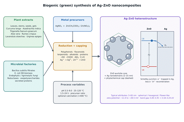
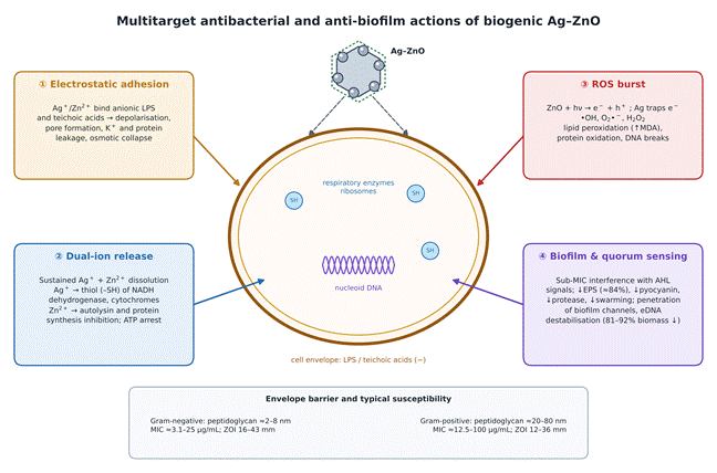
Article metrics
View details
0
Downloads
0
Citations
10
Views

View Dimensions


View Plumx


View Altmetric


0
Save
0
Citation
10
View
0
Share