Cancer immunotherapy depends, in the end, on generating robust and durable tumor-specific adaptive immunity, and the tumor-draining lymph nodes (TDLNs) are where that program is actually written (Guo et al., 2024). Soluble vaccines falter because they are degraded, cleared, and poorly retained in nodes; nanovaccines answer this by packaging TAAs, neoantigens, or whole-tumor-cell lysates with immunostimulatory adjuvants inside nanoscale carriers (Li et al., 2025). The sections below move from the physics of drainage to the immunology of outcome, and the four synthesis tables (Table 1; Table 2; Table 3; Table 4) and four figures (Figure 1; Figure 2; Figure 3; Figure 4) anchor each step.
3.1. Physicochemical rules governing lymphatic drainage and APC internalization
The passage of parenterally injected nanovaccines from the interstitium into afferent lymphatics, and their subsequent retention in draining nodes, is regulated by hydrodynamic diameter, surface charge, shape, elasticity, and targeting ligands, as summarized in Table 1 and Figure 1 (Farooq et al., 2025).
3.1.1. Size-dependent drainage kinetics
Size is the principal gating parameter. Interstitial tissue, a tangle of collagen and anionic hyaluronic acid, presents lymphatic endothelial gaps of roughly 20–100 nm (Farooq et al., 2025; Li et al., 2025). Ultra-small particles (<10 nm) diffuse into blood capillaries and are cleared renally
Table 1. Physicochemical parameters and lymphatic disposition of representative engineered nanovaccines. For each platform the table lists carrier composition, hydrodynamic size and morphology, surface charge and functional modifications, administration route and target cells, and the principal lymph-node drainage and antigen-presenting-cell uptake outcomes reported. The systems span passive size-optimized (OVA-GNPs, SPPA, AlbiVax), charge- and elasticity-tuned (SMONV), and actively targeted (M-LNP, PLGA-aCD40, PLO) designs, illustrating how size, charge, geometry, and ligands jointly govern nodal delivery.
|
Nanoparticle system & composition
|
Size & morphology
|
Surface charge & modifications
|
Route & target cells
|
Key lymphatic drainage & APC uptake outcomes
|
Key references (APA)
|
|
PLO nanovaccine (PLGA core loaded with ovalbumin peptide and Lycium barbarum polysaccharide NLBPE1)
|
~120 nm; spherical core–shell
|
−18.4 mV; polysaccharide coating
|
Subcutaneous (tail-base); CD11c+ DCs
|
Sustained nodal retention to 48 h; 3-fold higher retention than soluble peptide; TLR-mediated DC activation and cross-presentation.
|
Bao et al. (2026)
|
|
OVA-GNPs (gold nanoparticles conjugated with ovalbumin antigen)
|
10, 22, and 33 nm spherical
|
Near-neutral to slightly negative
|
Subcutaneous (footpad); popliteal-node DCs
|
22 and 33 nm give optimal interstitial convection and higher popliteal-node accumulation than 10 nm particles, which diffuse into blood.
|
Farooq et al. (2025); Guo et al. (2024)
|
|
Soft organosilica nanovaccines (SMONV; deformable mesoporous organosilica)
|
~80 nm; elastic soft meshwork
|
Neutral to slightly negative
|
Intradermal; dermal and draining-LN DCs
|
Mechanical elasticity enhances interstitial deformation and lymphatic entry versus rigid silica, giving ~10-fold higher DC cross-presentation.
|
Li et al. (2025)
|
|
Mannose-modified LNPs (M-LNP; ionizable lipid nanoparticle encapsulating mRNA)
|
~95–110 nm; spherical lipid bilayer
|
+2.1 mV; mannose ligands
|
Intramuscular/subcutaneous; CLR+ DCs
|
Mannose targeting drives receptor-mediated endocytosis into CD8a+ DCs, enabling endosomal escape, mRNA translation, and MHC-I presentation.
|
Jin et al. (2024)
|
|
SPPA triple-activating complex (Soluplus matrix co-delivering lipid-neoantigens, 3M-052, 2'3'-cGAMP)
|
~45–60 nm self-assembled aggregates
|
−12.3 mV
|
Subcutaneous; invasive and node-resident APCs
|
Small size enables rapid interstitial transit to inguinal nodes, lower hepatic clearance, and strong CD80/CD86 upregulation.
|
Rachamala et al. (2026)
|
|
AlbiVax nanocomplexes (in vivo albumin-binding lipophilic peptide–adjuvant conjugates)
|
~15–25 nm albumin complexes in situ
|
Anionic lipophilic tail
|
Subcutaneous (tail-base); endogenous albumin
|
Albumin binding prevents systemic wash-out, extends drainage >10-fold, and directs accumulation into subcapsular sinuses and paracortical zones.
|
Zhu et al. (2017)
|
|
PLGA-aCD40 mAb nanovaccine (PLGA core conjugated with anti-CD40 agonist antibody)
|
~145 nm; spherical polymeric
|
−14.2 mV; surface aCD40 mAb
|
Subcutaneous; CD40+ mature DCs
|
Active anti-CD40 targeting promotes receptor-mediated nodal retention, accelerating DC maturation and CD8+ cross-presentation.
|
Huang et al. (2026)
|
Table 2. Antigen–adjuvant formulation architectures and immunological signaling. The table maps representative nanovaccine platforms to their antigen payload, adjuvant combination and innate signaling pathway, delivery vector or membrane coating, and the resulting antigen-presenting-cell mechanism and cross-presentation route. Platforms range from biomimetic membrane-coated and carrier-free systems to dual TLR/STING and outer-membrane-vesicle designs, showing how payload breadth and adjuvant synergy together shape MHC-I cross-presentation and CD8+ T-cell priming.
|
Nanovaccine platform
|
Antigen payload type
|
Adjuvant combination & pathway
|
Delivery vector / coating
|
Immunological mechanism & cross-presentation
|
Key references (APA)
|
|
CCM-biomimetic vesicles (cancer-cell-membrane-coated nanoparticles)
|
Whole-tumor-cell membrane antigens and patient neoantigens
|
MPLA (TLR4) or CpG-ODN (TLR9)
|
Autologous B16F10/4T1 membrane over PLGA core
|
Homotypic targeting preserves native antigens; TLR4/9 engagement upregulates MHC-I/II and primes cytotoxic responses.
|
Guo et al. (2024); Ye et al. (2026)
|
|
Carrier-free F-CpG nanovaccine (self-assembled peptide–adjuvant complex)
|
Obsl1 tumor neoantigen peptide (83.19 wt%)
|
Fluorinated CpG (F-CpG; TLR9)
|
Carrier-free fluorinated self-assembly
|
Ultra-high loading eliminates polymer toxicity; F-CpG enhances uptake and cytosolic diffusion for MHC-I cross-presentation.
|
Huang et al. (2026); Li et al. (2025)
|
|
CMNPs biomimetic mRNA vaccine (neutrophil-targeted LNPs)
|
Full-length mRNA encoding tumor antigen epitopes
|
Ionizable lipid + STING via released dsDNA
|
Neutrophil-membrane-coated liposomal nanoparticle
|
Targets cancer-associated neutrophils and node DCs; ROS and tumor dsDNA activate cGAS-STING to prime CD8+ CTLs.
|
Li et al. (2026)
|
|
Probiotic OMV nanovaccine (outer membrane vesicles from engineered probiotics)
|
Recombinant viral/tumor antigens on membrane
|
LPS fragments + flagellin (TLR4/5)
|
Inhalable/parenteral bacterial OMV
|
PAMPs trigger mucosal and systemic DC activation, inducing dual mucosal IgA and systemic memory T-cell responses.
|
Huang et al. (2026); Ye et al. (2026)
|
|
SPPA neoantigen system (Soluplus polymeric self-assembly)
|
Lipid-conjugated neoantigen peptides (Adpgk/Mut30)
|
TLR7/8 (3M-052) + STING (2'3'-cGAMP)
|
Soluplus amphiphilic polymeric core
|
Simultaneous TLR7/8 and STING engagement drives NF-κB/IRF3, elevated IL-12 and IFN-β, and profound CD8+ CTL expansion.
|
Rachamala et al. (2026)
|
|
PLO botanical polysaccharide vaccine (PLGA polymer nanocomplex)
|
Model OVA protein / melanoma peptides
|
Lycium barbarum polysaccharide (NLBPE1; TLR4/2)
|
PLGA polymeric nanoparticle matrix
|
Natural polysaccharide acts as non-toxic adjuvant, augmenting DC cross-presentation and nodal antigen retention.
|
Bao et al. (2026)
|
|
AlbiVax nanocomplex (in vivo self-assembling peptide formulation)
|
Lipophilic neoantigen peptides (e.g., Adpgk)
|
Lipophilic CpG-DNA motifs (TLR9)
|
Endogenous serum albumin chaperone
|
Fatty-acid modification binds circulating albumin, trafficking to nodes to activate subcapsular macrophages and DCs without cytokine toxicity.
|
Zhu et al. (2017)
|
(Guo et al., 2024; Huang et al., 2026), whereas constructs in the 10–100 nm window, optimally 20–50 nm, enter initial lymphatics by convection and drain passively into nodes (Farooq et al., 2025; Uti et al., 2026). Ovalbumin-functionalized gold nanoparticles of 22 and 33 nm, for instance, accumulated in popliteal nodes far more than their 10 nm counterparts, which diffused into blood instead (Guo et al., 2024). Above 100–200 nm, passive entry collapses; particles form a depot from which tissue-resident DCs slowly ferry cargo nodeward over days rather than hours (Golshan et al., 2017). Comparative PLGA nanovaccines illustrate the steepness of this threshold: 83 nm formulations drained well, while 103 and 122 nm variants did not (Li et al., 2025) (Table 1).
3.1.2. Surface charge, matrix sequestration, and charge-reversal
Charge sets the balance between mobility and uptake. Near-neutral or anionic surfaces minimize electrostatic binding to the anionic extracellular matrix and transit efficiently, but resist direct endocytosis by APCs (Farooq et al., 2025). Cationic particles bind APC membranes avidly and aid endosomal disruption, yet become entrapped locally and opsonized systemically (Li et al., 2025). Charge-reversal chemistry resolves the conflict: a slightly negative surface during drainage flips to cationic in the acidic subcapsular sinus or endosome, triggering DC internalization on arrival (Li et al., 2025) (Figure 1).
3.1.3. Geometry, elasticity, and active receptor targeting
Shape and deformability tune intranodal fate. Gold nanostars localize to B-cell follicles and favor humoral responses, while nanocages persist in paracortical T-cell zones and amplify cellular immunity (Huang et al., 2026). Soft, elastic mesoporous organosilica nanovaccines deform through endothelial gaps more readily than rigid particles, yielding roughly a 10-fold rise in cross-presentation markers (Li et al., 2025). Layered onto this, ligand decoration adds selectivity: mannose engages C-type lectin receptors on APCs (Jin et al., 2024), and anti-CD40 antibody grafted onto PLGA drives CD80/CD86 upregulation and cross-presentation (Huang et al., 2026) (Table 1).
3.2. Antigenic repertoires and adjuvant co-delivery architecture
Potent CTL responses require antigen and adjuvant to reach the cytosol of node-resident APCs together, a logic captured in Table 2 and Figure 2.
3.2.1. Whole-cell lysates, biomimetic coatings, and carrier-free platforms
Single-peptide vaccines are vulnerable to antigen-loss escape under tumor heterogeneity (Ye et al., 2026). Whole-tumor-cell lysates and cancer-cell membranes supply a broad, patient-specific antigen repertoire, and biomimetic coatings preserve membrane antigens in native conformation while homing receptors aid migration (Guo et al., 2024; Li et al., 2026; Ye et al., 2026). Carrier-free designs go further: complexing fluorinated CpG with tumor peptide reached antigen loading of 83.19 wt%, removing carrier toxicity while boosting uptake (Huang et al., 2026; Li et al., 2025) (Table 2).
3.2.2. Dual-adjuvant synergies and plant-derived adjuvants
Pairing adjuvants within one carrier engages complementary innate pathways. Combining TLR7/8 or TLR4 agonists with cGAS-STING activators drives type I interferon and proinflammatory cytokine output (Huang et al., 2026; Rachamala et al., 2026). The SPPA platform co-delivers lipid-conjugated neoantigen peptides with 3M-052 (TLR7/8) and 2'3'-cGAMP (STING), upregulating MHC-I/II and costimulatory molecules (Rachamala et al., 2026). Naturally derived polysaccharides also serve: the Lycium barbarum fraction NLBPE1 in PLGA (the PLO nanovaccine) promoted DC maturation and tripled nodal antigen retention relative to free peptide (Bao et al., 2026) (Table 2).
3.3. Theranostic modalities for in vivo spatiotemporal tracking
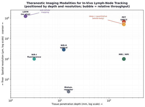
Replacing post-mortem harvests with non-invasive readouts is what makes delivery a controllable variable; the modalities and their trade-offs are compared in Table 3 and positioned in Figure 3 (Uti et al., 2026).
3.3.1. Optical and near-infrared (NIR-I/NIR-II) fluorescence
NIR fluorophores (DiD, Rhodamine-PE, Cy5.5, ICG) and rare-earth or upconversion nanoprobes visualize drainage in real time (Bao et al., 2026; Xiang et al., 2015). DiD-labeled PLO nanovaccines reached inguinal nodes within 2 h and were retained for 48 h, while free dye cleared hepatically (Bao et al., 2026). NIR-II imaging (1000–1700 nm) reduces scattering and autofluorescence, enabling light-driven gold-nanorod/R848 systems to track ablation,
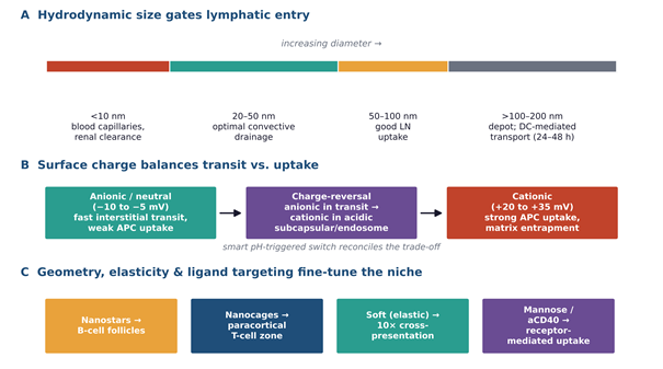
Figure 1. Physicochemical determinants of nanovaccine lymph-node trafficking. (A) Hydrodynamic diameter gates lymphatic entry: particles below ~10 nm partition into blood capillaries and are renally cleared, the 20–50 nm band drains optimally by interstitial convection, 50–100 nm retains good nodal uptake, and constructs above 100–200 nm form an injection-site depot that relies on slow dendritic-cell transport. (B) Surface charge balances interstitial transit against antigen-presenting-cell uptake, with pH-triggered charge-reversal reconciling the two. (C) Geometry, elasticity, and ligand decoration fine-tune the intranodal niche. Synthesized from Farooq et al. (2025), Guo et al. (2024), Huang et al. (2026), and Li et al. (2025).

Figure 2. Antigen–adjuvant co-delivery architecture and innate signaling. Broad antigen sources (whole-tumor-cell lysate or membrane, multi-epitope neoantigens, carrier-free peptide–adjuvant complexes) are paired with synergistic adjuvant combinations (TLR7/8 plus STING, CpG plus Mn2+, MPLA or polysaccharide adjuvants) within polymeric, lipid, biomimetic, or self-assembling vectors. Convergent uptake by node-resident dendritic cells, with endosomal escape to the cytosol, drives NF-κB and IRF3 activation, upregulation of MHC-I/II and costimulatory molecules, type I interferon and IL-12 output, and MHC-I cross-presentation. Synthesized from Huang et al. (2026), Rachamala et al. (2026), Bao et al. (2026), and Zhu et al. (2017).
antigen capture, and nodal migration (Wang et al., 2026) (Table 3).
3.3.2. Nuclear imaging (PET/SPECT) and high-resolution intranodal mapping
PET and SPECT deliver quantitative, whole-body 3D tracking; Copper-64 via NOTA chelation permits %ID/g measurement in sentinel nodes (Zhu et al., 2017). Ex vivo, nodes cleared by passive CLARITY (PACT) and imaged by 3D light-sheet fluorescence microscopy localized albumin-binding complexes (AlbiVax) to subcapsular sinuses and paracortical zones (Zhu et al., 2017) (Table 3; Figure 3).
3.3.3. Magnetic imaging (MRI/MPI) and genetic reporters
Superparamagnetic iron oxide nanoparticles act as T2 MRI and MPI tracers, allowing radiation-free tracking of DC migration over days, and nanoscale coordination-polymer nanovaccines extend the inorganic-core toolkit (Kemp & Kwon, 2021; Yang et al., 2025; Zhao et al., 2019). Firefly luciferase mRNA in LNPs adds a functional genetic readout of in vivo translation (Jin et al., 2024) (Table 3).
3.4. Immunological cascades, TME remodeling, and TLS neogenesis
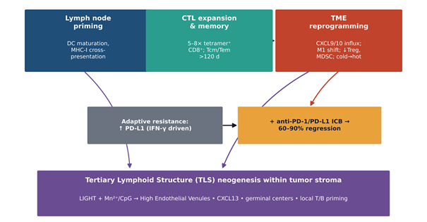
The ultimate test is whether nodal trafficking becomes systemic, tumor-eradicating immunity; Table 4 and Figure 4 trace that path.
3.4.1. DC maturation, cross-presentation, and memory
On nodal entry, nanovaccines are captured by subcapsular sinus macrophages and CD11c+ DCs (Bao et al., 2026; Farooq et al., 2025). Adjuvant signaling via endosomal TLRs or cytosolic STING drives NF-κB and IRF3 translocation, DC maturation, and IL-12/IFN-β secretion (Jin et al., 2024; Rachamala et al., 2026). Endosomal escape routes antigen into proteasomal processing and MHC-I cross-presentation, expanding tetramer-positive CD8+ CTLs and seeding central and effector memory pools (Bao et al., 2026; Wang et al., 2026) (Figure 4).
3.4.2. TME reprogramming and checkpoint synergy
Activated CTLs and IFN-γ/TNF-α convert “cold” tumors to “hot” (Huang et al., 2026). Neutrophil-targeted biomimetic vaccines (CMNPs) trigger ROS-mediated dsDNA release and tumor-intrinsic cGAS-STING, polarizing N1 neutrophils (Li et al., 2026). Because monotherapy often upregulates PD-L1, pairing with anti-PD-1/PD-L1 blockade overcomes anergy and depletes Tregs and MDSCs (Jin et al., 2024; Rachamala et al., 2026) (Table 4).
3.4.3. Tertiary lymphoid structure neogenesis
Advanced biomimetic systems can nucleate TLS inside tumors. Delivering LIGHT with dual Mn2+/CpG adjuvants induces high endothelial venule neogenesis and CXCL13 release, driving germinal-center maturation and ectopic lymphoid hubs that sustain local immunity (Wang et al., 2026; Wen et al., 2023) (Table 4; Figure 4).
3.5. Translational challenges and future theranostic horizons
Preclinical promise notwithstanding, translation faces familiar hurdles. Fluorophore leaching and tracer dissociation demand covalently bonded radiotracers or stable inorganic cores (Farooq et al., 2025; Uti et al., 2026). Hepatic and splenic RES clearance caps the dose reaching nodes (Rachamala et al., 2026). Multicomponent platforms require GMP compliance, reproducibility, and immunotoxicological profiling (Uti et al., 2026; Zhu et al., 2017). Looking ahead, machine learning for neoantigen prediction and property tuning, coupled with 3D PET/CT and NIR-II readouts, points toward digitally controlled, patient-tailored vaccination (Uti et al., 2026; Ye et al., 2026).