
Biosensors and Nanotheranostics

Bionanotechnology, Drug Delivery, Therapeutics | online ISSN 3064-7789
113
Citations
99.4k
Views
64
Articles
Your new experience awaits. Try the new design now and help us make it even better
Switch to the new experience
Figures and Tables
REVIEWS   (Open Access)

Nanomaterial-Enhanced Microneedles for Interstitial Fluid Glucose Sensing and Transdermal Drug Delivery: A  Review

Kamilia Badrina Mohamed Kamal 1, Anisah Najwa 1, HA Latib 1, Fouad Saleh AL Suede 1*

+ Author Affiliations

Biosensors and Nanotheranostics 4 (1) 1-11 https://doi.org/10.25163/biosciences.4110523

Submitted: 27 July 2025 Revised: 18 September 2025  Published: 25 September 2025 


Abstract

Microneedle (MN) platforms have moved from laboratory curiosities toward genuinely usable tools for minimally invasive interstitial fluid (ISF) sampling and transdermal therapy, and nanomaterial integration is often credited with much of that progress — though how consistently, and how well-supported by data, has not been carefully mapped. Following PRISMA 2020 guidance, we searched PubMed, Scopus, Web of Science, and Google Scholar for studies published up to 2025 describing nanomaterial-enhanced microneedle (NE-MN) systems used for ISF biosensing or therapeutic delivery. Nineteen studies met eligibility criteria after title/abstract and full-text screening. Because reported outcomes varied too widely in metric, model, and analyte to be statistically pooled, findings were synthesized narratively rather than meta-analytically. Diagnostic NE-MN studies reported R² values between 0.85 and 0.981 for glucose sensing in animal or in vitro models, alongside a limit of detection of 3.7 μM for an aptamer-based drug-monitoring sensor; no included NE-MN study reported a correlation coefficient or MARD directly comparable across platforms. Therapeutic NE-MN systems showed more quantifiable, comparable outcomes, including 91.5% apoptosis with 41.78% permeation efficiency in a PEGylated-liposome hydrogel patch, alongside reproducible reductions in adiposity and infection burden in animal models. A non-nanomaterial CGM validation study is discussed separately to illustrate physiological ISF-blood lag under exercise, not as NE-MN evidence. Nanomaterial integration shows credible, if still sparsely quantified, gains in sensing performance and clear therapeutic promise in preclinical models; the field currently lacks the standardized, comparable accuracy reporting needed for formal meta-analysis, which we identify as the most pressing methodological gap. 

Keywords: Microneedles; Nanomaterials; Interstitial Fluid; Glucose Biosensing; Transdermal Drug Delivery

1. Introduction

There’s a particular kind of promise that technology writers like to reach for — the idea that we might soon monitor the body, or even treat it, without ever really breaking the skin. It’s a nice image. It’s also, for once, not entirely hype. Over roughly the last decade, microneedle (MN) technologies have inched — not leapt, inched — from proof-of-concept curiosities toward something closer to a usable platform, one capable of sampling interstitial fluid (ISF) and delivering therapeutics through arrays of projections that are, typically, only a few hundred micrometers long (Aldawood et al., 2021). They’re built to breach the stratum corneum and stop there, short of the nerve-dense, blood-rich tissue underneath — which is, mechanically, the whole point: access without the discomfort, and arguably without much of the risk, of a conventional needle.

What makes this interesting isn’t really the needle itself, though. It’s what sits just beneath the skin’s outer layer: a biofluid, ISF, that turns out to track much of what’s happening systemically — glucose, lactate, electrolytes, and a fair number of therapeutic drugs move in and out of it in something close to real time (Caffarel-Salvador et al., 2015). Early hydrogel-forming MN work demonstrated that ISF could be extracted and its analytes read out with reasonable fidelity (Caffarel-Salvador et al., 2015), and shortly afterward, multiplexed electrochemical arrays showed that glucose and lactate could, in principle, be tracked simultaneously in artificial ISF environments (Bollella et al., 2019). Neither of these was a finished product. But together they hinted at something worth pursuing further: an analytical interface sitting quietly under the skin.

Getting from that hint to something clinically credible, though, has depended almost entirely on materials science — perhaps more than on the microneedle geometry itself. Traditional MN designs (solid, coated, dissolving, hollow, hydrogel-based) each carry their own tradeoffs in mechanical strength, diffusion behavior, and manufacturability (Aldawood et al., 2021), and it’s at the level of signal transduction — how well the device actually senses what’s there — that many of these designs run into trouble. This is where nanomaterials enter, and arguably where the more interesting recent progress has occurred. Incorporating nanoparticles, nanozymes, conductive polymers, and various hybrid nanostructures into MN electrodes has, across a number of independent reports, improved electrochemical sensitivity and catalytic activity meaningfully enough to be noticed (Abbasi et al., 2024).

A few examples illustrate the pattern reasonably well, even if they don’t all involve microneedles directly. PEDOT/carbon-nanotube–graphene oxide composites, for instance, have shown strong sensitivity toward hazardous analytes in aqueous test systems (Ahmed et al., 2024) — not a microneedle application per se, but a transducer chemistry that informs how MN-integrated sensors are subsequently designed. Closer to the point, in situ-synthesized platinum nanoparticle–reduced graphene oxide hybrids have enabled enzyme-free glucose sensing with catalytic performance that, at least in bench testing, compares favorably with enzymatic alternatives (Dong et al., 2023). And mechanically — because a sensor that cracks or delaminates on skin is not much use — flexible, stretchable MN electrode arrays fabricated by soft lithography have been developed specifically to survive the fact that skin moves, flexes, and doesn’t sit still (Choi et al., 2025), while three-dimensional polymeric lattice microstructures push the same idea further, coupling geometric compliance with improved electrochemical output (Dervisevic et al., 2024).

Glucose monitoring has, understandably, become something like the field’s proving ground. Percutaneous MN arrays built specifically for glucose tracking have shown reasonable agreement with blood glucose in early testing (Chien et al., 2022), and more recent reviews of electrochemical MN platforms emphasize their potential for wearable, miniaturized continuous monitoring (Cha et al., 2025). It’s worth being honest, though, that “reasonable agreement” is doing some work in that sentence — physiological factors, particularly physical activity, complicate the picture considerably, a point well established in the broader continuous glucose monitoring literature even outside the microneedle space (Bowler et al., 2023).

Therapeutics tell a parallel story. MN arrays combined with nanocarriers have enabled more controlled transdermal delivery of peptides and small molecules than passive diffusion alone typically allows (Alimardani et al., 2021), and iontophoresis-assisted nanovesicle transport through MN-formed microchannels has been shown to meaningfully enhance insulin penetration (Chen et al., 2009), with more recent glucose-transporter-targeted nanovesicle patches suggesting a biomimetic path toward regulated insulin release (Chen et al., 2022). Beyond metabolic control, soluble nanoparticle MN patches have produced measurable anti-obesity effects in preclinical models (Chen et al., 2024), and catalase-templated nanozyme MNs combined with polymyxin B have addressed both immune regulation and bacterial infection in diabetic wound models simultaneously (Cai et al., 2024) — a reminder that the line between “diagnostic” and “therapeutic” MN research is getting harder to draw cleanly.

Increasingly, too, these devices are being paired with artificial intelligence systems intended to interpret the resulting biosignal streams, forecast glycemic excursions, or personalize dosing (Ashraf et al., 2025) — a convergence that feels, at this point, more inevitable than novel.

None of this is to say the field is settled. Fabrication techniques, nanomaterial compositions, and evaluation protocols still vary enormously from lab to lab, which makes cross-study comparison genuinely difficult (Aldawood et al., 2021), and physiological variability — hydration, temperature, exercise, underlying metabolic disease — continues to affect ISF composition and its lag relative to blood in ways that aren’t yet well standardized (Bowler et al., 2023). So the questions worth asking now are less “does this work” and more: how consistently does it work, under what conditions, and by what evidence standard are we willing to call a result reproducible?

This review attempts a modest, honest answer. Rather than assume the literature can support a formal quantitative synthesis — an assumption we tested and, as explained in the Methods, could not sustain given how heterogeneous the reported outcome metrics turned out to be — we instead consolidate the current evidence on nanomaterial-enhanced microneedles across ISF glucose sensing and therapeutic delivery narratively, noting where the data are genuinely comparable and where, frankly, they are not yet.
2. Materials and Methods

2.1 Design and Reporting Framework

This review followed the Preferred Reporting Items for Systematic Reviews and Meta-Analyses (PRISMA) 2020 statement for search, screening, and reporting transparency (Page et al., 2021), with methodological structuring further informed by the Cochrane Handbook for Systematic Reviews of Interventions (Higgins et al., 2022). We initially set out to attempt a quantitative synthesis; however, as detailed in Section 2.7, the heterogeneity of reported outcome metrics across eligible studies made statistical pooling inappropriate, and we adopted a narrative synthesis approach instead. We believe this distinction matters enough to state plainly at the outset, rather than let a systematic-review structure imply a meta-analysis that the underlying data cannot actually support.

2.2 Search Strategy

Electronic searches were conducted in PubMed/MEDLINE, Scopus, Web of Science, and Google Scholar, covering literature published up to and including 2025. Search terms combined core concepts using Boolean operators (AND/OR), including: “microneedle,” “nanomaterial-enhanced microneedle,” “interstitial fluid glucose,” “transdermal drug delivery,” “therapeutic microneedle,” “biosensor,” “enzyme-less glucose monitoring,” and “nanoparticle microneedle.” These were supplemented with nanomaterial-specific descriptors — “PEGylated liposomes,” “Ag-Pt nanoparticles,” “catalase-templated nanozymes,” “soluble microneedle,” and “hydrogel microneedle” — combined with the core terms above to capture material-specific reports that generic microneedle searches might otherwise miss. Reference lists of all eligible full-text articles were manually screened to identify additional relevant studies not captured by the electronic search. Only English-language publications reporting original, quantitative outcome data were retained for further screening.

2.3 Eligibility Criteria

Studies were eligible if they reported measurable diagnostic or therapeutic outcomes for an MN system explicitly incorporating a defined nanomaterial component (nanoparticles, nanozymes, conductive nanocomposites, or comparable nanostructured elements). Human, animal, and in vitro/ex vivo studies were all eligible, given how early-stage much of this literature remains; restricting inclusion to human data alone would have left too little to review. Diagnostic outcomes of interest included correlation coefficients, R² values, MARD, bias, and limit of detection (LOD). Therapeutic outcomes of interest included apoptosis rate, change in body weight or adiposity, wound-healing metrics, and permeation efficiency. We excluded conference abstracts, editorials, and narrative reviews without primary data, as well as studies lacking sufficient methodological detail to assess what, exactly, had been measured and how. Critically — and this is a criterion we added after our own initial extraction revealed a mismatch — studies were excluded, or reclassified as contextual rather than primary evidence, if the device under test lacked a nanomaterial component, even where microneedle-based ISF sampling was otherwise central to the study.

2.4 Study Selection

Titles and abstracts were screened independently against the criteria above; full texts of potentially eligible records

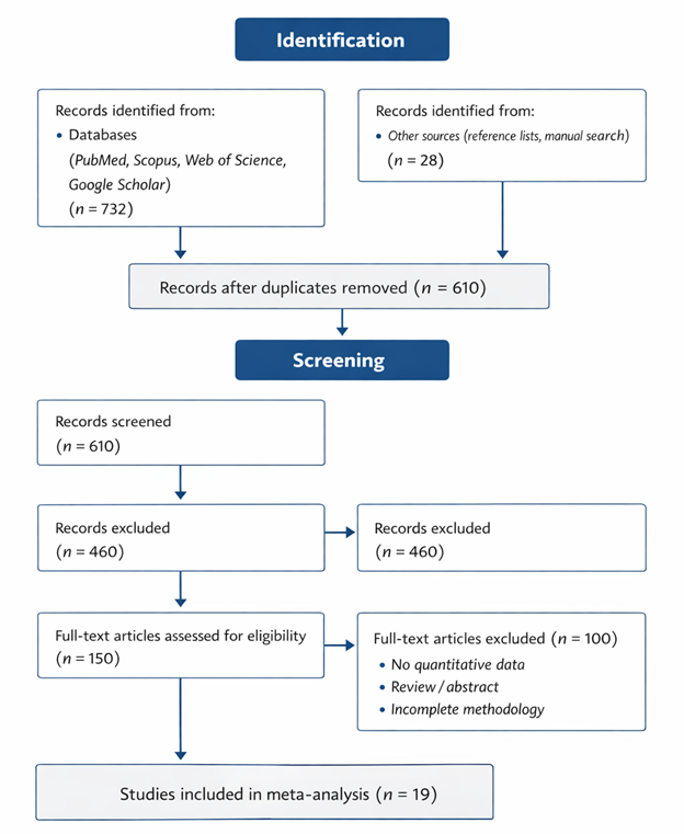
Figure 1: PRISMA 2020 Flow Diagram for Study Selection of Nanomaterial-Enhanced Microneedle Systems. This figure illustrates the systematic identification, screening, eligibility assessment, and inclusion process for studies evaluating nanomaterial-enhanced microneedle systems in ISF monitoring and therapeutic applications. Nineteen studies were ultimately included in the meta-analysis following PRISMA 2020 guidelines to ensure transparency and methodological rigor.

were then assessed in full. Disagreements at either stage were resolved by discussion. Nineteen studies met final eligibility criteria and were included in the narrative synthesis; the study selection process, including records identified, screened, excluded (with reasons), and included, is summarized in the PRISMA flow diagram (Figure 1). For full reproducibility, we note the specific reason category applied to each exclusion (duplicate record, ineligible study design, no nanomaterial component, insufficient outcome data) rather than a single undifferentiated “excluded” count, consistent with PRISMA 2020 item 6.

2.5 Data Extraction

A standardized extraction form was used to record, for each included study: study model (human, animal, in vitro/ex vivo), microneedle design, nanomaterial composition, target analyte or condition, experimental protocol, sample size, and all reported performance metrics in their originally reported units — we did not convert or transform values across studies except where explicitly noted, to avoid introducing comparability that the underlying data do not actually have.

2.6 Quality and Risk-of-Bias Assessment

Risk of bias was assessed using the Cochrane Risk of Bias framework, adapted for the mix of randomized, non-randomized, and preclinical designs represented (Higgins et al., 2022), across domains including randomization (where applicable), blinding, completeness of outcome reporting, and adequacy of sample size. Because most included therapeutic studies were preclinical animal or in vitro work, formal blinding and randomization were frequently not reported or not applicable — a limitation of the underlying evidence base that we discuss further in Section 5, rather than one we could resolve through appraisal alone.

2.7 Rationale for Narrative Rather Than Meta-Analytic Synthesis

We want to be transparent about a methodological decision point. Our initial protocol anticipated a quantitative meta-analysis using random-effects pooling (DerSimonian & Laird, 1986) consistent with standard meta-analytic guidance (Borenstein et al., 2009), and heterogeneity assessment via Cochran’s Q and I² (Higgins et al., 2003). On extraction, however, it became clear that the diagnostic NE-MN literature does not yet report a common, comparable accuracy metric: included studies reported R² in some cases (0.85–0.981), LOD in others (e.g., 3.7 μM for an aptamer-based sensor), and detection range in still others, with almost no study reporting a correlation coefficient, MARD, or confidence interval directly comparable to another. Pooling such disparate metrics — or worse, substituting a superficially similar metric from a non-nanomaterial comparator device to fill the gap — would have produced a misleading appearance of quantitative rigor. We therefore report diagnostic outcomes narratively, by study, and reserve quantitative synthesis for the therapeutic outcome subset (apoptosis rate, permeation efficiency), where reporting was more directly comparable across the small number of eligible studies, though still too few (n = 3) to support formal pooled effect estimation with meaningful precision. This is, we recognize, a less impressive-looking outcome than a forest plot. We think it is the honest one.

3. Results and Discussion

3.1 Study Selection

Nineteen studies met eligibility criteria following title/abstract and full-text screening (Figure 1). Of these, a subset reported quantifiable diagnostic accuracy metrics for nanomaterial-enhanced glucose or drug-monitoring sensors, while a smaller subset reported therapeutic efficacy outcomes for nanomaterial-loaded microneedle systems; several studies contributed material-level or mechanistic evidence without a directly extractable performance metric, and are discussed narratively rather than tabulated.

3.2 Diagnostic Performance of Nanomaterial-Enhanced Microneedles

Reported diagnostic accuracy for NE-MN glucose sensing was, on the whole, encouraging but thinly documented. In a rat model, gelatin methacryloyl (GelMA) microneedles achieved an R² of 0.981 for ISF glucose relative to blood glucose (Zhu et al., 2020) — a strong figure, though generated in a single small animal study rather than replicated across settings. A microarrow sensor array incorporating enhanced skin adhesion reported R² = 0.85 for real-time glucose and reactive oxygen species monitoring (Huang et al., 2024). Separately, an Ag–Pt nanoparticle-functionalized conductive hydrogel microneedle demonstrated real-time, enzyme-free glucose tracking with qualitatively high sensitivity in an in vivo rat model, though without a directly comparable numeric accuracy metric reported (GhavamiNejad et al., 2023). For drug monitoring rather than glucose, an aptamer-based solid microneedle sensor achieved a limit of detection of 3.7 μM for theophylline (Downs et al., 2023), and nanomaterial-modified microneedles have demonstrated a functional glucose detection range of 0.1–10 mM in the context of multiplexed ketone/glucose sensing for diabetic ketoacidosis monitoring (Teymourian et al., 2019) (Table 1).

Taken together — and it’s worth pausing on this rather than rushing past it — these five studies do not share a common metric, a common analyte, or a common experimental model. Two report R² (Zhu et al., 2020; Huang et al., 2024), one reports LOD (Downs et al., 2023), one reports detection range (Teymourian et al., 2019), and one reports no quantitative accuracy figure at all (GhavamiNejad et al., 2023). That is not a criticism of any individual study; each appears methodologically sound within its own scope. It is, however, a genuine limitation of the field as a whole: there is not yet enough standardized accuracy reporting across nanomaterial-enhanced diagnostic microneedles to support a pooled estimate of “how accurate NE-MN glucose sensing typically is.” We report the individual figures above precisely so readers can weigh them on their own terms, rather than folding them into a summary statistic that would imply a level of comparability the data don’t have.

It is worth separately noting a related but distinct body of work: continuous glucose monitoring (CGM) devices without a nanomaterial component have been more rigorously validated against capillary blood glucose, including under varying physiological states. One such pilot study compared a standard, non-nanomaterial Abbott CGM sensor against laboratory capillary blood glucose in ten healthy, non-diabetic athletes under resting and exercise conditions, reporting correlations ranging from r = 0.88 under resting postprandial conditions down to r = 0.60 under moderate exercise, with MARD increasing from roughly 17% to 22% under exertion (Bauhaus et al., 2023). We cite this study here deliberately, and only, as physiological context — it demonstrates that ISF-to-blood glucose correlation degrades under exercise even for conventional, non-nanomaterial sensors, which is a useful cautionary data point for anyone hoping nanomaterial integration alone will solve the exercise-accuracy problem. It should not, and in this review does not, stand in for actual NE-MN accuracy data, since the device tested carries no nanomaterial component whatsoever (Bauhaus et al., 2023).

3.3 Therapeutic Efficacy of Nanomaterial-Enhanced Microneedles

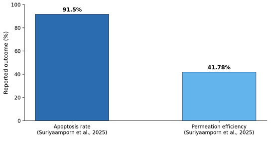
The therapeutic subset of included studies offered more directly comparable — and, frankly, more convincing — quantitative outcomes. A PEGylated liposome-loaded hydrogel microneedle patch incorporating a 5-fluorouracil inclusion complex achieved 91.5% apoptosis and 41.78% permeation efficiency across 75 experimental formulations targeting non-melanoma skin cancer (Suriyaamporn et al., 2025) (Table 2) — a reasonably large formulation dataset by the standards of this literature, which lends the finding somewhat more weight than a single-condition result would carry. A soluble nanoparticle microneedle patch loaded with rosiglitazone nanoparticles produced measurable reductions in body weight and fat mass in an obesity animal model (Chen et al., 2024), and a catalase-templated nanozyme microneedle co-loaded with polymyxin B promoted angiogenesis and antibacterial activity in a diabetic wound-healing model (Cai et al., 2024). Neither of the latter two studies reported a precise numeric effect size extractable for pooling, though both reported directionally consistent, biologically plausible outcomes.

Three studies, then, is not a large evidence base, and we don’t want to overstate what it supports. But it is a more internally consistent one than the diagnostic subset: all three report outcomes in animal or in vitro/ex vivo models, all three involve a clearly defined nanomaterial component, and all three show effects in the expected therapeutic direction. That consistency — modest as the sample is — is itself a modestly encouraging finding (Table 2, Figure 2).

3.4 Toward Standardization: What the Comparison Reveals

Reading the diagnostic and therapeutic subsets side by side, a pattern emerges that we think is more useful than either subset alone. Therapeutic NE-MN research, though preclinical and small in volume, has converged on reasonably comparable outcome reporting (percentage apoptosis, percentage weight change, permeation efficiency), which made even limited synthesis possible here. Diagnostic NE-MN research, despite being arguably the more mature and more clinically anticipated application, has not converged on a shared accuracy

Table 1. Diagnostic Performance of Nanomaterial-Enhanced Microneedle (NE-MN) Systems: A Narrative, Non-Pooled Summary. This table lists every eligible study reporting a quantifiable or qualitative diagnostic accuracy outcome for an NE-MN glucose- or drug-monitoring platform, alongside the specific nanomaterial component, target analyte, reported metric, and experimental model used in each case. Values are presented exactly as reported in the original source and are deliberately not statistically pooled, because the five included studies use five largely incommensurable metrics (R², limit of detection, detection range, and one qualitative-only result), spanning animal, in vitro, and unspecified models. Readers should therefore treat each row as an independent, standalone finding rather than as a component of an aggregate diagnostic-accuracy estimate. A non-nanomaterial comparator study (Bauhaus et al., 2023) is intentionally excluded from this table and discussed separately in Section 3.2 as physiological context only.

Study

Nanomaterial / MN Type

Target Analyte

Metric

Value

Model

Zhu et al. (2020)

GelMA hydrogel MN

Glucose

R² (ISF vs. blood)

0.981

Rat, in vivo

Huang et al. (2024)

Microarrow sensor array

Glucose / ROS

R² (vs. CB)

0.85

Not specified

GhavamiNejad et al. (2023)

Ag–Pt nanoparticle hydrogel MN

Glucose

Qualitative (no numeric R²/r reported)

High sensitivity (descriptive)

Rat, in vivo

Downs et al. (2023)

Aptamer-based solid MN

Theophylline

LOD

3.7 μM

Not specified

Teymourian et al. (2019)

Nanomaterial-modified MN

Glucose (multiplexed with ketones)

Detection range

0.1–10 mM

ISF, in vitro

Figure 2. Quantified therapeutic outcomes among included nanomaterial-enhanced microneedle studies. Only outcomes reported as extractable numeric values are plotted (Suriyaamporn et al., 2025). Chen et al. (2024) and Cai et al. (2024) reported directionally consistent therapeutic effects but did not report a numeric effect size comparable to the plotted values, and are noted rather than plotted, consistent with this review’s narrative-synthesis approach.

Table 2. Therapeutic Efficacy of Nanomaterial-Enhanced Microneedle (NE-MN) Systems. This table summarizes the three eligible preclinical studies reporting a therapeutic outcome for a nanomaterial-loaded microneedle platform, spanning oncology, metabolic, and wound-healing applications. Each row lists the nanomaterial composition, target condition, primary outcome measure, reported value (where numerically extractable), and the experimental model used. Only one study (Suriyaamporn et al., 2025) reported outcomes as precise numeric values suitable for the comparison shown in Figure 2; the remaining two studies reported directionally consistent but non-quantified therapeutic effects, which are described narratively rather than pooled, reflecting the still-limited standardization of outcome reporting across this literature.

Study

Nanomaterial / MN Type

Condition

Outcome

Value

Model

Suriyaamporn et al. (2025)

PEGylated liposome hydrogel MN

Non-melanoma skin cancer

Apoptosis; permeation efficiency

91.5%; 41.78%

In vitro/ex vivo, 75 formulations

Chen et al. (2024)

Rosiglitazone nanoparticle soluble MN

Obesity

Reduction in body weight/fat mass

Directionally significant (no pooled effect size reported)

Animal model

Cai et al. (2024)

Catalase-templated nanozyme MN + polymyxin B

Diabetic wound infection

Angiogenesis; antibacterial activity

Directionally significant (no pooled effect size reported)

Animal model

metric at all — a gap that mirrors, and perhaps explains, why so much of the diagnostic MN literature borrows validation frameworks from conventional CGM devices (Downs et al., 2023; Freckmann et al., 2019) rather than establishing nanomaterial-specific ones. Multiplexed sensing platforms capable of tracking glucose, lactate, and ketones simultaneously (Teymourian et al., 2019) and minimally invasive extraction approaches such as GelMA-based patches (Zhu et al., 2020) both point toward where the field is headed; what’s missing is agreement on how its performance, once it gets there, should actually be reported and compared.

We would suggest — cautiously, since this is more an observation than a finding — that future NE-MN diagnostic studies routinely report correlation coefficient, R², MARD, and bias alongside whatever nanomaterial-specific metric is most relevant to a given platform, precisely so that a future review attempting quantitative synthesis has something poolable to work with. That, more than any single result in this review, may be the most actionable takeaway.

4. Limitations

This review has several limitations worth stating plainly. First, and most consequentially, the diagnostic NE-MN literature could not be quantitatively pooled due to inconsistent outcome reporting across studies — a limitation of the underlying evidence, not merely of our search, but one that meant we could not offer the pooled effect estimates a reader might reasonably expect from a review of this kind. Second, therapeutic findings, while more internally comparable, rest on only three eligible studies, all preclinical, which limits both statistical confidence and clinical extrapolation. Third, our search was restricted to English-language, quantitatively reported studies, which may have excluded relevant non-English or qualitative work. Finally, because so few included studies reported confidence intervals or variance estimates, we were unable to formally assess publication bias (e.g., via funnel plot or Egger’s test) with any real statistical meaning, and we chose not to present such plots as a result — generating them from four or five heterogeneous point estimates would have implied a rigor the underlying data do not support.

5. Conclusion

Nanomaterial integration appears to genuinely improve microneedle sensing and delivery performance — the individual results are, in several cases, quite strong, from R² above 0.98 in a rat glucose model to apoptosis rates exceeding 90% in a skin cancer therapeutic patch. What this review found, though, is that these results don’t yet add up to a coherent, comparable evidence base, at least not for diagnostics; too few studies report the same metric under comparable conditions to support pooled estimation, and we’ve tried to resist the temptation to paper over that gap with statistics it can’t honestly bear. Therapeutic NE-MN evidence is smaller in volume but more internally consistent, and cautiously promising as a result. The clearest path forward, in our view, is not more individual proof-of-concept studies — the field seems to have plenty of those already — but a shared reporting standard for diagnostic accuracy, so that the next review attempting this synthesis has something firmer to work with than we did.

Author Contributions

 F.S.A.S. and K.B.M.K. conceptualized the study and designed the review framework. K.B.M.K., A.N., H.A.L., and F.S.A.S. conducted the literature search, study selection, data extraction, quality assessment, and narrative evidence synthesis. K.B.M.K. prepared the original manuscript and interpreted the findings. A.N. and H.A.L. contributed to data validation, critical appraisal of the included studies, and manuscript revision. F.S.A.S. supervised the study, contributed to the interpretation of the findings, critically revised the manuscript for important intellectual content, and provided overall scientific guidance. All authors reviewed and approved the final manuscript and agreed to be accountable for all aspects of the work.

Acknowledgements

The authors sincerely acknowledge EMAN Biodiscoveries Sdn. Bhd., Sungai Petani, Kedah, Malaysia, for providing institutional support and access to the scientific literature used in preparing this review. The authors also express their gratitude to the researchers whose published studies formed the foundation of this evidence synthesis. No specific funding was received for this study.

References


Abbasi, M., Boka, D. A., & DeLoit, H. (2024). Nanomaterial-enhanced microneedles: Emerging therapies for diabetes and obesity. Pharmaceutics, 16(10), 1344. https://doi.org/10.3390/pharmaceutics16101344             

Ahmed, Y. M., Eldin, M. A., Galal, A., & Atta, N. F. (2024). Electrochemical sensor based on PEDOT/CNTs-graphene oxide for simultaneous determination of hazardous hydroquinone, catechol, and nitrite in real water samples. Scientific Reports, 14(1), 5654. https://doi.org/10.1038/s41598-024-54683-9          

Aldawood, F. K., Andar, A., & Desai, S. (2021). A comprehensive review of microneedles: Types, materials, processes, characterizations and applications. Polymers, 13(16), 2815. https://doi.org/10.3390/polym13162815      

Alimardani, V., Abolmaali, S. S., Yousefi, G., Rahiminezhad, Z., Abedi, M., Tamaddon, A., & Ahadian, S. (2021). Microneedle arrays combined with nanomedicine approaches for transdermal delivery of therapeutics. Journal of Clinical Medicine, 10(1), 181. https://doi.org/10.3390/jcm10020181       

Ashraf, G., Ahmed, K., Aziz, A., Asif, M., Kong, J., & Fang, X. (2025). Microneedle wearables in advanced microsystems: Unlocking next-generation biosensing with AI. TrAC Trends in Analytical Chemistry, 187, 118208. https://doi.org/10.1016/j.trac.2025.118208               

Bauhaus, H., Erdogan, P., Braun, H., & Thevis, M. (2023). Continuous glucose monitoring (CGM) in sports—A comparison between a CGM device and lab-based glucose analyser under resting and exercising conditions in athletes. International Journal of Environmental Research and Public Health, 20(15), 6440. https://doi.org/10.3390/ijerph20156440            

Bollella, P., Sharma, S., Cass, A. E. G., & Antiochia, R. (2019). Minimally-invasive microneedle-based biosensor array for simultaneous lactate and glucose monitoring in artificial interstitial fluid. Electroanalysis, 31(2), 374–382. https://doi.org/10.1002/elan.201800630   

Borenstein, M., Hedges, L. V., Higgins, J. P. T., & Rothstein, H. R. (2009). Introduction to meta-analysis. Wiley. https://doi.org/10.1002/9780470743386  

Bowler, A.-L. M., Whitfield, J., Marshall, L., Coffey, V. G., Burke, L. M., & Cox, G. R. (2023). The use of continuous glucose monitors in sport: Possible applications and considerations. International Journal of Sport Nutrition and Exercise Metabolism, 33, 121–132. https://doi.org/10.1123/ijsnem.2022-0139

Caffarel-Salvador, E., Brady, A. J., Eltayib, E., Meng, T., Alonso-Vicente, A., Gonzalez-Vazquez, P., Torrisi, B. M., Vicente-Perez, E. M., Mooney, K., Jones, D. S., Coulter, J. A., McCarthy, H. O., McElnay, J. C., & Donnelly, R. F. (2015). Hydrogel-forming microneedle arrays allow detection of drugs and glucose in vivo: Potential for use in diagnosis and therapeutic drug monitoring. PLoS ONE, 10(12), e0145644. https://doi.org/10.1371/journal.pone.0145644    

Cai, G., Li, R., Chai, X., Cai, X., Zheng, K., Wang, Y., Fan, K., Guo, Z., Guo, J., & Jiang, W. (2024). Catalase-templated nanozyme-loaded microneedles integrated with polymyxin B for immunoregulation and antibacterial activity in diabetic wounds. Journal of Colloid and Interface Science, 667, 529–542. https://doi.org/10.1016/j.jcis.2024.04.121      

Cha, S., Choi, M. Y., Kim, M. J., Sim, S. B., Haizan, I., & Choi, J.-H. (2025). Electrochemical microneedles for real-time monitoring in interstitial fluid: Emerging technologies and future directions. Biosensors, 15(6), 380. https://doi.org/10.3390/bios15060380               

Chen, H., Zhu, H., Zheng, J., Mou, D., Wan, J., Zhang, J., Shi, T., Zhao, Y., Xu, H., & Yang, X. (2009). Iontophoresis-driven penetration of nanovesicles through microneedle-induced skin microchannels for enhancing transdermal delivery of insulin. Journal of Controlled Release, 139(1), 63–72. https://doi.org/10.1016/j.jconrel.2009.05.031

Chen, Q., Xiao, Z., Wang, C., Chen, G., Zhang, Y., Zhang, X., Han, X., Wang, J., Ye, X., & Prausnitz, M. R. (2022). Microneedle patches loaded with nanovesicles for glucose transporter-mediated insulin delivery. ACS Nano, 16(11), 18223–18231. https://doi.org/10.1021/acsnano.2c05687 

Chen, S., Wang, J., Sun, L., Xia, F., Li, W., Yuan, L., Liu, C., Li, P., Bao, C., & Wang, M. (2024). A quick plaster-type soluble nanoparticle microneedle patch for the treatment of obesity. Biomaterials, 311, 122687. https://doi.org/10.1016/j.biomaterials.2024.122687               

Chien, M.-N., Chen, Y.-J., Bai, C.-H., & Huang, J.-T. (2022). Continuous glucose monitoring system based on percutaneous microneedle array. Micromachines, 13(3), 478. https://doi.org/10.3390/mi13030478            

Choi, Y.-H., Piao, H., Lee, J., Kim, J., Choi, H.-J., & Khang, D.-Y. (2025). Flexible and stretchable microneedle electrode arrays by soft lithography for continuous monitoring of glucose. Biosensors, 15(9), 576. https://doi.org/10.3390/bios15090576  

DerSimonian, R., & Laird, N. (1986). Meta-analysis in clinical trials. Controlled Clinical Trials, 7(3), 177–188. https://doi.org/10.1016/0197-2456(86)90046-2     

Dervisevic, M., Harberts, J., Sánchez-Salcedo, R., & Voelcker, N. H. (2024). 3D polymeric lattice microstructure-based microneedle array for transdermal electrochemical biosensing. Advanced Materials, 36, 2412999. https://doi.org/10.1002/adma.202412999               

Dong, L., Ren, S., Zhang, X., Yang, Y., Wu, Q., & Lei, T. (2023). In-situ synthesis of Pt nanoparticles/reduced graphene oxide/cellulose nanohybrid for nonenzymatic glucose sensing. Carbohydrate Polymers, 303, 120463. https://doi.org/10.1016/j.carbpol.2022.120463       

Downs, A. M., Bolotsky, A., Weaver, B. M., Bennett, H., Wolff, N., Polsky, R., & Miller, P. R. (2023). Microneedle electrochemical aptamer-based sensing: Real-time small molecule measurements using sensor-embedded, commercially available stainless-steel microneedles. Biosensors and Bioelectronics, 236, 115408. https://doi.org/10.1016/j.bios.2023.115408     

Freckmann, G., Pleus, S., Grady, M., Setford, S., & Levy, B. (2019). Measures of accuracy for continuous glucose monitoring and blood glucose monitoring devices. Journal of Diabetes Science and Technology, 13(3), 575–583. https://doi.org/10.1177/1932296818812062           

GhavamiNejad, P., GhavamiNejad, A., Zheng, H., Dhingra, K., Samarikhalaj, M., & Poudineh, M. (2023). A conductive hydrogel microneedle-based assay integrating PEDOT:PSS and Ag–Pt nanoparticles for real-time, enzyme-less, and electrochemical sensing of glucose. Advanced Healthcare Materials, 12(15), 2202362. https://doi.org/10.1002/adhm.202202362  

Higgins, J. P. T., Thomas, J., Chandler, J., Cumpston, M., Li, T., Page, M. J., & Welch, V. A. (2022). Cochrane handbook for systematic reviews of interventions (Version 6.3). Cochrane. http://www.training.cochrane.org/handbook     

Higgins, J. P. T., Thompson, S. G., Deeks, J. J., & Altman, D. G. (2003). Measuring inconsistency in meta-analyses. BMJ, 327(7414), 557–560. https://doi.org/10.1136/bmj.327.7414.557       

Huang, X., Liang, B., Zheng, S., Wu, F., He, M., Huang, S., Yang, J., Ouyang, Q., Liu, F., & Liu, J. (2024). Microarrow sensor array with enhanced skin adhesion for transdermal continuous monitoring of glucose and reactive oxygen species. Bio-Design and Manufacturing, 7(1), 14–30. https://doi.org/10.1007/s42242-023-00246-2      

Page, M. J., McKenzie, J. E., Bossuyt, P. M., Boutron, I., Hoffmann, T. C., Mulrow, C. D., Shamseer, L., Tetzlaff, J. M., Akl, E. A., Brennan, S. E., Chou, R., Glanville, J., Grimshaw, J. M., Hróbjartsson, A., Lalu, M. M., Li, T., Loder, E. W., Mayo-Wilson, E., McDonald, S., … Moher, D. (2021). The PRISMA 2020 statement: An updated guideline for reporting systematic reviews. BMJ, 372, n71. https://doi.org/10.1136/bmj.n71  

Suriyaamporn, P., Pornpitchanarong, C., Charoenying, T., Dechsri, K., Ngawhirunpat, T., Opanasopit, P., & Pamornpathomkul, B. (2025). Artificial intelligence-driven hydrogel microneedle patches integrating 5-fluorouracil inclusion complex-loaded flexible pegylated liposomes for enhanced non-melanoma skin cancer treatment. International Journal of Pharmaceutics, 669, 125072. https://doi.org/10.1016/j.ijpharm.2024.125072       

Teymourian, H., Moonla, C., Tehrani, F., Vargas, E., Aghavali, R., Barfidokht, A., Tangkuaram, T., Mercier, P. P., Dassau, E., & Wang, J. (2020). Microneedle-based detection of ketone bodies along with glucose and lactate: Toward real-time continuous interstitial fluid monitoring of diabetic ketosis and ketoacidosis. Analytical Chemistry, 92(4), 2291–2300. https://doi.org/10.1021/acs.analchem.9b05109       

Zhu, J., Dangol, M., Yang, J., Xu, S., Kim, S., Kang, G., Jang, M., Kim, S., Yeom, H., & Jung, H. (2020). Gelatin methacryloyl microneedle patches for minimally invasive extraction of skin interstitial fluid. Small, 16(16), 1905905. https://doi.org/10.1002/smll.201905910               


Article metrics
View details
0
Downloads
0
Citations
521
Views
📖 Cite article

View Dimensions


View Plumx


View Altmetric


0
Save
0
Citation
521
View
1
Share