
Biosensors and Nanotheranostics

Bionanotechnology, Drug Delivery, Therapeutics | online ISSN 3064-7789
159
Citations
101.4k
Views
64
Articles
Your new experience awaits. Try the new design now and help us make it even better
Switch to the new experience
Figures and Tables
REVIEWS   (Open Access)

Electrochemical Biosensors for Single-Cell Microfluidics: Trapping and Detection Strategies and the Reporting Gap Limiting Clinical Translation

Lamiah Hossain 1
 

+ Author Affiliations

Biosensors and Nanotheranostics 4 (1) 1-10 https://doi.org/10.25163/biosensors.4110710

Submitted: 10 May 2025 Revised: 05 July 2025  Published: 14 July 2025 


Abstract

Single-cell analysis is now central to modern diagnostics because bulk measurements average away the cellular heterogeneity that often drives disease. Electrochemical biosensors integrated with microfluidic architectures that manipulate femtoliter volumes provide one of the most direct routes to capturing this heterogeneity at single-cell resolution. This review synthesizes the literature on microfluidic electrochemical biosensing platforms for single-cell analysis, focusing on cell-trapping mechanisms, biorecognition immobilization strategies, and detection modalities. Rather than pooling quantitative outcomes across studies, we compare the qualitative strengths, trade-offs, and translational barriers reported across hydrodynamic, acoustic, dielectrophoretic, and optical trapping approaches, and across amperometric, fluorescence-based, and mass-spectrometric detection schemes. Hydrodynamic and dielectrophoretic trapping consistently achieve higher capture efficiency at the cost of throughput, while acoustic approaches are consistently reported as gentler on cell viability. Hybrid detection strategies that combine electrochemical readouts with fluorescence or mass spectrometry partially resolve the sensitivity-throughput trade-off, though inconsistent reporting standards across studies limit direct comparison. We argue that inconsistent reporting, not any single technical limitation, is the primary obstacle between proof-of-concept devices and routine clinical tools, particularly in liquid biopsy applications, and we identify where standardized reporting and cross-laboratory validation are most needed.

Keywords: Electrochemical biosensors; Microfluidics; Single-cell analysis; Cell trapping; Liquid biopsy

1. Introduction

It is tempting, when surveying the last two decades of analytical chemistry, to reach for words like “revolution” — and in the case of single-cell analysis, the term is not entirely undeserved, even if it undersells how uneven the progress has actually been. Electrochemical biosensors, broadly defined as devices that couple a biological recognition element (an enzyme, an antibody, a strand of nucleic acid) with a transducer capable of converting a biochemical event into a measurable electrical signal, have existed in recognizable form for decades (Bartlett, 2008; Gil & de Melo, 2010). What has changed more recently is where, and how finely, these devices can be applied — down, in some cases, to the scale of a single cell.

Most biological measurement, historically, has been an exercise in averaging. A population of a million cells is homogenized, lysed, or otherwise treated as a single measurable unit, and whatever signal emerges is reported as though it represented every cell equally. It rarely does. Junker and van Oudenaarden (2014) made this point clearly a decade ago: rare cellular states — a handful of resistant cancer cells, a small subpopulation of activated immune cells — can carry outsized clinical or biological significance precisely because they are diluted into invisibility by bulk averaging. Single-cell analysis exists, in large part, as a corrective to this blind spot.

Microfluidic systems are, the natural instrument for this kind of work, if only because they operate at volumes — femtoliters to nanoliters — that finally approach the physical size of the cells themselves (Ren et al., 2013). This is not simply a matter of convenience; working at matched scales allows for a degree of fluidic control, and a signal-to-noise ratio, that bulk-scale instrumentation cannot easily replicate. When electrochemical biosensors are miniaturized and embedded within these microfluidic architectures, the resulting devices can track biochemical changes in something close to real time, with a sensitivity that would have seemed optimistic to describe even fifteen years ago (Monosik et al., 2012).

There is, however, a persistent and somewhat under-discussed complication here: the physics of the sensor surface itself changes as devices shrink. At the micro- and nanoscale, thin biocompatible coatings — those produced by spin coating, for instance — tend to favor adsorption-controlled kinetics over the diffusion-limited behavior seen in bulkier systems, which in principle should yield faster, more reproducible responses (Schubert & Dunkel, 2003; De Wael et al., 2010). In principle. In practice, keeping a biorecognition element active and properly oriented on a solid surface, without letting it leach away or lose its native conformation, has proven to be one of the more stubborn barriers to translating these sensors out of the lab (Garcia-Galan et al., 2011; Brady & Jordaan, 2009). Gelatin-based matrices, among other options, are attractive because their hydrophilic networks bind biomolecules well, but the swelling behavior of gelatin in aqueous environments more or less demands additional crosslinking chemistry just to keep the enzyme where it is supposed to be (Bigi et al., 2001).

Before a cell can be interrogated, of course, it first has to be caught — and held in place long enough for a measurement to mean anything. Several distinct engineering philosophies have emerged for this. Hydrodynamic traps rely on little more than microfabricated geometry and fluidic resistance to steer cells into position, which is elegant mainly because it requires no external field or force at all (Lin et al., 2013; Sochol et al., 2012). Acoustic approaches, by contrast, use standing surface acoustic waves to nudge cells toward nodes of minimal energy, a method generally regarded as gentler on cell viability than most alternatives (Chen et al., 2014). Dielectrophoretic trapping takes a different route again, exploiting how polarizable particles behave in non-uniform electric fields to sort cells by dielectric signature rather than size alone (Bhattacharya et al., 2014; Huang et al., 2014). Optical tweezers offer perhaps the finest spatial control of any of these methods, though the heat generated by a focused laser beam remains a real concern for sensitive cell types (Ramser & Hanstorp, 2010).

Once trapped, cells can be probed in more than one way, and increasingly, in more than one way at once. Amperometric detection remains a workhorse for quantifying electroactive species such as neurotransmitters released during cellular signaling (Keithley et al., 2013; Larsen et al., 2012), while capillary electrophoresis coupled with laser-induced fluorescence adds a separation dimension well suited to metabolic or microRNA profiling (Ban et al., 2013). Mass spectrometry, when it can be coupled to microfluidic separation, offers the richest chemical picture of all, at the cost of considerably more instrumental complexity (Mellors et al., 2010; Aerts et al., 2014).

None of this, though, has closed the gap between a working prototype and a clinically deployable tool — a gap that becomes especially visible in liquid biopsy, where circulating tumor cells, exosomes, or pathogenic bacteria must be found against an overwhelming background of normal cells, often in a matter of minutes rather than hours (Neoh et al., 2018; Qiu et al., 2020; Sierra et al., 2020). Meeting the field’s own throughput-sensitivity-purity criteria simultaneously, rather than one at a time, has proven difficult enough that multi-modal platforms combining several separation and detection principles are now a fairly active area of their own (Antfolk & Laurell, 2017; Liu & Wang, 2022; Fang et al., 2021). This review does not attempt to quantitatively pool outcomes across these disparate platforms — the underlying studies are simply too heterogeneous in design and reporting for that to be meaningful. Instead, it aims to lay out, as clearly as the literature allows, what design choices seem to matter, where the trade-offs sit, and where the field still owes itself more consistent reporting before these platforms can move convincingly from the bench toward the clinic.

2. Methods

2.1 Review Design

This paper is a narrative review rather than a systematic review or meta-analysis. The literature search followed a structured, reproducible protocol; the synthesis is qualitative. We did not extract standardized effect sizes, pool outcomes statistically, or generate forest or funnel plots, because heterogeneity in device design, reporting units, and validation conditions across the available literature makes such pooling unreliable. Readers seeking a quantitative meta-analysis of this literature will not find one here. What follows is a structured, citation-anchored synthesis organized around three recurring design themes: cell trapping, biorecognition immobilization, and detection strategy.

2.2 Literature Search Strategy

Searches were conducted in PubMed/MEDLINE, Scopus, and Web of Science, covering records from January 2000 through December 2023. The search string combined controlled vocabulary and free-text terms: ("electrochemical biosensor*" OR "amperometric biosensor*") AND ("microfluidic*" OR "lab-on-a-chip") AND ("single-cell analysis" OR "single cell" OR "cell trapping" OR "liquid biopsy"). Searches were restricted to English-language, full-text-available records. Reference lists of retrieved articles were hand-searched for additional sources, which surfaced foundational works predating the primary search string (e.g., Bartlett, 2008; Schubert & Dunkel, 2003).

2.3 Screening and Eligibility Criteria

Titles and abstracts were screened first, followed by full-text assessment against explicit inclusion and exclusion criteria. Included were original research articles reporting experimental (not purely simulated) work on electrochemical biosensors integrated with microfluidic platforms, addressing capture, detection, or manipulation of individual cells, conducted in vitro or in biological fluids. Excluded were purely computational or simulation studies without experimental validation, reviews and opinion pieces (cited separately as background where relevant), and studies restricted to bulk-cell rather than single-cell measurement.

2.4 Data Organization

For each included study, we recorded the trapping mechanism, the biorecognition element and its immobilization strategy, the detection modality, and the stated clinical or environmental application, where reported. These data were organized qualitatively into the comparative summaries in Table 1 and Table 2, rather than pooled numerically, for the reasons stated in Section 2.1.

2.5 Reproducibility Statement

The search string, database list, date range, and eligibility criteria specified above are sufficient for another reviewer applying the same string to the same databases over the same date range to retrieve a substantially overlapping set of records. Residual differences would result from database indexing updates and reviewer judgment at the full-text screening stage, an inherent feature of narrative synthesis.

3. The Convergence of Electrochemical Biosensors and Microfluidic Single-Cell Platforms

Population averages obscure the differences that matter most in biology: a single cell's metabolic state, its signaling output, its response to a given stressor. A growing body of work pairs microfluidic architectures with electrochemical biosensing to resolve biology one cell at a time rather than as an average. This workflow — sample introduction, single-cell trapping, biorecognition, detection, and signal interpretation — is summarized in Figure 1. This convergence is not simply miniaturization for its own sake; it reflects a shift in what analytical chemistry is being asked to do: resolve picoliter volumes, detect molecular events with high precision, and do so in ways relevant to clinical diagnostics, neurobiology, and food safety.

3.1 Isolating the Individual: Architectures for Cell Capture

Single-cell platform performance depends on the ability to isolate one target cell from a heterogeneous biological background. Microfluidics provides the geometric and fluidic precision required: channels scaled to cell dimensions, forces tuned to sort without destroying. Hydrodynamic, dielectrophoretic, magnetic, and acoustic forces have each been used for this purpose, with distinct advantages and limitations.

Inertial microfluidics is a workhorse for high-throughput sample preparation. It balances inertial lift forces against Dean drag forces within curved or spiral channels, pushing cells into predictable streamlines according to size and deformability (Gou et al., 2018). Spiral microchannels use this principle to focus rare targets such as circulating tumor cells while depleting surrounding blood cells with high efficiency (Kalyan et al., 2021). This label-free approach preserves cellular integrity, so downstream measurements reflect the cell's actual state rather than an artifact of the isolation process.

Where a single separation principle is insufficient, researchers combine several. Multi-Modal Microfluidics (M³) platforms layer inertial focusing atop bio-affinity filtration or similar sequential steps to meet the "TSP" criteria — Throughput, Sensitivity, and Purity — simultaneously rather than as a trade-off (Liu & Wang, 2022). Raw clinical samples arrive in volumes that exceed what microscale sensors can process; bridging that mismatch is a prerequisite for moving single-cell analysis from the laboratory into routine hospital use.

3.2 Electrochemical Transduction: What Happens When You Scale Down

Once a cell is captured, the central task becomes detecting the minute quantities of molecule it secretes or contains. Electrochemical biosensors are used here because of their low cost, amenability to miniaturization, and sensitivity, which competing modalities have struggled to match (De Wael et al., 2012). The basic architecture pairs a recognition element, often an enzyme or antibody, with a transducer that converts a biological interaction into an electrical signal.

The physical scale of the electrode shapes sensor behavior. Work on enzyme-gelatin matrices, using catalase or cytochrome c as model systems, shows that film preparation method affects performance as much as the chemistry within it (De Wael et al., 2012). Thick, drop-dried films are diffusion-controlled, with signal bottlenecked by the rate at which molecules traverse the matrix. Thinning these films to the sub-micrometer scale via spin coating shifts the governing kinetics to adsorption-controlled behavior, producing a more stable, more reproducible response — a requirement when working in sub-nanoliter volumes.

Conductive polymers extend this approach. PEDOT: tosylate enables all-polymer, low-noise sensing devices that integrate readily into microfluidic chips, making real-time monitoring of exocytosis and related dynamic cellular processes more tractable.

3.3 Synthetic Receptors: The Case for Imprinted Polymers

Natural biological receptors, chiefly antibodies, are prized for specificity but are fragile. In industrial or food-processing environments, where harsh chemical or thermal conditions are common, antibodies do not perform reliably. This has driven researchers toward Imprinted Polymers as a more resilient synthetic alternative (Arreguin-Campos et al., 2021).

Molecularly Imprinted Polymers, described as artificial antibodies, are built by polymerizing monomers around a template molecule; removing the template leaves a cavity shaped to the target's contours and chemistry, suited to small molecules such as pesticides, toxins, or pharmaceutical residues (Arreguin-Campos et al., 2021). For larger targets — whole bacteria, or proteins too bulky for a molecular cavity — Surface Imprinted Polymers place recognition sites at the polymer surface rather than embedding them within it, simplifying template removal and speeding rebinding, which matters for real-time sensing.

Pairing SIPs with electrochemical transducers has been demonstrated against pathogens such as E. coli and Pseudomonas aeruginosa in matrices including apple juice and milk. One capacitive sensor built around microcontact imprinting reached a detection limit of 70 CFU/mL, a figure that rivals traditional culture-based methods while delivering results in a fraction of the time.

3.4 Applications Across DisciplinesThis convergence extends across several disciplines, with a consistent underlying logic: isolate, then interrogate electrochemically. In neurobiology, microfluidic platforms have quantified the total vesicular content of single PC12 cells, showing that exocytosis is frequently a partial process, with vesicles releasing approximately 40% of their chemical payload rather than emptying completely. In oncology, liquid biopsy — detecting rare circulating tumor cells from a standard blood draw — offers a non-invasive

Figure 1. End-to-End Workflow of a Microfluidic Electrochemical Single-Cell Biosensing Platform. This schematic traces the five sequential stages of a typical platform: biological sample introduction, microfluidic single-cell trapping, biorecognition element immobilization, electrochemical (or hybrid) detection, and signal readout/clinical interpretation. It is a conceptual diagram intended to orient the reader to where in the pipeline the trapping mechanisms (Section 3.1) and detection modalities (Section 4.3) discussed in the text actually sit relative to one another. The figure is illustrative and is not derived from a specific experimental dataset.

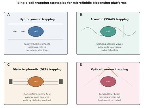
Figure 2. Physical Principles of the Four Single-Cell Trapping Mechanisms Compared in This Review. This figure illustrates, panel by panel, how each trapping mechanism physically immobilizes a single cell: (A) hydrodynamic trapping via passive fluidic resistance in microfabricated traps, (B) acoustic (SSAW) trapping via standing waves that guide cells to pressure nodes without labeling, (C) dielectrophoretic (DEP) trapping via a non-uniform electric field that polarizes and captures cells by dielectric contrast, and (D) optical tweezer trapping via a focused laser beam offering precise but heat-sensitive control. The four panels correspond directly to the mechanisms compared quantitatively in Table 1. The figure is schematic and not derived from a specific dataset.

route to tracking cancer progression; integrated platforms such as the CTC-iChip combine inertial focusing with electrochemical characterization to flag metastatic markers, information that can help clinicians tailor treatment to individual patients.

Food safety applications are organized around the "ASSURED" criteria — Affordable, Sensitive, Specific, User-friendly, Rapid, Equipment-free, and Deliverable (Arreguin-Campos et al., 2021). Electrochemical MIP sensors built to these standards detect trace contaminants such as malathion in olive oil or kanamycin in milk, extending rapid, on-site testing into settings where laboratory infrastructure is limited. Cardiomyocyte physiology has traditionally relied on fluorescent dyes and optogenetic tools (Broyles et al., 2018); electrochemical sensors provide a complementary approach, tracking ionic movement and metabolic byproducts such as hydrogen peroxide, a recognized marker of oxidative stress in excitable tissue.

3.5 Remaining Barriers

The field has not solved several important problems. Biocompatibility remains a persistent concern: many stimuli-responsive materials used in 3D-printed microactuators require pH conditions above 9, which are lethal to living cells. This constraint requires materials with gentler activation thresholds before these systems can be used with live samples (Lao et al., 2021). Fabrication presents a related challenge. Two-Photon Polymerization achieves resolution down to 100 nm, but the process remains too slow for mass production (Lao et al., 2021; Burrow & Gaylord, 2011). Closing this gap will depend on sustained collaboration among engineers, clinicians, and industry partners.

The combination of electrochemical biosensors with microfluidic single-cell platforms is among the most promising directions in analytical science, not because any single component is novel, but because high-throughput inertial separation, robust imprinted-polymer recognition, and finely tuned thin-film transduction together produce capability exceeding the sum of their parts. Translating this promise into standard clinical or industrial practice will depend on progress in biocompatible materials and scalable, multi-modal fabrication.

4. Capture Efficiency, Immobilization, and Hybrid Detection in Single-Cell Biosensing Platforms

4.1 Cell-Trapping Mechanisms: Capture Efficiency versus Viability

The clearest theme in this literature is a direct trade-off between capture efficiency and cell viability, present across nearly every trapping method examined (Table 1; Figure 2). Hydrodynamic traps rely on channel geometry and fluidic resistance and require no electric field or acoustic transducer, only precise microfabrication (Lin et al., 2013; Sochol et al., 2012). This simplicity limits throughput, because cells must be delivered slowly enough for the geometry to function. Acoustic trapping, using standing surface waves, is consistently reported to be benign for cell viability because it avoids direct electrical or optical contact with the cell (Chen et al., 2014), making it a reasonable choice where downstream viability matters as much as capture. Dielectrophoretic trapping is the most selective method reviewed, distinguishing cells by dielectric properties rather than size or shape alone; several studies report high capture rates for specific cell types such as breast cancer cell lines (Bhattacharya et al., 2014; Huang et al., 2014), though primary, patient-derived cells do not always behave as predictably as cultured lines under the same field conditions. Optical tweezers provide the finest spatial precision of any method surveyed. Still, the heat generated by a focused beam is a genuine constraint for temperature-sensitive cells, which limits this approach in high-throughput contexts (Ramser & Hanstorp, 2010).

4.2 Immobilization Strategies for Biorecognition ElementsA second recurring theme concerns how the biorecognition element is anchored to the sensor surface. Covalent attachment and entrapment strategies both appear across the reviewed studies, aimed at keeping an enzyme, antibody, or aptamer catalytically active and properly oriented without leaching into solution (Garcia-Galan et al., 2011; Brady & Jordaan, 2009). Gelatin-based matrices are common: their hydrophilic character makes them effective hosts for biomolecules, but this property causes swelling in aqueous buffers, requiring additional crosslinking, commonly glutaraldehyde-based, to hold the matrix and enzyme together (Bigi et al., 2001; De Wael et al., 2010). Electrode-scaling technique also matters: nanostructured or composite electrode surfaces, produced through methods like spin coating, shift the dominant reaction kinetics from diffusion-limited toward adsorption-controlled behavior, which several authors link to faster

Table 1. Capture Efficiency, Viability, and Throughput Trade-offs Across Single-Cell Trapping Mechanisms. This table compares the four principal cell-trapping strategies used in microfluidic electrochemical single-cell platforms — hydrodynamic, acoustic (SSAW), dielectrophoretic (DEP), and optical tweezer trapping — against their underlying physical principle and three performance dimensions: relative capture efficiency, relative effect on cell viability, and relative throughput. Ratings (e.g., Moderate, High) are qualitative syntheses drawn from the representative studies cited in the rightmost column, not pooled statistical values. The table is intended to show that no single mechanism optimizes all three dimensions simultaneously; each involves a defined trade-off. Representative studies are provided so readers can trace each qualitative rating back to its primary source.

Mechanism

Underlying Principle

Relative Capture Efficiency

Relative Effect on Viability

Relative Throughput

Representative Studies

Hydrodynamic

Channel geometry and fluidic resistance position cells passively

Moderate

Minimal disturbance

Moderate

Lin et al. (2013); Sochol et al. (2012)

Acoustic (SSAW)

Standing acoustic waves guide cells to pressure nodes

Moderate–High

Low impact

Moderate–High

Chen et al. (2014)

Dielectrophoretic (DEP)

Non-uniform electric fields exploit dielectric contrast

High (cell-type dependent)

Field-exposure dependent

Moderate

Bhattacharya et al. (2014); Huang et al. (2014)

Optical tweezers

Focused laser beam provides direct spatial manipulation

High (single-cell precision)

Heat-sensitive; variable

Low

Ramser & Hanstorp (2010)

Table 2. Strengths, Limitations, and Representative Evidence for Single-Cell Detection Modalities. This table compares four detection approaches used downstream of cell capture—amperometry, capillary electrophoresis with laser-induced fluorescence (CE-LIF), mass spectrometry, and hybrid multi-modal detection—by their underlying measurement principle, key strength, and key limitation. It illustrates the central sensitivity-versus-throughput trade-off discussed in Section 4.3: single-modality methods each sacrifice something (analyte range, labeling requirements, or instrumental complexity), while hybrid platforms partially reconcile these trade-offs at the cost of added system complexity. As in Table 1, descriptors are qualitative syntheses of the cited literature rather than pooled quantitative outcomes.

Modality

Principle

Key Strength

Key Limitation

Representative Studies

Amperometry

Measures current from electroactive species at the electrode

Direct, real-time quantification

Limited to electroactive analytes

Keithley et al. (2013); Larsen et al. (2012)

CE-LIF

Electrophoretic separation with laser-induced fluorescence detection

Resolves multiple analytes; high sensitivity

Requires fluorescent labeling in most cases

Ban et al. (2013)

Mass spectrometry

Ionization and mass analysis of separated intracellular content

Broad, unbiased chemical coverage

High instrumental complexity and cost

Mellors et al. (2010); Aerts et al. (2014)

Hybrid (multi-modal)

Combines two or more of the above within one platform

Mitigates sensitivity–throughput trade-off

Added design and integration complexity

Antfolk & Laurell (2017); Liu & Wang (2022)

and more reproducible signal generation (Schubert & Dunkel, 2003; Monosik et al., 2012). Whether this holds consistently across cell types and sample matrices is not established in the qualitative literature alone.

4.3 Detection Modalities and the Case for Hybrid Platforms

The choice of detection modality determines what can be learned from a captured cell (Table 2). Amperometric detection is the most direct method for quantifying electroactive species, with an established record for measuring neurotransmitter release and similar signaling events at the single-cell level (Keithley et al., 2013; Larsen et al., 2012). Capillary electrophoresis with laser-induced fluorescence adds a separation step that amperometry alone cannot provide, making it suited to metabolic profiling or microRNA characterization where multiple analytes must be resolved (Ban et al., 2013). Mass spectrometry, when coupled to microfluidic separation, provides the most comprehensive chemical readout of the three, capturing a broader range of intracellular content than either electrochemical or fluorescence methods alone (Mellors et al., 2010; Aerts et al., 2014), though the added instrumental complexity explains why MS-coupled platforms remain less common outside specialized laboratories. Across the studies reviewed, hybrid detection — pairing amperometry with fluorescence, for instance — is increasingly used to escape the sensitivity-versus-throughput limitation of single-modality platforms.

4.4 From Bench to Bedside: Liquid Biopsy and Clinical Translation

Liquid biopsy demonstrates the practical stakes of this technology. Detecting circulating tumor cells, exosomes, or pathogenic bacteria against an overwhelming background of normal cells and proteins requires throughput, sensitivity, and purity together; few devices achieve all three simultaneously (Neoh et al., 2018; Qiu et al., 2020; Sierra et al., 2020). This is why multi-modal microfluidic (M3) platforms, which combine size-based filtration with immunomagnetic enrichment or similar sequential strategies, have become a more active area of development (Liu & Wang, 2022; Fang et al., 2021): no single separation principle carries the entire analytical burden alone. Antfolk and Laurell (2017) reach a similar conclusion in their review of continuous-flow separation methods for rare cells and bioparticles in blood: combining complementary physical principles, rather than optimizing one in isolation, is the more effective route to devices that handle clinically relevant sample volumes in clinically relevant timeframes.

4.5 Where the Field Falls Short: Reporting and Standardization

The primary limitation across the literature surveyed here is not technical but methodological: studies vary in which metrics they report, how those metrics are defined, and under what sample conditions — cultured cell lines, spiked blood, or complex biological fluids — they were obtained. This variability prevents direct, quantitative comparison across studies, which is why this review does not attempt a pooled statistical synthesis. Junker and van Oudenaarden's (2014) broader point about single-cell biology — that population averages obscure biologically meaningful variation — applies here as well: a field built around resolving heterogeneity at the cellular level has not yet resolved the heterogeneity in its own reporting practices. Standardized metrics and cross-laboratory validation are likely to matter more for near-term clinical translation than any single new trapping or detection innovation.

5. Conclusion

No single trapping mechanism or detection modality is unambiguously superior for microfluidic single-cell electrochemical biosensing; each represents a defined compromise among capture efficiency, throughput, and cell viability. Hydrodynamic and dielectrophoretic trapping favor capture precision, acoustic methods favor gentleness, and hybrid detection schemes offer the most promising, though not yet fully validated, route to reconciling sensitivity with throughput. Clinical translation is limited less by any single unsolved engineering problem than by uneven reporting conventions that make cross-study comparison difficult. Standardized metrics, broader validation in complex biological matrices rather than cultured cell lines alone, and closer collaboration across laboratories are achievable next steps toward routine diagnostic use.


Author Contributions

L.H. conceptualized the study, designed the review framework, conducted the literature search, study selection, data extraction, critical appraisal of the included studies, and qualitative evidence synthesis. L.H. interpreted the findings, prepared the original manuscript, revised the article critically for important intellectual content, approved the final version for publication, and agreed to be accountable for all aspects of the work.

Acknowledgements

The author sincerely acknowledges the Department of Biomedical Engineering, SUNY University at Buffalo, for providing academic support and access to the scientific literature utilized in preparing this review. The author also extends appreciation to the researchers whose published studies formed the foundation of this evidence synthesis. No specific funding was received for this study.

References


Aerts, J. T., Louis, K. R., Crandall, S. R., et al. (2014). Patch clamp electrophysiology and capillary electrophoresis-mass spectrometry metabolomics for single cell characterization. Analytical Chemistry, 86(6), 3203–3208. https://doi.org/10.1021/ac500168d       

Antfolk, M., & Laurell, T. (2017). Continuous flow microfluidic separation and processing of rare cells and bioparticles found in blood. Analytica Chimica Acta, 965, 9–35. https://doi.org/10.1016/j.aca.2017.02.017          

Arreguin-Campos, R., Jiménez-Monroy, K. L., Diliën, H., Cleij, T. J., van Grinsven, B., & Eersels, K. (2021). Imprinted polymers as synthetic receptors in sensors for food safety. Biosensors, 11(2), 46. https://doi.org/10.3390/bios11020046    

Ban, E., Chae, D. K., & Song, E. J. (2013). Determination of micro-RNA in cardiomyoblast cells using CE with LIF detection. Electrophoresis, 34(4), 598–604. https://doi.org/10.1002/elps.201200442             

Bartlett, P. N. (2008). Bioelectrochemistry: Fundamentals, experimental techniques and applications. John Wiley & Sons. https://doi.org/10.1002/9780470753842          

Bhattacharya, S., Chao, T. C., Ariyasinghe, N., et al. (2014). Selective trapping of single mammalian breast cancer cells by insulator-based dielectrophoresis. Analytical and Bioanalytical Chemistry, 406(7), 1855–1865. https://doi.org/10.1007/s00216-013-7598-2      

Bigi, A., Cojazzi, G., Panzavolta, S., Rubini, K., & Roveri, N. (2001). Mechanical and thermal properties of gelatin films at different degrees of glutaraldehyde crosslinking. Biomaterials, 22, 763–768. https://doi.org/10.1016/S0142-9612(00)00236-2 

Brady, D., & Jordaan, J. (2009). Advances in enzyme immobilization. Biotechnology Letters, 31, 1639–1650. https://doi.org/10.1007/s10529-009-0076-4    

Broyles, C. N., Robinson, P., & Daniels, M. J. (2018). Fluorescent, bioluminescent, and optogenetic approaches to study excitable physiology in the single cardiomyocyte. Cells, 7(6), 51. https://doi.org/10.3390/cells7060051 

Burrow, G. M., & Gaylord, T. K. (2011). Multi-beam interference advances and applications: Nano-electronics, photonic crystals, metamaterials, subwavelength structures, optical trapping, and biomedical structures. Micromachines, 2(2), 221–257. https://doi.org/10.3390/mi2020221   

Chen, A., Byvank, T., Chang, W. J., et al. (2013). On-chip magnetic separation and encapsulation of cells in droplets. Lab on a Chip, 13(6), 1172–1181. https://doi.org/10.1039/c2lc41201b            

Chen, Y., Li, S., Gu, Y., et al. (2014). Continuous enrichment of low-abundance cell samples using standing surface acoustic waves (SSAW). Lab on a Chip, 14(5), 924–930. https://doi.org/10.1039/C3LC51001H        

De Wael, K., De Belder, S., Van Vlierberghe, S., Van Steenberge, G., Dubruel, P., & Adriaens, A. (2010). Electrochemical study of gelatin as a matrix for the immobilization of horse heart cytochrome c. Talanta, 82, 1980–1985. https://doi.org/10.1016/j.talanta.2010.08.019       

De Wael, K., De Belder, S., Pilehvar, S., Van Steenberge, G., Herrebout, W., & Heering, H. A. (2012). Enzyme-gelatin electrochemical biosensors: Scaling down. Biosensors, 2(1), 101–113. https://doi.org/10.3390/bios2010101

Fang, Y. L., Wang, C. H., Chen, Y. S., et al. (2021). An integrated microfluidic system for early detection of sepsis-inducing bacteria. Lab on a Chip, 21(1), 113–121. https://doi.org/10.1039/D0LC00966K     

García-Galán, C., Berenguer-Murcia, A., Fernández-Lafuente, R., & Rodrigues, R. C. (2011). Potential of different enzyme immobilization strategies to improve enzyme performance. Advanced Synthesis & Catalysis, 353, 2885–2904. https://doi.org/10.1002/adsc.201100534       

Gil, E. D., & de Melo, G. R. (2010). Electrochemical biosensors in pharmaceutical analysis. Brazilian Journal of Pharmaceutical Sciences, 46(3), 375–391. https://doi.org/10.1590/S1984-82502010000300002      

Gou, Y., Jia, Y., Wang, P., & Sun, C. (2018). Progress of inertial microfluidics in principle and application. Sensors, 18(6), 1762. https://doi.org/10.3390/s18061762    

Huang, C. J., Liu, C. X., & Stakenborg, J. L. T. (2014). Single cell viability observation in cell dielectrophoretic trapping on a microchip. Applied Physics Letters, 104(1), 013703. https://doi.org/10.1063/1.4861135           

Junker, J. P., & van Oudenaarden, A. (2014). Every cell is special: Genome-wide studies add a new dimension to single-cell biology. Cell, 157(1), 8–11. https://doi.org/10.1016/j.cell.2014.02.010

Kalyan, S., Torabi, C., Khoo, H., Sung, H. W., Choi, S.-E., Wang, W., Treutler, B., Kim, D., & Hur, S. C. (2021). Inertial microfluidics enabling clinical research. Micromachines, 12(3), 257. https://doi.org/10.3390/mi12030257

Keithley, R. B., Rosenthal, A. S., Essaka, D. C., et al. (2013). Capillary electrophoresis with three-color fluorescence detection for the analysis of glycosphingolipid metabolism. Analyst, 138(1), 164–170. https://doi.org/10.1039/C2AN36286D

Lai, H. C., Wang, C. H., Liou, T. M., & Lee, G. B. (2014). Influenza A virus-specific aptamers screened by using an integrated microfluidic system. Lab on a Chip, 14(12), 2002–2013. https://doi.org/10.1039/C4LC00187G   

Lao, Z., Xia, N., Wang, S., Xu, T., Wu, X., & Zhang, L. (2021). Tethered and untethered 3D microactuators fabricated by two-photon polymerization: A review. Micromachines, 12(4), 465. https://doi.org/10.3390/mi12040465

Larsen, S. T., Heien, M. L., & Taboryski, R. (2012). Amperometric noise at thin film band electrodes. Analytical Chemistry, 84(18), 7744–7749. https://doi.org/10.1021/ac301136x          

Lin, L. Y., Chu, Y. S., Thiery, J. P., Lim, C. T., & Rodriguez, I. (2013). Microfluidic cell trap array for controlled positioning of single cells on adhesive micropatterns. Lab on a Chip, 13(4), 714–721. https://doi.org/10.1039/c2lc41070b              

Liu, J., Yin, L. F., Dai, Y. R., Jiang, F., & Niu, J. F. (2012). Application of electrochemical enzyme biosensor in environmental pollution monitoring. Progress in Chemistry, 24(1), 131–143.          

Liu, Y., & Wang, W. (2022). Multi-modal microfluidics (M3) for sample preparation of liquid biopsy: Bridging the gap between proof-of-concept demonstrations and practical applications. Micromachines, 13(2), 209. https://doi.org/10.3390/mi13020209 

Mellors, J. S., Jorabchi, K., Smith, L. M., & Ramsey, J. M. (2010). Integrated microfluidic device for automated single cell analysis using electrophoretic separation and electrospray ionization mass spectrometry. Analytical Chemistry, 82(3), 967–973. https://doi.org/10.1021/ac902218y    

Monosik, R., Ukropcova, D., Stredansky, M., & Sturdik, E. (2012). Multienzymatic amperometric biosensor based on gold and nanocomposite planar electrodes for glycerol determination in wine. Analytical Biochemistry, 421(1), 256–261. https://doi.org/10.1016/j.ab.2011.10.020          

Neoh, K. H., Hassan, A. A., Chen, A., et al. (2018). Rethinking liquid biopsy: Microfluidic assays for mobile tumor cells in human body fluids. Biomaterials, 150, 112–124. https://doi.org/10.1016/j.biomaterials.2017.10.006    

Qiu, J., Xu, J., Zhang, K., et al. (2020). Refining cancer management using integrated liquid biopsy. Theranostics, 10(5), 2374–2384. https://doi.org/10.7150/thno.40677    

Ramser, K., & Hanstorp, D. (2010). Optical manipulation for single-cell studies. Journal of Biophotonics, 3(3), 187–206. https://doi.org/10.1002/jbio.200910050            

Ren, K. N., Zhou, J. H., & Wu, H. K. (2013). Materials for microfluidic chip fabrication. Accounts of Chemical Research, 46(11), 2396–2406. https://doi.org/10.1021/ar300314s     

Schubert, D. W., & Dunkel, T. (2003). Spin coating from a molecular point of view: Its concentration regimes, influence of molar mass and distribution. Materials Research Innovations, 7(5), 314–321. https://doi.org/10.1007/s10019-003-0270-2      

Sierra, J., Marrugo-Ramírez, J., Rodríguez-Trujillo, R., et al. (2020). Sensor-integrated microfluidic approaches for liquid biopsy applications in early detection of cancer. Sensors, 20(5), 1317. https://doi.org/10.3390/s20051317   

Sochol, R. D., Dueck, M. E., Li, S., Lee, L. P., & Lin, L. W. (2012). Hydrodynamic resettability for a microfluidic particulate-based arraying system. Lab on a Chip, 12(23), 5051–5056. https://doi.org/10.1039/c2lc40704c


Article metrics
View details
0
Downloads
0
Citations
845
Views
📖 Cite article

View Dimensions


View Plumx


View Altmetric


0
Save
0
Citation
845
View
0
Share