1. Introduction
Cardiovascular disease has long been framed as a condition of later life—something that unfolds slowly, almost predictably, over decades. Yet premature coronary artery disease (pCAD) unsettles that narrative. It appears earlier than expected, often in individuals who are still navigating the most productive decades of adulthood. And perhaps more concerning, it frequently advances quietly. By the time classical symptoms demand attention, structural and molecular injury may already be well underway.
This tension—between biological onset and clinical recognition—has prompted a gradual but unmistakable shift in cardiovascular diagnostics. Traditional tools remain indispensable. Electrocardiography, angiography, and serum enzyme assays have saved countless lives. But they are, in essence, reactive instruments. They confirm damage; they visualize obstruction; they quantify injury. What they do less effectively is reveal the earliest molecular tremors that precede overt pathology. Recent advances in sensor technology suggest that this diagnostic gap may not be insurmountable (Cricco et al., 2025). The question, however, is not merely technological. It is conceptual: can we meaningfully detect—and interpret—subclinical molecular signals before coronary disease becomes anatomically apparent?
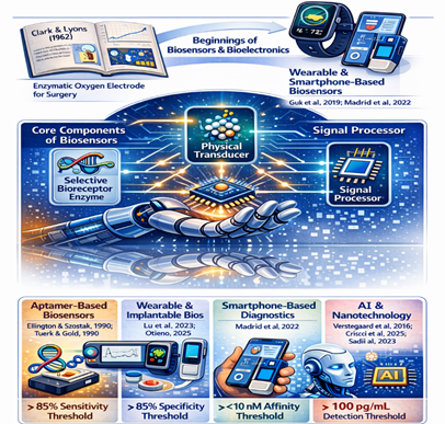
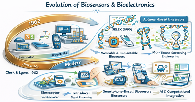
The intellectual roots of biosensing stretch back more than half a century. In 1962, Clark and Lyons introduced an electrode system designed for continuous monitoring during cardiovascular surgery, quietly establishing the structural template for modern biosensors (Clark & Lyons, 1962). The architecture was elegantly simple: a biological recognition element positioned in intimate contact with a transducer capable of converting biochemical events into measurable signals. That blueprint, though refined and miniaturized over decades, remains intact.
What has changed—sometimes dramatically—is the context in which these systems operate. Wearable bioelectronics now enable real-time physiological monitoring beyond the confines of clinical laboratories (Guk et al., 2019). Portable platforms, some integrated with smartphones, compress complex analytical processes into user-accessible formats (Citartan & Tang, 2019). Even saliva, once overlooked as a diagnostic medium, has emerged as a viable biofluid reflecting systemic health states (Ilea et al., 2019). Collectively, these developments suggest that continuous, non-invasive cardiovascular monitoring is no longer speculative. It is, at least technologically, plausible.
Still, biosensors are only as effective as their recognition elements. For decades, antibodies dominated molecular detection strategies. Their specificity is formidable, and immunosensors remain central to biomarker detection (Karachaliou & Livaniou, 2023). Yet antibodies are biologically derived and can exhibit variability, stability limitations, and batch-dependent inconsistencies. In contrast, aptamers—synthetic, single-stranded DNA or RNA oligonucleotides—offer a different proposition.
First described through in vitro selection techniques in 1990 (Ellington & Szostak, 1990), aptamers are generated via the SELEX process, which iteratively enriches nucleic acid sequences capable of binding specific targets with high affinity. Over successive rounds of selection and amplification, molecular recognition becomes refined rather than incidental. Early demonstrations of cell-based SELEX further illustrated that aptamers could be tailored against complex biological targets, including whole cells and membrane-associated proteins (Ara et al., 2012; Amraee et al., 2017). This adaptability has since broadened their diagnostic reach.
The appeal of aptamers lies partly in their structural behavior. Unlike antibodies, aptamers fold into intricate three-dimensional conformations dictated by nucleotide sequence and environmental conditions. These conformations create binding pockets capable of recognizing proteins, ions, and small molecules with impressive specificity (Kara et al., 2023). They are thermally stable, chemically synthesizable, and amenable to controlled modification. Importantly, their production does not rely on biological hosts, reducing variability between batches.
In cardiovascular diagnostics, where biomarkers such as cardiac troponin I (cTnI) circulate at extremely low concentrations during early myocardial stress, sensitivity is critical. Electrochemical aptasensors have demonstrated the capacity to detect cTnI with remarkable analytical performance (Jo et al., 2015). Beyond single-analyte detection, multi-analyte electrochemical aptasensors now enable simultaneous monitoring of multiple biomarkers—an increasingly relevant feature in multifactorial conditions like coronary artery disease (Grabowska et al., 2022).
The evolution of aptasensor design has also been iterative rather than linear. Dual-signaling strategies that integrate “signal on/off” and labeling approaches have enhanced detection fidelity (Cao et al., 2017). Electrochemical impedance-based platforms have proven effective in detecting disease-associated proteins in infectious contexts, demonstrating adaptability across biomarker classes (Chakma et al., 2018). Optical systems, including FRET-based DNA aptasensors, have further expanded the methodological repertoire, even within intracellular environments (Ghosh et al., 2020). Reviews of optical and electrochemical aptasensors underscore the rapid diversification of detection modalities (Farid et al., 2023).
Nanotechnology has arguably accelerated this progress. Metal nanoparticles and carbon-based nanomaterials enhance electron transfer kinetics and increase surface area, amplifying sensor responsiveness (Fritea et al., 2021). More recent platforms integrate flexible materials and unconventional conductors; for example, modified electrochemical paper-based analytical devices have enabled simultaneous detection of homocysteine and C-reactive protein in saliva, biomarkers closely associated with cardiovascular risk (Diao et al., 2024). Parallel efforts have explored CRISPR/Cas12a-based electrochemical biosensors capable of highly sensitive cTnI detection, suggesting that nucleic acid engineering and aptamer science may increasingly converge (Chen et al., 2022).
The conceptual trajectory, then, appears to be one of convergence rather than replacement. Immunosensors continue to evolve (Karachaliou & Livaniou, 2023), while aptamer-based platforms introduce synthetic precision and modularity. In oncology, aptamer ligands have successfully identified cell-surface markers such as troponin T on tumor endothelial cells (Ara et al., 2014), illustrating their potential for membrane-targeted diagnostics. Such versatility hints at broader applicability within cardiovascular pathology, where endothelial dysfunction and membrane-associated markers often precede overt plaque formation.
Yet enthusiasm warrants caution. Analytical performance under controlled laboratory conditions does not automatically translate into clinical reliability. Stability over time, calibration drift, regulatory approval pathways, and user adherence all shape real-world deployment. Even the most sensitive device must ultimately integrate into existing care infrastructures and earn patient trust.
This review therefore examines aptamer-based biosensors within the specific context of early pCAD detection. Rather than assuming technological inevitability, it seeks to weigh experimental promise against translational realities. By tracing developments from foundational biosensor architecture to contemporary multi-analyte electrochemical platforms, the review aims to clarify where evidence is strongest—and where questions remain unresolved. The conceptual framework underlying aptamer-based cardiovascular diagnostics, including molecular recognition mechanisms and integration into advanced biosensing architectures, is illustrated in Figure 1.
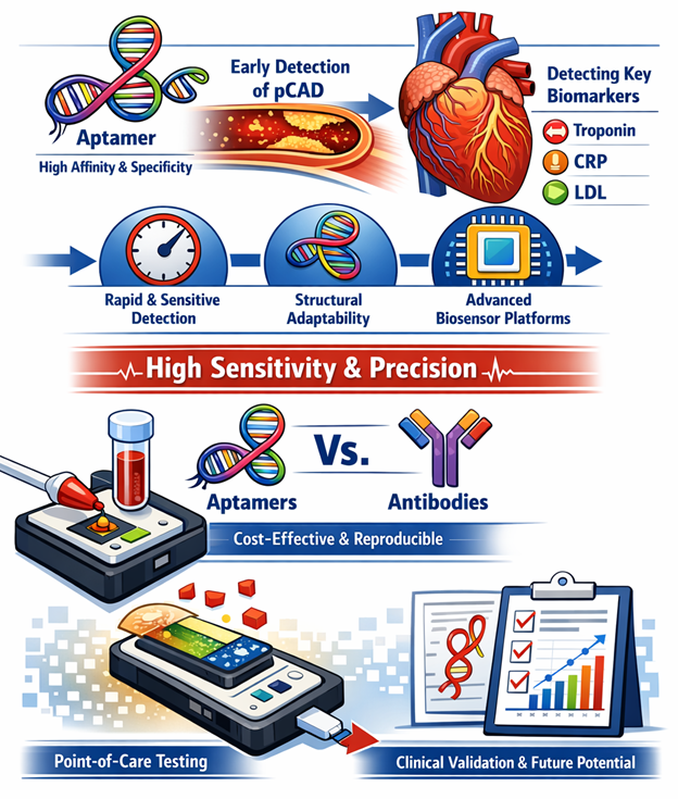
Figure 1. Conceptual Overview of Aptamer-Based Biosensors for Early Detection of Premature Coronary Artery Disease (pCAD). This graphical illustration summarizes the molecular recognition capability, high binding affinity, and structural adaptability of aptamers, highlighting their integration into advanced biosensing platforms for early cardiovascular biomarker detection. The figure also contrasts aptamers with conventional antibody-based systems and emphasizes their suitability for rapid, sensitive, and point-of-care diagnostic applications.
Perhaps the most significant shift is not technical but philosophical. If molecular signals of coronary pathology can be detected before structural damage accumulates, cardiovascular care may move incrementally from reaction to anticipation. Whether aptamer-based biosensors will fully realize that aspiration remains uncertain. But the trajectory—spanning nucleic acid engineering, nanomaterials, wearable electronics, and multiplexed diagnostics—suggests that early, non-invasive detection of premature coronary artery disease is no longer a distant abstraction. It is, cautiously, within reach.