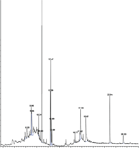
EMAN RESEARCH PUBLISHING | <p>Anti-angiogenesis as a possible mechanism of action for anti-tumor (potential anti-cancer) activity of <em>Crinum asiaticum</em> leaf methanol extract</p>
Inflammation Cancer Angiogenesis Biology and Therapeutics | Impact 0.1 (CiteScore) | Online ISSN  2207-872X
RESEARCH ARTICLE   (Open Access)

Anti-angiogenesis as a possible mechanism of action for anti-tumor (potential anti-cancer) activity of Crinum asiaticum leaf methanol extract

Sa’adiah Mohd. Yusoff  a*, Mohd Zaini Asmawi a, Amin Malik Shah Abdul Majid a, b, Mohamed Khadeer A. Basheer c, Shazmin  Kithur Mohamed a, Muhammad Asif a, Seyedeh Fatemeh Jafari a, Hussein Mahfoudh Baharetha a, d

+ Author Affiliations

Journal of Angiotherapy 1 (1) 012-017 https://doi.org/10.25163/angiotherapy.11000210419100517

Submitted: 15 February 2017 Revised: 10 April 2017  Published: 10 May 2017 


Abstract

Most chemotherapeutic agents can destroy tumours and retard cancer growth, but may damage normal cells and tissues as well. Thus, new anti-cancer drugs derived from natural products are expected to play an important role in the development of more effective and safer strategies to inhibit the progress of cancer, without inducing cell lethality in the healthy surrounding tissues. Crinum asiaticum is used traditionally to treat inflammations and tumours. The aim of this work is to ascertain the scientific viability of its use in traditional medicine as an anti-tumour (potential anti-cancer) agent. Due to its anti-inflammatory properties, it is hypothesised that the anti-tumour activity may be due to anti-angiogenic activity. Thus, in the present study, an attempt has been made to study the anti-angiogenic activity of the Crinum asiaticum leaf methanol extract (CALME) of the Malaysian species. Rat aortic ring assay results showed that CALME prevented new blood vessel formation from the aortic ring explants, with IC50 11.58 µg/ml. The effect of CALME on EAhy 926 cell proliferation (inhibition) had also been investigated, and the MTT results showed that CALME induces cytotoxic effects in EAhy 926 cells, since the IC50 value was found to be 12.18 μg/ml (active cytotoxicity). CALME inhibited endothelial cell migration, at a dose of approximately 12 μg/ml. This dose is similar to the dose at which cytotoxicity is observed in cell. CALME also inhibited the release of the proangiogenic cytokine, VEGF, but not significantly. GC-MS data confirmed the presence of lycorine in CALME. In conclusion, the present work supports the traditional use and previously related works on the plant, which confirm that CALME exhibits anti-angiogenic (potential anti-cancer) activity. However, the anti-angiogenic effect demonstrated by CALME is due to the cytotoxic nature of the extract, and less related to inhibition of one or more of angiogenesis process steps.

Keywords: anti-tumour; anti-angiogenic; Crinum asiaticum; aortic ring assay; EAhy 926 cell proliferation inhibition; cytotoxic; cell migration; VEGF; GC-MS; lycorine.

Introduction

GO

Angiogenesis plays a critical role in tumour growth. Without it, neoplastic tissues cannot expand beyond 1 to 2 mm3 (Folkman and Cotran, 1976). Cancer cells within the tumour can use the newly formed blood vessels as a port to metastasize to other localities (Weidner et al., 1991). Since the interdependency and close relationship between angiogenesis, cancer growth and metastasis has been well-established, much efforts have been invested into the development and/or discovery of anti-angiogenic compounds to treat cancer as well as a variety of other angiogenic related ailments. Most chemotherapeutic agents can destroy tumours and retard cancer growth, but may damage normal cells and tissues as well. Thus, new anti-cancer drugs derived from natural products are expected to play an important role in the development of more effective and safer strategies to inhibit the progress of cancer, without causing toxicity to healthy cells and tissues (Sporn 1999).

Crinum asiaticum, a large, white-flowered, lily-like plant, commonly found along sandy sea-shores, and sometimes planted inland, is occasionally cultivated for its beautiful and showy flowers (Figure 1). The plant is known throughout all of Malaysia as tembaga suasa, bakong (melong), bawang tanah, or bawang hitam. It can also be found on the strand from South East Asia to western Polynesia. As mentioned in the Pharmacopoeia of India, the plant leaves and bulbs are the parts used medicinally. The chemical substance acting emetically, was reported to be an alkaloid, lycorine (1 to 1.8 %), which is allied to emetine  (Medicinal plants of the Philippines, p. 171). Both lycorine and palmilycorine were isolated from the fruits of the plant. Lycorine was also detected in the plant leaves and roots. In in vivo experiments, only lycorine had a pronounced inhibitory effect on the growth of tumour cells, while Palmilycorine showed in vitro (cytotoxic) effects (Shibnath, G. et. al, 1985; Fennell and Van Staden, 2001).

Sa’adiah et al-2017-Figure-1

Figure 1 |  The plant Crinum asiaticum

The plant is used traditionally to treat inflammations and tumours. Thus, the aim of this work is to ascertain the scientific viability of its use in traditional medicine as an anti-cancer agentDue to its anti-inflammatory properties (Samud AM, et. al., 1999), it is hypothesized that the anti-cancer activity may be due to its anti-angiogenic activity, since the latter is known to be influenced by inflammation. anti-angiogenic (potential anti-cancer) activity of Crinum asiaticum leaf methanol extract (CALME) of the Malaysian species.

Materials and methods

GO

GC-MS Measurement

Mass spectra were obtained using a GC/MS 6890N/5973 Inert (Agilent Technologies). The samples were prepared in GC grade methanol and were injected directly into the ESI source.

Cell culture and Reagents

Dulbecco’s Modified Eagle Medium (DMEM), heat inactivated foetal bovine serum (HIFBS), penicillin/streptomycin, and trypsin were obtained from Gibco, Life technology, UK. Phosphate Buffered Saline (PBS), sodium chloride, MTT reagent, betulinic acid, suramin, thrombin, fungizone, glutamine, gentamycin, aminocaproic acid and aprotinin were purchased from Sigma – Aldrich, Germany. Fibrinogen was acquired from Calbiochem, USA. All chemicals used in this study were of analytical grade. EAhy 926 cell line was provided by American Type Culture Collection, Rockville, MD, USA.

Plant collection and authentication

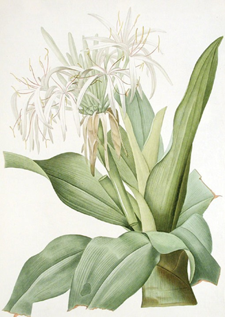
Samples of the plant, Crinum Asiaticum (locally known as tembaga suasa) were collected in several places, around Seberang Prai and Balik Pulau, Penang Island. The plant material was sent to the herbarium unit of the School of Biology, USM, for authentication, and a voucher specimen (no. 11537) of the plant (leaves and flowers) was deposited at both the herbarium unit (School of Biology) and herbal room of the School of Pharmaceutical Sciences, USM.

Preparation of plant extract

The plant samples were first cut into small pieces, then air-dried followed by heat-dried in the oven, at about 300C, for roughly a week. The dried material was then ground into fine powder, using an electric grinder. They were then subjected to methanol extraction, using a Soxhlet apparatus, to yield the crude extract, which was stored at -20oC until further use.

In vitro anti-angiogenic activity of CALME

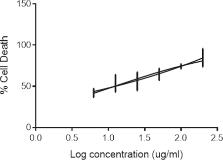
EAhy-926 cell proliferation assay

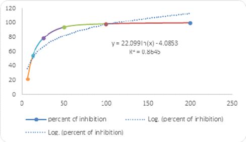
EAhy-926 cells were maintained in DMEM supplemented with 20% of HIFBS, 1% of fungizone, 1% of glutamine, 0.6% of gentamycin and 0.1% of aminocaproic acid. The cells were seeded in 96-well plates, at a density of 2 x 10⁴ cells/well in 100 µl of growth media and kept overnight to facilitate attachment. The cells were exposed to CALME (6.25 to 200 µg/ml) for 48 h. After incubation, the viability of EAhy-926 cells was assessed by MTT (3-4,5 dimethylthiazol-2,5 diphenyl tetrazolium bromide) assay (Mosmann T.,1983). 20 µl of MTT solution (5 mg/ml in PBS) was added to all wells. After 4-hour incubation at 370C in a 5% CO2 atmosphere, the supernatant (medium or extract) was aspirated from the wells as completely as possible, without disturbing the formazan crystals.  A 100 micro-litre volume ofsolvent (DMSO) was then added to each well. After a few minutes at room temperature, to ensure that all crystals were dissolved, the absorbance in each well was measured at 570nm on a multi-well spectrophotometer (ELISA reader).
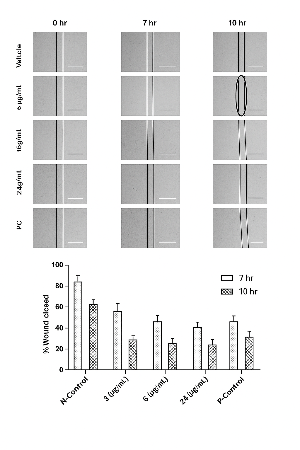
Migration assay

The assay was carried out as described by Liang et al. (2007). Briefly, EAhy-926 cells were seeded in a 6-well plate till the formation of a confluent monolayer, after which a wound was created with 200 µl micropipette tip. The detached cells were removed by washing twice with PBS and the plates were treated with CALME. The wounds were photographed after 7 and 10 h. The width of the cell-free wound was measured under an inverted light microscope equipped with Leica Quin computerized imaging system. 7 fields per well were photographed and a minimum of 7 readings per field were measured.

Human VEGF (Vascular Endothelial Growth Factor) inhibition assay

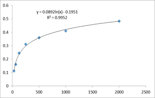
The assay was conducted as per the protocol given in the product (DuoSet® ELISA Development System) information leaflet. Briefly, 60-70% confluent cultures of HUVECs were treated with CALME at 4.15µg/ml (IC50) and 7.47µg/ml (IC90), for 6 h, and cell lysates were prepared using the supplied cell lysis buffer. This assay employs a human VEGF-specific antibody coated on a 96-well plate. Standards and HUVEC lysates were pipetted into the plate so that the VEGF present in the samples would bind the immobilized antibodies. The wells were washed and biotinylated antihuman VEGF antibody was added before the wells were washed again. Then, HRP-conjugated streptavidin was added to the wells. After washing, the colouring agent Tetra Methyl Benzidine substrate solution was pipetted to the wells. The Stop Solution (H2SO4) was added to each well, and the intensity of the colour was measured at 450 nm. At the time of the experiment, a calibration curve of VEGF standard was prepared. The concentration of VEGF in cell lysates was calculated using the log-log regression equation of the best fit line of the standard calibration curve; (y = 0.0892x, R= 0.995). The experiment was repeated two times in triplicates.

Rat aortic ring assay (ex vivo)

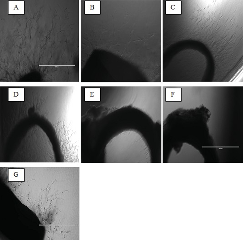
The assay was carried out by utilizing normal, healthy, male Sprague Dawley (Rattus norvegicus) rats of 8-12 weeks old. Sprague Dawley rats are preferred because rats are bigger in size, easily available, easy to handle and the aorta is longer than mice. Male rats are preferable than female ones, to avoid the hormonal disturbance that may occur during the menstrual cycle. The rats were euthanized with 5% CO2 before their aorta excised. The aorta was subsequently cleaned, cut into rings at approximately 1 mm. The rings were placed into a 48-well plate, filled with a special nutrient medium, to which thrombin was added to form the first layer. The extract of different concentrations was then prepared with a second layer of medium. After five days of incubation, the explants were visualized, to study the growth of micro-vessels, which occurred spontaneously at a basal rate, from the cut surfaces of the aortic rings (Nicosia et al., 1992).

Statistical analysis

Statistical analysis involved use of Graph Pad Prism 5. Data are given as the mean + S.E.M. (standard error mean) and statistics were performed using ANOVA (analysis of variance).

Results

GO

 GC-MS analysis of CALME

Fig. 2 below shows the GC-MS chromatogram of CALME. The GC-MS results confirmed the presence of the active alkaloid lycorine in the local leaf extract, as found in the Indian species.

Inhibition of EAhy-926 cell proliferation

To characterize the anti-angiogenic activity of CALME, we first determined whether CALME inhibited endothelial cell proliferation. Results showed that CALME caused inhibition of EAhy-926 cell proliferation, with an IC50 of 12.18 µg/ml (active cytotoxicity) as shown in Fig. 3. IC50 value was found to be 12.18 µg/ml (active cytotoxicity).

Sa’adiah et al-2017-Figure-2

Figure 2 |  The CALME ?ngerprint with GC-MS analysis. Pk: 15; RT: 18.67; Area%: 10.59; Library/ID: NIST02.Lycorine ((3,4-Dimethyl-phenyl)-(2,6-dimethyl-pyrimidin-4-yl)-amine); Ref: 110373, 74433; CAS: 000476-28-897, 1000296-56-445


Figure 3 |  Effect of CALME on proliferation of EAhy-926 cells. IC50 value was found to be 12.18 µg/ml (active cytotoxicity).Inhibitory effect of CALME on EAhy-926 cell migration

CALME inhibited EAhy 926 cell migration in a dose-dependent manner as shown in Table 1, Fig. 4 and 5 below. CALME showed remarkable inhibition of EAhy-926 cell migration at 24 µg/ml, by 48% after 10 h. At 12µg/ml, CALME demonstrated a significant inhibitory effect (34%) after 10 h.

Table 1: Effect of CALME on EAhy 926 cell migration

Treatment

% Wound closed


7hr

10hr

Negative-Control

70

88

3 (µg/mL)

61

64

6  (µg/mL)

41

63

12  (µg/mL)

37

54

24  (µg/mL)

36

40

Positive-Control (PC)

27

31

Figure 4 |   Effect of CALME on EAhy 926 cell migration. The photomicrograph shows the cells after treatment iwth di?erent dosage of CALME and the histogram shows the percentage of wound closure at 7 h and 10 h of treatment.

Effect of CALME on VEGF Expression in EAhy 926 Cells

Table 1 |  Effect of CALME treatment on the release of the proangiogenic cytokine, VEGF 

CALME (µg/mL)

VEGF (pg/mL)

Inhibition (%)

Negative(control)

45.73  ± 1.92

-

IC50

44.14 ± 0.64

3.47 ± 1.40

IC90

44.00  ± 0.42

3.78 ± 0.91


Figure 5 |  Dose response curve of VEGF standard    

Table 2  shows the percentage of inhibition of blood vessel outgrowth observed in rat aortic ring assay. IC50 = 11.58 ug/mL (IC50 from MTT assay = 12.18 ug/mL).  CALME inhibited the release of the proangiogenic cytokine, VEGF, as shown in the table above, although not significantly.

Effect of CALME on sprouting of micro-vessels in rat aorta

Table 2 |  Percentage inhibition of blood vessel outgrowth observed in rat aortic ring assay.

CALME (µg/ml)

Blood Vessel Inhibition (%)

6.25

22.47 ± 13.09

12.50

54.39 ± 10.45

25.00

78.52 ± 5.98

50.00

94.22 ± 12.34

100.00

98.64 ± 10.50

Positive Control (BA)

51.56 ± 7.46


Figure 6 |  Photomicrograph of rat aorta rings after treatment with CALME. A | Negative (0.5 % DMSO); B | 6.25 µg/mL CALME; C | 12.50 µg/mL CALME; D | 25.00 µg/mL CALME; E | 50.00 µg/mL CALME; F | 100.00 µg/mL CALME; G | Positive (Betulinic Acid 3 µg/mL).   

Figure 7 |  Percentage inhibition of blood vessel outgrowth in rat aortic ring assay. 


Discussion

GO

Most chemotherapeutic agents can destroy tumors and retard cancer growth, but may damage normal cells and tissues as well. Thus, new anti-cancer drugs from natural products are expected to play an important role in the development of more effective and safer strategies to inhibit the progress of cancer without causing toxicity to healthy cells and tissues (Sporn 1999). The main difference between cytotoxic chemotherapy and anti-angiogenic therapy is the target; the chemotherapy agents target tumour cells themselves, but antiangiogenic compounds target endothelial cells. Accordingly, anti-angiogenic compounds have many advantages over conventional cytotoxic agents. In fact, since endothelial cells are highly genetically stable with a  low mutation rate, the probability to develop resistance against anti-angiogenesis agents is very low (Folkman, 2003). Moreover, little or no toxicity is associated with anti-angiogenic therapy. This advantage is expect ed as anti-angiogenic compounds target specific immature characteristics of tumour vasculature (Kerbel, 2000). Another important advantage is that endothelial cells are highly accessible to therapeutic agents, because the endothelial cells are directly exposed to blood borne substances (Schliemann and Neri, 2007).

In this study, rat aortic ring assay was used as a screening model for the anti-angiogenesis activity of CALME. The results showed that CALME prevented new blood vessel formation from the rat aortic ring explants, with an IC50 11.58µg/ml. To ascertain whether the observed effect on rat aortic ring assay was due to inhibition of endothelial cells proliferation or not, the effect of CALME on EAhy 926 proliferation was investigated. The MTT results showed that CALME is a cytotoxic agent against EAhy 926, as the IC50 value was found to be 12.18 µg/ml (active cytotoxicity). CALME also inhibited 100% of the angiogenesis process in the ex vivo rat aortic ring model at a dose of about 24 µg/ml. Taken together, the results of the rat aortic ring assay and the MTT assay on EAhy 926 suggest that the anti-angiogenic effect demonstrated by CALME is due to the cytotoxic nature of the extract, and may be less related to inhibition of one or more of the angiogenesis process steps such as endothelial cell migration, endothelial cell differentiation and/or inhibition of VEGF signalling. In order to investigate the angiogenesis inhibitory mechanisms of CALME, we have studied its effects on two major steps in angiogenesis namely, endothelial cell migration and VEGF expression. CALME inhibited cell migration, at a dose of about 12 µg/ml. This dose is similar to the cytotoxic concentration (IC50) in endothelial cells. CALME also inhibited the release of the proangiogenic cytokine, VEGF, as shown in the results above, but not significantly. Thus, these results confirmed that the anti-angiogenic effect demonstrated by CALME is due to the cytotoxic nature of the extract, and less related to inhibition of one or more of other angiogenesis process steps, especially VEGF expression. Finally, GC-MS data confirmed the presence of the active alkaloid, lycorine, in CALME.

Conclusion

GO

In conclusion, the present work supports the traditional use and the previously related works on the plant, which confirm that CALME exhibits anti-angiogenic (and potential anti-cancer) effects. A significant inhibition (IC50 11.58 µg/ml) of micro-vessel formation was observed in treated aortic explants. The in vitro inhibition of proliferation, migration, and VEGF production in EAHY cells by the extract, further support its anti-angiogenic activity. However, the anti-angiogenic effect demonstrated by CALME is due to the cytotoxic nature of the extract, and less related to inhibition of one or more of other angiogenesis process steps. Also, GC-MS data confirmed the presence of lycorine in CALME.

Acknowledgements

GO

The authors would like to express their gratitude to University Science Malaysia for research grant (no.131/0210/0350) and facilities.

Author Contribution

GO

Sa'adiah M. Y. performed all aspects of the project; Mohd Zaini A. supervised the project; Amin M. S. A. M. planned and supervised the project; Mohamed
Khadeer A. B. planned the project; Shazmin  K. M.   conducted the cell cultures; Muhammad Asif performed the rat aorta ring and VEGF assays; Seyedeh
Fatemeh J. assesed the rat aorta ring & cell migration assays and Hussein M. Baharetha conducted the VEGF assay.

References


Medicinal Plants of the Philippines, 171.

Fennell C.W., Van Staden J. (2001) ‘Crinum species in traditional and modern medicine’, J. Ethnopharmacol. 78, 15-26.

Folkman, J. & Cotran, R. (1976). 'Relation of vascular proliferation to tumour growth', Int  Rev Exp Pathol, 16, 207-48.

Folkman, J. (2003), 'Angiogenesis and apoptosis', Seminars in Cancer Biology, 13(2), 159-167.

Kerbel, R. S. (2000), 'Tumor angiogenesis: past, present and the near future', Carcinogenesis, 21(3), 505-515.

Liang CC, Park AY, Guan JL (2007) In vitro scratch assay: a convenient and inexpensive method for analysis of cell migration in vitro, Nat. Protoc. 2, 329-333.

Mossmann, T. (1983). Rapid colorimetric assay for cellular growth and survival: application to proliferation and cytotoxicity assays, J. Immunol. Methods, 65, 55-63.

Mohd. Yusoff, S. (2016). Abstract for poster presentation at the 2nd. AMDI International Bio-health Sciences Conference. Malaysia: Journal of Biomedical and Clinical Sciences

Nicosia RF. (1992). Large-vessel endothelium switches to a microvascular phenotype during angiogenesis in collagen gel culture of rat aorta, Atherosclerosis, 95 (2-3), 191-9.

Pharmacopoeia of India, 234.

Samud AM. (1999). Anti-inflammatory activity of Crinum Asiaticum and its effect on bradykinin-induced contractions on isolated uterus, Immunopharmacol. 43, 311-16.

Schliemann, C. & Neri, D. (2007) 'Antibody-based targeting of the tumor vasculature', Biochim Biophys Acta, 1776(2), 175-92.

Shibnath G. et. al., (1985), Phytochemistry 24, 2703.

Sporn MB. (1999). Prevention of cancer in the next millennium: report of the chemoprevention working group to the American Association for cancer research, Cancer Research 59, 4743-58.

Weidner, N., Semple, J. P., Welch, W. R. & Folkman, J. (1991). 'Tumour angiogenesis and metastasis-correlation in invasive breast carcinoma', N Engl J Med, 324(1), 1-8.

Committee on Publication Ethics

PDF
Abstract
Export Citation

View Dimensions


View Plumx


View Altmetric


Save
0
Citation
1143
View

Share